
The Characterization and Optimization of an Underwater Gamma-Ray Detection System (DUGS)

1
Faculty of Military Science, Stellenbosch University, Saldanha 7395, South Africa
2
Department of Electrical and Information Engineering, University of Nairobi, Nairobi P.O. Box 30197-00100, Kenya
*
Author to whom correspondence should be addressed.
J. Mar. Sci. Eng. 2023, 11(1), 171; https://0-doi-org.brum.beds.ac.uk/10.3390/jmse11010171
Submission received: 22 November 2022
/
Revised: 3 January 2023
/
Accepted: 6 January 2023
/
Published: 10 January 2023
(This article belongs to the Special Issue Application of Coastal/Ocean Sensors and Systems)
Abstract
:Sedimentation can cause numerous problems in rivers, estuaries, harbors, and coastal areas. It is therefore important to trace and model the movement of sediments. The natural physical, chemical, and biological components of the aquatic sediment generally relate to these features in its terrestrial catchment area. It consequently contains the naturally occurring radionuclides of thorium, uranium, and potassium, which can be used as tracers. To achieve this aim, a delta underwater gamma system (DUGS) was developed to map the radionuclides in aquatic sediments. Though the system has been tested for radiometric accuracy, the low concentrations of natural radionuclides in aquatic sediments and the attenuation by the water and detector enclosure necessitated an evaluation to determine the detection efficiency of the detector along with optimal operational parameters. These included spectra accumulation time and underwater speed. The DUGS was consequently used to determine and optimize these parameters for mobile measurement of aquatic sediments. The acquired gamma spectra were also analyzed using full spectrum and energy window analysis to determine the optimal method for extracting the activity concentrations of the nuclides in the sediments.
1. Introduction
Understanding and modelling sedimentation processes are imperative when dealing with destructive erosion and deposition in coastal areas. These destructive processes are enhanced by global warming and the increase in human activities. Artificial tracers, which include anthropogenic nuclides, are traditionally employed to study sediment transport [1]. The use of the dominant naturally occurring radionuclides as tracers was consequently proposed by Bezuidenhout [2] as an alternative to the use of artificial radionuclides. These nuclides occur naturally in all rocks and soils and therefore pose no environmental risk in contrast to artificially introduced tracers. The naturally occurring radionuclides are mainly potassium (40K), uranium (238U), and thorium (232Th), and they occur in sediment in varying concentrations [3]. The concentrations mainly depend on the origin of the sediment but are also influenced by geographical factors and physical characteristics of the sediment. The distribution of natural radionuclides can therefore illustrate sediment transport patterns in harbors, deltas, and other coastal areas.
The measurement of naturally occurring radionuclide concentrations in sediment, however, poses various challenges. The concentrations of naturally occurring nuclides are typically determined by the characteristic gamma-ray emissions from the progenies in the decay chains of these nuclides. Several underwater gamma detectors have been developed for mainly detecting radon [4] and cesium [5], submarine exploration [6], and marine contamination [7]. The challenging methods of surveying sediment in aquatic environments, however, require a unique approach. Firstly, a mobile or towed system is required [8,9]. Many recent studies involving in-situ gamma ray spectrometry of aquatic radionuclides have focused on the stationary deployment of these systems for long-term or continuous measurements of cesium (137Cs) and radon progenies [10,11]. These systems are also relatively expensive, mainly due to the use of bismuth germanium oxide (BGO) crystals commonly used for the detectors in these systems.
A tailor-made underwater gamma detection system, called the delta underwater gamma system (DUGS), was consequently developed. This system was tested for radiometric accuracy and calibrated before being field tested in the Berg River Estuary in South Africa [12]. Even though the system demonstrated promising results in extracting the gamma ray energies, the sediments in the estuary were characterized by low concentrations of natural radionuclides. Additionally, their gamma radiation was attenuated by the water and the detector enclosure, which necessitated an evaluation to improve efficiency and optimize measurements. Though efficiency analyses for gamma ray systems are commonly available, the attenuation characteristics of the water and system enclosure had to be determined, along with its detection efficiency when measuring sediments. The survey speed and accumulation time of the system were also determined, along with the ideal spectral analysis method for sediments. This could either be full spectrum or energy window analyses, or a combination thereof, as possibilities for optimal extraction of nuclide concentrations in sediments. This study, therefore, uses the DUGS system to investigate, determine, characterize, and optimize these factors for mobile measurement of aquatic sediments.
2. System Architecture and Measurement Configuration
The DUGS consists of a 3″ × 3″ NaI(Tl) scintillation detector, photomultiplier tube, off-the-shelf TB-5 multichannel analyzer (MCA) from Amptek®, interface logic, and accompanying power electronics all enclosed in a waterproof enclosure [12]. The system was designed to be dragged among the aquatic sediment and should therefore not only be able to withstand the hydrostatic pressure, but also the impact from rock outcrops and debris. Grade 316 stainless steel was consequently selected for its strength, pressure, and corrosion tolerance. To minimize the gamma ray attenuation from the enclosure, the thinnest possible wall thickness was selected for the section of the enclosure where the detector is located. A commercial stainless-steel cylindrical pipe was selected for this section, and due to its fabrication tolerances, the minimum wall thickness was found to be approximately 1.5 mm. To improve the hydrodynamic drag of the system, and to provide additional protection against impact, the front of the detector system was equipped with a stainless-steel cone of 4 mm wall thickness. The system is pulled by a rope that connects the cone to the boat. Communication with the detector system is established through Ethernet over a Cat5e cable, which also delivers power to the system through power-over-Ethernet (PoE). To prevent excessive roll that could potentially damage the power and data cable entering the enclosure through the center of the cone, a wing with a righting buoy was attached to the top of the enclosure. The righting buoy can also be used for system recovery. The spectral data were analyzed using customized software running on either a laptop or tablet. Figure 1 shows an isometric view of the enclosure, along with a simplified block diagram of its components.
3. Attenuation Characteristics and Efficiency
3.1. Materials and Methods
Kilel et al. [12] determined the attenuation of the DUGS enclosure by comparing the relative count rates of different photopeaks at various thicknesses of 316 stainless steel. Thereafter, the gamma ray spectra of black sands, which simulated the seabed sediment, were compared with and without the enclosure to determine the observability of the individual photopeaks. Lastly, the mass attenuation coefficients of different gamma ray energies were determined using the US National Institute of Standards and Technology (NIST)’s XCOM software [13], which were then compared to experimental values. The gamma ray count rates for different energies were also determined and compared when placing the detector horizontally (180°) as opposed to its normal vertical position (90°). Even though no differences were found between the two measurement geometries, the detection efficiency of the detector was not determined.
The detection efficiency of NaI(Tl) detectors is largely dependent on gamma ray energy, source location, activity, source, and detector geometry, all of which are quantities that are situational. The absolute detection efficiency for NaI(Tl) detectors therefore cannot be represented by a single quoted value. Various studies have shown that the geometric efficiency of 3” × 3” NaI(Tl) detectors reduces to less than 10% if the source-to-detector distance exceeds 10 cm [14]. Geometric efficiency is defined as the ratio between the number of radiation quanta incidents on the detector compared to the total radiation quanta emitted by the source. The absolute efficiency, which is defined as the ratio between the number of pulses recorded by the detector to the number of radiation quanta emitted by the source, has also been shown to decrease with an increase in source-to-detector distance. [15,16]. The source-to-detector distance has also been shown to affect the detector’s intrinsic efficiency [17], specifically if the source is located off-center [18]. Typically, a detector’s intrinsic efficiency relates to its ability to convert impinging radiation into recorded pulses, but a slight dependence on the distance between the source and detector remains. This is because the path length of the radiation through the detector will be affected by this spacing [19]. Toivonen [20] also showed that the counting efficiency of a detector depends on the off-center angle of the source relative to the beam of the detector.
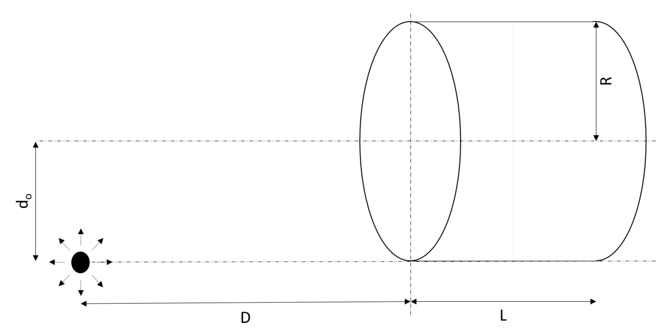
Provision, therefore, needed to be made for the measurement of sediment located at an off-center position to the detector entrance window, as seen in Figure 2. The gamma rays that are emitted by the sediment can be approximated as a point source, which is located at a distance D from the entrance window of the detector. This source is then located at an off-center distance of d0 when the detector is dragged horizontally among the sediment. An increase in D will also significantly increase the gamma ray attenuation by the water [21]. The attenuation of an incident gamma ray (I0) is given by Equation (1) [22]
with I the attenuated intensity, μ the mass attenuation coefficient, and D the distance between the source and the detector.
An experiment was consequently designed to determine the detection efficiency of a 3″ × 3″ NaI(Tl) detector used in this configuration. This was done by placing a uranium (238U) source in an off-center position to the entrance window of the detector, as shown in Figure 3. The system was then submerged in seawater and suspended approximately 2 m below the water surface and well above the seabed. This suspended position prevents the marine sediment and cosmic radiation from affecting the measurements. The distance between the detector and the source was systematically increased starting from the entrance window of the detector (D = 0). Gamma ray spectra at increasing distances (D) were accumulated for periods of 2 min. The distance was increased until the low energy 214Bi peak of 609 keV was no longer visible on the spectrum. The background spectrum was also acquired for a period of 5 min. A window analysis was then performed to extract the count rates of the most prominent 238U peaks for the measurements at different distances. The background was subtracted, and the results were plotted. Functions were then fitted to the data and their coefficients were extracted. These values are the combined efficiency coefficients of the system and represent the attenuation due to source-detector solid angle, off-center distance, and water, respectively.
3.2. Results and Discussion
Kilel et al. [12] found that the relative count rates (I/I0), as expected, reduce with increasing stainless-steel thickness. For 1.5 mm, the thickness of the DUGS enclosure, an attenuation of 13.87% and 8.67% can be observed for 351.9 keV and 1764.5 keV energies, respectively. Despite this attenuation, the gamma ray energies from the black sand spectra were easily identified. Additionally, the experimental mass attenuation coefficients showed a good correlation with the XCOM calculated values. Low energy photons were found to have a higher mass attenuation coefficient than the higher energy photons, but photopeaks above 300 keV are typically used in sediment spectra [23] and sediment measurements are therefore less affected by this attenuation. In terms of the detector’s measuring geometry, no noticeable difference in count rates was detected between the detector’s horizontal and vertical orientation. These findings, therefore, indicate that the only factor that could severely affect measurements is detection efficiency.
Figure 4 illustrates the count rates in the 40K photopeak, the highest energy photopeak of 232Th, and the prominent 238U photopeaks, as the source-to-detector distance (D) is increased at an off-center distance (D0) of 38.1 mm from the entrance window. The functions fitted to the data are also shown in Figure 4 and indicate an exponential decrease in count rates in each photopeak as the distance is increased. A similar trend is also observed in the total count rates. While these functions bear similarity to Equation (1), they not only include the attenuation from the water, but also the reduction in counting efficiency as the source-to-detector is increased along with a reduction in solid angle due to the off-center location of the source. This exponential decrease in count rates can therefore be represented by Equation (2):
with R the attenuated count rates, R0 the count rates measured when the source is at the detector’s entrance window, D the distance between the source and the detector, and μE the efficiency coefficient of the system at the different photopeak energies.
Typically, the mass attenuation coefficients are expected to decrease with an increase in photo energy [24]. It is therefore surprising that the efficiency coefficients of all the energy peaks are within 10% of each other, as can be seen in Figure 5. Further study is required to explain this phenomenon, but it could be related to the detector’s intrinsic efficiency and its dependence on detector-to-source distance. Assuming a point source, numerous Monte Carlo simulations have shown that the detector’s intrinsic efficiency decreases with an increase in distance (D), reaching a minimum at D/R ≅ 0.7, with R the radius of the detector. After this point, any further increase in distance will increase the intrinsic efficiency, even exceeding the initial intrinsic efficiency at D/R > 10 [17]. A similar ratio was also found for multiple off-axis positions [18]. A possible explanation is that at this distance, the directions of the photons impinging on the surface of the detector are parallel to its axis. This implies that all photons will travel the same distance inside the detector. Therefore, for a given energy, the intrinsic efficiency calculation becomes independent of distance and off-axis angle. The increase in the distance also increases the probability of Compton scattering in the water from the higher energies into the lower energy peaks.
Regardless of the cause, from an in-situ measurement perspective, uniform attenuation is highly beneficial since all gamma energies emitted by the sediment would experience a similar attenuation.
4. Survey Speed and Accumulation Time of the DUGS
4.1. Materials and Methods
As the DUGS is being dragged among the sediment, gravity and two diagonally upward forces act on the system. The drag from the righting buoy and pull from the boat consequently oppose gravity and depending on the water depth and speed of the boat, the DUGS risks being lifted off the sediment. Loss of contact with the sediment can be monitored by observing changes in the total counts measured. A significant drop in the total counts could be an indication that the DUGS is floating and not in contact with the sediments on the seafloor. To prevent this from happening, a 2.4 m chain was added to the nose cone of the DUGS, from which the system is dragged. A test was performed with and without the chain to determine the optimal traveling speed for the boat.
A parameter that closely relates to the traveling speed of the boat is the accumulation time of the system. The traveling speed combined with the accumulation time determine the system’s spatial resolution. While each sample contains the radiometric information of the measured area, the geolocation information is only added once the sample is saved. This implies that while longer accumulation times decrease the statistical uncertainty of the measurements, it also reduces the spatial resolution.
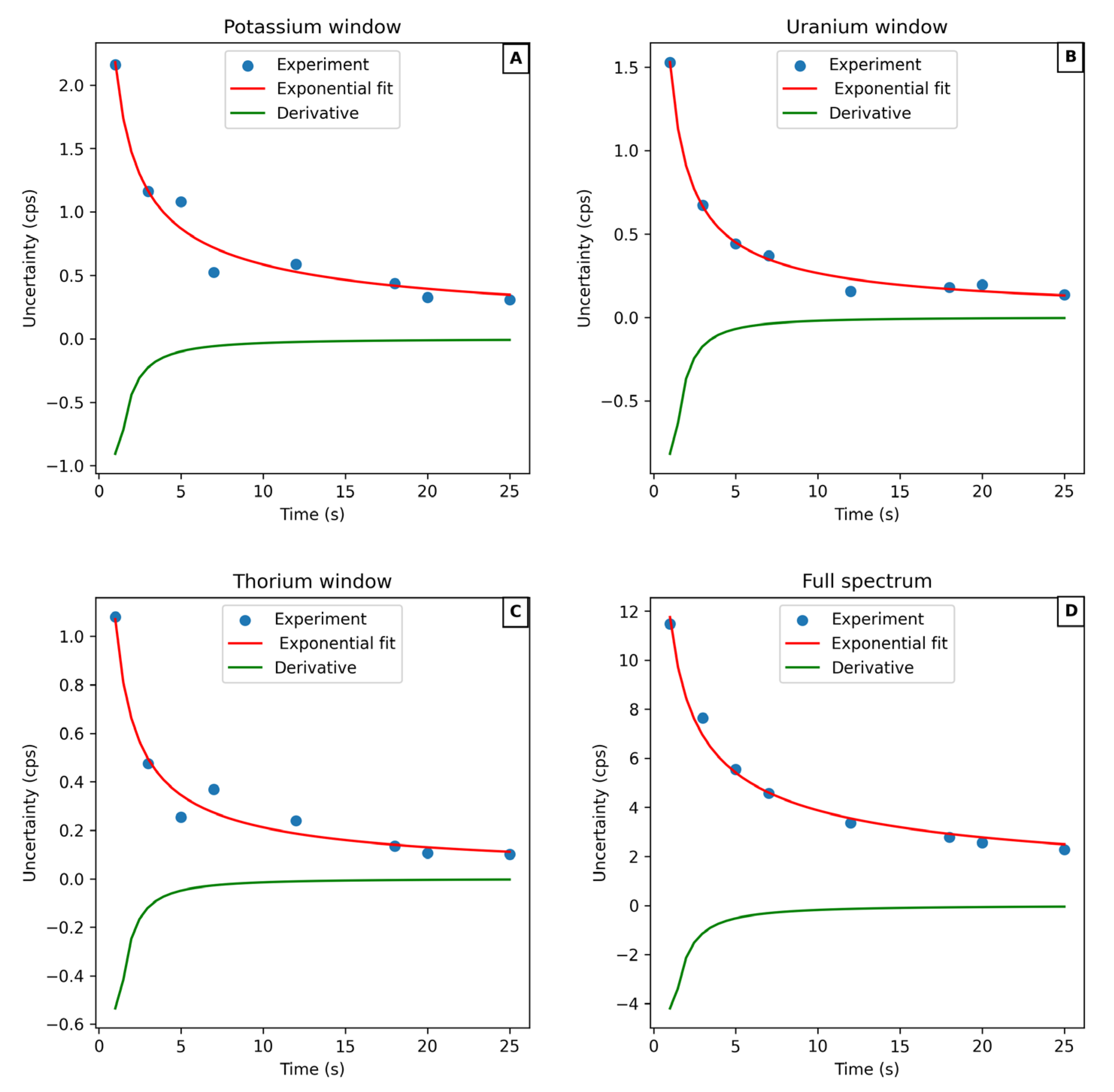
The optimal accumulation time was determined by surveying sediments in the Berg River Estuary. The DUGS was calibrated before the measurements and the energy windows of uranium, thorium, and potassium were selected. The spectra were then accumulated and saved for different periods: 1 s, 2 s, 3 s, 4 s, 5 s, 7 s, 12 s, 15 s, 18 s, 20 s, and 25 s. The count rates in the energy windows for uranium (1764 keV), thorium (2615 keV), and potassium (1460 keV) were then extracted, along with the total count rates. The uncertainties for these count rates were calculated and plotted as a function of accumulation time. The slope at the different accumulation times was then obtained by deriving the respective curves. The derivatives were used to estimate the optimal accumulation time for the DUGS.
4.2. Results and Discussion
The optimum speed for underwater in-situ measurements using DUGS was determined to be 2 knots (1 ms−1) without the chain. At this speed, contact between the DUGS and sediments was ensured. As expected, the weight of the chain greatly increases the measuring speed without the risk of losing contact with the sediment, but low speeds have the advantage of improved spatial resolution. A similar study done by Van Wijngaarden et al. [21] towed MEDUSA (multi-detector system for underwater sediment activity) at a speed of 2 ms−1 with an integration time of 10 s. Therefore, a speed of 1 ms−1 is within the range of other similar studies.
The count rates of the spectra at different accumulation times were extracted and the uncertainty was calculated. Figure 6 shows the variation of the count rate uncertainties at different accumulation times in selected energy windows. As expected, the results showed a high uncertainty in the count rates at 1 s, which was greatly reduced at 25 s. It also showed that the uncertainty decreases sharply between 1 s and 5 s, after which it starts to stabilize. Increasing the accumulation times beyond this point, therefore, does not significantly improve the uncertainty. Short accumulation times will also decrease the spatial resolution of the system. The optimal accumulation time is therefore the time at which the gradient of the uncertainty plots starts approaching zero. The derivatives of the plots are also shown in Figure 6, and the optimal accumulation time is seen to be at 5 s. These measurements were carried out on low activity concentrations. Figure 7 shows the total activity extracted for each accumulation time.
Spatial resolution directly relates to the accumulation time. A spatial resolution of 5 m is therefore obtained at an accumulation time of 5 s. This is significantly better than the spatial resolution of 20 m reported by Venema and De Meijer [25]. The DUGS, therefore, yields an improved spatial and more detailed distribution of the sediments.
5. Comparison of the Energy Window and Full Spectrum Analysis Methods
5.1. Materials and Methods
To determine the optimal method for extracting the activity concentrations of the nuclides in the sediments a comparison between the window and full spectrum analysis had to be performed. To achieve this, the DUGS was used to survey the Berg River Estuary on the west coast of South Africa in April 2021. The results from these measurements were analyzed using these two methods and a comparison was made.
The survey area extended 6 km upstream from the Berg River mouth to the town of Velddrif. The delta is classified as a tidal estuary [26] and low concentrations of naturally occurring radionuclides were consequently expected due to the continuous influence of tidal ocean water with low levels of radioactivity. This made the Berg River data ideal to compare the analyses of methods when low natural nuclide concentrations occur in sediment. The in-situ spectra were analyzed using an energy window method described by Bezuidenhout [27], as well as with a full spectrum approach as proposed by Caciolli [28]. The window analysis method adopted three centroids, namely 1460 keV, 1764 keV, 2614 keV for potassium, uranium, and thorium, respectively. The lower and upper limits of each nuclide’s energy window was selected 7% below and above its centroid. The concentrations of potassium, uranium and thorium were extracted from each in-situ spectrum employing the two methods and GPS coordinates were linked to the concentrations.
The full spectrum analyses were done by binning each of the three calibration spectra of thorium, uranium, and potassium and each of the in-situ spectra into 1 keV bins. The three calibration spectra were acquired on standard radiation reference pads of the Nuclear Energy Corporation of South African (NECSA) in Pretoria. The acquired calibration spectra were then normalized to one Becquerel per kilogram and used as standard spectra. The full spectrum fit was conducted over 2700 bins (n), starting at 300 keV and ending at 3000 keV. The three standard spectra were combined into a 2700 × 3 (Si(n)) matrix and an in-situ spectrum in a 2700 × 1 (R(n)) matrix. The concentration activities (Ai) were extracted in a 1 × 3 matrix (Equation (3))
where R and S represent the in-situ and standard matrix spectra, respectively, each matrix with n bins. The activity concentrations (Ai) and standard spectra matrixes of potassium, uranium, and thorium are labeled by the index i = 1 to 3, respectively. This matrix equation can then be rewritten as A = S × R−1 and the activity concentrations are then extracted.
Layers of the concentrations of potassium, uranium, and thorium were constructed in QGIS, and these were overlaid on a Google Earth image of the Berg River Estuary. Quantitative and qualitative evaluations were conducted on the extracted activity concentrations of the nuclides and the analysis methods were compared. The activity concentrations for each method were then plotted in frequency histograms.
5.2. Results and Discussion
The overlays of the uranium concentrations for the energy window and full spectrum analysis methods are plotted in Figure 8. It is evident from the figure that the number of points with zero uranium concentrations is significantly lower in the full spectrum overlay than in the energy windows overlay. The patterns of uranium in the sediment are also more visible in the full spectrum analysis. Significantly elevated uranium concentrations are clearly noticeable close to the riverbanks in the upper part of the river where the influence of marine sediment is less. The full spectrum overlay, however, also demonstrates this better.
The averages and standard deviations of the nuclide concentrations for the different methods are listed in Table 1. All the values of uranium and thorium are remarkably higher when analyzed with the full spectrum method. This can be attributed to the fact that the windows analysis only utilizes three relatively small energy windows around 1460 keV, 1764 keV and 2615 keV for 40K, 238U and 232Th, respectively to extract the activity concentrations. This is an indication that the full spectrum method provides a superior analysis method compared to the energy windows method, especially when investigating nuclides with low concentrations.
The concentrations of potassium, uranium, and thorium for the two methods were also plotted against one another and the coefficients of determination of 0.86, 0.24, and 0.34 were found for the different nuclides, respectively. A plot of the potassium concentrations for the two methods is illustrated in Figure 9. It is evident from the coefficient of determination that the potassium concentrations compare relatively well, contrary to the uranium and thorium concentrations. This may be due to relatively high concentrations of potassium and relatively low concentrations of uranium and thorium in aquatic sediment. The lower concentrations are more prone to statistical variation, which favors the full spectrum method were more counts are integrated compared to the energy windows methods.
Frequency diagrams of the thorium concentrations that were extracted using the energy windows method and the full spectrum method are plotted in Figure 10. The frequency diagrams of the uranium concentrations show similar characteristics. Uranium and thorium have relatively low concentrations in sediment compared to potassium. Some of the in-situ spectra consequently have only one or two counts within an energy window over a 5 s acquisition period. This results in large statistical uncertainties in the extracted activity concentrations of these nuclides when using the energy windows method. The full spectrum analysis method conversely utilizes all the counts of an in-situ spectrum, resulting in a significant reduction in uncertainties, as well as more accurate results. This again illustrates that the full spectrum method produces better data than the energy windows method, specifically when low nuclide concentrations are measured.
6. Conclusions
A previous study [12] determined that a 1.5 mm stainless-steel enclosure does not significantly contribute towards the attenuation of gamma ray energies, especially at the energies of interest to sediment mapping studies. It also found no discernable difference in the gamma ray spectra acquired using a NaI(Tl) detector in horizontal and vertical orientations. This study further evaluated this detector’s measurement efficiency for measuring aquatic sediments. Approximating the sediments as a point source, it was found that the count rates decreased exponentially with an increase in the distance from the detector. This exponential decrease was similar for all energy peaks, implying a consistent detection efficiency for all photopeaks when measuring aquatic sediment. This is advantageous since all gamma-ray energies emitted by the sediments will experience similar attenuation, determined by the efficiency coefficient, before reaching the detector. It was also found that all gamma rays emitted by sediments beyond 30 cm from the detector would be attenuated to such an extent that they would not be detected. Assuming a 4π geometry, a system like the DUGS is, therefore, able to detect the gamma rays from sediments in the 30 cm spherical area surrounding the system.
The optimum speed for in-situ radiometric measurements using DUGS was determined to be 1 ms−1 since it ensures that the detector is in contact with the sediments. Using the uncertainties of the measurements, the optimal accumulation time was also determined at 5 s, which corresponds to a spatial resolution of 5 m. Comparing this to an earlier study that reported a spatial resolution of 20 m [25], this is a significant improvement and enhances the accuracy of the spatial distribution of natural radionuclides measured in the sediments. It was also found that an increase in accumulation time above 5 s would not significantly reduce the uncertainty of the measurements.
In-situ gamma ray spectra were analyzed using the energy window and full spectrum methods. The concentrations of potassium for the two methods compared well, but the uranium and thorium concentrations were incomparable due to the inferiority of the energy windows method when low counts are recorded. In this study, the full spectrum analysis was found to produce better-quality results when compared to the energy windows method. The full spectrum analysis method is consequently proposed for measurements of nuclides in aquatic sediment.
Author Contributions
Conceptualization, J.B.; methodology, J.B., R.R.l.R. and K.K.K.; software R.R.l.R. and K.K.K.; validation, J.B., R.R.l.R. and K.K.K.; formal analysis, J.B., R.R.l.R. and K.K.K.; investigation, J.B., R.R.l.R. and K.K.K.; resources, J.B., R.R.l.R. and K.K.K.; writing—original draft preparation, J.B., R.R.l.R. and K.K.K.; writing—review and editing, J.B., R.R.l.R. and K.K.K.; visualization, J.B., R.R.l.R. and K.K.K.; supervision, J.B. and R.R.l.R.; project administration, J.B.; funding acquisition, J.B. All authors have read and agreed to the published version of the manuscript.
Funding
This work was partially funded by the IAEA under CRP-22074 and RAF 0052.
Institutional Review Board Statement
Not applicable.
Informed Consent Statement
Not applicable.
Data Availability Statement
Not applicable.
Conflicts of Interest
The authors declare no conflict of interest.
References
- IAEA. Radiotracer and Sealed Source Techniques for Sediment Management: Report of the CM, 21–25 April 2008; IAEA: Vienna, Austria, 2008. [Google Scholar]
- Bezuidenhout, J. The investigation of natural radionuclides as tracers for monitoring sediment processes. J. Appl. Geophys. 2020, 181, 104135. [Google Scholar] [CrossRef]
- Macdonald, W.G.; Rozendaal, A.; de Meijer, R.J. Radiometric characteristics of heavy mineral deposits along the west coast of South Africa. Miner. Depos. 1997, 32, 371–381. [Google Scholar] [CrossRef]
- Tsabaris, C.; Bagatelas, C.; Dakladas, T.; Papadopoulos, C.T.; Vlastou, R.; Chronis, G.T. An autonomous in situ detection system for radioactivity measurements in the marine environment. Appl. Radiat. Isot. 2008, 66, 1419–1426. [Google Scholar] [CrossRef] [PubMed]
- Tsabaris, C.; Zervakis, V.; Kaberi, H.; Delfanti, R.; Georgopoulos, D.; Lampropoulou, M.; Kalfas, C.A. 137Cs vertical distribution at the deep basins of the North and Central Aegean Sea, Greece. J. Environ. Radioact. 2014, 132, 47–56. [Google Scholar] [CrossRef] [PubMed]
- Zhang, G.L.; Zhang, W.J.; Li, H.L.; Cao, W.Z.; Da Wang, B.; Guo, W.S.; Gao, P. Waterproofing behavior of sealing gaskets for circumferential joints in shield tunnels: A full-scale experimental investigation. Tunn. Undergr. Sp. Technol. 2021, 108, 103682. [Google Scholar] [CrossRef]
- Kim, J.H.; Park, K.H.; Joo, K.S. Development of low-cost, compact, real-time, and wireless radiation monitoring system in underwater environment. Nucl. Eng. Technol. 2018, 50, 801–805. [Google Scholar] [CrossRef]
- Zhang, Y.; Wu, B.; Liu, D.; Zhang, Y.; Cheng, Y. Development and deployment of an autonomous sensor for the in-situ radioactivity measurement in the marine environment. Appl. Radiat. Isot. 2018, 142, 181–186. [Google Scholar] [CrossRef]
- Osvath, I.; Povinec, P.P. Seabed γ-ray spectrometry: Applications at IAEA-MEL. J. Environ. Radioact. 2001, 53, 335–349. [Google Scholar] [CrossRef]
- Naumenko, A.; Andrukhovich, S.; Kabanov, V.; Kabanau, D.; Kurochkin, Y.; Martsynkevich, B.; Shoukavy, D.; Shpak, P. Autonomous NaI(Tl) gamma-ray spectrometer for in situ underwater measurements. Nucl. Instrum. Methods Phys. Res. Sect. A Accel. Spectrometers Detect. Assoc. Equip. 2018, 908, 97–109. [Google Scholar] [CrossRef]
- Tsabaris, C.; Ballas, D. On line gamma-ray spectrometry at open sea. Appl. Radiat. Isot. 2005, 62, 83–89. [Google Scholar] [CrossRef]
- Kilel, K.K.; Bezuidenhout, J.; Gatari, M.J.; le Roux, R.R.; Kaniu, M.I. A low-cost delta underwater gamma system (DUGS) for in-situ measurement of natural radionuclides in aquatic sediments. J. Radioanal. Nucl. Chem. 2022, 1–9. [Google Scholar] [CrossRef]
- Berger, M.J.; Hubbell, J.H.; Seltzer, S.M.; Chang, J.; Coursey, J.S.; Sukumar, R.; Zucker, D.S.; Olsen, K. XCOM: Photon Cross Sections Database. NIST. Available online: https://www.nist.gov/pml/xcom-photon-cross-sections-database (accessed on 28 July 2021).
- Gürler, O.; Yalçin, S.; Tarim, U.A. A Quick Method to Calculate NaI(Tl) Detector Efficiency Depending on Gamma ray Energy and Source-to-detector Distance. Celal Bayar Üniversitesi Fen Bilim. Derg. 2018, 14, 195–199. [Google Scholar]
- Nakamura, T. Monte Carlo calculation of efficiencies and response functions of NaI(Tl) crystals for thick disk gamma-ray sources and its application to Ge(Li) detectors. Nucl. Instr. Methods 1972, 105, 77–89. [Google Scholar] [CrossRef]
- Yalcin, S.; Gurler, O.; Kaynak, G.; Gundogdu, O. Calculation of total counting efficiency of a NaI(Tl) detector by hybrid Monte-Carlo method for point and disk sources. Appl. Radiat. Isot. 2007, 65, 1179–1186. [Google Scholar] [CrossRef]
- Jehouani, A.; Ichaoui, R.; Boulkheir, M. Study of the NaI(Tl) efficiency by Monte Carlo method. Appl. Radiat. Isot. 2000, 53, 887–891. [Google Scholar] [CrossRef]
- Ogundare, F.O.; Oniya, E.O.; Balogun, F.A. Dependence of NaI(Tl) detector intrinsic efficiency on source-detector distance, energy and off-axis distance: Their implications for radioactivity measurements. Pramana 2008, 70, 863–874. [Google Scholar] [CrossRef]
- Knoll, G.F. Radiation Detection and Measurement; John Wiley & Sons: Hoboken, NJ, USA, 2010; ISBN 0470131489. [Google Scholar]
- Toivonen, H.; Dowdall, M. Angular insensitivity of a gamma spectrometer for in-field applications. Appl. Radiat. Isot. 2023, 191, 110561. [Google Scholar] [CrossRef]
- van Wijngaarden, M.; Venema, L.B.; de Meijer, R.J.; Zwolsman, J.J.G.; Van Os, B.; Gieske, J.M.J. Radiometric sand-mud characterisation in the Rhine-Meuse estuary part A. Fingerprinting. Geomorphology 2002, 43, 87–101. [Google Scholar] [CrossRef]
- Hubbell, J.H. Photon mass attenuation and energy-absorption coefficients. Int. J. Appl. Radiat. Isot. 1982, 33, 1269–1290. [Google Scholar] [CrossRef]
- Tsabaris, C.; Zervakis, V.; Georga, H.; Pappa, F.K.; Alexakis, S.; Krasakopoulou, E.; Patiris, D.L. In situ characterization using natural radio-tracers in a submarine freshwater spring, Kiveri, Greece. J. Environ. Radioact. 2021, 233, 106583. [Google Scholar] [CrossRef]
- Agar, O.; Tekin, H.O.; Sayyed, M.I.; Korkmaz, M.E.; Culfa, O.; Ertugay, C. Experimental investigation of photon attenuation behaviors for concretes including natural perlite mineral. Results Phys. 2019, 12, 237–243. [Google Scholar] [CrossRef]
- Venema, L.B.; De Meijer, R.J. Natural radionuclides as tracers of the dispersal of dredge spoil dumped at sea. J. Environ. Radioact. 2001, 55, 221–239. [Google Scholar] [CrossRef] [PubMed]
- Galloway, W.E. Process framework for describing the morphologic and stratigraphic evolution of deltaic depositional systems. In Deltas: Models for Exploration; Houston Geological Society: Houston, TX, USA, 1975; pp. 87–98. [Google Scholar]
- Bezuidenhout, J. In situ gamma ray measurements of radionuclides at a disused phosphate mine on the West Coast of South Africa. J. Environ. Radioact. 2015, 150, 1–8. [Google Scholar] [CrossRef] [PubMed]
- Caciolli, A.; Baldoncini, M.; Bezzon, G.P.; Broggini, C.; Buso, G.P.; Callegari, I.; Colonna, T.; Fiorentini, G.; Guastaldi, E.; Mantovani, F.; et al. A new FSA approach for in situ γ ray spectroscopy. Sci. Total Environ. 2012, 414, 639–645. [Google Scholar] [CrossRef] [Green Version]
Figure 1.
Isometric view of the DUGS with a simplified block diagram of its system components.

Figure 2.
The geometrical configuration of a point source at an off-center position (d0) and a distance (D) from the bare-surface NaI(Tl) detector.
Figure 2.
The geometrical configuration of a point source at an off-center position (d0) and a distance (D) from the bare-surface NaI(Tl) detector.

Figure 3.
A photo of the DUGS with a 238U source mounted at an off-center distance (d0). The distance from the detector (D) was systematically increased and the spectra recorded until the prominent 609 keV peak of 214Bi was no longer visible.
Figure 3.
A photo of the DUGS with a 238U source mounted at an off-center distance (d0). The distance from the detector (D) was systematically increased and the spectra recorded until the prominent 609 keV peak of 214Bi was no longer visible.

Figure 4.
Graphs illustrating the decrease in count rates as the distance to the detector is increased at an off-center distance of 38.1 mm. Also graphed are the functions fitted to the data, which show an exponential decrease in count rates for all gamma energies.
Figure 4.
Graphs illustrating the decrease in count rates as the distance to the detector is increased at an off-center distance of 38.1 mm. Also graphed are the functions fitted to the data, which show an exponential decrease in count rates for all gamma energies.

Figure 5.
The extracted efficiency coefficients for the different energy photopeaks of 40K, 238U and 232Th.
Figure 5.
The extracted efficiency coefficients for the different energy photopeaks of 40K, 238U and 232Th.

Figure 6.
The uncertainty in count rate as a function of accumulation time for the (A) uranium, (B) thorium, and (C) potassium windows. The uncertainty in the full spectrum is shown in (D). The uncertainty was determined by calculating the standard deviation of the count rates.
Figure 6.
The uncertainty in count rate as a function of accumulation time for the (A) uranium, (B) thorium, and (C) potassium windows. The uncertainty in the full spectrum is shown in (D). The uncertainty was determined by calculating the standard deviation of the count rates.

Figure 7.
Total count rates measured at different accumulation times on the riverbed at the Berg River Estuary, South Africa.
Figure 7.
Total count rates measured at different accumulation times on the riverbed at the Berg River Estuary, South Africa.

Figure 8.
Google Earth images with uranium concentration overlays that were extracted using the energy windows method (left) and the full spectrum method (right).
Figure 8.
Google Earth images with uranium concentration overlays that were extracted using the energy windows method (left) and the full spectrum method (right).

Figure 9.
A graph of the potassium concentrations that were extracted using the full spectrum method plotted against the potassium concentrations of the energy windows method.
Figure 9.
A graph of the potassium concentrations that were extracted using the full spectrum method plotted against the potassium concentrations of the energy windows method.

Figure 10.
Frequency diagrams of thorium concentrations that were extracted using the energy windows method (left) and the full spectrum method (right).
Figure 10.
Frequency diagrams of thorium concentrations that were extracted using the energy windows method (left) and the full spectrum method (right).


Table 1.
The averages and standard deviations of the potassium (K), uranium (U), and thorium (Th) concentrations in Bq/kg as extracted by means of the energy window and full spectrum analysis methods.
Table 1.
The averages and standard deviations of the potassium (K), uranium (U), and thorium (Th) concentrations in Bq/kg as extracted by means of the energy window and full spectrum analysis methods.
| Energy Window | Full Spectrum | |||||
|---|---|---|---|---|---|---|
| Nuclide | K | U | Th | K | U | Th |
| Average | 143.2 | 6.8 | 10.8 | 122.8 | 21.6 | 14.9 |
| Standard Dev | 76.6 | 14.5 | 13.4 | 73.5 | 21.7 | 14.2 |
Disclaimer/Publisher’s Note: The statements, opinions and data contained in all publications are solely those of the individual author(s) and contributor(s) and not of MDPI and/or the editor(s). MDPI and/or the editor(s) disclaim responsibility for any injury to people or property resulting from any ideas, methods, instructions or products referred to in the content. |
© 2023 by the authors. Licensee MDPI, Basel, Switzerland. This article is an open access article distributed under the terms and conditions of the Creative Commons Attribution (CC BY) license (https://creativecommons.org/licenses/by/4.0/).
Share and Cite
MDPI and ACS Style
Bezuidenhout, J.; le Roux, R.R.; Kilel, K.K. The Characterization and Optimization of an Underwater Gamma-Ray Detection System (DUGS). J. Mar. Sci. Eng. 2023, 11, 171. https://0-doi-org.brum.beds.ac.uk/10.3390/jmse11010171
AMA Style
Bezuidenhout J, le Roux RR, Kilel KK. The Characterization and Optimization of an Underwater Gamma-Ray Detection System (DUGS). Journal of Marine Science and Engineering. 2023; 11(1):171. https://0-doi-org.brum.beds.ac.uk/10.3390/jmse11010171
Chicago/Turabian StyleBezuidenhout, Jacques, Rikus R. le Roux, and Kennedy K. Kilel. 2023. "The Characterization and Optimization of an Underwater Gamma-Ray Detection System (DUGS)" Journal of Marine Science and Engineering 11, no. 1: 171. https://0-doi-org.brum.beds.ac.uk/10.3390/jmse11010171
Note that from the first issue of 2016, this journal uses article numbers instead of page numbers. See further details here.