Article

Primary Structure and Conformation of a Tetrodotoxin-Binding Protein in the Hemolymph of Non-Toxic Shore Crab Hemigrapsus sanguineus

1
Department of Agro-Food Science, Niigata Agro-Food University, 2416 Hiranedai, Tainai, Niigata 959-2702, Japan
2
Department of Food Science and Technology, Tokyo University of Marine Science and Technology, 4-5-7 Konan, Minato, Tokyo 108-8477, Japan
3
Department of Ocean Sciences, Tokyo University of Marine Science and Technology, 4-5-7 Konan, Minato, Tokyo 108-8477, Japan
4
Noto Marine Laboratory, Institute of Nature and Environmental Technology, Kanazawa University, Noto-Cho, Ogi, Ishikawa 927-0553, Japan
5
Department of Life Science, Graduate School of Engineering Science, Akita University, 1-1 Tegatagakuen-machi, Akita 010-8502, Japan
*
Author to whom correspondence should be addressed.
J. Mar. Sci. Eng. 2023, 11(1), 181; https://0-doi-org.brum.beds.ac.uk/10.3390/jmse11010181
Submission received: 14 December 2022 / Revised: 31 December 2022 / Accepted: 6 January 2023 / Published: 11 January 2023
(This article belongs to the Special Issue Chemistry, Toxicology and Etiology of Marine Biotoxins)

Abstract

:
Tetrodotoxin (TTX)-binding proteins are present in toxic TTX-bearing animals, such as pufferfish and gastropods. These may prevent autotoxicity. However, TTX-binding proteins are also found in the nontoxic marine shore crab, Hemigrapsus sanguineus. Here, we isolated the TTX-binding protein, HSTBP (Hemigrapsus sanguineus TTX-binding protein), from the hemolymph of H. sanguineus and elucidated its primary structure using cDNA cloning. HSTBP, a 400 kDa acidic glycoprotein by gel filtration high-performance liquid chromatography, comprises 3 subunits, 88 kDa (subunit-1), 65 kDa (subunit-2), and 26 kDa (subunit-3) via sodium dodecyl sulfate-polyacrylamide gel electrophoresis under reduced conditions. The open reading frame of the cDNA comprises 5049 base pairs encoding 1683 amino acid residues, and the mature protein contains 1650 amino acid residues from Arg34 to Ser1683. The three subunits are arranged in tandem in the following order: subunit-3 (Arg34-Gln261), subunit-1 (Asp262-Phe1138), and subunit-2 (Val1139-Ser1683). A BLAST homology search showed weak similarity of HSTBP to clotting proteins of crustaceans (29–40%). SMART analysis revealed a von Willebrand factor (vWF)-type (⇒delete hyphen) D domain at Phe1387-Gly1544. We confirmed that the recombinant protein of HSTBP subunit-2 containing the vWF-type (⇒delete hyphen) D domain bound to TTX at a molecular ratio of 1:1.

1. Introduction

Tetrodotoxin (TTX), a potent neurotoxin, is a selective sodium channel blocker that prevents the influx of sodium ions through voltage-gated sodium (Nav) channels in most neurons and muscle cells. It causes paralysis with rapid and progressive muscular weakness [1,2]. TTX was initially discovered in pufferfish from the family Tetraodontidae and is widely found in various taxa, including marine and terrestrial eukaryotes, as well as bacteria [3,4]. Some TTX-bearing animals possess large amounts of TTX, exceeding their lethal doses. The minimum lethal dose of the toxic pufferfish Takifugu niphobles (presently Takifugu alboplumbeus) by intraperitoneal injection is 35 mouse unit (MU)/g body weight (equivalent to 7.7 µg TTX/g body weight) [5], while the toxicity level of T. niphobles (T. alboplumbeus) is over 1000 MU/g in the liver and ovary [6]. Here, one mouse unit (MU) is defined as the amount of TTX required to kill a 20 g mouse in 30 min via intraperitoneal injection and is equivalent to 0.22 µg TTX [7]. The minimum lethal dose of the xanthid crab, Atergatis floridus, injected at the chela is 50–100 MU/g body weight (equivalent to 11–22 µg TTX/g body weight) [8]. The toxicity of the crab was more than 100 MU/g with the highest score at 9000 MU/g, which coexisted with TTX and paralytic shellfish toxins (PSTs) [9,10,11]. Although how TTX-bearing organisms acquire, accumulate, and retain high concentrations of TTX in their bodies is poorly understood, it likely relates to unique mechanisms that prevent autotoxicity, such as the resistance of Nav channels to TTX and TTX-binding proteins. In the former, the skeletal muscle Nav1.4 channels in TTX-bearing animals are mutated in the P-loop regions of Nav1.4, reducing the binding affinity for TTX [12,13]. The latter is that TTX-binding proteins bind to TTX and neutralize it. TTX-binding proteins have been identified in pufferfish, gastropods, and shore crabs.
Matsui et al. [14] purified a TTX-binding protein from the plasma T. niphobles (T. alboplumbeus) and analyzed a partial amino acid sequence of the purified protein. Then, Yotsu-Yamashita et al. [15] elucidated the primary structure and characteristics of the TTX-binding protein from the plasma Takifugu pardalis and identified it as pufferfish saxitoxin (STX) and tetrodotoxin-binding protein (PSTBP). PSTBP is a glycoprotein with a molecular mass of 200 kDa consisting of 104 kDa subunits that binds to not only TTX but also STX, the representative principle of paralytic shellfish toxins (PSTs) (⇒Change to “PSTs”), preferring STX to TTX. PSTBP is widely distributed in the genus Takifugu of pufferfish and among most organisms (⇒Change to “tissues of T. pardalis”); therefore, it may function as a carrier protein for TTX [16]. Recently, other TTX-binding proteins were found in the ovaries of the pufferfish T. pardalis and Takifugu flavidus. They were identified as the vitellogenin subfragment, von Willebrand factor (vWF) type D domain, implicating TTX transport from the liver to the ovary because vitellogenin is synthesized in the liver and moves to the ovary via circulating blood [17,18]. Additionally, peroxiredoxin-1, a class of mammalian antioxidants from the pufferfish Takifugu bimaculatus binds to TTX [19].
The occurrence of TTX-binding high molecular weight substances (HMWS) has been reported in the muscles from five species of Taiwanese gastropods, with TTX-binding capacities of 0.12–0.65 MU/mg protein (equivalent to 0.026–0.14 µg TTX/mg protein) [20]. HMWS with a molecular mass of 434 kDa, including two subunits, 272 and 47 kDas, was purified from the muscle of the toxic gastropod Nitica line (⇒Change to “lineata”). The purified HMWS showed an increased TTX binding capacity of 4.2 MU/mg protein (equivalent to 0.92 µg TTX/mg protein), although the primary structure and the features of HMWS are unclear.
The shore crab, Hemigrapsus sanguineus, has a TTX-binding protein in the hemolymph, despite being non-toxic [21]. Yamamori et al. [22] investigated the resistance of non-toxic marine crabs to TTX and revealed that H. sanguineus was much more resistant to TTX than other crab species. However, the nerves of H. sanguineus were significantly sensitive to TTX, as those of other species. In contrast, intraperitoneal or intravenous injection of the hemolymph into mice prior to TTX injection effectively protected them from the lethal activity of TTX. Furthermore, Shiomi et al. [23] demonstrated the neutralizing effect of hemolymph against TTX in mice in three different ways: simultaneous injection of hemolymph and TTX, pre-injection of hemolymph, and post-injection of hemolymph. Shiomi et al. [23] also obtained TTX-binding HMWS with a molecular mass over 2000 kDa by gel filtration on a Sepharose 6B column. Thereafter, we purified and characterized the TTX-binding protein by successive ultrafiltration, lectin affinity column chromatography, and gel filtration high performance liquid chromatography (HPLC) [21]. The TTX-binding protein is an acidic glycoprotein with a molecular mass of 400 kDa and isoelectric point (pI) of 3.5. This selectively binds to TTX with a neutralizing ability of 6.7 MU/mg protein (equivalent to 1.5 µg TTX/mg protein) but not to PSTs.
In this study, we evaluated the TTX-binding activity of H. sanguineus hemolymph using liquid chromatography-tandem mass spectrometry (LC-MS/MS). Next, we isolated the TTX-binding protein, elucidated its primary protein structure by cDNA cloning, and characterized it. The mature protein contains 1650 amino acid residues, comprising three subunits: 98.8, 60.8, and 25.5 kDas. We then produced a recombinant protein of subunit-2 (60 kDa) and confirmed the binding of the protein with TTX. This study provides a reference for applying the protein to possible antidotes for TTX intoxication because of its selective binding to TTX and its effective neutralizing ability against TTX.

2. Materials and Methods

2.1. Materials

Hemigrapsus sanguineus were collected from Tokyo Bay, Tokyo Metropolis, Japan. The hemolymph was extracted from the hemocoel with a syringe, the inside of which was wetted with 0.1 M sodium oxalate to prevent coagulation and centrifuged at 18,800× (Add “×”) g for 15 min at 4 °C. The supernatant was used as the hemolymph. Hemolymph samples were pooled to purify the TTX-binding protein. To determine the TTX-binding activity of each crab, hemolymph samples were individually prepared from each specimen of five female and five male crabs. The details of the specimens used are listed in Table 1. Viscera obtained from the specimens were used for extracting total RNA.
TTX was partially purified from the ovary of T. pardalis by the method of Nagashima et al. [24], with a specific toxicity of 2800 MU/mg. Crystalline TTX (Fuji Film Wako Pure Chemical Industries, Osaka, Japan) was used as the standard for LC-MS/MS analysis. All other reagents used were of analytical grade.

2.2. Isolation of TTX-Binding Protein

TTX-binding proteins were isolated according to a previous method [21]. Six hundred-µg TTX was added to the pooled hemolymph sample (95 mL, 5300 mg protein). The mixture was allowed to stand for 30 min at 4 °C and ultrafiltered through a 5k molecular weight cut-off filter (Ultracel 5 kDa, Millipore, MA, USA). After the concentrate was diluted with 0.5 M NaCl-0.01 M sodium phosphate buffer (pH 7.4), it was salted out with 50% saturation ammonium sulfate and centrifuged at 18,800× g for 15 min to precipitate. The precipitate (3970 mg protein) was dissolved in 0.5 M NaCl-0.01 M sodium phosphate buffer (pH 7.4) and subjected to a Con A-Sepharose column (1.5 × 30 cm, GE Healthcare Bio-Sciences, Piscataway, NJ, USA) equilibrated with the saline buffer containing 1 mM CaCl2 and 1 mM MgCl2. The column was washed with the buffer and then eluted with 0.15 M methyl-α-D-mannopyranoside-0.5 M NaCl-0.01 M sodium phosphate buffer (pH 7.4) at a flow rate of 20 mL/h. The eluate was collected in 4 mL portions.
Active fractions containing TTX-binding proteins were collected and concentrated by ultrafiltration using a 5k filter. The TTX-binding protein was run through a MonoQ 5/50 GL anion exchange column (0.5 × 5.0 cm, GE Healthcare Bio-Sciences) and eluted with a linear gradient of 0.2–1.0 M NaCl in 0.01 M sodium phosphate buffer (pH 7.4) at a flow rate of 0.5 mL/min. The TTX-binding protein was purified by preparative electrophoresis (AE-6750, Atto, Tokyo, Japan) and re-chromatographed with a MonoQ 5/50 GL column (0.5 × 5.0 cm, GE Healthcare Bio-Sciences) with the same conditions as described above. The TTX-binding protein was monitored by measuring the absorbance at 280 nm and TTX-binding activity, as described below. Hereafter, the isolated TTX-binding protein is referred to as Hemigrapsus sanguineus TTX-binding protein (HSTBP).

2.3. N-Terminal Amino Acid Sequencing of TTX-Binding Protein, HSTBP

The isolated TTX-binding protein was subjected to SDS-PAGE on a polyacrylamide gel (E-D310L, Atto) and electrically transferred onto a polyvinylidene difluoride membrane (Clearblot P, Atto) according to the manufacturer’s instructions. Amino acid sequence analysis of the TTX-binding protein was performed using an automatic gas-phase sequencer (492HT; Applied Biosystems, Foster City, CA, USA).

2.4. Molecular Cloning of TTX-Binding Protein, HSTBP

Total RNA was extracted from 2 g of the shore crab viscera with TRIzol reagent (Invitrogen, Carlsbad, CA, USA). mRNA was purified from total RNA (1.5 mg) using an mRNA purification kit (GE Healthcare Bio-Sciences) and converted to cDNA, followed by ligation of a Marathon cDNA adaptor using a Marathon cDNA Amplification kit (Clontech Laboratories, Palo Alto, CA, USA) according to the manufacturer’s instructions.
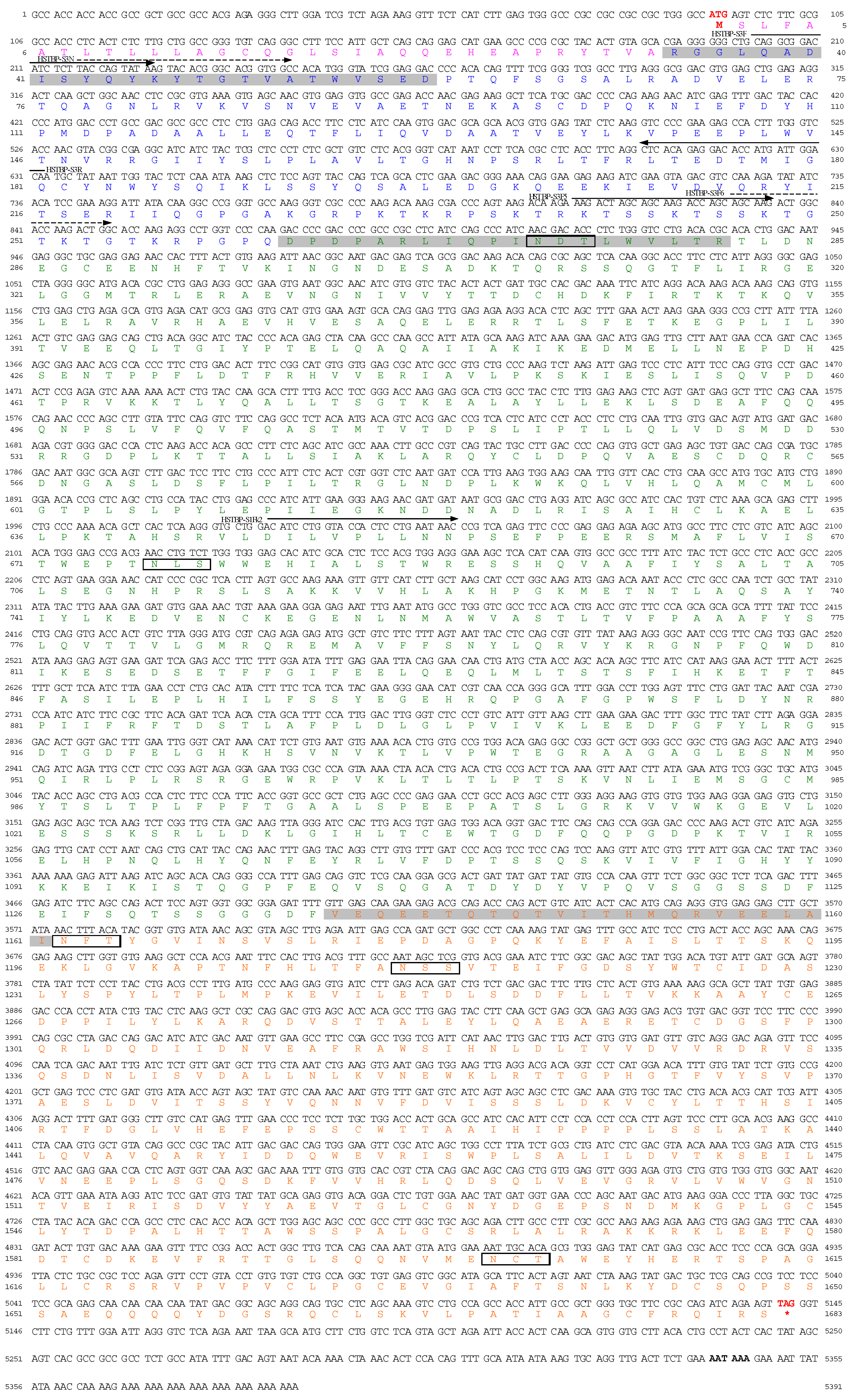
Degenerate primers were designed based on the amino acid sequence of the subfragments of HSTBP and are shown in Table 2. The forward primer, HSTBP-S3F, and reverse primer, a Marathon adaptor primer (AP1), were used for the 3′ rapid amplification of cDNA ends [RACE] (Thermo Fisher Scientific, Waltham, MA, USA). Amplification was carried out using TaKaRa Taq HS DNA polymerase (Takara Bio Inc., Shiga, Japan) under the following conditions: 35 cycles of 98 °C for 10 s, X °C for 30 s, and 72 °C for 2.5 min, in which the annealing temperature, X was adapted between 50 and 65 °C. The subsequent nested PCR used the degenerate primers HSTBP-S3N (forward primer) and AP2-2 (reverse primer) for 3′RACE. 5′RACE fragment of subunit 3 of HSTBP (HSTPB-sub3) was amplified with HSTBP-S3R and AP1, using a Mighty Amp DNA polymerase (Takara Bio Inc.) under the following conditions: 98 °C for 2 min and 35 cycles of 98 °C for 10 s, 59 °C for 15 s, and 68 °C for 2 min. The downstream sequences were determined by 3′RACE using forward primers (HSTBP-S3F5, HSTBP-S3F6, and HSTBP-S1Fk2) and reverse primer, AP1, respectively. The PCR products were subcloned into the pT7Blue T-vector (Novagen, Madison, WI, USA) and cloned into Escherichia coli JM109 competent cells (Nippon Gene Co., Ltd., Tokyo, Japan). The cloned plasmids were sequenced using a BigDye Terminator v3.1 Cycle Sequencing Kit (Thermo Fisher Scientific) and 3130 Genetic Analyzer (Thermo Fisher Scientific). The strategy used for HSTBP cDNA cloning is illustrated in Figure S1.

2.5. Computational Analyses

The amino acid sequences were analyzed for similarity using the standard protein BLAST search (https://blast.ncbi.nlm.nih.gov/Blast.cgi, accessed on 14 December 2022). Signal peptides in the N-terminal segments of HSTBP and other clotting proteins were predicted using the SignalP 6.0 server (https://services.healthtech.dtu.dk/service.php?SignalP, accessed on 14 December 2022) [25]. Multiple amino acid sequence alignments of HSTBP and other clotting proteins were performed with the Clustal W algorithm [26] using the MEGA XI software (https://www.megasoftware.net/, accessed on 14 December 2022) [27]. Molecular Evolution Genetics Analyses Version XI) software [27]. The theoretical molecular mass and electric point (pI) were estimated using DNASIS pro (Hitachi Software Engineering, Tokyo, Japan). The SMART program (http://smart.embl-heidelberg.de/, accessed on 14 December 2022) [28] was used to determine the conserved domains of HSTBP.
The three-dimensional structures of HSTBP sub-2 (Val1139-Ser1683) and vWF type D domain TfVWF from T. flavidus were predicted using Colabfold (version 1.4.0) [29]. Structural models were viewed using PyMOL.

2.6. Recombinant Protein of TTX-Binding Protein Subunit 2 (rHSTBP-sub2) and Its Binding with TTX

A partial gene fragment corresponding to HSTBP subunit 2 (HSTBP sub-2) was synthesized based on the cDNA sequence (accession number LC733238) using an artificial gene synthesis system (Eurofins Genomics K.K., Tokyo, Japan). Codon usage of the fragment sequence was optimized for E. coli. To insert the restriction enzyme sites, the fragment was amplified by PCR using primers containing NdeI (forward primer, 5′-GTCGTCATATGGTCGAACAGGAAGAAAC-3′) and BamH1 (reverse primer, 5′-CAGCCGGATCCTTAACTGCGGATTTGGCGAAAAC-3′) recognition sequences. The amplicon was cloned into the pGEM®-T vector (Promega, Madison, WI, USA) and transformed into DH5α competent E. coli (Nippon gene Co., Ltd.) following the manufacturer’s instructions. The plasmid and empty pET16b vector (Merck Millipore, Burlington, MA, USA) were digested with NdeI and BamHI (Nippon Gene Co., Ltd.). Both the fragment and linearized pET16b vector were ligated and transformed into DH5α competent E. coli (Nippon gene Co., Ltd.) to produce the expression construct.
The expression construct was transformed into Rosetta-Gami B (DE3) pLysS E. coli (Merck Millipore). rHSTBP sub-2 was overexpressed by incubation at 37 °C for 4 h in a shaking incubator (150 rpm) with 100 µg/mL of ampicillin, 34 µg/mL of chloramphenicol, and 0.3 mM isopropyl β-D-thiogalactopyranoside (IPTG) after 2 h-preculture in the same conditions, except for IPTG. The cells were harvested by centrifugation (3000× g, 4 °C, 20 min) and the inclusion body was collected by centrifugation (10,000× g, 4 °C, 15 min) after treatment with 0.15 M NaCl-0.05 M Tris-HCl buffer (pH 8.0) containing 1% Triton-X100, 0.001% RNase, and 0.001% DNase I (Nippon gene). The inclusion body was solubilized with 6 M guanidine hydrochloride-0.15 M NaCl-0.05 M Tris-HCl buffer (pH 8.0) and applied to a Ni-IMAC resin (Bio-Rad Laboratories, Hercules, CA, USA). The recombinant protein was eluted with 0.5 M imidazole-0.15 M NaCl-0.05 M Tris-HCl buffer (pH 8.0) after washing with 0.025 M imidazole-0.15 M NaCl-0.05 M Tris-HCl buffer (pH 8.0).
The rHSTBP sub-2 fraction was reduced with β-mercaptoethanol and refolded by a dilution and dialysis as follows: the protein fraction was added dropwise into 25 volumes of the saline buffer containing 0.5 M L-arginine, 1 mM glutathione, and 0.1 mM glutathione disulfide with stirring. The diluted fraction was dialyzed twice against 0.5 M L-arginine-0.15 M NaCl-0.05 M Tris-HCl buffer (pH 8.0) and ultrafiltered with an Ultracel 5 kDa ultrafiltration disc (Merck Millipore) to replace the buffer by 0.02 M Tris-acetate buffer (pH 7.4). The rHSTBP sub-2 was mixed with TTX and allowed to stand for 3 h at 4 °C. The mixture was applied to a Sephacryl S-300 column (2 × 29 cm, GE Healthcare Bio-Sciences) and eluted with 0.02 M Tris-acetate buffer (pH 7.4) at a flow rate of 0.5 mL/min. The eluate was fractionated in a 4-mL portion. Each 1 mL aliquot of the fractions was mixed with 10 µL of acetic acid and heated in a boiling water bath for 10 min to liberate TTX bound to rHSTBP sub-2. TTX was analyzed by LC-MS/MS, as described below.

2.7. Measurement of TTX Binding Activity

For the hemolymph, 500-µL aliquots of the samples were mixed with 20 µg TTX and allowed to stand for 60 min at 4 °C. The mixture was ultrafiltered with an Amicon Ultra-0.5 mL centrifugal filter Ultracel-3K (nominal molecular weight cut-off 3000, Merck Millipore) to remove free TTX. Milli-Q water was added to the centrifugal filter, and ultrafiltration was repeated twice to concentrate to 100 µL. TTX bound to the TTX-binding protein was separated from the protein by adding 100 µL of 0.1% (v/v) acetic acid and heating in a boiling water bath for 10 min.
For the active fractions from the purification steps, 100-µL aliquots of the fractions were mixed with an equal volume of 0.1% (v/v) acetic acid and heated in a boiling water bath for 10 min.
TTX was determined using a Waters Acquity UPLC and TDQ triple-quadrupole tandem mass spectrometry (Waters, Midford, MA, USA) [30]. A TSKgel Amide-80 column (2.0 × 150 mm, 3-µm particle size; Tosoh, Tokyo, Japan) was maintained at 25 °C and eluted with 0.016 M ammonium formate (pH 5.5): acetonitrile (40:60, v/v) at a flow rate of 0.2 mL/min. The eluate was introduced into the ion source of electrospray ionization-mass spectrometry, ionized by the positive mode, and detected in multiple reaction monitoring mode, m/z 320 > 162, with a collision energy of 45 eV.
Student’s t-test was used to test for significant differences, with a significance level of 5%.

2.8. Analytical Methods

The molecular mass of the isolated TTX-binding protein was determined by gel filtration HPLC on a TSKgel G300SWXL column (0.78 × 30 cm, Tosoh) with 0.5 M NaCl-0.01 M sodium phosphate buffer (pH 7.4) at a flow rate of 0.5 mL/min. Four reference proteins (Bio-Rad Laboratories), thyroglobulin (670 k), bovine gamma globulin (158 k), chicken ovalbumin (44 k), and equine myoglobin (17 k) were used to calibrate the column. TTX-binding protein was estimated by SDS-PAGE on a C520L gel (Atto). Prior, the protein was dissolved in 2× buffer containing 0.1 M dithiothreitol and 2% SDS and heated in a boiling water bath for 5 min. The proteins were stained with Rapid CBB KANTO (Kanto Chemical Co., Inc., Tokyo, Japan). Precision Plus Protein Standards (Bio-Rad Laboratories) were used as the reference. Native-PAGE was performed using a C7.5 gel (Atto).
Protein levels were measured using bovine serum albumin as a standard protein using the Lowry method [31]. The protein concentration of rHSTBP sub-2 eluted by Sephacryl S-300 column chromatography was measured with a protein assay kit using a Qubit 4 fluorometer (Thermo Fisher Scientific).

3. Results

3.1. TTX-Binding Activity of the H. sanguineus Hemolymph

As shown in Table 1, all the hemolymph samples used were bound to TTX. TTX-binding activity ranged from 1.20 to 1.55 µg TTX/mL. There was no difference in TTX-binding activity among individuals or between female and male samples (p > 0.05). The former ranged from 1.20 to 1.40 µg TTX/mL (mean ± S.D. 1.32 ± 0.09 µg TTX/mL) and the latter from 1.20 to 1.55 µg TTX/mL (mean ± S.D. 1.36 ± 0.15 µg TTX/mL). No relationship was observed between TTX-binding activity and crab size (carapace width).

3.2. Isolation of TTX-Binding Protein

TTX-binding protein was isolated using five purification steps. In the first step of salting out, TTX-binding protein was recovered from the 30 to 50% saturation ammonium sulfate fraction. The TTX-binding protein was subjected to lectin affinity chromatography on a Con A-Sepharose column and eluted in the methyl-α-D-mannopyranoside eluate fraction (data not shown). Next, the active fraction containing the TTX-binding protein was chromatographed by anion-exchange HPLC on a MonoQ 5/50 GL column and eluted with 0.6–0.8 M NaCl in 0.01 M sodium phosphate buffer (pH 7.4) (data not shown). Finally, the TTX-binding protein was re-chromatographed using a MonoQ 5/50 HPLC after preparative electrophoresis. TTX-binding protein was detected at retention times of 44–46 min (Fraction I) and 48–52 min (Fraction II) (Figure 1). Native PAGE analysis revealed that both fractions produced only one band, confirming their homogeneity (Figure 1). Fraction I showed the TTX-binding activity of 0.22 µg TTX/mg protein and Fraction II of 0.33 µg TTX/mg protein. Because Fraction II had higher TTX-binding activity and more TTX-binding protein than Fraction I, Fraction II was identified as H. sanguineus TTX-binding protein (HSTBP) and used in further experiments.

3.3. Characterization of TTX-Binding Protein, HSTBP

The molecular mass of the isolated TTX-binding protein, HSTBP, was measured at 400 kDa using gel filtration HPLC on a TSKgel G3000SWXL column (Figure 2A,B). In contrast, the molecular masses estimated by SDS-PAGE were approximately 88, 65, and 26 kDa (Figure 2C). These results suggested that HSTPB has a dimeric conformation consisting of three subunits with molecular masses of 88 (HSTBP sub-1), 65 (HSTPB sub-2), and 26 kDa (HSTBP sub-3).
Based on the chromatographic results of the purification procedures, HSTBP is most likely an acidic glycoprotein. HSTBP was adsorbed onto an anion exchange MonoQ 5/50 GL column. It was also retained on the lectin affinity Con A-Sepharose column and eluted with an eluent containing methyl-α-D-mannopyranoside. The molecular mass of HSTBP sub-2 (65 kDa) was decreased to 63 kDa after digestion with the glycopeptidase, PNGase F (Figure 2D), indicating the presence of an N-linked carbohydrate side chain. However, that of the other two subunits, HSTBP sub-1 (88 kDa) and HSTBP sub-3 (26 kDa), did not change.

3.4. cDNA Cloning and Molecular Properties of HSTBP

The N-terminal amino acid sequences of HSTBP sub-1, sub-2, and sub-3 were determined as 1DPDPARLIQPINDTLWVLTR20, 1VEQEETQTQTVITHMQRVEELAINF25, and 1RGGLQADISYQYKYTGTVATWVSED25, respectively (Table 3).
A cDNA fragment was amplified by 3′RACE using the degenerate primers HSTBP-S3F and HSTBP-S3N. These were designated from the amino acid sequence of HSTBP sub-3 as forward primers and AP1 and AP2 as reverse primers (Figure S1). Based on the nucleotide sequence of the PCR product (682 bp), the remaining 5′region sequence was determined by 5′RACE using AP1 and HSTBP-S3R. The PCR product (632 bp) contained a 5′-untranslated region (Figure 3). Next, a cDNA fragment was amplified by 3′RACE using two primer sets: HSTBP-S3F5/AP1 and HSTPB-S3F6/AP1. A PCR product of 1289 bp was obtained, which included the nucleotides encoding 20-amino acids of the N-terminus of HSTBP sub-1. The remaining 3′region was identified by 3′RACE using HSTBP-S1Fk2 and AP1 primers. The 3364 bp PCR product was obtained, which contained 23-amino acid residues of the N-terminus of HSTBP sub-2 and 3′-untranslated region. Here, the nucleotide sequence of the full-length HSTBP cDNA (5391 bp) was determined (accession number LC733238). The accuracy of this sequence was verified using re-cloning experiments. The initial ATG codon is present at nucleotides 91–93. In the 3′-untranslated region, a polyadenylation signal AAT AAA was identified in nucleotides 5341–5346, and a poly(A) tail at nucleotide 5368. An open reading frame (ORF) comprised of 5049 bp, encoding a precursor protein of 1683 amino acid residues from the putative initiating Met1 to the putative last Ser1683 was also identified. The amino acid sequences of the N-terminal proteins of HSTBP sub-1, HSTBP sub-2, and HSTBP sub-3, determined by protein sequencing, were all found at positions 34–58 (HSTBP sub-3), 262–281 (HSTBP sub-1), and 1139–1163 (HSTBP sub-2) of the deduced molecule (Figure 3).
In Figure 4, the deduced amino acid sequences of the TTX-binding protein were aligned with the clotting proteins of shrimp and crab from the suborder Pleocyemata. SignalP-6.0 Server predicted that HSTBP comprised a signal peptide (Met1 to Gly17) and a mature protein starting with Leu18. However, HSTBP sub-3 began with Arg34, suggesting the absence of a peptide from Leu18 to Ala33 by translational modification. The mature protein of HSTBP likely consisted of 1650 amino acid residues from Arg34 to Ser1683. The theoretical molecular masses of HSTBP and its subunits (HSTBP sub-1, HSTBP sub-2, and HSTBP sub-3) were calculated at 185,080.6 Da, 98,822.3 Da, 60,784.6 Da, and 25,509.7 Da, respectively, using the DNASIS Pro (Hitachi Software Engineering). The isoelectric point (pI) of HSTBP was estimated to be 5.26 using DNASIS pro (Hitachi Software Engineering). A BLAST homology search showed weak identity of the deduced amino acid sequence of HSTBP to clotting proteins of crustaceans, Portunus trituberculatus (accession number XP_045118682.1) (40.2%), Scylla paramamosain (accession number AYK27566.1) (39.9%), Pacifastacus leniusculus (accession number AAD16454.1) (29.9%), Homarus americanus (accession number KAG7156252.1) (29.1%), and Procambarus clarkii (accession number AYD41596.1) (29.1%).
Analysis using the SMART program showed a lipoprotein N-terminal domain at Leu37-Thr704 and vWF type D domain at Phe1387-Gly1544. A three-dimensional structure model of HSTBP sub-2 containing the vWF type D domain was constructed using Colabfold (Figure 5A). It comprised three independent domains (N-domain, M-domain, and C-domain), and the M-domain corresponded to the vWF type D domain (Figure 5A). Figure 5B compares the 3D structural models of the vWF type D domains between HSTBP sub-2 and TfVWF from the pufferfish Takifugu flavidus liver [18]. The key amino acids Asp115, Val117, and Lys122 in TfVWF were predicted to mediate binding to TTX using in silico structural and docking analyses [18]. However, they were not conservative in the HSTBP sub-2.

3.5. Binding of rHSTBP-sub2 with TTX

The rHSTBP-sub2 was successfully produced by the E. coli expression system and solubilized using the L-arginine-aided refolding procedure. It produced a single band on SDS-PAGE with a molecular mass of 60 kDa, indicating its homogeneity (Figure 6A). The rHSTBP sub-2 was combined with TTX and subjected to gel filtration chromatography on a Sephacryl S-300 column. The elution profile is shown in Figure 6B. TTX was detected in two fractions, Fr. 11 and Frs. 24–45. The former fraction contained TTX bound to a high molecular weight substance, rHSTBP-sub2, and the latter fraction contained TTX in free form. Based on the concentrations of proteins and TTX in Fr. 11 and the molecular masses of rHSTBP sub-2 (60 kDa) and TTX (319 Da), TTX-binding activity of rHSTBP sub-2 was measured at 5.51 µg TTX/mg, equivalent to 1:1 stoichiometry TTX/protein.

4. Discussion

We isolated the TTX-binding protein HSTBP from the hemolymph of H. sanguineus by salting out with ammonium sulfate, lectin affinity chromatography on a Con A-Sepharose column, ion-exchange HPLC on a MonoQ 5/50 GL column, and preparative electrophoresis. HSTBP is an acidic glycoprotein with a molecular mass of 400 kDa by gel filtration HPLC on a TSKgel G3000SWXL. This protein comprised three subunits of 88 (subunit-1), 65 (subunit-2), and 26 kDa (subunit-3) by SDS-PAGE under reduced conditions, suggesting that HSTBP is a homodimer. We elucidated the primary structure of the protein using cDNA cloning. The ORF of the cDNA consisted of 5049 bp encoding 1683 amino acid residues, and the mature protein contained 1650 amino acid residues from Arg34 to Ser1683; cDNA cloning showed that the three subunits were arranged in tandem in the following order: subunit-3 (Arg34-Gln261 with a calculated molecular mass of 25,509.7), subunit-1 (Asp262-Phe1138 with the calculated molecular mass of 98,822.3), and subunit-2 (Val1139-Ser1683 with the calculated molecular mass of 60,784.6). Subunit-1 may be cleaved by post-translational modification because the molecular mass estimated by SDS-PAGE was approximately 10 kDa smaller than that calculated. Subunit-2 may have an N-type sugar chain because the molecular mass estimated by SDS-PAGE was higher than that predicted and decreased in molecular mass by deglycosylation with PNGase F. Furthermore, the subunits may have been bound by disulfide bonds. On SDS-PAGE, HSTBP segregated into three subunits under reduced conditions, while it did not enter the polyacrylamide gel under non-reduced conditions due to aggregation. These results imply the formation process of HSTBP as follows: after translation, the TTX-binding protein (Met1-Ser1683) with a molecular mass of 188,565.4 is cleaved off the signal peptide (Met1-Gly17) and the subsequent peptide (Leu18-Ala33), glycosylated with the subunit-2 region, separated into three subunits, and cleaved off the C-terminal region of subunit-1. These modified subunits may then rearrange via disulfide bonds to form dimers.
A previous study determined the molecular mass of the TTX-binding protein to be approximately 400 kDa by gel filtration on a TSKgel G3000SW and approximately 72 and 82 kDa by SDS-PAGE, indicating that the TTX-binding protein comprised at least two kinds of subunits with molecular masses of approximately 72 and 82 kDa [21]. In contrast, this study showed three subunits with molecular masses of 88, 65, and 26 kDa on SDS-PAGE; cDNA cloning verified that the TTX-binding protein comprised three subunits. The previous study might have overlooked the third subunit (subunit-3 with a molecular mass of 26 kDa in the present study) because a high-molecular-weight calibration kit containing myosin (212 kDa), macroglobulin (170 kDa), β-galactosidase (116 kDa), transferrin (96 kDa), and glutamic dehydrogenase (53 kDa) was used as a marker. The shore crab hemolymph has a TTX-binding protein other than HSTBP, as determined by ion-exchange HPLC on a MonoQ 5/50 GL column (Figure 1). This protein could be an isoform of HSTBP because it exhibited the same pattern as HSTBP on SDS-PAGE (Figure 2). Further studies are necessary to elucidate the sequences of other TTX-binding proteins.
SMART program analysis identified a vWF type D domain in HSTBP subunit 2. It has been reported that the vWF type D domain may be involved in the toxification of pufferfish. Yin et al. [17] demonstrated the presence of a pufferfish toxin-binding protein in the ovaries of T. pardalis (Takifugu pardalis ovary toxin-binding protein with a molecular mass of 10 kDa, TPOBP-10) and identified it as a vitellogenin-1-like protein [Takifugu rubripes] subdomain, a vWF type D domain. Furthermore, Qiao et al. [18] confirmed that the vWF type D domain bound TTX using a recombinant protein of TfVWF from T. flavidus liver. Surface plasma resonance analysis evaluated the weak affinity of the protein to TTX with an equilibrium dissociation constant of 2.92 × 10⁻³ M. Although kinetic analysis has not been performed in this study, it is evident that HSTBP sub-2 binds TTX at a 1:1 molar ratio. The binding mechanism of the TTX-binding protein HSTBP remains to be elucidated in the context of TfVWF in the pufferfish liver.
Barber et al. [32] observed that the small shore crab Hemigrapsus oregonesis along the coasts of British Columbia, Canada, developed resistance to STX when exposed to the red tide bloom of Gonyaulax catenella (presently Alexandrium pacificum [Group V]). In contrast, crabs did not resist TTX administration. Notably, the shore crabs belonging to the genus Hemigrasus showed differing resistance activities to the toxins between the two species of H. sanguineus and H. oregonesis: the former exclusively resisted TTX, whereas the latter resisted only STX. H. oregonesis induced the production of a STX-resistant protein complex with a molecular mass of 145 kDa in the viscera by exposure to the wild toxic dinoflagellate bloom and by artificial treatment with STX in a dose-dependent manner, suggesting the involvement of the STX-resistant protein in detoxifying activity of crabs [33]. However, the TTX-binding protein of H. sanguineus hemolymph is not likely associated with TTX toxification, but by binding the toxin. Further investigation is in progress on the disposition of the TTX-binding protein in shore crab tissues and the distribution among shore crab species to clarify the physiological role of the protein.
Otherwise, TTX-binding protein could be a promising target for developing an antidote and applied to diagnostics for TTX intoxication because of its unique properties of specific binding to TTX and neutralization against TTX toxicity. Furthermore, it would be applicable to biochemical research to purify, identify, and analyze TTX in biological samples because we previously developed affinity chromatography using partially purified TTX-binding protein as a ligand [34].

5. Conclusions

This study revealed a novel TTX-binding protein, HSTBP, from the hemolymph of non-toxic shore crab H. sanguineus. The protein comprises three subunits, Arg34-Gln261 (subunit-3), Asp262-Phe1138 (subunit-1), and Val1139-Ser1683 (subunit-2). HSTBP showed a weak similarity (29–40%) to clotting proteins of crustaceans and conserved vWF type D domain at Phe1387-Gly1544 in subunit-2. The recombinant protein of subunit-2 bound TTX at a molecular ratio of 1:1. Therefore, HSTBP may neutralize TTX and prevent the lethal toxicity of TTX. It could be applicable to possible antidotes for TTX intoxication.

Supplementary Materials

The following supporting information can be downloaded at: https://0-www-mdpi-com.brum.beds.ac.uk/article/10.3390/jmse11010181/s1, Figure S1: Strategy of 5′ RACE and 3′ RACE.

Author Contributions

Conceptualization, Y.N., Y.K. and M.O.; methodology, Y.N., Y.K., M.O. and S.I.; software, M.O., A.Y.-K. and K.F.; validation, Y.N., S.I.; formal analysis, investigation, K.F., Y.K. and Y.N.; data curation, Y.N.; writing—original draft preparation, Y.N.; writing—review and editing, S.I., M.O., Y.K. and A.Y.-K.; supervision, Y.N.; project administration, Y.N. All authors have read and agreed to the published version of the manuscript.

Funding

This study was partly supported by Fundamental Research (C) 19K06241, grants-in-aid for scientific research from the Ministry of Education, Culture, Sports, Science and Technology, Japan. This study was partly performed under the cooperative research program Institute of Nature and Environmental Technology, Kanazawa University <17024>.

Institutional Review Board Statement

Not applicable.

Informed Consent Statement

Not applicable.

Data Availability Statement

The data presented in this study are available on request from the corresponding author.

Conflicts of Interest

The authors declare no conflict of interest.

References

  1. Lee, C.H.; Rube, P.C. Interaction between voltage-gated sodium channels and the neurotoxin, tetrodotoxin. Channels 2008, 2, 407–412. [Google Scholar] [CrossRef] [PubMed]
  2. Bucciarelli, G.M.; Lechner, M.; Fontes, A.; Kats, L.B.; Eisthen, H.L.; Shaffer, H.B. From poison to promise: The evolution of tetrodotoxin and its potential as a therapeutic. Toxins 2021, 13, 517. [Google Scholar] [CrossRef] [PubMed]
  3. Bane, V.; Lehane, M.; Dikshit, M.; O’Riordan, A.; Furey, A. Tetrodotoxin: Chemistry, toxicity, source, distribution and detection. Toxins 2014, 6, 693–755. [Google Scholar] [CrossRef] [PubMed] [Green Version]
  4. Lago, J.; Rodriguez, L.P.; Blanco, L.; Vieites, J.M.; Cabado, A.G. Tetrodotoxion, an extremely potent marine neurotoxin: Distribution, toxicity, origin and therapeutical uses. Mar. Drugs 2015, 13, 6384–6406. [Google Scholar] [CrossRef] [PubMed]
  5. Saito, T.; Noguchi, T.; Harada, T.; Murata, O.; Abe, T.; Hashimoto, K. Resistibility of toxic and nontoxic pufferfish against tetrodotoxin. Bull. Jap. Soc. Sci. Fish. 1985, 51, 1371. [Google Scholar] [CrossRef] [Green Version]
  6. Noguchi, T.; Arakawa, O. Tetrodotoxin-Distribution and accumulation in aquatic organs, and cases of human intoxication. Mar. Drugs 2008, 6, 220–242. [Google Scholar] [CrossRef] [Green Version]
  7. Kodama, M.; Sato, S. Standard Methods of Analysis in Food Safety Regulation, Chemistry, Ministry of Health Labour and Welfare; Japan Food Hygiene Association: Tokyo, Japan, 2015; pp. 813–820. (In Japanese) [Google Scholar]
  8. Koyama, K.; Noguchi, T.; Uzu, A.; Hashimoto, K. Resistibility of toxic and nontoxic crabs against paralytic shellfish poison and tetrodotoxin. Bull. Jap. Soc. Sci. Fish. 1983, 49, 485–489. [Google Scholar] [CrossRef]
  9. Shiomi, K.; Inaoka, H.; Yamanaka, H.; Kikuchi, T. Occurrence of a large amount of gonyautoxins in a xanthid crab Atergatis floridus from Chiba. Bull. Jap. Soc. Sci. Fish. 1982, 48, 1407–1410. [Google Scholar] [CrossRef]
  10. Noguchi, T.; Uzu, A.; Koyama, K.; Maruyama, J.; Nagashima, Y.; Hashimoto, K. Occurrence of tetrodotoxin as the major toxin in a xanthid crab Atergatis floridus. Bull. Jap. Soc. Sci. Fish. 1983, 49, 1887–1892. [Google Scholar] [CrossRef] [Green Version]
  11. Noguchi, T.; Koyama, K.; Uzu, A.; Hashimoto, K. Local variation of toxicity and toxin composition in a xanthid crab Atergatis floridus. Bull. Jap. Soc. Sci. Fish. 1983, 49, 1883–1886. [Google Scholar] [CrossRef]
  12. Lu, J.; Zheng, J.; Xu, Q.; Chen, K.; Zhang, C. Adaptive evolution of the vertebrate skeletal muscle sodium channel. Gen. Mol. Biol. 2011, 34, 323–328. [Google Scholar] [CrossRef] [PubMed] [Green Version]
  13. Chen, R.; Chung, S.-H. Mechanism of tetrodotoxin block and resistance in sodium channels. Biochem. Biophys. Res. Commun. 2014, 446, 370–374. [Google Scholar] [CrossRef]
  14. Matsui, T.; Yamamori, K.; Furukawa, K.; Kono, M. Purification and some properties of a tetrodotoxin binding protein from the blood plasma of kusafugu, Takifuru niphobles. Toxicon 2000, 38, 463–468. [Google Scholar] [CrossRef]
  15. Yotsu-Yamashita, M.; Sugimoto, A.; Terakawa, T.; Shoji, Y.; Miyazawa, T.; Yasumoto, T. Purification, characterization, and cDNA cloning of a novel soluble saxitoxin and tetrodotoxin binding protein from plasma of the puffer fish, Fugu paradalis. Eur. J. Biochem. 2001, 268, 5937–5946. [Google Scholar] [CrossRef]
  16. Yotsu-Yamashita, M.; Yamaki, H.; Okoshi, N.; Araki, N. Distribution of homologous proteins to puffer fish saxitoxin and tetrodotoxin binding protein in the plasma of puffer fish and among the tissues of Fugu pardalis examined by Western blot analysis. Toxicon 2010, 55, 1119–1124. [Google Scholar] [CrossRef]
  17. Yin, X.; Kiriake, A.; Ohta, A.; Kitani, Y.; Ishizaki, S.; Nagashima, Y. A novel function of vitellogenin subdomain, vWF type D, as a toxin-binding protein in the pufferfish Takifugu pardalis ovary. Toxicon 2017, 136, 56–66. [Google Scholar] [CrossRef] [PubMed]
  18. Qiao, K.; Jaing, C.; Xu, M.; Chen, B.; Qiu, W.; Su, Y.; Hao, H.; Lin, Z.; Cai, S.; Su, J.; et al. Molecular characterization of the von Willebrand factor type D domain of vitellogenin from Takifugu flavidus. Mar. Drugs 2021, 19, 181. [Google Scholar] [CrossRef]
  19. Qiao, K.; Wang, C.; Huang, L.; Feng, H.; Chen, B.; Xu, M.; Su, Y.; Liu, S.; Pan, N.; Su, J.; et al. Molecular characterization of a new tetrodotoxin-binding protein, peroxiredoxin-1, from Takifuru bimaculatus. Intl. J. Mol. Sci. 2022, 23, 3071. [Google Scholar] [CrossRef]
  20. Hwang, P.-A.; Tsai, Y.-H.; Lin, H.-P.; Hwang, D.-F. Tetrodotoxin-binding protein isolated from five species of toxic gastropods. Food Chem. 2007, 103, 1153–1158. [Google Scholar] [CrossRef]
  21. Nagashima, Y.; Yamamoto, K.; Shimakura, K.; Shiomi, K. A tetrodotoxin-binding protein in the hemolymph of shore crab Hemigrapsus sanguineus: Purification and properties. Toxicon 2002, 40, 753–760. [Google Scholar] [CrossRef]
  22. Yamamori, K.; Yamaguchi, S.; Maehara, E.; Matsui, T. Tolerance of shore crabs to tetrodotoxin and saxitoxin and antagonistic effect of their body fluid against the toxins. Nippon. Suisan Gakkaishi 1992, 58, 1157–1162. [Google Scholar] [CrossRef] [Green Version]
  23. Shiomi, K.; Yamaguchi, S.; Kikuchi, T.; Yamamori, K.; Matsui, T. Occurrence of tetrodotoxin-binding high molecular weight substances in the body fluid of shore crab (Hemigrapsus sanguineus). Toxicon 1992, 30, 1529–1537. [Google Scholar] [CrossRef]
  24. Nagashima, Y.; Toyoda, M.; Hasobe, M.; Shimakura, K.; Shiomi, K. In vitro accumulation of tetrodotoxin in pufferfish liver tissue slices. Toxicon 2003, 41, 569–574. [Google Scholar] [CrossRef]
  25. Petersen, T.N.; Brunak, S.; von Heijne, G.; Nielsen, H. SignalP 40: Discriminating signal peptides from transmembrane regions. Nat. Methods 2011, 8, 785–786. [Google Scholar] [CrossRef] [PubMed]
  26. Thompson, J.D.; Higgins, D.G.; Gibson, T.J. CLUSTAL W: Improving the sensitivity of progressive multiple sequence alignment through sequence weighting, position-specific gap penalties and weight matrix choice. Nucleic Acids Res. 1994, 22, 4673–4680. [Google Scholar] [CrossRef] [Green Version]
  27. Kumar, S.; Stecher, G.; Li, M.; Knyaz, C.; Tamura, K. MEGA X: Molecular evolutionary genetics analysis across computing platforms. Mol. Biol. Evol. 2018, 35, 1547–1549. [Google Scholar] [CrossRef] [PubMed]
  28. Letunic, I.; Bork, P. 20 years of the SMART protein domain annotation resource. Nucleic Acids Res. 2018, 46, D493–D496. [Google Scholar] [CrossRef]
  29. Mirdita, M.; Schütze, K.; Moriwaki, Y.; Heo, L.; Ovchinnikov, S.; Steinegger, M. ColabFold: Making protein folding accessible to all. Nat. Methods 2022, 19, 679–682. [Google Scholar] [CrossRef]
  30. Nagashima, Y.; Ohta, A.; Yin, X.; Ishizaki, S.; Matsumoto, T.; Doi, H.; Ishibashi, T. Difference in the uptake of tetrodotoxin and saxitoxins into liver tissue slices among pufferfish, boxfish and porcupinefish. Mar. Drugs 2018, 16, 17. [Google Scholar] [CrossRef] [Green Version]
  31. Lowry, O.H.; Rosebrough, N.J.; Farr, A.L.; Randall, R.L. Protein measurement with the Folin phenol reagent. J. Biol. Chem. 1951, 193, 256–275. [Google Scholar] [CrossRef]
  32. Barber, K.G.; Kitts, D.D.; Townsley, P.M. Response of the shore crab Hemigrapsus oregonesis to saxitoxin injections. Bull. Environ. Contam. Toxicol. 1988, 40, 190–197. [Google Scholar] [CrossRef] [PubMed]
  33. Barber, K.G.; Kitts, D.D.; Townsley, P.M.; Smith, D.S. Appearance and partial purification of a high molecular weight protein in crabs exposed to saxitoxin. Toxicon 1988, 26, 1027–1034. [Google Scholar] [CrossRef] [PubMed]
  34. Nagashima, Y.; Watanabe, K.; Arita, J.; Shimakura, K.; Shiomi, K. Affinity chromatography using tetrodotoxin-binding high molecular weight substances in the shore crab Hemigrapsus sanguineus as ligands: Usefulness in analyzing tetrodotoxin in biological samples. J. Nat. Toxins 1997, 6, 293–306. [Google Scholar]
Figure 1. MonoQ anion-exchange HPLC of active fraction obtained from preparative electrophoresis. Sample: active fraction obtained from preparative electrophoresis; column: MonoQ5/50 GL (0.5 × 5 cm); mobile phase: 0.2–2.0 M NaCl-0.01 M sodium phosphate buffer (pH 7.4); flow rate: 0.5 mL/min; detection: absorbance at 280 nm; inlet: Native-PAGE; I; peak I obtained from Mono Q anion-exchange HPLC, II; peak II obtained from MonoQ anion-exchange HPLC.
Jmse 11 00181 g001
Figure 2. Molecular mass of TTX-binding protein: (A) gel filtration HPLC on a TSKgel G3000SWXL column (0.78 × 30 cm); (B) calibration curve for the determination of TTX-binding proteins by gel filtration HPLC; ●: TTX-binding protein, ◇: standard proteins, a: thyroglobulin (670 kDa), b: bovine γ-globulin (158 kDa), c: chicken ovalbumin (44 kDa), d: equine myoglobin (17 kDa); (C) SDS-PAGE. I; peak I obtained from MonoQ anion-exchange HPLC, II; peak II obtained from MonoQ anion-exchange HPLC; ①, ②, and ③ indicate subunits-1, -2, and -3, respectively; (D) SDS-PAGE before (−) and after (+) deglycosylation treatment with PNGase F; ①, ②, and ③ indicate subunits-1, -2, and -3, respectively. Transferrin was used as a control of N-glycosylated protein.
Jmse 11 00181 g002
Figure 3. Nucleotide and deduced amino acid sequence of cDNA. N-terminal amino acid sequences of TTX-binding protein subunits are shown in gray. Underlines indicate primers. Protein N-glycosylation site is boxed. Amino acids marked in magenta, blue, green, and red represent signal peptides and cleaved peptides, subunit 3, subunit 1, and subunit 2, respectively.
Jmse 11 00181 g003
Figure 4. Deduced amino acid sequence alignment of TTX binding protein, HSTBP, with clotting proteins of shrimp and crab from the suborder Pleocyemata. Accession numbers for clotting proteins (DDBJ/EMBL/GenBank databases): XP_045118682.1 for Portunus trituberculatus; AYK27566.1 for Scylla paramamosain; KAG7156252.1 for Hormarus americanus; AAD16454.1 for Pacifastacus leniusculus; AYD41596.1 for Procambarus clarkii. The residues identical with those of HSTBP are shown by gray and gaps by dashes. Signal peptides are boxed in straight lines. Cysteine residues are highlighted in white on a black background. Putative N-glycosylation sites are indicated by closed circles. Amino acid numbers are shown at the left and right.
Jmse 11 00181 g004
Figure 5. (A) Three-dimensional structure of HSTBP sub-2; (B) three-dimensional structures of vWF type D domain of HSTBP sub-2 (left) and TfVWF (right).
Jmse 11 00181 g005
Figure 6. Recombinant protein of TTX-binding protein subunit-2, rHSTBP sub-2: (A) SDS-PAGE, lane 1: marker; lane 2: sample; white arrow indicates recombinant protein of TTX-binding protein subunit-2, rHSTBP sub-2; (B) Sephacryl S-300 column chromatography; Sample: recombinant protein of TTX-binding protein subunit-2, rHSTBP sub-2, mixed with TTX; column: Sephacryl S-300 (2 × 29 cm); mobile phase: 0.02 M Tris-acetic acid (⇒Change to ”acetate”) buffer (pH 7.4); flow rate: 0.5 mL/min. Fraction: 4 mL/Fr.
Jmse 11 00181 g006
Table 1. TTX-binding activity of the shore crab hemolymph sample.
Sample No.SexCarapace Width (cm)TTX-Binding Ability (µg TTX/mL)
1Female2.71.20
2Female2.51.25
3Female3.31.31
4Female2.51.40
5Female3.11.40
6Male3.81.20
7Male3.81.28
8Male3.91.29
9Male3.51.49
10Male3.21.55
Mean ± S.D. 1.34 ± 0.12
Table 2. Primers used in this study. ⇒Need space between “Base” and “Tm ( C )”.
SequenceBaseTm (°C)
Forward primer
HSTBP-S3F5′-CARGCIGAYATIWSITAYCARTAYAA-3′2653
Nested forward primer
HSTBP-S3N5′-TAYCARTAYAARTAYACIGGIACIGTIGC-3′2957
5′-RACE reverse primer
HSTBP-S3R5′-TGTCCAATCATGGTGTCCTCTGTGAG-3′2661
3′-RACE forward primer
HSTBP-S3F55′-CTAGCAGCAAGACCAGCAGCAAG-3′2362
HSTBP-S3F65′-AGCAAGACTGGCACCAAGACTGG-3′2362
HSTBP-S1Fk25′-CATCCTGGTACCACTCCTGAATAACC-3′2662
Marathon adaptor primer
AP15′-CCATCCTAATACGACTCACTATAGGGC-3′2762
AP2-25′-ACTATAGGGCTCGAGCGGCCGCC-3′2367
Sequence primer
T7 promoter5′-TAATACGACTCACTATAGGG-3′2052
SP6 promoter5′-TATTTAGGTGACACTATAG-3′1947
HSTBP-S1IF5′-CAGATCACAGCGAGAACACG-3′2057
HSTBP-S1IR5′-CAGGAAGGAGTCAAGACTTGC-3′2158
HSTBP-S2IF5′-CTGCAGGTGACCACTGTCTTAG-3′2260
HSTBP-S2IR5′-CGTGACTGCTATAGTGAGTAGG-3′2258
HSTBP-S2IF25′-CTAACCAGCACAAGCTTCATCC-3′2258
HSTBP-S2IR25′-CATGATACTCCCACGCTGTG-3′2057
HSTBP-S2IF35′-GAGTACAGGCTTGTGTTTGATCC-3′2358
HSTBP-S2IR35′-AACATCCACCACAGTCAAGTCC-3′2258
Table 3. N-terminal amino acid sequences of TTX binding protein subunits.
Subunit No.Molecular Mass (kDa)Amino Acid Sequences
188DPDPARLIQPINDTLWVLTR
265VEQEETQTQTVITHMQRVEELAINF
326RGGLQADISYQYKYTGTVATWVSED
Disclaimer/Publisher’s Note: The statements, opinions and data contained in all publications are solely those of the individual author(s) and contributor(s) and not of MDPI and/or the editor(s). MDPI and/or the editor(s) disclaim responsibility for any injury to people or property resulting from any ideas, methods, instructions or products referred to in the content.

