Article

Sustainable Green Synthesis of Yttrium Oxide (Y2O3) Nanoparticles Using Lantana camara Leaf Extracts: Physicochemical Characterization, Photocatalytic Degradation, Antibacterial, and Anticancer Potency

by
Rajakumar Govindasamy
1,
Mydhili Govindarasu
2,
Salman S. Alharthi
3,
Preeyanghaa Mani
4,
Neppolian Bernaurdshaw
4,
Thandapani Gomathi
5,
Mohammad Azam Ansari
6,
Mohammad N. Alomary
7,
Banan Atwah
8,
M. Shaheer Malik
9,
V. Devi Rajeswari
10,
Kaliaperumal Rekha
11,
Saleh A. Ahmed
9,12,* and
Muthu Thiruvengadam
13,14,*
1
Collaborative Innovation Center for Advanced Organic Chemical Materials Co-Constructed by the Province and Ministry, Ministry of Education Key Laboratory for the Synthesis and Application of Organic Functional Molecules and College of Chemistry and Chemical Engineering, Hubei University, Wuhan 430062, China
2
Molecular Oncology Laboratory, Department of Biochemistry, Periyar University, Salem 636011, Tamil Nadu, India
3
Department of Chemistry, College of Science, Taif University, P.O. Box 110999, Taif 21944, Saudi Arabia
4
Nanotechnology & SRM Research Institute, SRM Institute of Science and Technology, Kattankulathur, Chennai 603203, India
5
Department of Chemistry, D.K.M. College for Women, Vellore 632001, Tamil Nadu, India
6
Department of Epidemic Disease Research, Institute for Research and Medical Consultations (IRMC), Imam Abdulrahman Bin Faisal University, Dammam 31441, Saudi Arabia
7
National Centre for Biotechnology, King Abdulaziz City for Science and Technology (KACST), P.O. Box 6086, Riyadh 11442, Saudi Arabia
8
Laboratory Medicine Department, Faculty of Applied Medical Sciences, Umm Al-Qura University, Makkah P.O. Box 7607, Saudi Arabia
9
Department of Chemistry, Faculty of Applied Sciences, Umm Al-Qura University, Makkah 21955, Saudi Arabia
10
Department of Biomedical Sciences, School of Biosciences and Technology, VIT, Vellore 632014, Tamil Nadu, India
11
Department of Environmental and Herbal Science, Tamil University, Thanjavur 613005, Tamil Nadu, India
12
Department of Chemistry, Faculty of Science, Assiut University, Assiut 71516, Egypt
13
Department of Applied Bioscience, School of Life and Environmental Science, Konkuk University, Seoul 05029, Korea
14
Department of Microbiology, Saveetha Dental College, Saveetha Institute of Medical and Technical Sciences (SIMATS), Chennai 600077, India
*
Authors to whom correspondence should be addressed.
Nanomaterials 2022, 12(14), 2393; https://0-doi-org.brum.beds.ac.uk/10.3390/nano12142393
Submission received: 9 June 2022 / Revised: 9 July 2022 / Accepted: 9 July 2022 / Published: 13 July 2022
(This article belongs to the Special Issue Nanostructured Materials for Energy Applications)

Abstract

:
Due to their appropriate physicochemical properties, nanoparticles are used in nanomedicine to develop drug delivery systems for anticancer therapy. In biomedical applications, metal oxide nanoparticles are used as powerful and flexible multipurpose agents. This work described a green synthesis of Y2O3 nanoparticles (NPs) using the sol-gel technique with the use of aqueous leaf extracts of Lantana camara L (LC). These nanoparticles were characterized with the aid of different methods, including UV, X-ray diffraction (XRD), Fourier transformed infrared spectroscopy (FTIR), transmitted electron microscopy (TEM), and photocatalytic degradation. Y2O3 nanoparticles showed excellent antibacterial activity against Gram-positive Bacillus subtilis and Gram-negative Escherichia coli with a 10 to 15 mm inhibitory zone. Green Y2O3 NPs were released with a 4 h lag time and 80% sustained release rate, indicating that they could be used in drug delivery. In addition, the bioavailability of green Y2O3 NPs was investigated using cell viability in cervical cancer cell lines. These green-synthesized Y2O3 NPs demonstrated photocatalytic degradation, antibacterial, and anticancer properties.

1. Introduction

Cervical cancer is the 4th majorly diagnosed cancer in women worldwide, with approximately 604,000 new cases and 342,000 global mortalities in 2020 [1,2,3,4]. Although the incidence of cervical cancer decreased over the last few decades, the trend in young women has been primarily influenced by a distant stage and cervical carcinoma [5,6,7]. Advanced cervical carcinoma is the most aggressive type of ovarian cancer, occurring in around 70% of cases in both developed and developing countries [8]. The major causes of cervical cancer are hormonal changes, obesity, overweight, weakened immune system, human papilloma viral infection, talcum powder, family history, and age, as identified by the International Cancer Research Agency [9]. Various methods, such as surgical therapy, chemotherapy, and radiotherapy, are used to treat cervical cancer [10,11]. Nanostructure-based diagnostics and treatment might be capable of overcoming the limitations of traditional methodologies, while simultaneously improving chemotherapeutic drug efficiency. Nanoparticles (NPs) are less than 100 nm in size and are used to shuttle drugs with specific target particles. These NPs are targeted at specific infected sites to deliver effectively targeted cancer treatments [12,13,14].
The most critical components in nanotechnology research are metal oxide nanomaterials. They are significant because of their catalytic, magnetic, electrical, and mechanical properties. Oxidative nanostructures are favorable in a wide range of thermal, photonic, mechanical, power, electrostatic, physicochemical, and gas-sensitive applications due to their enormous surface-to-volume ratios [15,16,17]. The development of profitable and low-cost ways to enhance the efficacy of anticancer therapy is urgently needed. Over the past 20 years, significant progress in nanotechnology and nanoscience has been achieved, providing us with a new perspective to overcome the drawbacks of conventional cancer therapy [18,19]. Metal oxide nanoparticles are used as powerful and flexible multipurpose agents [20]. Diverse metal oxides and hydroxide-derived nanomaterials, such as zinc oxide, copper oxide, Europium (III) hydroxide [21], and iron oxide nanoparticles [22], have been used in the production of bioengineering scaffolds [23,24].
Green synthesis is considered the most economical, sustainable, efficient, and reliable approach to nanoparticle synthesis [25,26]. This technique does not require toxic substances, high temperatures, or high pressure and does not adversely affect public health and the environment [27]. Green chemistry, which uses plant biomass or plant minerals, has become a substitute for nanoparticle non-toxic, eco-friendly, and sustainable production [28]. The development of nanomaterials thus plays a vital role in improving anti-inflammatory features with low toxicity [29]. Plants are a great source of bioactive metabolites, such as pentacyclic triterpenoids, phytosterols, polyphenols, steroids, saponins, iridoid glycosides, oligosaccharides, tannins, phenolic acids, phycobatannin, anthocyanins, and proanthocyanidins [30]. Using bioactive phytochemical compounds, scientists investigated plant chemistry, toxicology, and pharmacology [31].
Lantana camara L., commonly called “Unnichedi” (Tamil) and “pulikampa” (Telugu), is a species of flowering plant of the Verbenaceae family that is widely recognized as a significant weed in India. Lantana camara (LC) is an important plant containing alkaloids, terpenoids, phenolics, flavonoids, and other phytochemicals. L. camara is often used to treat various diseases in traditional medicine, including paludism, cancer, hypertension, tetanus, neoplasm, dermatitis, cut-offs, catarrh, abdominal viscera, measles, chickenpox, bronchitis, and fever. L. camara also has antifungal, anti-proliferative, antibacterial, nematicidal, germicidal, anti-ulcerogenic [32,33], antidiabetic [34], immunosuppressive [35], anthelminthic, anti-protozoal, antimicrobial [36], anti-inflammatory, and antipyretic activity [37].
Yttrium oxide (Y2O3), a widespread rare earth metal, is significant for future use due to its thermal stability and chemical and mechanical reliability. Yttrium oxide is used in biomedical images, materials science, synthesis of inorganic compounds, optics, electricity, biology applications [38,39], and photodynamic therapy. Furthermore, yttrium (Yb) and the widely doped rare earth metals erbium (Er) and europium (Eu) are non-cytotoxic when utilized in vivo. In this respect, Y2O3 nanoparticles of different sizes and morphologies were developed through various methods, such as sonochemical, solvothermal, hydrothermal, electrochemical, sol-gel, and thermal decomposition [16,40,41,42]. However, most of these approaches include costly and toxic chemicals as stabilizing or capping agents; their environmental applications are limited. Interestingly, a promising method to overcome these limitations was developed that involves the utilization of plant extracts.
In recent years, Y2O3 has been considered a crucial component of rare-earth substances and an excellent potential component in manufacturing optoelectronic equipment and chemical catalysis. The dielectric constant of Y2O3 is high, and the material has high thermal stability [43,44]. It can be used as a highly efficient stabilizer and functional composite materials, such as yttria-stabilized zirconia [45]. It is also widely employed as a host for rare-earth doping and is interested in potential biological and photodynamic image processing applications. Y2O3 NPs reduce oxidative stress-induced apoptosis and pancreas damage caused by diazinon in the rat pancreatic islets [46,47]. Plant metabolites can reduce cap metal ions in nanoparticles and contribute to their absorption [48]. This intrinsic ability arises from phytochemicals that could be suppressed with reduction and capping agents [49]. Therefore, L. camara leaf extract could meet the growing need for an alternative method of environment-friendly and economical synthesis of Y2O3 nanoparticles.
Photodegradation is a promising alternative because it produces hydroxyl free radicals, which can degrade many dyes [50,51,52]. The Y2O3 NPs are excellent substrates for rare earth metals and have strong luminescence effectiveness, and they could be utilized in photocatalytic therapies and biomedical diagnostics [53,54]. This study aimed to develop a new strategy for the green synthesis of Y2O3 NPs using L. camara aqueous leaf extract. Then, characterization of the products was carried out to see cell viability, biocatalytic, antibacterial, and anticancer activity in human cervical cancer HeLa cells.

2. Materials and Methods

2.1. Chemicals and Reagents

The following chemicals and reagents were purchased from commercial sources and used as received without further purification: Yttrium (III) acetate hydrate 99.0%, oleylamine of the technical grade 70%, ammonia hydroxide of reagent grade 30%, yttrium (III) nitrate of the hexahydrate 99.8%, anhydrous of the reagent grade of the ammonium hydroxide 99.5%, chloral hydrate 99.8%, n-hexane, gelatine solution, sodium hydroxide, Dulbecco modified eagle media (DMEM), fetal bovine serum (FBS), and MacConkey agar medium.

2.2. Preparation of Leaf Extracts

Lantana camara L. fresh leaves were collected and washed with tap water and then washed with double distilled water until no impurities remained. The L. camara leaves were dried in the shade for ten days to remove residual moisture. The dried leaves were pulverized in a sterile electric blender to obtain a fine powder and stored in an airtight bottle avoiding sunlight for further use. Then, 10 g of leaf powder was mixed thoroughly with 200 mL of double distilled water and heated for 10 min at 60 °C. This was followed by cooling and filtration through Whatman No.1 filter paper to afford the leaf extract [55,56]. The filtered extract was collected and kept for further studies.

2.3. Y2O3 Nanoparticles Synthesis and Characterization

Nanoparticles of Y2O3 were synthesized in the presence of gelatin through precipitation with ammonium hydroxide. In this method, 75 mL of 0.1 M yttrium nitrate hexahydrate (Y(NO3)3·6H2O) aqueous solution was added to 100 mL of ammonium hydroxide, and the solutions were mixed and stirred by REMI Electromagnetic stirrer (Chennai, Tamil Nadu, India). The precipitate formed was kept for 4 h at room temperature, washed several times with de-ionized water, centrifuged at 8000 rpm, and finally resuspended in ethanol. The obtained slurry was dried in a hot air oven for 24 h at 70 °C and pulverized with mortar and pestle. The powder was calcined to crystal-clear Y2O3 nanocrystals at 650 °C for four h [16,57]. The UV–vis spectrum was recorded with a spectrophotometer (Shimadzu UV 1800, Torrance, CA, USA) at 37 °C. XRD characterized the LC Y2O3 nanoparticles on a Bruker D8 automated multipurpose powder X-Ray Diffractometer with Cu-K radiation of wavelength 1.5406 nm. The Infrared Spectra of the LC Y2O3 NPs were obtained using an FTIR-spectrophotometer (Therma Nicolet Corp, MA, USA). In contrast, a field emission gun was used for TEM analysis at a 200 kV accelerating voltage in a FETEM apparatus (JEOL FETEM, 200 kV, Tokyo, Japan) [58].

2.4. Photocatalytic Evaluation

The photo-degradation of Rhodamine B (RhB) revealed the photoelectrocatalytic activity of the LC Y2O3 NPs. The photodegradation was examined by a 250W Xenon lamp with high pressure, with the liquid approximately 10 cm from the Xenon bulb illumination. In a traditional photodegradation procedure, 20 mg of LC Y2O3 NPs were added to a 20 mL aqueous solution of 10 µM RhB (10 ppm). To obtain dye adsorption-desorption equilibrium on the catalyst, the solution was stirred for 120 min in the dark before being subjected to visible light for different time intervals. Approximately 4mL of the solution was taken every 30 min throughout photocatalytic degradation and centrifuged at 750 rpm for 5 min to remove the excess catalytic nanoparticles. Subsequently, the amount of RhB was instantly observed using a UV-Vis spectrophotometer (Shimadzu UV 1800, Torrance, CA, USA) to determine the absorption in the spectral region of 500–750 nm [59]. Under the same conditions, the spontaneous photocatalytic degradation of RhB (i.e., photolysis) was investigated without a photocatalyst [60]. The following formula can be used to determine the photocatalytic degradation rate of the catalyst.
D e g r a d a t i o n   e f f i c i e n c y   % = C i C f C i × 100
where Ci represents the initial concentration of RhB, and Cf represents the final concentration of the dye after a specified reaction time (min).

2.5. Antibacterial Activity

The agar diffusion method was used in the microbiological experiment [46]. The antibacterial effect of the LC Y2O3 NPs was examined using microbes such as E. coli (MTCC 732) and Bacillus subtilis (MTCC 5981). The bacterial strain was acquired from the MTCC (Microbial-Type-Culture-Collection) at IMTECH in Chandigarh, India. The microbiological suspension culture was maintained for 24 h in nutrient broth; the prepared MacConkey agar medium was heated at 120 °C for 15 min, and 20 mL of sterile agar medium was poured into Petri plates. The bacterial culture was spread uniformly over agar plates using a cotton swab stick. Following inoculation, various concentrations (50, 100, 150, and 200 µmol/L) of LC Y2O3 NPs dissolved in DMSO and sonicated for 15 min were placed onto the sterile discs (6 mm). The microbes were then inoculated into discs or plates and kept at 37 °C for 24 h for bacterial species with a triplicate test performed for each microbe. The size of the inhibition zone formed on each disc was determined (mm).

2.6. Cytotoxicity Assay

Human HeLa cell lines were obtained from the National Centre for Cell Science (NCCS) in Pune, India. DMEM was used to maintain the added cells with 10% FBS. In the median, penicillin (100 μg/mL) and streptomycin (100 μg/mL) were added to avoid bacterial contamination. The HeLa cells were positioned in 24 wells (1 × 10⁵ cells/well) and incubated in a 5% CO2 incubator at 37 °C. After the cells were placed in wells and kept in the incubator for 24 h, nanoparticles were added at concentrations of 1.56, 3.12, 6.25, 12.5, 25, 50, and 100 µg/mL. The samples were then removed from the well and washed with DMEM. 3-(4,5-Dimethyl-2-thiazolyl)-2,5-diphenyl tetrazolium-bromide (0.5% MTT reagent) was added (10 μL/well) to each well and incubated for 4 h. The spectrophotometer measured the optical density at 540 nm (Shimadzu UV 1800, Torrance, CA, USA) [59]. A nanoparticle’s half-maximal inhibitory concentration (IC50) measures whether it can prevent a specific biological or metabolic function [60]. The following formula was used to determine the percentage of inhibition of proliferation:
V i a b i l i t y   c e l l s   i n h i b i t i o n   % = 100 A t A b A c A b × 100 %
At = absorption of test compound, Ab = absorption of blank, Ac = absorption of control.

2.7. In Vitro Drug Release

The dialysis technology examined drug release rate from synthesized samples [60]. In brief, the dialysis tube was filled with 2 mL of the LC leaf extract, Y2O3 NPs, and LC Y2O3 NPs dispersion (≈10 mg of NPs) (12 kDa, Sigma, Aldrich, MA, USA). As recommended to test the dissolution of vagina products, the dialysis tube was placed in 15 mL SVF (pH < 4.2) and kept at 37 °C and 100 rpm. Then, 1 mL sample was removed from the medium at specific time intervals and replaced with a fresh medium (SVF: Stromvascular Fraction, pH ≈ 4.2) to provide the sample’s persistence. A UV-Visible spectrometer was used to measure the concentration in each sample at 298 nm (Shimadzu UV, 1800, Torrance, CA, USA).

3. Results

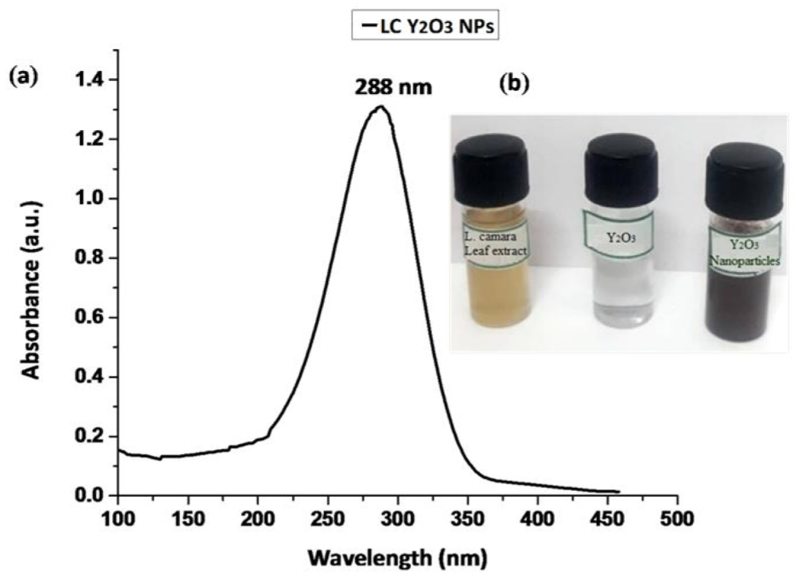
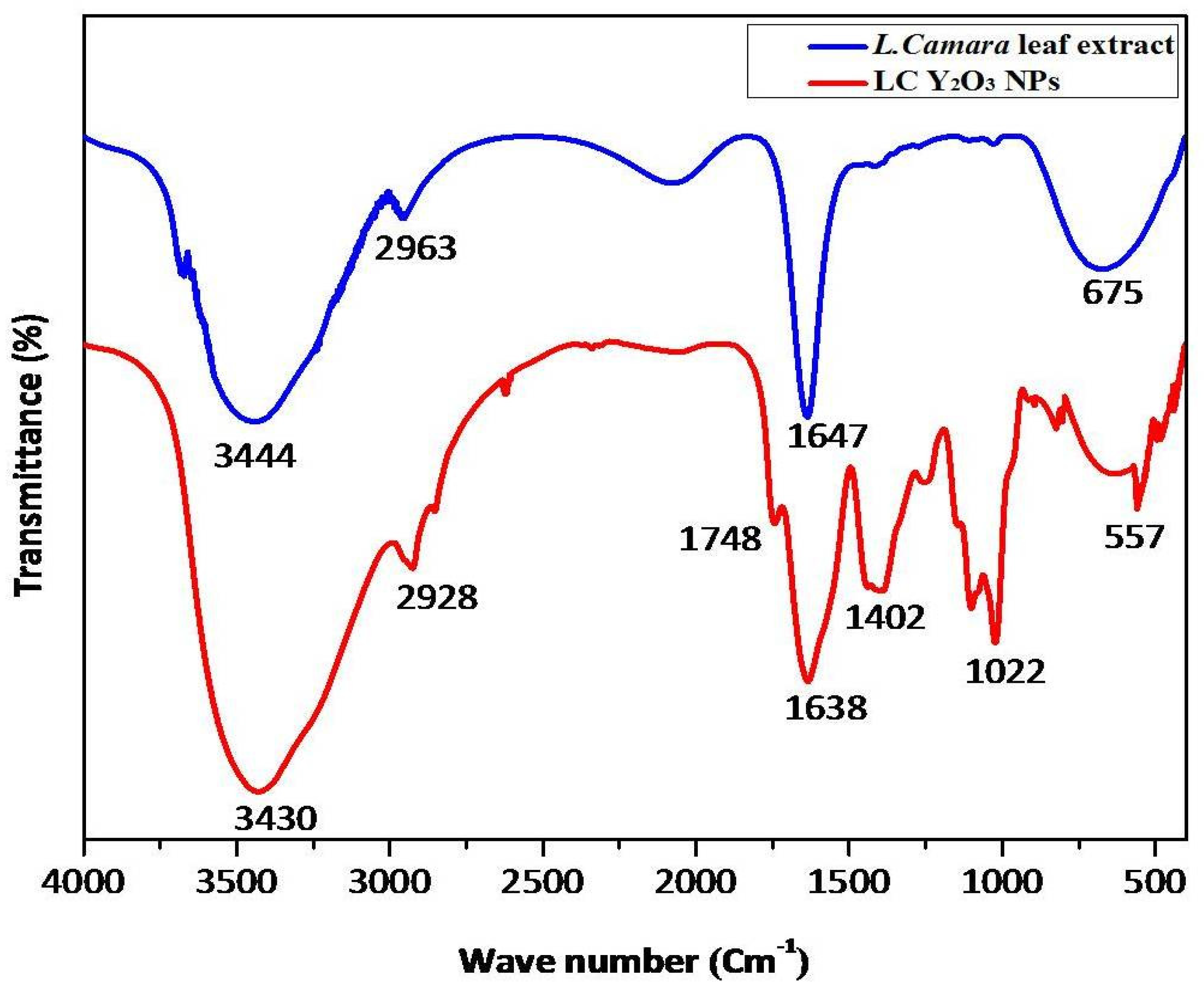
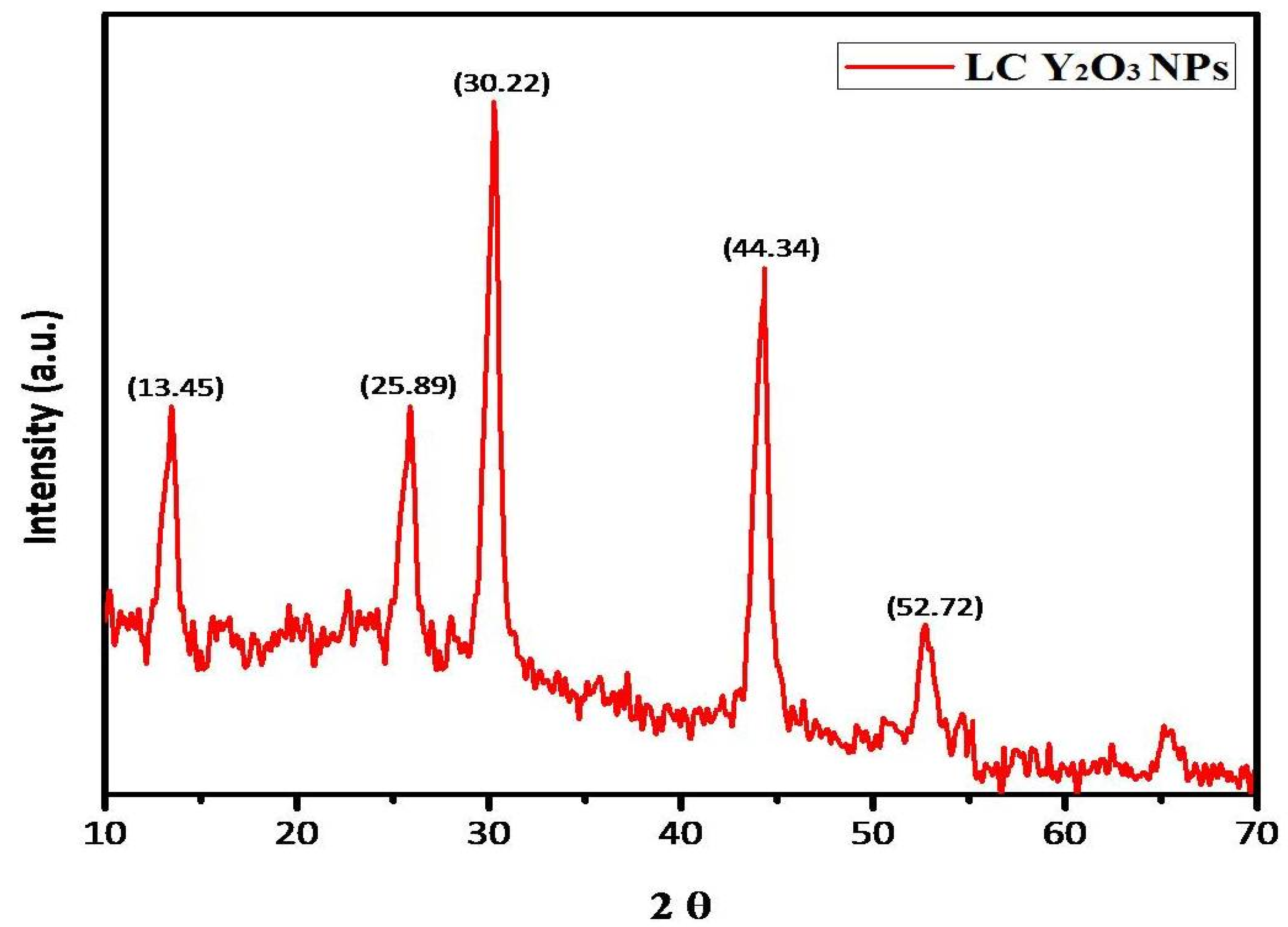
3.1. Characterization of LC Y2O3 NPs

The effect of green synthesized LC Y2O3 NPs was studied using UV-Visible spectroscopy. As demonstrated in Figure 1, the LC Y2O3-loaded NPs exhibited broad UV absorption with enhanced intensity at 288 nm. Similar results were reported for rare earth oxides [61] and organic semiconductor-coated silver particles [62], where such effect was attributed to absorption-induced scattering. The presence of multifunctional groups in the L. camara leaf extract and green synthesized LC Y2O3 NPs were investigated using FTIR. The infrared spectrum of L. camara leaf extract illustrates vibrational bands at 3444, 2963, 1647, and 675 cm−1. The absorption peaks of L. camara leaf extract at 3444 and 2963 cm−1 were attributed to O–H stretching and C–H antisym and sym stretching vibrations, respectively. The absorption peaks of L. camara leaf extract at 1647 and 675 cm−1 bands may be assigned to C=O in ketone esters and C–C–CHO bending in aldehydes, respectively. The absorption band at 1050 cm−1 was obtained by fabricating the green synthesized Y2O3 NPs [63,64,65], whereas the very wide band at 3425 cm−1 is attributed to the C–H stretching of H–bonded alcohols and phenols [66,67]. Similar results reported that the 600–400 cm−1 band is assigned to the M–O region (Y–O), whereas the 617 cm−1 band confirms the YO NPs’ extending vibration [68]. FTIR results of green synthesized LC Y2O3 NPs revealed 3430 cm−1 is assigned to the NH stretch with primary amines and 2928 cm−1 to C–H antisym and sym stretching vibrations. On the other hand, absorption bands at 1748 cm−1 and 1638 cm−1 are attributed to the C=O in esters and C=O stretch secondary amides, respectively. FTIR spectrum of green synthesized LC Y2O3 NPs showed bands at 1402 cm−1 due to O–H in carboxylic acids, 1022 cm−1 to CH–OH in cyclic alcohols with C=O stretch vibrations. Furthermore, the FTIR peaks observed at 557 cm−1 were ascribed to Y–O band stretching (Figure 2). The Y–O band (557 cm−1) indicates that the Yttria phase was formed at 600 °C [69].
The 2θ values of the green synthesized XRD detected Y2O3 NPs. Indeed, diffraction peaks of LC Y2O3 NPs at 2θ values of 13.45°, 25.89°, 30.22°, 44.34°, and 52.72° were indexed as 202, 211, 222, 431, and 440 (JCPDS card no. 83-0927) [70]. The phase evaluation of all synthesized samples was performed with XRD. Figure 3 provides XRD patterns of powders calcinated at different temperatures. In various crystalline phases, Y2O3 NPs could be found as polymorphs. For all samples, the XRD analysis confirms the formation of a single-phase orthorhombic Y2O3 structure, revealing the successful synthesis of Y2O3 NPs. According to the method of Hanawalt (222) and (431), the cubic Y2O3 stage was characterized by sharp peak diffraction angles of 30.22° and 44.34°, respectively [71].
Meanwhile, TEM images show these particles’ tendency to form agglomerates ranging from 20 nm to 45 nm. The Y2O3 NPs display high crystallinity in the TEM images in Figure 4a. Moreover, the coating layer of Y2O3 is very thin in the composite samples, which agrees with the lack of diffraction peaks related to the formation of Y2O3 particles in Figure 4a. The images show crystalline particles with nearly orthorhombic structure and some hexagonal particles. Figure 4a shows a TEM image of green synthesized LC Y2O3 NPs with irregular shapes. The Scherrer equation estimated the crystallite size of green synthesized LC Y2O3 NPs at 30 nm.

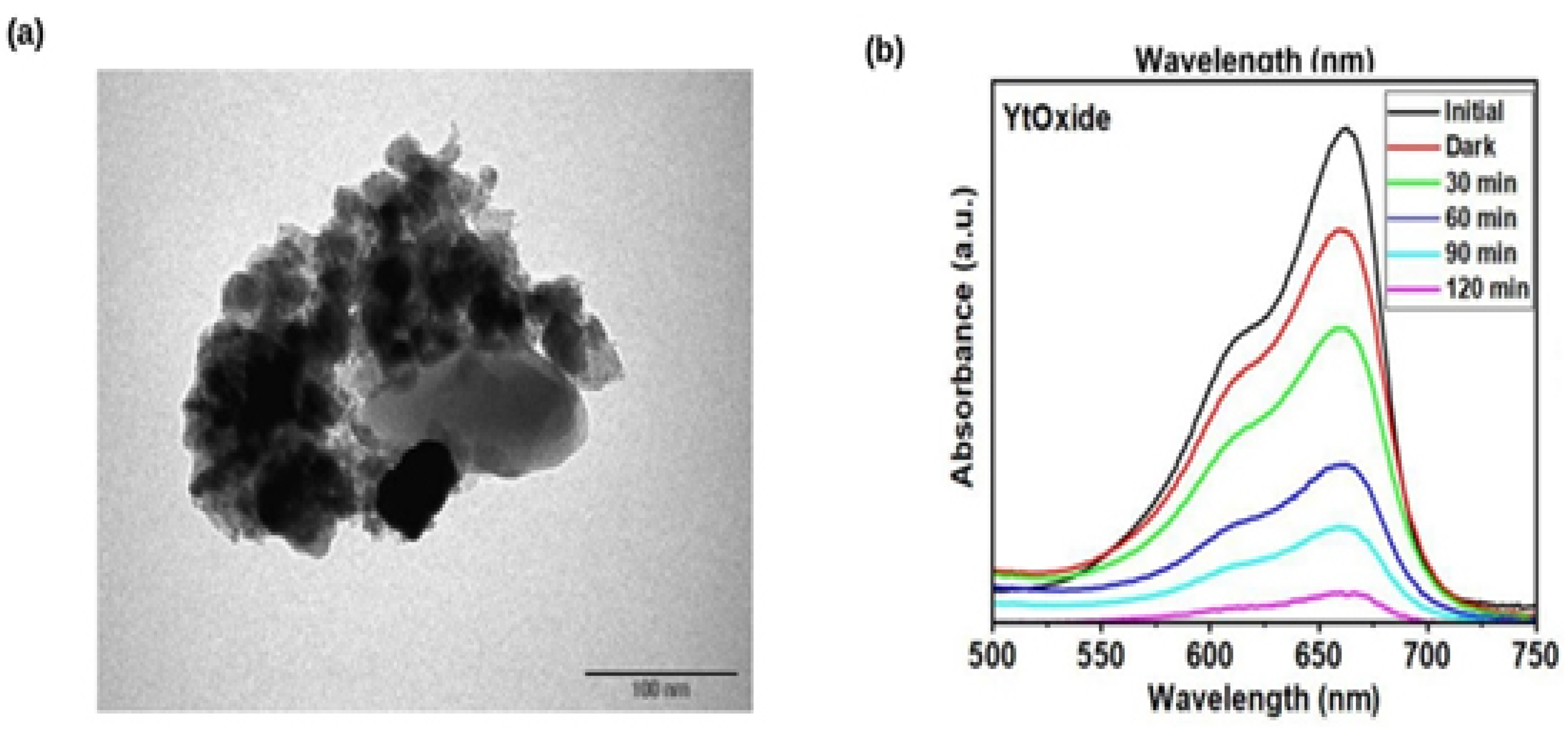
3.2. Photocatalytic Activity of Green Synthesized LC Y2O3 NPs

The photodegradation of dyes without a catalyst was evaluated by exposing the blank dye to illumination. For 120 min, absorbance measurements on blank dye were conducted at different time intervals. The degradation of Rhodamine B dye (RhB) was used to illustrate the photocatalytic efficiency of LC Y2O3 NPs. In the absence of the catalyst, only 18% of the dye degraded after 60 min of exposure, indicating that the catalyst played a significant role in dye photocatalytic degradation [67]. A reduction in peak intensity over time indicates that the dye concentration was considerably reduced and that the catalyst was photoactive. The efficiency of LC Y2O3 NPs as a photocatalyst was investigated using LC Y2O3 NPs for photocatalytic degradation of Rhodamine B. Based on these findings, 92% of the dye was degraded within 60 min, as depicted in Figure 4b. When photons with higher intensity than the bandgap energy of LC Y2O3 NPs were absorbed, electrons were excited to the conduction band, causing a vacancy in the valence band. The electrons in conduction produce (O2•) free-radicals by transferring an electron to the oxygen molecule. In contrast, the vacancy in the valence band generates hydroxyl radicals (•OH), which react with water molecules [72]. These produced reactive oxygen species (ROS), which destroyed the Rhodamine B dye. The reactions result in dye decomposition in the presence of eco-friendly synthesized LC Y2O3 NPs.

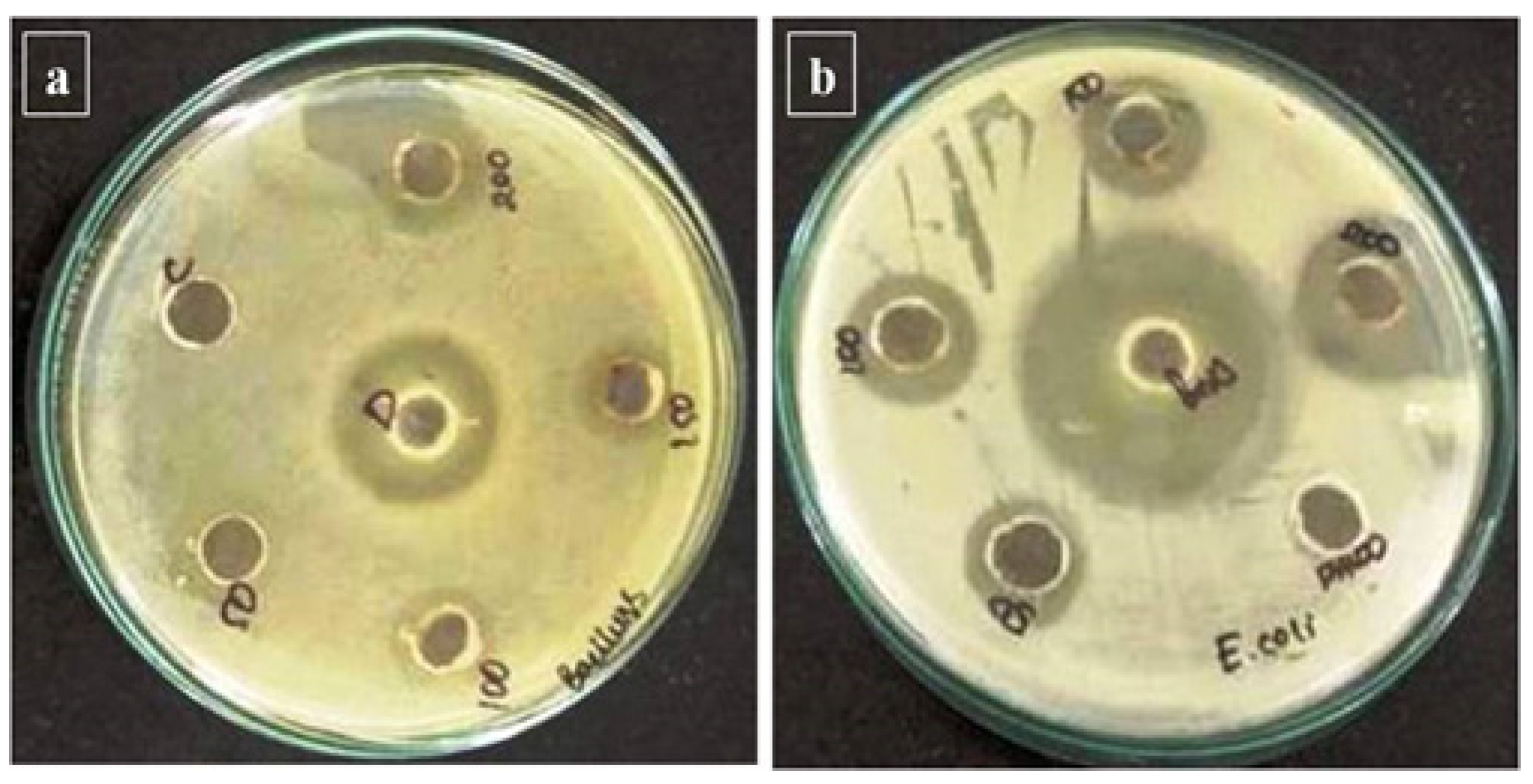
3.3. Green-Synthesized LC Y2O3 NPs Antibacterial Activity

The antibacterial activity of green-synthesized Y2O3 NPs produced in LC aqueous leaf extracts (50–200 µL) was studied against gram-positive and negative bacterial strains. The green synthesized LC Y2O3 NPs with typical antibacterial activity (the zone of inhibition) ranged from 10 to 15 mm for the selected bacterial strains. Bacillus was found to have the highest activity (15 mm inhibition). The diameter of the zone of inhibition was 15 and 11 for Bacillus and E. coli (11mm), respectively (Figure 5a,b). Compared with the antibiotics and LC Y2O3 NPs showed an excellent inhibition zone. In leaf synthesized Y2O3 NPs, there was a slight increase in the inhibition zone compared to the control. The results show that the new LC Y2O3 NPs are promising antimicrobials for plant pathogens. Previously reported Y2O3 NPs were effective against E. coli, P. aeruginosa, S. marcens, and S. aureus [73].

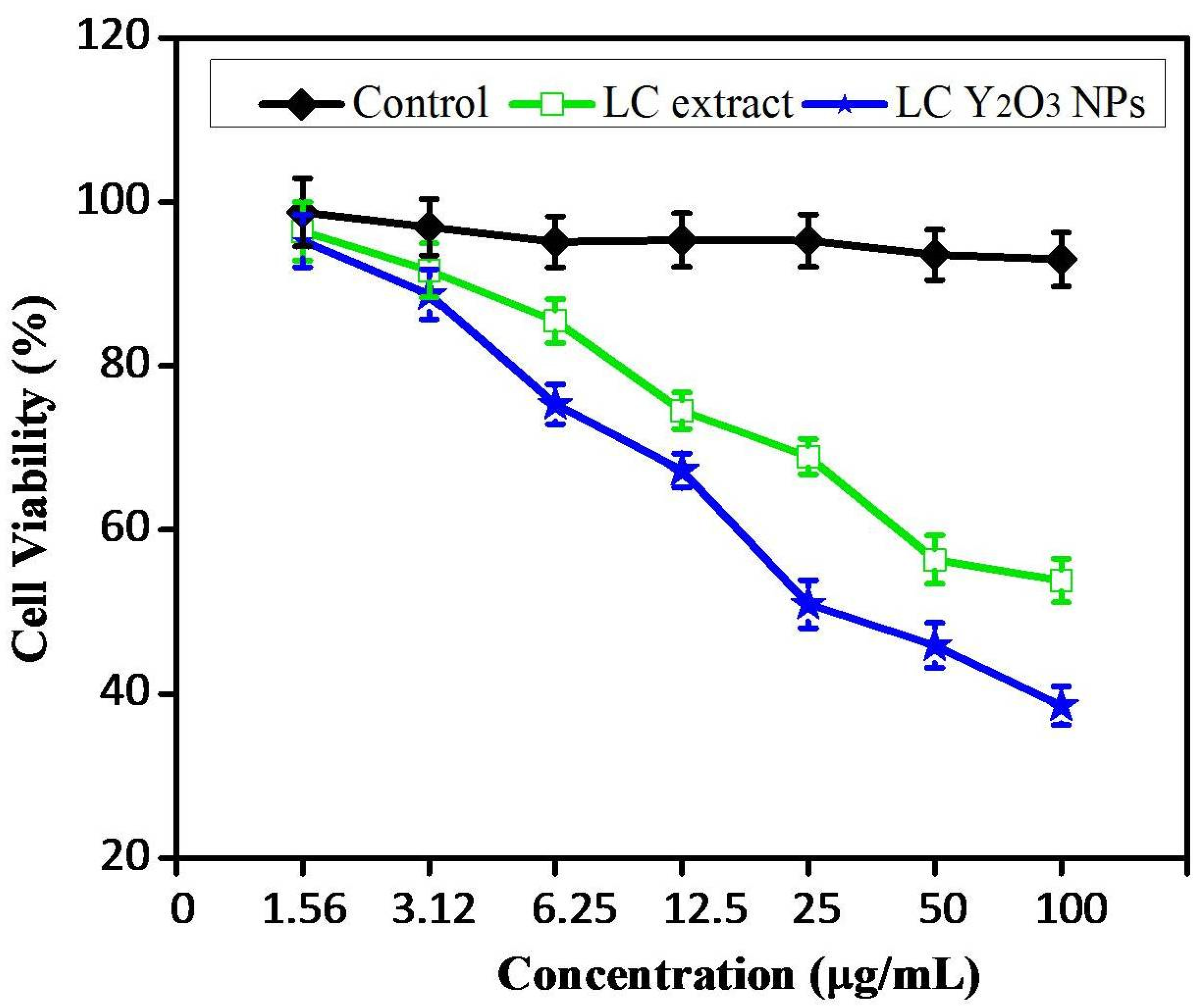
3.4. Cytotoxicity of Y2O3 NPs in HeLa Cell Lines

The MTT assay evaluated the cytotoxicity of different doses of synthesized leaf extract and LC Y2O3 NPs (Figure 6). These results showed that treating cells with 25 µg/mL of synthetic Y2O3 NPs reduces cell viability by 52.6% compared to the control and reduces significantly to around 40.2% (Figure 6). The treatment with 50 μg/mL of green synthesized LC Y2O3 NPs decreased the cell viability by about 59%, and treatment with 100 µg/mL of Y2O3 NPs reduced the cell viability by around 32%.
Y2O3 NPs are particularly important for medical applications because they are considered drug-supply candidates. The major mechanism is Y2O3 NPs penetrating the cell wall and causing cell breathing distress. In the microorganism’s cell wall, Y2O3 NPs also penetrate the cell and destroy it by combining the action of sulfur and phosphorus compounds, such as proteins and deoxyribonucleic acid. Moreover, different concentrations (0, 1.56, 3.12, 6.25, 12.5, 25, 50, and 100 μg/mL) of NPs were used to treat the HeLa cells at 24 h. The results showed time- and dose-dependent declines in cell viability decreased by 53 to 98% in the 24 h treatments. These findings suggest that cell viability decreases when the concentration of NPs increases. The green synthesized LC Y2O3 NPs IC50 value against HeLa cell lines was determined after 24 h and was found to be 25 µg/mL.

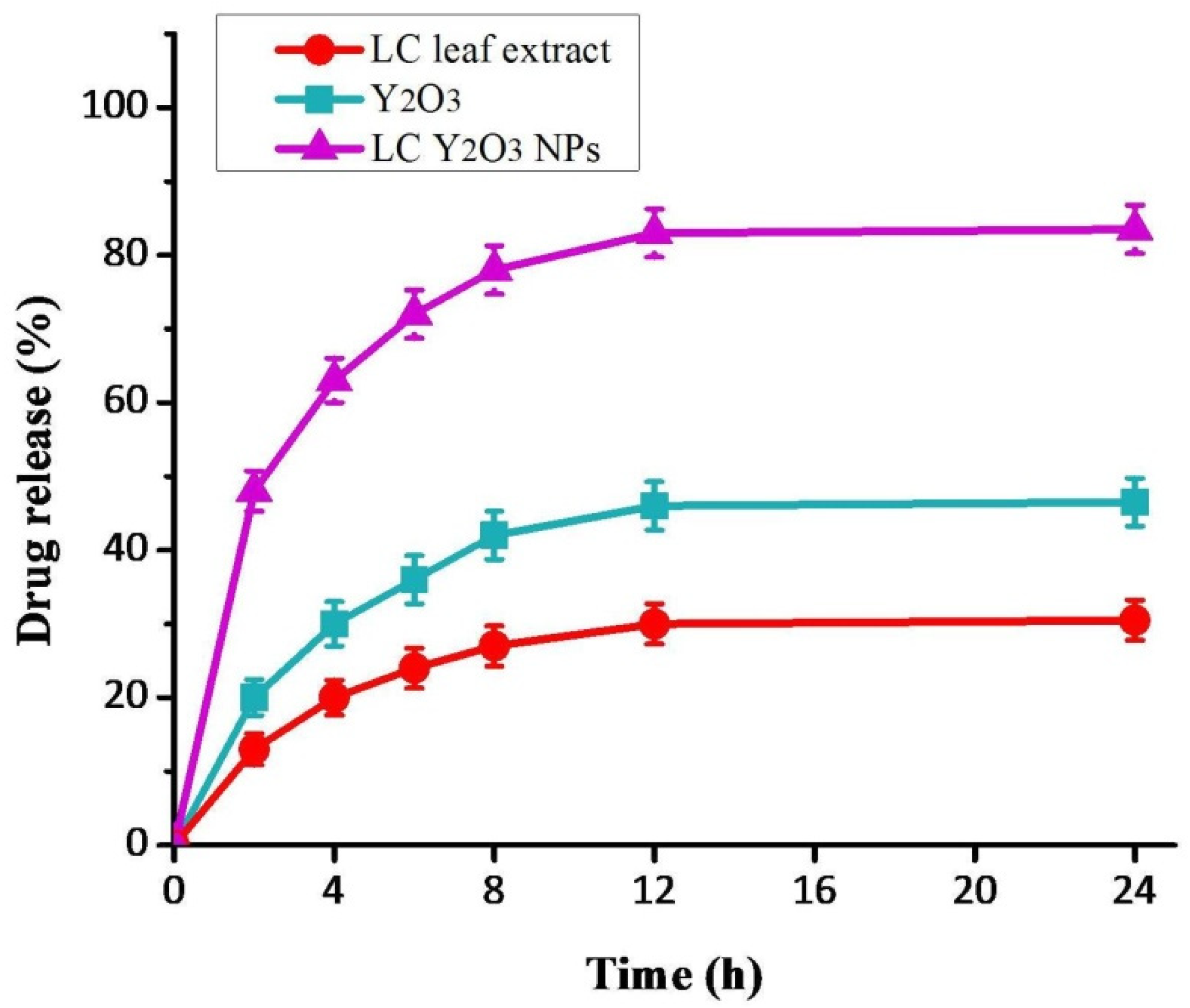
3.5. In Vitro Drug Release

Nanoparticles could be used as anticancer drugs in the treatment. They can penetrate cancer cells for targeted therapy by a smaller number of NPs. Large vascular pores deliver oxygen and nutrients to cancer sites and inflammatory tissues in these locations, allowing NPs to pass through and accumulate [73,74]. The curve would have an initial release, but the not whole drug was released before 4 h. The contrast to the behavior of this release can be explained by the reservoir function of the NPs, which release the drugs (Figure 7). Otherwise, the medicine is released immediately from NPs, as it is in the biochemical condition in these formulations [75]. This release pattern is advantageous for formulating various pharmaceutical drugs, especially where the preferential release rate is more desirable, such as in anticancer therapy.

4. Conclusions

In conclusion, we synthesized Y2O3 NPs from L. camara aqueous leaf extract with high surface reactivity and biocompatibility. These nanoparticles were characterized using various techniques, such as X-ray diffraction and FTIR, which revealed that they were pure single-phase and crystalline LC Y2O3 NPs with orthorhombic shapes. TEM analysis of green synthetic LC Y2O3 powder confirmed the production of nanoparticles with an average size of 30 nm. The zone of inhibition of LC Y2O3 NPs was found to be Bacillus (15 nm) and E. coli (11 mm). The results of this study revealed that the green synthesized LC Y2O3 NPs are effective at degrading dyes from water and preventing bacterial growth. Thus, these NPs possess potential in water filtration, coatings, and the food-manufacturing sector. The cytotoxicity effects of LC Y2O3 NPs nanoparticles against HeLa cell lines were dose-dependent and can be used to treat cervical cancer in humans. Due to their strong surface reactivity, these NPs could be employed in cancer therapy to deliver targeted drug loading. These characteristics make the green synthesized Y2O3 NPs an appropriate option for use in medicinal applications. Furthermore, detailed in vivo studies are needed to determine the safety and effectiveness of these green synthesized LC Y2O3 nanoparticles.

Author Contributions

Conceptualization, M.T. and R.G.; Methodology S.A.A., N.B., T.G., M.A.A., S.S.A. and P.M.; Investigation, R.G.; Data analysis, M.N.A., B.A., M.S.M., S.A.A., V.D.R. and K.R.; Writing original draft M.T., M.G. and R.G. All authors have read and agreed to the published version of the manuscript.

Funding

This research received no external funding.

Institutional Review Board Statement

Not applicable.

Informed Consent Statement

Not applicable.

Data Availability Statement

Not applicable.

Acknowledgments

The authors would like to acknowledge the Deanship of Scientific Research at Umm Al-Qura University for supporting this work by grant code: 22UQU4320545DSR19. This work was financially supported by Taif University Researchers Supporting Project number (TURSP-2020/90), Taif University, Taif, Saudi Arabia.

Conflicts of Interest

The authors declare no conflict of interest.

References

  1. Andleeb, A.; Andleeb, A.; Asghar, S.; Zaman, G.; Tariq, M.; Mehmood, A.; Nadeem, M.; Hano, C.; Lorenzo, J.M.; Abbasi, B.H. A systematic review of biosynthesized metallic nanoparticles as a promising anti-cancer-strategy. Cancers 2021, 13, 2818. [Google Scholar] [CrossRef] [PubMed]
  2. Sung, H.; Ferlay, J.; Siegel, R.L.; Laversanne, M.; Soerjomataram, I.; Jemal, A.; Bray, F. Global Cancer Statistics 2020: GLOBOCAN Estimates of Incidence and Mortality Worldwide for 36 Cancers in 185 Countries. CA Cancer J. Clin. 2021, 71, 209–249. [Google Scholar] [CrossRef] [PubMed]
  3. Al Sufyani, M.N.; Hussien, N.A.; Hawsawi, Y.M. Characterization and Anticancer Potential of Silver Nanoparticles Biosynthesized from Olea chrysophylla and Lavandula dentata Leaf Extracts on HCT116 Colon Cancer Cells. J. Nanomater. 2019, 2019, 7361695. [Google Scholar] [CrossRef] [Green Version]
  4. Ratan, Z.A.; Haidere, M.F.; Nurunnabi, M.; Shahriar, S.M.; Ahammad, A.J.; Shim, Y.Y.; Reaney, M.J.; Cho, J.Y. Green chemistry synthesis of silver nanoparticles and their potential anticancer effects. Cancers 2020, 12, 855. [Google Scholar] [CrossRef] [Green Version]
  5. Sudhakar, A. History of cancer, ancient and modern treatment methods. J. Cancer Sci. Ther. 2009, 1, 1–4. [Google Scholar] [CrossRef] [Green Version]
  6. AbuMousa, R.A.; Baig, U.; Gondal, M.A.; Dastageer, M.A.; AlSalhi, M.S.; Moftah, B.; Alqahtani, F.Y.; Akhter, S.; Aleanizy, F.S. Investigation of the survival viability of cervical cancer cells (HeLa) under visible light-induced photo-catalysis with facile synthesized WO3/ZnO nanocomposite. Saudi J. Biol. Sci. 2020, 27, 1743–1752. [Google Scholar] [CrossRef]
  7. Islami, F.; Miller, K.D.; Siegel, R.L. National and state estimates of lost earnings from cancer deaths in the United States. JAMA Oncol. 2019, 5, e191460. [Google Scholar] [CrossRef]
  8. Fontham, E.T.; Wolf, A.M.; Church, T.R.; Etzioni, R.; Flowers, C.R.; Herzig, A.; Guerra, C.E.; Oeffinger, K.C.; Shih, Y.C.T.; Walter, L.C.; et al. Cervical cancer screening for individuals at average risk: 2020 guideline update from the American Cancer Society. CA Cancer J. Clin. 2020, 70, 321–346. [Google Scholar] [CrossRef]
  9. Ding, W.; Liang, Z.; El-Kott, A.F.; El-Kenawy, A.E. Investigation of anti-human ovarian cancer effects of decorated Au nanoparticles on Thymbra spicata extract modified Fe3O4 nanoparticles. Arab. J. Chem. 2021, 14, 103205. [Google Scholar] [CrossRef]
  10. Chen, J.; Li, Y.; Fang, G.; Cao, Z.; Shang, Y.; Alfarraj, S.; Alharbi, S.A.; Li, J.; Yang, S.; Duan, X. Green synthesis, characterization, cytotoxicity, antioxidant, and anti-human ovarian cancer activities of Curcumae kwangsiensis leaf aqueous extract green-synthesized gold nanoparticles. Arab. J. Chem. 2021, 14, 103000. [Google Scholar] [CrossRef]
  11. Aka, E.K.; Horo, A.; Fanny, M.; Koffi, A.; Olou, L.; Konan, P.; Toure-Ecra, A.; Kone, M. The delay, symptoms, and survival of Ivorian adolescent girls and young adults with uterine cervical cancer. Gynecol. Obstet. Clin. Med. 2021, 1, 153–159. [Google Scholar] [CrossRef]
  12. Monge-Fuentes, V.; Muehlmann, L.A.; de Azevedo, R.B. Perspectives on the application of nanotechnology in photodynamic therapy for the treatment of melanoma. Nano Rev. 2014, 5, 24381. [Google Scholar] [CrossRef] [PubMed] [Green Version]
  13. Vinardell, M.P.; Mitjans, M. Nanocarriers for delivery of antioxidants on the skin. Cosmetics 2015, 2, 342–354. [Google Scholar] [CrossRef] [Green Version]
  14. Prabhakar, U.; Maeda, H.; Jain, R.K.; Sevick-Muraca, E.M.; Zamboni, W.; Farokhzad, O.C.; Barry, S.T.; Gabizon, A.; Grodzinski, P.; Blakey, D.C. Challenges and key considerations of the enhanced permeability and retention effect for nanomedicine drug delivery in oncology. Cancer Res. 2013, 73, 2412–2417. [Google Scholar] [CrossRef] [Green Version]
  15. Elveny, M.; Khan, A.; Nakhjiri, A.T.; Albadarin, A.B. A state-of-the-art review on the application of various pharmaceutical nanoparticles as a promising technology in cancer treatment. Arab. J. Chem. 2021, 14, 103352. [Google Scholar] [CrossRef]
  16. Rajakumar, G.; Mao, L.; Bao, T.; Wen, W.; Wang, S.; Gomathi, T.; Gnanasundaram, N.; Rebezov, M.; Shariati, M.A.; Chung, I.M.; et al. Yttrium oxide nanoparticle synthesis: An overview of methods of preparation and biomedical applications. Appl. Sci. 2021, 11, 2172. [Google Scholar] [CrossRef]
  17. Ramana, C.V.; Atuchin, V.V.; Kesler, V.G.; Kochubey, V.A.; Pokrovsky, L.D.; Shutthanandan, V.; Becker, U.; Ewing, R.C. Growth and surface characterization of sputter-deposited molybdenum oxide thin films. Appl. Surf. Sci. 2007, 253, 5368–5374. [Google Scholar] [CrossRef]
  18. Rahman, M.; Kazmi, I.; Beg, S.; Hafeez, A.; Afzal, M.; Kumar, V.; Anwar, F.; Ahmad, F.J. Functionalized graphene-based nanomaterials for drug delivery and biomedical applications in cancer chemotherapy. In Nano Pharmacother; William Andrew Publishing: Norwich, NY, USA, 2019; Volume 13, pp. 429–460. [Google Scholar] [CrossRef]
  19. Ramana, C.V.; Carbajal-Franco, G.; Vemuri, R.S.; Troitskaia, I.B.; Gromilov, S.A.; Atuchin, V.V. Optical properties and thermal stability of germanium oxide (GeO2) nanocrystals with α-quartz structure. Mater. Sci. Eng. B 2010, 174, 279–284. [Google Scholar] [CrossRef]
  20. Augustine, R.; Dalvi, Y.B.; Nath, V.Y.; Varghese, R.; Raghuveeran, V.; Hasan, A.; Thomas, S.; Sandhyarani, N. Yttrium oxide nanoparticle loaded scaffolds with enhanced cell adhesion and vascularization for tissue engineering applications. Mater. Sci. Eng. C 2019, 103, 109801. [Google Scholar] [CrossRef]
  21. Augustine, R.; Dan, P.; Sosnik, A. Electrospun poly (vinylidene fluoride-trifluoroethylene)/zinc oxide nanocomposite tissue engineering scaffolds with enhanced cell adhesion and blood vessel formation. Nano Res. 2017, 10, 3358–3376. [Google Scholar] [CrossRef]
  22. Xia, Y.; Chen, H.; Zhang, F.; Wang, L.; Chen, B.; Reynolds, M.A.; Ma, J.; Schneider, A.; Gu, N.; Xu, H.H. Injectable calcium phosphate scaffold with iron oxide nanoparticles to enhance osteogenesis via dental pulp stem cells. Artif. Cells Nanomed. Biotechnol. 2018, 46, 423–433. [Google Scholar] [CrossRef] [PubMed] [Green Version]
  23. Garg, V.; Sengar, B.S.; Awasthi, V.; Kumar, A.; Singh, R.; Kumar, S.; Mukherjee, C.; Atuchin, V.V.; Mukherjee, S. Investigation of Dual-Ion Beam Sputter-Instigated Plasmon Generation in TCOs: A Case Study of GZO. ACS Appl. Mater. Interfaces 2018, 10, 5464–5474. [Google Scholar] [CrossRef] [PubMed]
  24. Dyshlyuk, L.; Babich, O.; Ivanova, S.; Vasilchenco, N.; Atuchin, V.; Korolkov, I.; Russakov, D.; Prosekov, A. Antimicrobial potential of ZnO, TiO2 and SiO2 nanoparticles in protecting building materials from biodegradation. Int. Biodeterior. Biodegrad. 2020, 146, 104821. [Google Scholar] [CrossRef]
  25. Augustine, R.; Dominic, E.A.; Reju, I.; Kaimal, B.; Kalarikkal, N.; Thomas, S. Investigation of angiogenesis and its mechanism using zinc oxide nanoparticle-loaded electrospun tissue engineering scaffolds. RSC Adv. 2014, 4, 51528–51536. [Google Scholar] [CrossRef]
  26. Ahtzaz, S.; Nasir, M.; Shahzadi, L.; Amir, W.; Anjum, A.; Arshad, R.; Iqbal, F.; Chaudhry, A.A.; Yar, M.; ur Rehman, I. A study on the effect of zinc oxide and zinc peroxide nanoparticles to enhance angiogenesis-pro-angiogenic grafts for tissue regeneration applications. Mater. Des. 2017, 132, 409–418. [Google Scholar] [CrossRef]
  27. Athinarayanan, J.; Periasamy, V.S.; Alshatwi, A.A. Eco-friendly synthesis and characterization of platinum-copper alloy nanoparticles induce cell death in human cervical cancer cells. Process Biochem. 2016, 51, 925–932. [Google Scholar] [CrossRef]
  28. Perveen, A.; Molardi, C.; Fornaini, C. Applications of laser welding in dentistry: A state-of-the-art review. Micromachines 2018, 9, 209. [Google Scholar] [CrossRef] [Green Version]
  29. Thompson, L.A.; Darwish, W.S. Environmental chemical contaminants in food: Review of a global problem. J. Toxicol. 2019, 2019, 2345283. [Google Scholar] [CrossRef] [Green Version]
  30. Bahrulolum, H.; Nooraei, S.; Javanshir, N. Green synthesis of metal nanoparticles using microorganisms and their application in the agri-food sector. J. Nanotech. 2021, 19, 86. [Google Scholar] [CrossRef]
  31. Nagajyothi, P.C.; Cha, S.J.; Yang, I.J.; Sreekanth, T.V.; Kim, K.J.; Shin, H.M. Antioxidant and anti-inflammatory activities of zinc oxide nanoparticles synthesized using Polygala tenuifolia root extract. J. Photochem. Photobiol. B Biol. 2015, 146, 10–17. [Google Scholar] [CrossRef]
  32. Wu, P.; Song, Z.; Wang, X.; Li, Y.; Li, Y.; Cui, J.; Tuerhong, M.; Jin, D.Q.; Abudukeremu, M.; Lee, D.; et al. Bioactive triterpenoids from Lantana camara showing anti-inflammatory activities in vitro and in vivo. Bioorg. Chem. 2020, 101, 104004. [Google Scholar] [CrossRef] [PubMed]
  33. SujimaAnbu, A.; Velmurugan, P.; Lee, J.H.; Oh, B.T.; Venkatachalam, P. Biomolecule-loaded chitosan nanoparticles induce apoptosis and molecular changes in cancer cell line (SiHa). Int. J. Biol. Macromol. 2016, 88, 18–26. [Google Scholar] [CrossRef] [PubMed]
  34. Ramana, C.V.; Mudavakkat, V.H.; Bharathi, K.K.; Atuchin, V.V.; Pokrovsky, L.D.; Kruchinin, V.N. Enhanced optical constants of nanocrystalline yttrium oxide thin films. Appl. Phys. Lett. 2011, 98, 031905. [Google Scholar] [CrossRef]
  35. Hussain, A.I.; Anwar, F.; Rasheed, S.; Nigam, P.S.; Janneh, O.; Sarker, S.D. Composition, antioxidant and chemotherapeutic properties of the essential oils from two Origanum species growing in Pakistan. Rev. Bras. Farmacogn. 2011, 21, 943–952. [Google Scholar] [CrossRef] [Green Version]
  36. Sathish, R.; Vyawahare, B.; Natarajan, K. Antiulcerogenic activity of Lantana camara leaves on gastric and duodenal ulcers in experimental rats. J. Ethnopharmacol. 2011, 134, 195–207. [Google Scholar] [CrossRef]
  37. Barros, L.M.; Duarte, A.E.; Morais-Braga, M.F.B.; Waczuk, E.P.; Vega, C.; Leite, N.F.; Alencar de Menezes, I.R.; Coutinho, H.D.M.; Rocha, J.B.T.; Kamdem, J.P. Chemical characterization and trypanocidal, leishmanicidal and cytotoxicity potential of Lantana camara L. (Verbenaceae) essential oil. Molecules 2016, 21, 209. [Google Scholar] [CrossRef] [Green Version]
  38. Venkatachalam, T.; Kumar, V.K.; Selvi, P.K.; Maske, A.O.; Anbarasan, V.; Kumar, P.S. Antidiabetic activity of Lantana camara Linn fruits in normal and streptozotocin-induced diabetic rats. J. Pharm. Res. 2011, 4, 1550–1552. [Google Scholar]
  39. Garg, S.K.; Shah, M.A.; Garg, K.M.; Farooqui, M.M.; Sabir, M. Antilymphocytic and immunosuppressive effects of Lantana camara leaves in rats. Indian J. Exp. Biol. 1997, 35, 1315–1328. [Google Scholar]
  40. Patil, S.P.; Kumbhar, S.T. Evaluation of terpene-rich extract of Lantana camara L. leaves for antimicrobial activity against mycobacteria using Resazurin Microtiter Assay (REMA). Beni-Suef Univ. J. Basic Appl. Sci. 2018, 7, 511–515. [Google Scholar] [CrossRef]
  41. Setua, S.; Menon, D.; Asok, A.; Nair, S.; Koyakutty, M. Folate receptor targeted rare-earth oxide nanocrystals for bi-modal fluorescence and magnetic imaging of cancer cells. Biomaterials 2010, 31, 714–729. [Google Scholar] [CrossRef]
  42. Srinivasan, R.; Rajeswari Yogamalar, N.; Elanchezhiyan, J.; Justin Joseyphus, R.; Chandra Bose, A. Structural and optical properties of europium doped yttrium oxide nanoparticles for phosphor applications. J. Alloys Compd. 2010, 496, 472–477. [Google Scholar] [CrossRef]
  43. Luca, A.D.; Kenel, C.; Pado, J.; Joglekar, S.S.; Dunand, D.C.; Leinenbach, C. Thermal stability and influence of Y2O3 dispersoids on the heat treatment response of an additively manufactured ODS Ni–Cr–Al–Ti γ/γ′ superalloy. J. Mater. Res. Technol. 2021, 15, 2883–2898. [Google Scholar] [CrossRef]
  44. Rubio, E.J.; Atuchin, V.V.; Kruchinin, V.N.; Pokrovsky, L.D.; Prosvirin, I.P.; Ramana, C.V. Electronic Structure and Optical Quality of Nanocrystalline YO Film Surfaces and Interfaces on Silicon. J. Phys. Chem. C 2014, 118, 13644–13651. [Google Scholar] [CrossRef]
  45. Gizowska, M.; Piątek, M.; Perkowski, K.; Konopka, G.; Witosławska, I. Fabrication of Nanoyttria by method of solution combustion synthesis. Nanomaterials 2020, 10, 831. [Google Scholar] [CrossRef]
  46. Hosseini, S.F.; Zandi, M.; Rezaei, M.; Farahmandghavi, F. Two-step method for encapsulation of oregano essential oil in chitosan nanoparticles: Preparation, characterization and in vitro release study. Carbohydr. Polym. 2013, 95, 50–56. [Google Scholar] [CrossRef] [PubMed]
  47. Khaksar, M.R.; Rahimifard, M.; Baeeri, M.; Maqbool, F.; Navaei-Nigjeh, M.; Hassani, S.; Moeini-Nodeh, S.; Kebriaeezadeh, A.; Abdollahi, M. Protective effects of cerium oxide and yttrium oxide nanoparticles on reduction of oxidative stress induced by sub-acute exposure to diazinon in the rat pancreas. J. Trace Elem. Med. Biol. 2017, 41, 79–90. [Google Scholar] [CrossRef]
  48. Kambale, E.K.; Nkanga, C.I.; Mutonkole, B.P.I.; Bapolisi, A.M.; Tassa, D.O.; Liesse, J.M.I.; Krause, R.W.; Memvanga, P.B. Green synthesis of antimicrobial silver nanoparticles using aqueous leaf extracts from three Congolese plant species (Brillantaisiapatula, Crossopteryxfebrifuga and Senna siamea). Heliyon 2020, 6, e04493. [Google Scholar] [CrossRef]
  49. Jeyaraj, M.; Gurunathan, S.; Qasim, M.; Kang, M.H.; Kim, J.H. A Comprehensive review on the synthesis, characterization, and biomedical application of platinum nanoparticles. Nanomaterials 2019, 9, 1719. [Google Scholar] [CrossRef] [Green Version]
  50. Hegazey, R.M.; Abdelrahman, E.A.; Kotp, Y.H.; Hameed, A.M.; Subaihi, A. Facile fabrication of hematite nanoparticles from Egyptian insecticide cans for efficient photocatalytic degradation of rhodamine B dye. J. Mater. Res. Technol. 2020, 9, 1652–1661. [Google Scholar] [CrossRef]
  51. Alharbi, A.; Abdelrahman, E.A. Efficient photocatalytic degradation of malachite green dye using facilely synthesized hematite nanoparticles from Egyptian insecticide cans. Spectrochim. Acta Part A Mol. Biomol. Spectrosc. 2020, 226, 117612. [Google Scholar] [CrossRef]
  52. Almehizia, A.A.; Al-Omar, M.A.; Naglah, A.M.; Bhat, M.A.; Al-Shakliah, N.S. Facile synthesis and characterization of ZnO nanoparticles for studying their biological activities and photocatalytic degradation properties toward methylene blue dye. Alex. Eng. J. 2021, 61, 2386–2395. [Google Scholar] [CrossRef]
  53. Zhao, B.; Mattelaer, F.; Rampelberg, G.; Dendooven, J.; Detavernier, C. Thermal and Plasma-Enhanced Atomic Layer Deposition of Yttrium Oxide Films and the Properties of Water Wettability. ACS Appl. Mater. Interfaces 2020, 12, 3179–3187. [Google Scholar] [CrossRef] [PubMed]
  54. Hare, J.I.; Lammers, T.; Ashford, M.B.; Puri, S.; Storm, G.; Barry, S.T. Challenges and strategies in anti-cancer nanomedicine development: An industry perspective. Adv. Drug Deliv. Rev. 2017, 108, 25–38. [Google Scholar] [CrossRef] [Green Version]
  55. Pirtarighat, S.; Ghannadnia, M.; Baghshahi, S. Green synthesis of silver nanoparticles using the plant extract of Salvia spinosa grown in vitro and their antibacterial activity assessment. J. Nanostruct. Chem. 2019, 9, 1–9. [Google Scholar] [CrossRef] [Green Version]
  56. Porosnicu, I.; Butnaru, C.M.; Tiseanu, I.; Stancu, E.; Munteanu, C.V.; Bita, B.I.; Duliu, O.G.; Sima, F. Y2O3 nanoparticles and X-ray radiation-induced effects in melanoma cells. Molecules 2021, 26, 3403. [Google Scholar] [CrossRef] [PubMed]
  57. Rajendran, A.; Alsawalha, M.; Alomayri, T. Biogenic synthesis of husked rice-shaped iron oxide nanoparticles using coconut pulp (Cocos nucifera L.) extract for photocatalytic degradation of Rhodamine B dye and their in vitro antibacterial and anticancer activity. J. Saudi Chem. Soc. 2021, 25, 101307. [Google Scholar] [CrossRef]
  58. Xu, J.; Zhu, J.; Niu, J.; Chen, M.; Yue, J. Efficient and stable photocatalytic hydrogen evolution activity of multi-hetero junction composite photocatalysts: CdS and NiS2 co-modified NaNbO3nanocubes. Front. Chem. 2020, 7, 880. [Google Scholar] [CrossRef]
  59. El-Sheekh, M.M.; Hassan, L.H.S.; Morsi, H.H. Assessment of the in vitro anticancer activities of cyanobacteria mediated silver oxide and gold nanoparticles in human colon CaCo-2 and cervical HeLa cells. Environ. Nanotechnol. Monit. Manag. 2021, 16, 100556. [Google Scholar] [CrossRef]
  60. Dixit, N.; Vaibhav, K.; Pandey, R.S.; Jain, U.K.; Katare, O.P.; Katyal, A.; Madan, J. Improved cisplatin delivery in cervical cancer cells by utilizing folate-grafted non-aggregated gelatin nanoparticles. Biomed. Pharmacother. 2015, 69, 1–10. [Google Scholar] [CrossRef]
  61. Benammar, I.; Salhi, R.; Deschanvres, J.L.; Maalej, R. The effect of rare earth element (Er, Yb) doping and heat treatment on suspension stability of Y2O3 nanoparticles elaborated by sol-gel method. J. Mater. Res. Technol. 2020, 9, 12634–12642. [Google Scholar] [CrossRef]
  62. Petoukhoff, C.E.; O’Carroll, D.M. Absorption-induced scattering and surface plasmon out-coupling from absorber-coated plasmonic metasurfaces. Nat. Commun. 2015, 6, 7899. [Google Scholar] [CrossRef] [PubMed] [Green Version]
  63. Golovnev, N.N.; Molokeev, M.S.; Vereshchagin, S.N.; Atuchin, V.V. Calcium and strontium thiobarbiturates with discrete and polymeric structures. J. Coord. Chem. 2013, 66, 4119–4130. [Google Scholar] [CrossRef]
  64. Narasaiah, P.; Mandal, B.; Sarada, N.C. Biosynthesis of copper oxide nanoparticles from Drypetessepiaria leaf extract and their catalytic activity to dye degradation. In IOP Conference Series: Materials Science and Engineering; IOP Publishing: Bristol, UK, 2017; Volume 263, p. 022012. [Google Scholar] [CrossRef]
  65. Lin, L.; Starostin, S.A.; Li, S.; Khan, S.A.; Hessel, V. Synthesis of yttrium oxide nanoparticles via a facile microplasma-assisted process. Chem. Eng. Sci. 2018, 178, 157–166. [Google Scholar] [CrossRef]
  66. Golovnev, N.N.; Molokeev, M.S.; Vereshchagin, S.N.; Atuchin, V.V.; Sidorenko, M.Y.; Dmitrushkov, M.S. Crystal structure and properties of the precursor [Ni(H2O)6](HTBA)2·2H2O and the complexes M(HTBA)2(H2O)2 (M=Ni, Co, Fe). Polyhedron 2014, 70, 71–76. [Google Scholar] [CrossRef]
  67. Hajizadeh-Oghaz, M.; Razavi, R.S.; Barekat, M.; Naderi, M.; Malekzadeh, S.; Rezazadeh, M. Synthesis and characterization of Y2O3 nanoparticles by sol-gel process for transparent ceramics applications. J. Sol-Gel Sci. Technol. 2016, 78, 682–691. [Google Scholar] [CrossRef]
  68. Zaidi, Z.; Vaghasiya, K.; Vijay, A.; Sharma, M.; Verma, R.K.; Vaidya, S. Hollow ZnO from assembly of nanoparticles: Photocatalytic and antibacterial activity. J. Mater. Sci. 2018, 53, 14964–14974. [Google Scholar] [CrossRef]
  69. Mitra, R.N.; Merwin, M.J.; Han, Z.; Conley, S.M.; Al-Ubaidi, M.R.; Naash, M.I. Yttrium oxide nanoparticles prevent photoreceptor death in a light-damage model of retinal degeneration. Free. Radic. Biol. Med. 2014, 75, 140–148. [Google Scholar] [CrossRef] [Green Version]
  70. Kannan, S.K.; Sundararajan, M. Biosynthesis of yttrium oxide nanoparticles using Acalypha indica leaf extract. Bull. Mater. Sci. 2015, 38, 945–950. [Google Scholar] [CrossRef]
  71. Carbonin, S.; Menegazzo, G. Teaching application of an X-ray powder diffraction data card file: XRD profile simulation and Hanawalt index tabulation. Comput. Geosci. 1996, 22, 935–942. [Google Scholar] [CrossRef]
  72. Selvaraj, V.; Bodapati, S.; Murray, E.; Rice, K.M.; Winston, N.; Shokuhfar, T.; Zhao, Y.; Blough, E. Cytotoxicity and genotoxicity caused by yttrium oxide nanoparticles in HEK293 cells. Int. J. Nanomed. 2014, 9, 1379–1391. [Google Scholar] [CrossRef] [Green Version]
  73. Emami Nejad, A.; Najafgholian, S.; Rostami, A.; Sistani, A.; Shojaeifar, S.; Esparvarinha, M.; Nedaeinia, R.; Haghjooy Javanmard, S.; Taherian, M.; Ahmadlou, M.; et al. The role of hypoxia in the tumor microenvironment and development of cancer stem cell: A novel approach to developing treatment. Cancer Cell Int. 2021, 21, 62. [Google Scholar] [CrossRef] [PubMed]
  74. Chen, C.; Wang, X.; Binder, K.; Ghahremanpour, M.M.; van der Spoel, D.; Pöschl, U.; Su, H.; Cheng, Y. Energetic analysis of succinic acid in water droplets: Insight into the size-dependent solubility of atmospheric nanoparticles. Atmos. Chem. Phys. Discuss. 2021, 21, 1–19. [Google Scholar] [CrossRef]
  75. WijiPrasetyaningrum, P.; Bahtiar, A.; Hayun, H. Synthesis and Cytotoxicity Evaluation of Novel Asymmetrical Mono-Carbonyl Analogs of Curcumin (AMACs) against Vero, HeLa, and MCF7 Cell Lines. Sci. Pharm. 2018, 86, 25. [Google Scholar] [CrossRef] [PubMed] [Green Version]
Figure 1. UV-vis absorption spectra of (a) green synthesized LC Y2O3 NPs suspended in ethanol and (b) the color changes following the synthesis of LC Y2O3 NPs.
Nanomaterials 12 02393 g001
Figure 2. FTIR spectrum of the L. camara leaf extract and green-synthesized LC Y2O3 NPs.
Nanomaterials 12 02393 g002
Figure 3. XRD pattern of green synthesized LC Y2O3 NPs (JCPDS card no. 83-0927).
Nanomaterials 12 02393 g003
Figure 4. (a) TEM image of the green synthesized LC Y2O3 NPs. (b) Photocatalytic degradation of Rhodamine B utilizing the green synthesized LC Y2O3 NPs.
Nanomaterials 12 02393 g004
Figure 5. The green synthesized LC Y2O3 NPs showed an improved zone of inhibition against (a) Bacillus, and (b) E. coli, compared to Y2O3 NPs.
Nanomaterials 12 02393 g005
Figure 6. Green LC Y2O3 NPs inhibit the cell viability of human cervical carcinoma HeLa cells. HeLa cells were treated for 24 h with various concentrations of LC extract and green LC Y2O3 NPs.
Nanomaterials 12 02393 g006
Figure 7. In vitro release of optimized LC Y2O3 NPs. The drug shows rapid NPs release and aqueous drug solution. Around 85% of the drug was released in the first 30 min of the study. A saturation status was then observed in the drug release. On the other hand, optimized LC Y2O3 NPs showed a more controlled drug release pattern than LC leaf extract and Y2O3 NPs.
Nanomaterials 12 02393 g007
Publisher’s Note: MDPI stays neutral with regard to jurisdictional claims in published maps and institutional affiliations.

