
One-Pot Synthesis of MnOx-SiO2 Porous Composites as Nanozymes with ROS-Scavenging Properties
 , , , , and
Abstract
:1. Introduction
2. Experimental Methods
2.1. Reagents and Materials
2.2. Synthesis
2.3. Materials Characterization
2.4. Determination of the Catalase Activity
2.5. Determination of the Superoxide Dismutase Activity
3. Results and Discussion
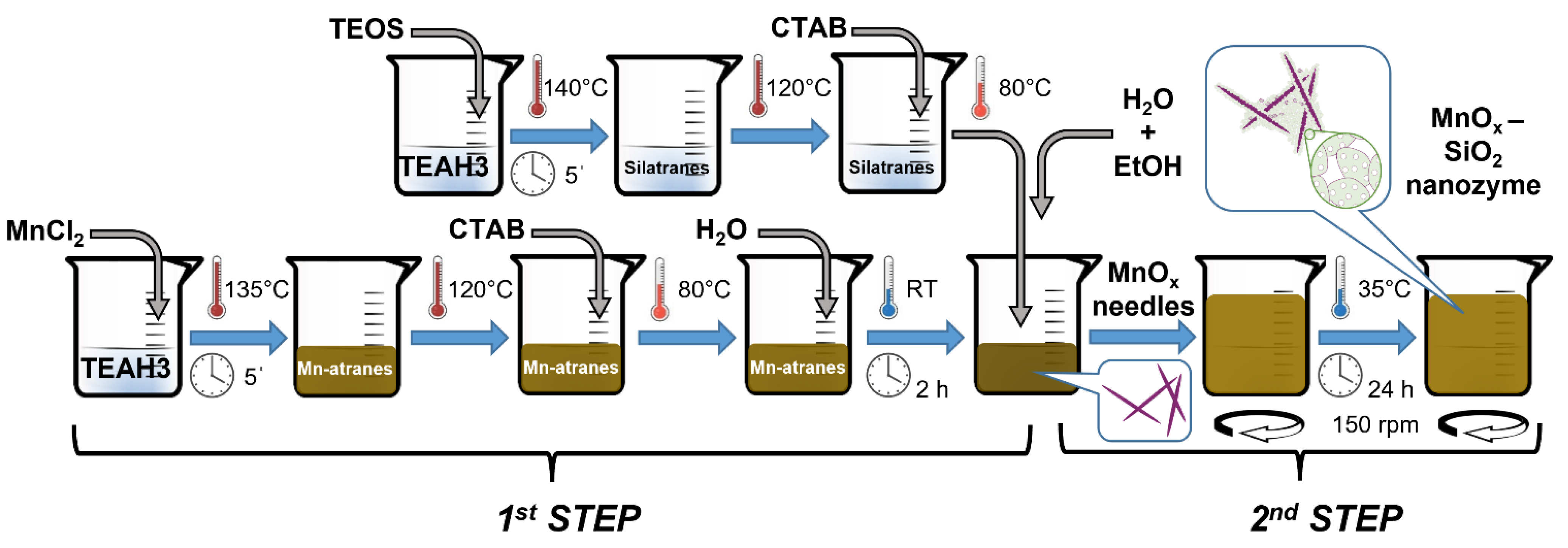
3.1. Synthesis Strategy
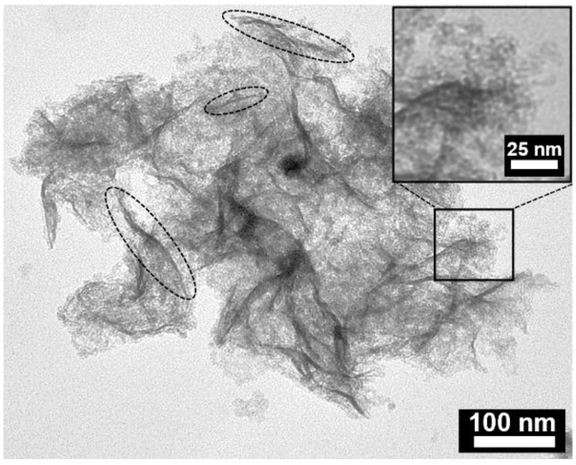
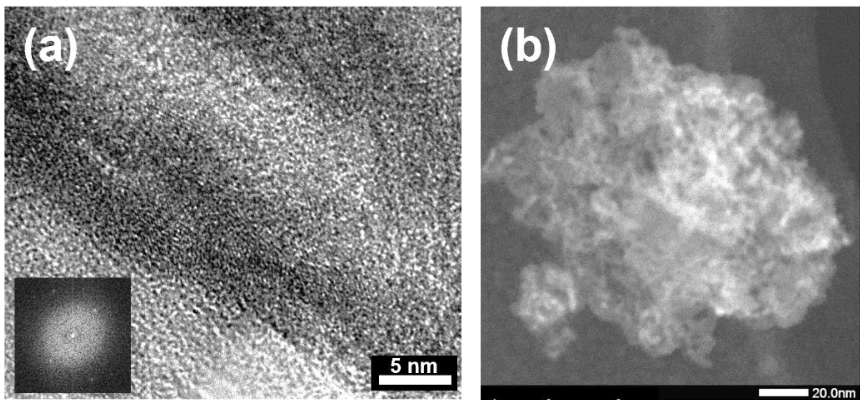
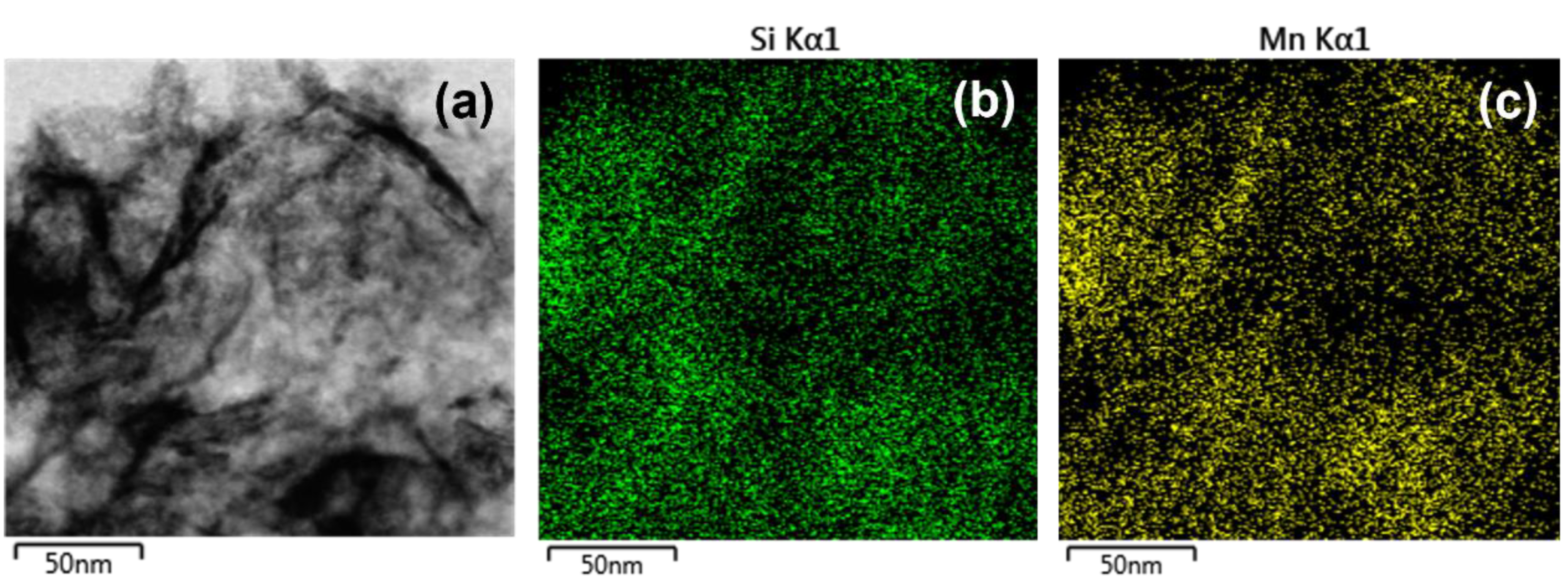
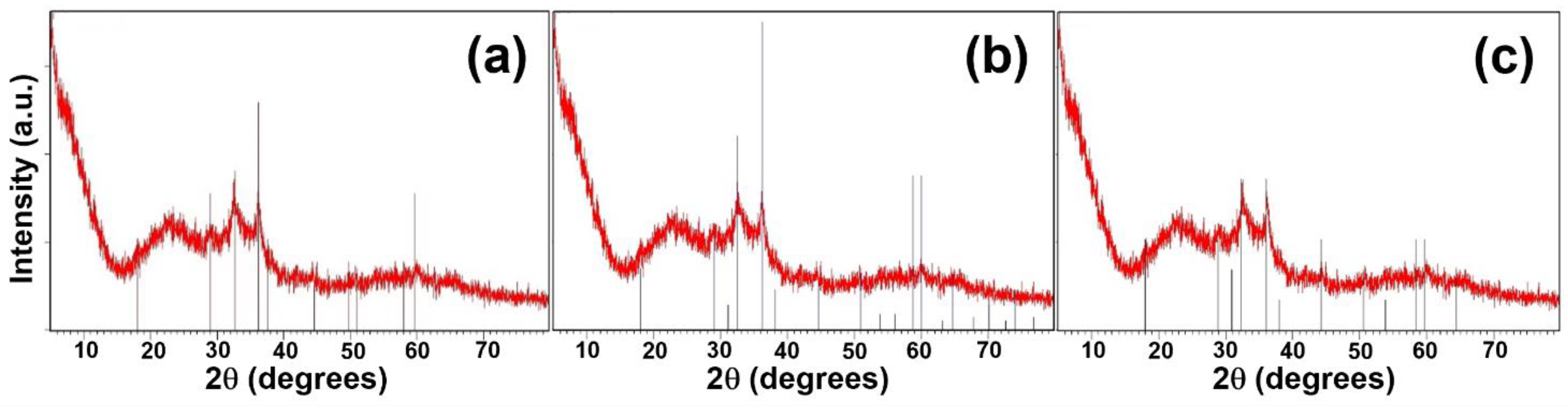
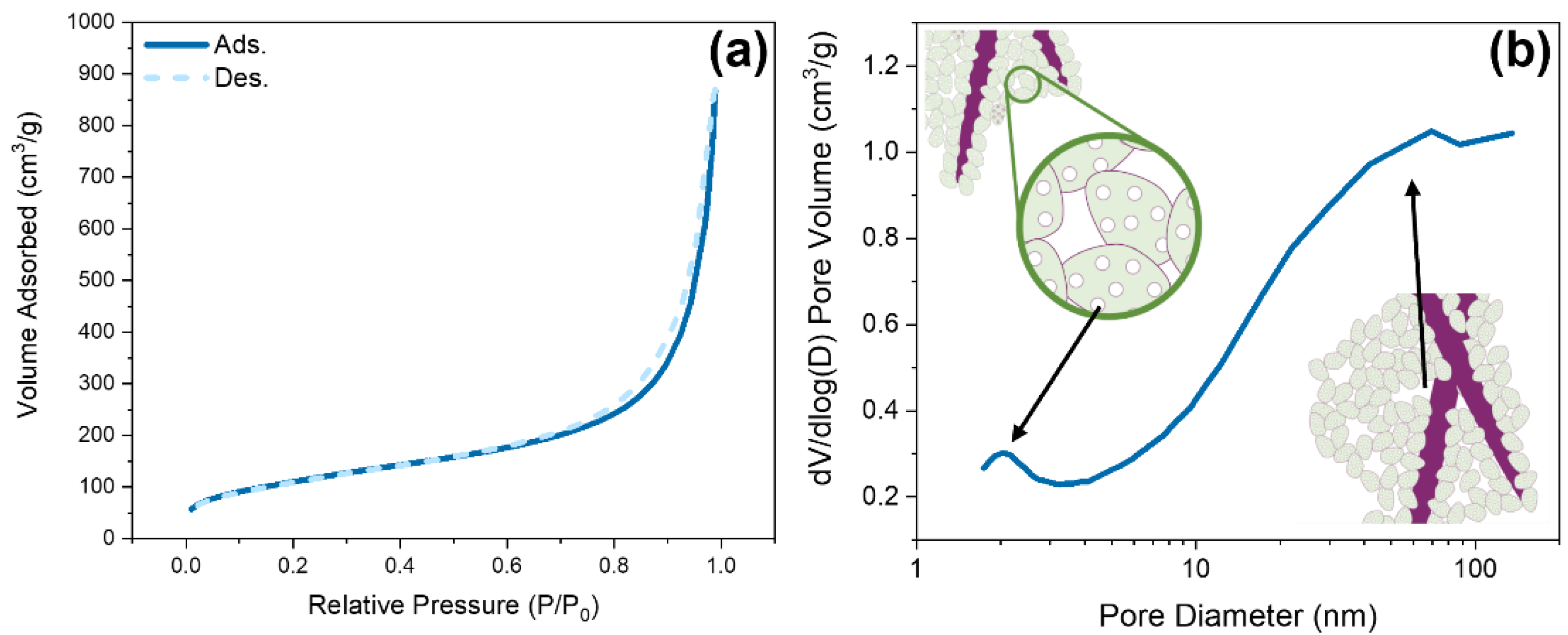
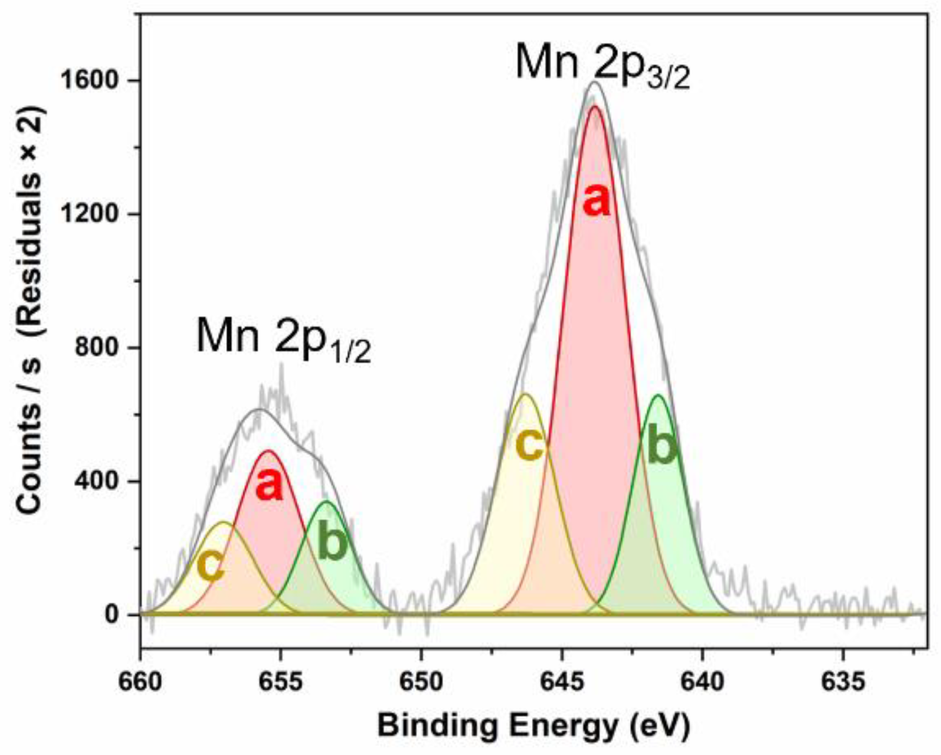
3.2. Nanozyme Characterization
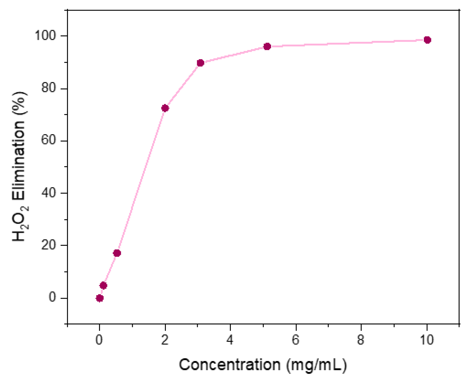
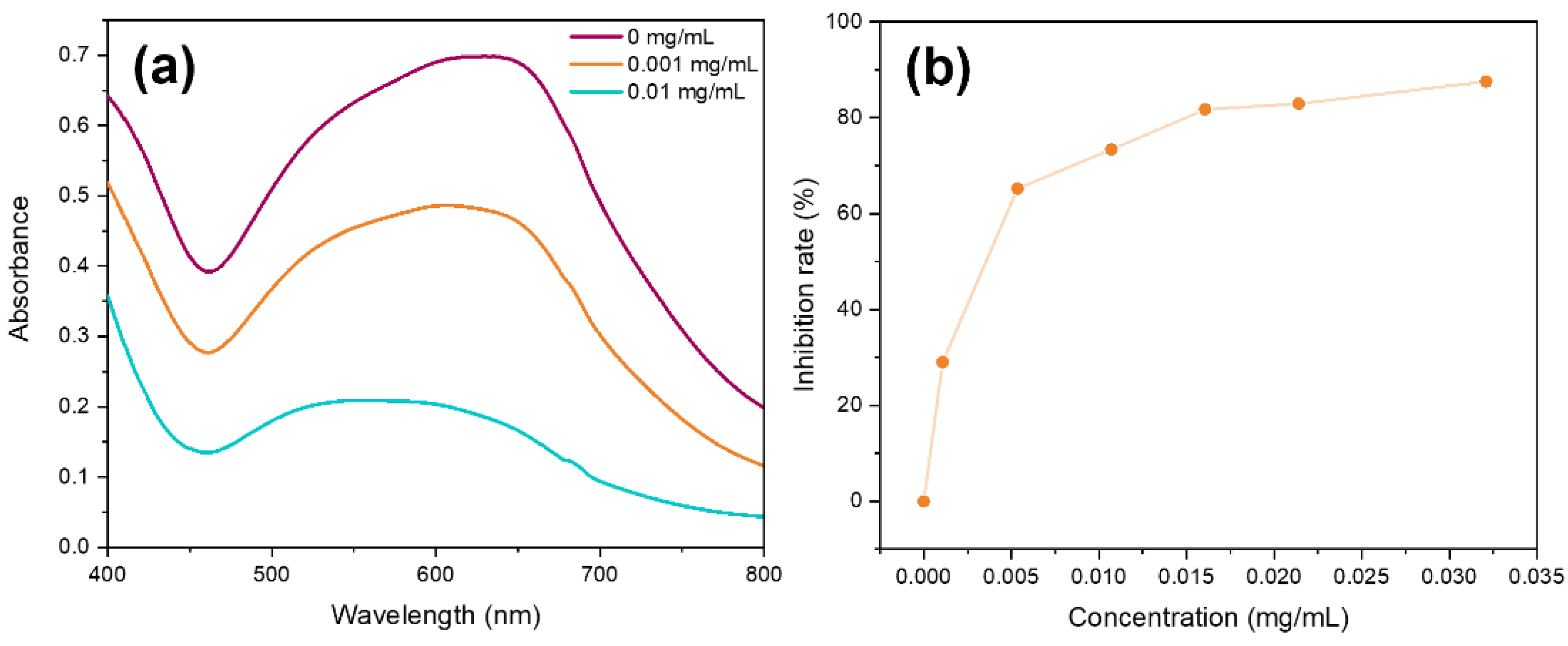
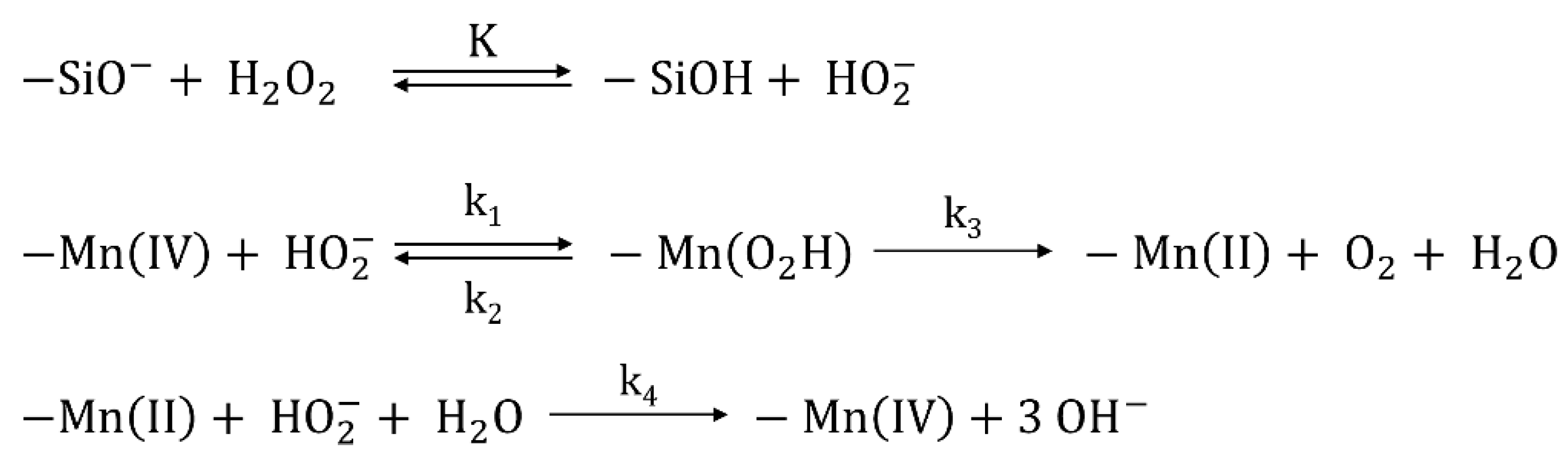
3.3. Multienzyme-like Activity of Mn-Nanozyme
4. Conclusions
Author Contributions
Funding
Data Availability Statement
Acknowledgments
Conflicts of Interest
References
- Wang, H.; Wan, K.; Shi, X. Recent Advances in Nanozyme Research. Adv. Mater. 2019, 31, 1–10. [Google Scholar] [CrossRef] [PubMed]
- Yu, Z.; Lou, R.; Pan, W.; Li, N.; Tang, B. Nanoenzymes in Disease Diagnosis and Therapy. Chem. Commun. 2020, 56, 15513–15524. [Google Scholar] [CrossRef] [PubMed]
- Huang, Y.; Ren, J.; Qu, X. Nanozymes: Classification, Catalytic Mechanisms, Activity Regulation, and Applications. Chem. Rev. 2019, 119, 4357–4412. [Google Scholar] [CrossRef] [PubMed]
- Singh, Y.P.; Patel, R.N.; Singh, Y.; Butcher, R.J.; Vishakarma, P.K.; Singh, R.K.B. Structure and Antioxidant Superoxide Dismutase Activity of Copper(II) Hydrazone Complexes. Polyhedron 2017, 122, 1–15. [Google Scholar] [CrossRef] [Green Version]
- Terra, W.S.; Ferreira, S.S.; Costa, R.O.; Mendes, L.L.; Franco, R.W.A.; Bortoluzzi, A.J.; Resende, J.A.L.C.; Fernandes, C.; Horn, A. Evaluating the Influence of the Diamine Unit (Ethylenediamine, Piperazine and Homopiperazine) on the Molecular Structure, Physical Chemical Properties and Superoxide Dismutase Activity of Copper Complexes. Inorg. Chim. Acta 2016, 450, 353–363. [Google Scholar] [CrossRef]
- Signorella, S.; Palopoli, C.; Ledesma, G. Rationally Designed Mimics of Antioxidant Manganoenzymes: Role of Structural Features in the Quest for Catalysts with Catalase and Superoxide Dismutase Activity. Coord. Chem. Rev. 2018, 365, 75–102. [Google Scholar] [CrossRef]
- Deawati, Y.; Onggo, D.; Mulyani, I.; Hastiawan, I.; Kurnia, D.; Lönnecke, P.; Schmorl, S.; Kersting, B.; Hey-Hawkins, E. Synthesis, Crystal Structures, and Superoxide Dismutase Activity of Two New Multinuclear Manganese(III)-Salen-4,4′-Bipyridine Complexes. Inorg. Chim. Acta 2018, 482, 353–357. [Google Scholar] [CrossRef]
- Domergue, J.; Pécaut, J.; Proux, O.; Lebrun, C.; Gateau, C.; Le Goff, A.; Maldivi, P.; Duboc, C.; Delangle, P. Mononuclear Ni(II) Complexes with a S3O Coordination Sphere Based on a Tripodal Cysteine-Rich Ligand: PH Tuning of the Superoxide Dismutase Activity. Inorg. Chem. 2019, 58, 12775–12785. [Google Scholar] [CrossRef]
- Singh, O.; Tyagi, N.; Olmstead, M.M.; Ghosh, K. The Design of Synthetic Superoxide Dismutase Mimetics: Seven-Coordinate Water Soluble Manganese(II) and Iron(II) Complexes and Their Superoxide Dismutase-like Activity Studies. Dalt. Trans. 2017, 46, 14186–14191. [Google Scholar] [CrossRef]
- Zhou, Y.H.; Liu, X.W.; Chen, L.Q.; Wang, S.Q.; Cheng, Y. Synthesis, Structure and Superoxide Dismutase-like Activity of Two Mixed-Ligand Cu(II) Complexes with N,N′-Bis(2-Pyridylmethyl)Amantadine. Polyhedron 2016, 117, 788–794. [Google Scholar] [CrossRef]
- Purtaş, S.; Köse, M.; Tümer, F.; Tümer, M.; Gölcü, A.; Ceyhan, G. A Novel Porphyrin Derivative and Its Metal Complexes: Electrochemical, Photoluminescence, Thermal, DNA-Binding and Superoxide Dismutase Activity Studies. J. Mol. Struct. 2016, 1105, 293–307. [Google Scholar] [CrossRef]
- Pelluau, T.; Sene, S.; Garcia-Cirera, B.; Albela, B.; Bonneviot, L.; Larionova, J.; Guari, Y. Multifunctionalized Mesostructured Silica Nanoparticles Containing Mn2 Complex for Improved Catalase-Mimicking Activity in Water. Nanomaterials 2022, 12, 1136. [Google Scholar] [CrossRef]
- Patriarca, M.; Daier, V.; Camí, G.; Rivière, E.; Hureau, C.; Signorella, S. Preparation, Characterization and Activity of CuZn and Cu2 Superoxide Dismutase Mimics Encapsulated in Mesoporous Silica. J. Inorg. Biochem. 2020, 207, 1–7. [Google Scholar] [CrossRef]
- Zhou, Y.; Liu, B.; Yang, R.; Liu, J. Filling in the Gaps between Nanozymes and Enzymes: Challenges and Opportunities. Bioconjug. Chem. 2017, 28, 2903–2909. [Google Scholar] [CrossRef]
- Zhang, R.; Yan, X.; Fan, K. Nanozymes Inspired by Natural Enzymes. Accounts Mater. Res. 2021, 2, 534–547. [Google Scholar] [CrossRef]
- Wang, X.; Hu, Y.; Wei, H. Nanozymes in Bionanotechnology: From Sensing to Therapeutics and Beyond. Inorg. Chem. Front. 2016, 3, 41–60. [Google Scholar] [CrossRef]
- Fan, Y.; Cao, X.D.; Hu, T.; Lin, X.; Dong, H.; Zou, X. Enhancement of Enzymatic Activity Using Microfabricated Poly(ε-Caprolactone)/Silica Hybrid Microspheres with Hierarchically Porous Architecture. J. Phys. Chem. C 2016, 120, 3955–3963. [Google Scholar] [CrossRef]
- Zahirinejad, S.; Hemmati, R.; Homaei, A.; Dinari, A.; Hosseinkhani, S.; Mohammadi, S.; Vianello, F. Nano-Organic Supports for Enzyme Immobilization: Scopes and Perspectives. Colloids Surf. B Biointerfaces 2021, 204, 111774. [Google Scholar] [CrossRef]
- Wu, J.; Wang, X.; Wang, Q.; Lou, Z.; Li, S.; Zhu, Y.; Qin, L.; Wei, H. Nanomaterials with Enzyme-like Characteristics (Nanozymes): Next-Generation Artificial Enzymes (II). Chem. Soc. Rev. 2019, 48, 1004–1076. [Google Scholar] [CrossRef]
- Wei, H.; Wang, E. Nanomaterials with Enzyme-like Characteristics (Nanozymes): Next-Generation Artificial Enzymes. Chem. Soc. Rev. 2013, 42, 6060–6093. [Google Scholar] [CrossRef]
- Liu, S.; Lu, F.; Xing, R.; Zhu, J.J. Structural Effects of Fe3O4 Nanocrystals on Peroxidase-like Activity. Chem. A Eur. J. 2011, 17, 620–625. [Google Scholar] [CrossRef]
- Gao, L.; Zhuang, J.; Nie, L.; Zhang, J.; Zhang, Y.; Gu, N.; Wang, T.; Feng, J.; Yang, D.; Perrett, S.; et al. Intrinsic Peroxidase-like Activity of Ferromagnetic Nanoparticles. Nat. Nanotechnol. 2007, 2, 577–583. [Google Scholar] [CrossRef]
- Wu, K.; Zhao, X.; Chen, M.; Zhang, H.; Liu, Z.; Zhang, X.; Zhu, X.; Liu, Q. Synthesis of Well-Dispersed Fe3O4 Nanoparticles Loaded on Montmorillonite and Sensitive Colorimetric Detection of H2O2 Based on Its Peroxidase-like Activity. New J. Chem. 2018, 42, 9578–9587. [Google Scholar] [CrossRef]
- Zhang, K.; Hu, X.; Liu, J.; Yin, J.J.; Hou, S.; Wen, T.; He, W.; Ji, Y.; Guo, Y.; Wang, Q.; et al. Formation of PdPt Alloy Nanodots on Gold Nanorods: Tuning Oxidase-like Activities via Composition. Langmuir 2011, 27, 2796–2803. [Google Scholar] [CrossRef]
- Cheng, Q.; Yang, Y.; Peng, Y.; Liu, M. Pt Nanoparticles with High Oxidase-Like Activity and Reusability for Detection of Ascorbic Acid. Nanomaterials 2020, 10, 1015. [Google Scholar] [CrossRef]
- An, P.; Rao, H.; Gao, M.; Xue, X.; Liu, X.; Lu, X.; Xue, Z. Simply Translating Mercury Detection into a Temperature Measurement: Using an Aggregation-Activated Oxidase-like Activity of Gold Nanoparticles. Chem. Commun. 2020, 56, 9799–9802. [Google Scholar] [CrossRef]
- Gil, D.; Rodriguez, J.; Ward, B.; Vertegel, A.; Ivanov, V.; Reukov, V. Antioxidant Activity of SOD and Catalase Conjugated with Nanocrystalline Ceria. Bioengineering 2017, 4, 1–9. [Google Scholar] [CrossRef] [Green Version]
- Yadav, N.; Singh, S. Polyoxometalate-Mediated Vacancy-Engineered Cerium Oxide Nanoparticles Exhibiting Controlled Biological Enzyme-Mimicking Activities. Inorg. Chem. 2021, 60, 7475–7489. [Google Scholar] [CrossRef] [PubMed]
- Adebayo, O.A.; Akinloye, O.; Adaramoye, O.A. Cerium Oxide Nanoparticles Attenuate Oxidative Stress and Inflammation in the Liver of Diethylnitrosamine-Treated Mice. Biol. Trace Elem. Res. 2020, 193, 214–225. [Google Scholar] [CrossRef] [PubMed]
- Zhang, D.Y.; Liu, H.; Li, C.; Younis, M.R.; Lei, S.; Yang, C.; Lin, J.; Li, Z.; Huang, P. Ceria Nanozymes with Preferential Renal Uptake for Acute Kidney Injury Alleviation. ACS Appl. Mater. Interfaces 2020, 12, 56830–56838. [Google Scholar] [CrossRef] [PubMed]
- Baldim, V.; Bedioui, F.; Mignet, N.; Margaill, I.; Berret, J.F. The Enzyme-like Catalytic Activity of Cerium Oxide Nanoparticles and Its Dependency on Ce3+ Surface Area Concentration. Nanoscale 2018, 10, 6971–6980. [Google Scholar] [CrossRef] [Green Version]
- Rodríguez-Carrillo, C.; Torres García, J.; Benítez, M.; El Haskouri, J.; Amorós, P.; Ros-Lis, J.V. Batch and Flow Synthesis of CeO2 Nanomaterials Using Solid-State Microwave Generators. Molecules 2022, 27, 2712. [Google Scholar] [CrossRef]
- Jiang, X.; Gray, P.; Patel, M.; Zheng, J.; Yin, J.J. Crossover between Anti- And pro-Oxidant Activities of Different Manganese Oxide Nanoparticles and Their Biological Implications. J. Mater. Chem. B 2020, 8, 1191–1201. [Google Scholar] [CrossRef]
- Cheng, Y.; Cheng, C.; Yao, J.; Yu, Y.; Liu, Y.; Zhang, H.; Miao, L.; Wei, H. Mn3O4 Nanozyme for Inflammatory Bowel Disease Therapy. Adv. Ther. 2021, 4, 1–9. [Google Scholar] [CrossRef]
- Zhu, X.; Liu, Y.; Yuan, G.; Guo, X.; Cen, J.; Gong, Y.; Liu, J.; Gang, Y. In Situ Fabrication of MS@MnO2hybrid as Nanozymes for Enhancing ROS-Mediated Breast Cancer Therapy. Nanoscale 2020, 12, 22317–22329. [Google Scholar] [CrossRef]
- Chen, Z.J.; Huang, Z.; Sun, Y.M.; Xu, Z.L.; Liu, J. The Most Active Oxidase-Mimicking Mn2O3 Nanozyme for Biosensor Signal Generation. Chem. A Eur. J. 2021, 27, 9597–9604. [Google Scholar] [CrossRef]
- Yin, Z.; Ji, Q.; Wu, D.; Li, Z.; Fan, M.; Zhang, H.; Zhao, X.; Wu, A.; Cheng, L.; Zeng, L. H2O2-Responsive Gold Nanoclusters @ Mesoporous Silica @ Manganese Dioxide Nanozyme for “off/On” Modulation and Enhancement of Magnetic Resonance Imaging and Photodynamic Therapy. ACS Appl. Mater. Interfaces 2021, 13, 14928–14937. [Google Scholar] [CrossRef]
- Marin, E.; Tapeinos, C.; Lauciello, S.; Ciofani, G.; Sarasua, J.R.; Larrañaga, A. Encapsulation of Manganese Dioxide Nanoparticles into Layer-by-Layer Polymer Capsules for the Fabrication of Antioxidant Microreactors. Mater. Sci. Eng. C 2020, 117, 111349. [Google Scholar] [CrossRef]
- Liu, L.; Wang, C.; Li, Y.; Qiu, L.; Zhou, S.; Cui, P.; Jiang, P.; Ni, X.; Liu, R.; Du, X.; et al. Manganese Dioxide Nanozyme for Reactive Oxygen Therapy of Bacterial Infection and Wound Healing. Biomater. Sci. 2021, 9, 5965–5976. [Google Scholar] [CrossRef]
- Yao, J.; Cheng, Y.; Zhou, M.; Zhao, S.; Lin, S.; Wang, X.; Wu, J.; Li, S.; Wei, H. ROS Scavenging Mn3O4 Nanozymes for: In Vivo Anti-Inflammation. Chem. Sci. 2018, 9, 2927–2933. [Google Scholar] [CrossRef]
- Zhang, Y.; Chen, L.; Sun, R.; Lv, R.; Du, T.; Li, Y.; Zhang, X.; Sheng, R.; Qi, Y. Multienzymatic Antioxidant Activity of Manganese-Based Nanoparticles for Protection against Oxidative Cell Damage. ACS Biomater. Sci. Eng. 2022, 8, 638–648. [Google Scholar] [CrossRef]
- Singh, N.; Savanur, M.A.; Srivastava, S.; D’Silva, P.; Mugesh, G. A Manganese Oxide Nanozyme Prevents the Oxidative Damage of Biomolecules without Affecting the Endogenous Antioxidant System. Nanoscale 2019, 11, 3958–3967. [Google Scholar] [CrossRef]
- Cabrera, S.; El Haskouri, J.; Guillem, C.; Latorre, J.; Beltrán-Porter, A.; Beltrán-Porter, D.; Marcos, M.D.; Amorós, P. Generalised Syntheses of Ordered Mesoporous Oxides: The Atrane Route. Solid State Sci. 2000, 2, 405–420. [Google Scholar] [CrossRef]
- El Haskouri, J.; Cabrera, S.; Gutierrez, M.; Beltrán-Porter, A.; Beltrán-Porter, D.; Marcos, M.D.; Amorós, P. Very High Titanium Content Mesoporous Silicas. Chem. Commun. 2001, 309–310. [Google Scholar] [CrossRef] [Green Version]
- El Haskouri, J.; Cabrera, S.; Guillem, C.; Latorre, J.; Beltrán, A.; Beltrán, D.; Dolores Marcos, M.; Amorós, P. Atrane Precursors in the One-Pot Surfactant-Assisted Synthesis of High Zirconium Content Porous Silicas. Chem. Mater. 2002, 14, 5015–5022. [Google Scholar] [CrossRef]
- El Haskouri, J.; Cabrera, S.; Gómez-García, C.J.; Guillem, C.; Latorre, J.; Beltrán, A.; Beltrán, D.; Marcos, M.D.; Amorós, P. High Cobalt Content Mesoporous Silicas. Chem. Mater. 2004, 16, 2805–2813. [Google Scholar] [CrossRef]
- Jia, M.J.; Valenzuela, R.X.; Amorós, P.; Beltrán-Porter, D.; El-Haskouri, J.; Marcos, M.D.; Corberán, V.C. Direct Oxidation of Isobutane to Methacrolein over V-MCM-41 Catalysts. Catal. Today 2004, 92, 43–47. [Google Scholar] [CrossRef]
- Fernández, L.; Garro, N.; Haskouri, J.E.; Pérez-Cabero, M.; Álvarez-Rodríguez, J.; Latorre, J.; Guillem, C.; Beltrán, A.; Beltrán, D.; Amorós, P. Mesosynthesis of ZnO-SiO2 Porous Nanocomposites with Low-Defect ZnO Nanometric Domains. Nanotechnology 2008, 19, 225603. [Google Scholar] [CrossRef]
- El Haskouri, J.; Ortiz de Zárate, D.; Pérez-Pla, F.; Cervilla, A.; Guillem, C.; Latorre, J.; Marcos, M.D.; Beltrán, A.; Beltrán, D.; Amorós, P. Improving Epoxide Production Using Ti-UVM-7 Porous Nanosized Catalystsy. New J. Chem. 2002, 26, 1093–1095. [Google Scholar] [CrossRef]
- Ortiz De Zárate, D.; Gómez-Moratalla, A.; Guillem, C.; Beltrán, A.; Latorre, J.; Beltrán, D.; Amorós, P. High-Zirconium-Content Nano-Sized Bimodal Mesoporous Silicas. Eur. J. Inorg. Chem. 2006, 2572–2581. [Google Scholar] [CrossRef]
- Huerta, L.J.; Amorós, P.; Beltrán-Porter, D.; Corberán, V.C. Selective Oxidative Activation of Isobutane on a Novel Vanadium-Substituted Bimodal Mesoporous Oxide V-UVM-7. Catal. Today 2006, 117, 180–186. [Google Scholar] [CrossRef]
- El Haskouri, J.; Dallali, L.; Fernández, L.; Garro, N.; Jaziri, S.; Latorre, J.; Guillem, C.; Beltrán, A.; Beltrán, D.; Amorós, P. ZnO Nanoparticles Embedded in UVM-7-like Mesoporous Silica Materials: Synthesis and Characterization. Phys. E Low-Dimensional Syst. Nanostructures 2009, 42, 25–31. [Google Scholar] [CrossRef]
- Candu, N.; Coman, S.M.; Parvulescu, V.I.; El Haskouri, J.; Amoros, P.; Beltran, D. Synthesis, Characterization and Catalytic Behavior of AlTf/UVM-7 as New Green Catalysts for the Glycols Etherification Reactions. Appl. Catal. A Gen. 2010, 372, 58–66. [Google Scholar] [CrossRef]
- Candu, N.; Musteata, M.; Coman, S.M.; Parvulescu, V.I.; El Haskouri, J.; Amoros, P.; Beltran, D. AlTf-UVM-7-Highly Active Catalysts for the Synthesis of Long Chain Symmetrical Ethers and Non-Ionic Surfactant Structures. Chem. Eng. J. 2010, 161, 363–370. [Google Scholar] [CrossRef]
- Garrido, M.D.; García-Llacer, C.; El Haskouri, J.; Marcos, M.D.; Sánchez-Royo, J.F.; Beltrán, A.; Amorós, P. Atrane Complexes Chemistry as a Tool for Obtaining Trimodal UVM-7-like Porous Silica. J. Coord. Chem. 2018, 71, 776–785. [Google Scholar] [CrossRef]
- Ortiz de Zárate, D.; Fernández, L.; Beltrán, A.; Guillem, C.; Latorre, J.; Beltrán, D.; Amorós, P. Expanding the Atrane Route: Generalized Surfactant-Free Synthesis of Mesoporous Nanoparticulated Xerogels. Solid State Sci. 2008, 10, 587–601. [Google Scholar] [CrossRef]
- Burguete, P.; Morales, J.M.; Fernández, L.; El Haskouri, J.; Latorre, J.; Guillem, C.; Pérez-Pla, F.; Cros, A.; Beltrán, D.; Beltrán, A.; et al. Layered-Expanded Mesostructured Silicas: Generalized Synthesis and Functionalization. Nanomaterials 2018, 8, 817. [Google Scholar] [CrossRef] [PubMed] [Green Version]
- El Haskouri, J.; Guillem, C.; Latorre, J.; Beltrán, A.; Beltrán, D.; Amorós, P. S+I- Ionic Formation Mechanism to New Mesoporous Aluminum Phosphonates and Diphosphonates. Chem. Mater. 2004, 16, 4359–4372. [Google Scholar] [CrossRef]
- Garrido, M.D.; El Haskouri, J.; Vie, D.; Beltrán, A.; Ros-Lis, J.V.; Marcos, M.D.; Moliner, N.; Amorós, P. Generalized “One-Pot” Preparative Strategy to Obtain Highly Functionalized Silica-Based Mesoporous Spherical Particles. Microporous Mesoporous Mater. 2022, 337, 111942. [Google Scholar] [CrossRef]
- Voronkov, M.G. Silatranes: Intra-Complex Heterocyclic Compounds of Pentacoordinated Silicon. Pure Appl. Chem. 1966, 13, 35–60. [Google Scholar] [CrossRef]
- Iler, V.R.K. The Chemistry of Silica. Solubility, Polymerization, Colloid and Surface Properties, and Biochemistry; John Wiley & Sons: New York, NY, USA, 1979. [Google Scholar]
- El Haskouri, J.; de Zárate, D.O.; Guillem, C.; Latorre, J.; Caldés, M.; Beltrán, A.; Beltrán, D.; Descalzo, A.B.; Rodríguez-López, G.; Martínez-Máñez, R.; et al. Silica-Based Powders and Monoliths with Bimodal Pore Systems. Chem. Commun. 2002, 2, 330–331. [Google Scholar] [CrossRef] [Green Version]
- El Haskouri, J.; Morales, J.M.; De Zárate, D.O.; Fernández, L.; Latorre, J.; Guillem, C.; Beltrán, A.; Beltrán, D.; Amorós, P. Nanoparticulated Silicas with Bimodal Porosity: Chemical Control of the Pore Sizes. Inorg. Chem. 2008, 47, 8267–8277. [Google Scholar] [CrossRef]
- Huerta, L.; Guillem, C.; Latorre, J.; Beltrán, A.; Martínez-Máñez, R.; Marcos, M.D.; Beltrán, D.; Amorós, P. Bases for the Synthesis of Nanoparticulated Silicas with Bimodal Hierarchical Porosity. Solid State Sci. 2006, 8, 940–951. [Google Scholar] [CrossRef]
- Pérez-Cabero, M.; Hungría, A.B.; Morales, J.M.; Tortajada, M.; Ramón, D.; Moragues, A.; El Haskouri, J.; Beltrán, A.; Beltrán, D.; Amorós, P. Interconnected Mesopores and High Accessibility in UVM-7-like Silicas. J. Nanoparticle Res. 2012, 14, 1045. [Google Scholar] [CrossRef]
- Zhang, L.; Liu, B.; Zhang, Y.; Han, G.; Huang, J.; Ye, J.; Li, Y. New Perspective on the Interface Reaction and Morphology Evolution in the Reduction of Manganese Silicate for Silicomanganese Alloy Production. Appl. Surf. Sci. 2021, 539, 148210. [Google Scholar] [CrossRef]
- Li, J.; Guo, J.; Shi, X.; Wen, X.; Chu, Y.; Yuan, S. Effect of Aluminum on the Catalytic Performance and Reaction Mechanism of Mn/MCM-41 for NH3-SCR Reaction. Appl. Surf. Sci. 2020, 534, 147592. [Google Scholar] [CrossRef]
- Aronson, B.J.; Blanford, C.F.; Stein, A. Synthesis, Characterization, and Ion-Exchange Properties of Zinc and Magnesium Manganese Oxides Confined within MCM-41 Channels. J. Phys. Chem. B 2000, 104, 449–459. [Google Scholar] [CrossRef]
- Solar, S.; Solar, W.; Getoff, N.; Holcman, J.; Sehested, K. Reactivity of OH and O- with Aqueous Methyl Viologen Studied by Pulse Radiolysis. J. Chem. Soc. Faraday Trans. Phys. Chem. Condens. Phases 1985, 81, 1101–1112. [Google Scholar] [CrossRef]
- Kwon, K.M.; Kim, I.G.; Nam, Y.S.; Choi, J.; Cho, W.I.; Oh, I.H.; Lee, K.B.; Jang, M.; Park, S.; Nah, I.W. Catalytic Decomposition of Hydrogen Peroxide Aerosols Using Granular Activated Carbon Coated with Manganese Oxides. J. Ind. Eng. Chem. 2018, 62, 225–230. [Google Scholar] [CrossRef]
- Pardhiya, S.; Priyadarshini, E.; Rajamani, P. In Vitro Antioxidant Activity of Synthesized BSA Conjugated Manganese Dioxide Nanoparticles. SN Appl. Sci. 2020, 2, 1–12. [Google Scholar] [CrossRef]
- Singh, N.; Savanur, M.A.; Srivastava, S.; D’Silva, P.; Mugesh, G. A Redox Modulatory Mn3O4 Nanozyme with Multi-Enzyme Activity Provides Efficient Cytoprotection to Human Cells in a Parkinson’s Disease Model. Angew. Chem. Int. Ed. 2017, 56, 14267–14271. [Google Scholar] [CrossRef]
- Robert, A.; Meunier, B. How to Define a Nanozyme. ACS Nano 2022, 16, 6956–6959. [Google Scholar] [CrossRef]


| Mesopore | Large Pore | ||||||
|---|---|---|---|---|---|---|---|
| Sample | Si/Mn 1 Nominal | Si/Mn 2 Real | BET area 3 (m2/g) | Size 4 (nm) | Volume 4 (cm3/g) | Size 4 (nm) | Volume 4 (cm3/g) |
| Mn nanozyme | 6.88 | 1.01 ± 0.06 | 405.1 | 2.95 | 0.32 | 26.66 | 1.20 |
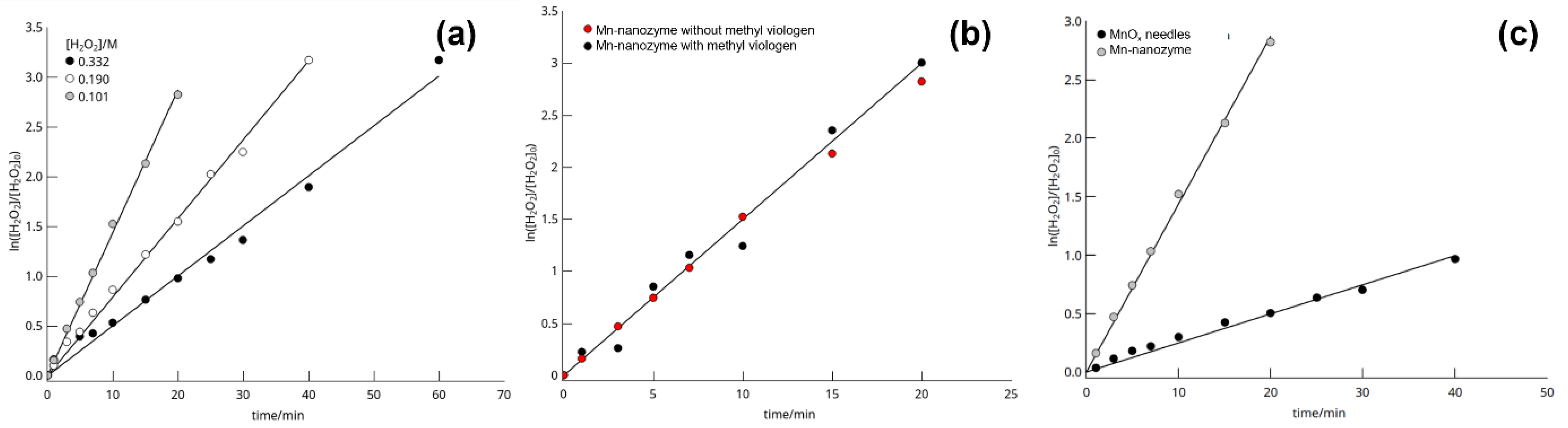
| Experiment | [H2O2]T (M) Initial | k × 102 (min−1) | R | Observations |
|---|---|---|---|---|
| 1 | 0.101 | 1.43 ± 0.02 | 0.998 | Reference reaction |
| 2 | 0.190 | 0.79 ± 0.01 | 0.992 | - |
| 3 | 0.332 | 0.50 ± 0.01 | 0.993 | - |
| 4 | 0.101 | 0.25 ± 0.06 | 0.991 | Uncoated MnOx needles |
| 5 | 0.101 | 1.50 ± 0.05 | 0.991 | Reaction in the presence of methylviologen |
Publisher’s Note: MDPI stays neutral with regard to jurisdictional claims in published maps and institutional affiliations. |
© 2022 by the authors. Licensee MDPI, Basel, Switzerland. This article is an open access article distributed under the terms and conditions of the Creative Commons Attribution (CC BY) license (https://creativecommons.org/licenses/by/4.0/).
Share and Cite
Garrido, M.D.; El Haskouri, J.; Marcos, M.D.; Pérez-Pla, F.; Ros-Lis, J.V.; Amorós, P. One-Pot Synthesis of MnOx-SiO2 Porous Composites as Nanozymes with ROS-Scavenging Properties. Nanomaterials 2022, 12, 3503. https://0-doi-org.brum.beds.ac.uk/10.3390/nano12193503
Garrido MD, El Haskouri J, Marcos MD, Pérez-Pla F, Ros-Lis JV, Amorós P. One-Pot Synthesis of MnOx-SiO2 Porous Composites as Nanozymes with ROS-Scavenging Properties. Nanomaterials. 2022; 12(19):3503. https://0-doi-org.brum.beds.ac.uk/10.3390/nano12193503
Chicago/Turabian StyleGarrido, M. Dolores, Jamal El Haskouri, María D. Marcos, Francisco Pérez-Pla, José Vicente Ros-Lis, and Pedro Amorós. 2022. "One-Pot Synthesis of MnOx-SiO2 Porous Composites as Nanozymes with ROS-Scavenging Properties" Nanomaterials 12, no. 19: 3503. https://0-doi-org.brum.beds.ac.uk/10.3390/nano12193503