
Tin/Tin Oxide Nanostructures: Formation, Application, and Atomic and Electronic Structure Peculiarities


1
Department Functional Interfaces, Leibniz Institute of Photonic Technology, Albert-Einstein Str. 9, 07745 Jena, Germany
2
Institute of Physical Chemistry, Friedrich Schiller University Jena, Helmholtzweg 4, 07743 Jena, Germany
*
Author to whom correspondence should be addressed.
Nanomaterials 2023, 13(17), 2391; https://0-doi-org.brum.beds.ac.uk/10.3390/nano13172391
Submission received: 28 June 2023
/
Revised: 18 August 2023
/
Accepted: 19 August 2023
/
Published: 22 August 2023
(This article belongs to the Special Issue Current Review in Nanofabrication and Nanomanufacturing)
Abstract
:For a very long period, tin was considered one of the most important metals for humans due to its easy access in nature and abundance of sources. In the past, tin was mainly used to make various utensils and weapons. Today, nanostructured tin and especially its oxide materials have been found to possess many characteristic physical and chemical properties that allow their use as functional materials in various fields such as energy storage, photocatalytic process, gas sensors, and solar cells. This review discusses current methods for the synthesis of Sn/SnO2 composite materials in form of powder or thin film, as well as the application of the most advanced characterization tools based on large-scale synchrotron radiation facilities to study their chemical composition and electronic features. In addition, the applications of Sn/SnO2 composites in various fields are presented in detail.
1. Introduction
Tin, one of the earliest metals known by human beings in 3500 BC, is still one of the most important metals today. In the very ancient period, soft copper was combined with a relative amount of tin to make bronze materials, which are much harder and can be applied in various tools and weapons with high strength and long lifetime. During recent centuries, with the development of fundamental scientific studies, a deeper understanding of tin’s chemical and physical properties has been constructed. Tin and its compounds such as tin oxides and tin sulfides, have been widely used as functional materials in electronics, chemical engineering, energy storage, and bio-photonics [1].
Among the wide range of tin-related materials, metallic tin and its oxides, which are the most usually existing tin states in nature, particularly attracting attention from scientists because of their unique structures and properties [2]. Metallic tin is a semimetal with atomic number 50 in the periodic table of elements. In general, there are three known tin oxide forms: SnO, SnO2, and Sn3O4. SnO is a p-type semiconductor with a dielectric constant of 15 and a bandgap about 2.4–2.7 eV [3]. At high temperature above 400 °C, SnO is thermodynamically unstable and disproportionate to metallic tin (0) and tin (IV) oxide. SnO2 is the most abundant and thermodynamically stable tin state in the nature. In ambient conditions, the crystal of SnO2 typically shows a tetragonal rutile structure. Stoichiometric SnO2 is generally be considered as an n-type semiconductor with a wide bandgap about 3.6 eV [4]. Sn3O4 is a metastable intermediate between SnO2 and SnO. It has been discussed that Sn3O4 has a layered structure which is held together by van der Waals forces [5]. As Sn3O4 has both Sn(IV) and Sn(II) ions, its electronic structure contains features form both SnO2 and SnO. In the reported literature, it is expected to be a p-type semiconductor with a bandgap in the range of 2.2–3.0 eV [5].
During the last couple of decades, nanoscale materials and nanostructures have attracted substantial attention due to the quantum size effect-induced novel properties when the size of a material is comparable to the Bohr radius [6,7]. Among all metal oxide semiconductors, nanomaterials of tin/tin oxides have received considerable notification and have found potential applications in various areas such as solar cells [8], lithium batteries [9], gas sensors [10], and catalysis [11,12], as shown in Figure 1. At the nanoscale, the optoelectronic properties of SnO2 such as optical bandgap, conductivity, and photoluminescence can be controlled via the incorporation of impurity or defects [13]. Moreover, Sn/SnO2 materials have high chemical and mechanical stability, have high abundance in nature, and are environmentally friendly. All these properties and advantages make tin/tin oxides promising candidates for constructing various next-generation energy storage and optoelectronic devices. In this review, various synthesis routes for fabricating nanostructured tin/tin oxides are presented, together with the introduction of state-of-the-art surface sensitive methods using large-scale synchrotron radiation characterization tools to study their unique atomic and electronic structure. Moreover, the different application potential for these functional materials is discussed. It is expected to provide an overview for the development of tin/tin oxide nanomaterials in recent years and promote related research.
2. Fabrication of Tin-Based Nanostructures and Thin Films
To further promote the widespread use of Sn/SnO2 composite (nano)powders and thin films, various fabrication methods have been developed with respect to the required properties, structures, and large-scale production. Among all fabrication methods, solid-state based, solution-based, and vapor-based methods can be used to classify the different routes.
2.1. Solid-State Methods
In solid-state methods, reactions between solid reactants are usually generated with additional heat or mechanic treatment. These methods are usually simple and efficient, with one synthesis step, whereby many materials can be obtained with high concentration. Furthermore, by adjusting different parameters, it is possible to control the size of the obtained materials.
Sinha et al. fabricated spherical Sn/SnO2 nanoparticles with 50 nm diameter on a gram level using a focused solar irradiation approach [14]. By stirring a strong NaOH solution with SnCl2 power, black microplates of SnO were observed. Upon further solar irradiation, a transformation into high-bandgap Sn/SnO2 nanoparticles was obtained. On the other hand, solid-state methods are mainly applied to synthesize pure tin oxide materials, such as SnO or SnO2. For example, Li et al. synthesized tin oxide nanoparticles in the size range of 3–15 nm through a convenient inexpensive and efficient one-step solid-state process [15]. Tin chloride, KCl, and KOH are typical reactants which can be mixed, accompanied by the emission of water vapor, followed by a wash and annealing step. According to the annealing temperature controlling, SnO2 nanoparticles with different sizes can be synthesized. The obtained SnO2 nanoparticles have high yield and gas sensitivity to EtOH, H2, and CO. Yang et al. applied a mechanochemical reaction between SnCl2 and Na2CO3 with NaCl as a diluent, followed by heat treatment at 600 °C to make tin oxide nanocrystals with an average crystal size of about 28 nm [16]. Chakravarty et al. also applied this method to synthesis mesoporous tin oxide in the range of 6–12 nm with a large surface area of 265 ± 16 m2 g−1. The so-obtained mesoporous tin oxide can be used as an advanced sorbent material for biological applications [17]. Apart from traditional solid-based methods to get 3D nanoparticles, more efforts are currently going toward fabricating 2D materials because of their unique physical and chemical properties. Jiang et al. obtained a 2D SnO nanosheet via mechanical exfoliation using Scotch tape, as shown in Figure 2 [18]. By adjusting the peeling process, nanolayers with varying thicknesses were formed, and the authors further found that the obtained nanostructures showed physical properties that were strongly thickness-dependent. The bandgap could be tuned from the IR range (0.60 eV) for bulk SnO to the UV range (3.65 eV) for the monolayer. On the other hand, it was reported that 2D SnO could be a good precursor for the synthesis of Sn/SnO2 composites, as presented in the next section related to wet chemical Sn/SnO2 composite formation. Additionally, it should be pointed out that classical solid-state methods such as mechanical milling can be used for the formation of Sn/SnO2 composites where tin oxide and metallic tin are mixed and milled in different stoichiometric ratios. Sivashanmugam et al. showed that a matrix in which metallic tin can be distributed without aggregation is essential for realizing Sn/SnO2 anodes with high cyclability [19].
2.2. Solution-Based Methods
Another popular route to synthesize Sn-based materials is based on the solution process, which usually includes an intensive chemical reaction in solution. The materials obtained using these methods are more uniformly distributed, and the experimental conditions are easily controlled.
One of the most important applications of Sn/SnO2 nanostructures is in electrodes for lithium-ion batteries, sensors, and supercapacitors. To increase the capacity, it is necessary to combine them with carbon materials, and solution processes provide an easy way to realize the synthesis. For example, Zhu et al. created a roughly 3 µm flower-like Sn/SnO2 graded-structure with excellent ethanol gas-sensing properties using 2D SnO sheets and a hydrothermal technique [20]. Hassan et al. applied an easy solution process using SnCl2·2H2O as a precursor and CMK-3 as a carbon framework [21]. After stirring, drying, and annealing processes, uniformly Sn/SnO2 embedded within the carbon pore walls could be obtained, exhibiting high and stable performance in lithium-ion batteries. Using a similar process, Wang et al. obtained evenly distributed Sn/SnO2 nanoparticles with 5 nm average diameter in multilayers of graphene sheets, presenting a hollow spherical structure [22].
Another technique to synthesize Sn/SnO2 nanoparticles is the electrochemical approach [23]. Saito et al. developed a surfactant-free direct-current electrolysis method using KCl as the electrolyte, as shown in Figure 3a–c [24]. By controlling the applied voltage and the concentration of the electrolyte, the obtained Sn/SnO2 particle size could be tuned from 200 to 1000 nm. Santiago-Giraldo et al. studied the reaction time when mixing SnCl2, cetyltrimethylammonium bromide, and ammonium hydroxide directly in an aqueous medium under reflux [25]. More photocatalytic activity was found with a longer reaction time.
In addition to the abovementioned methods, other solution-based ways include the hydrothermal process and sol–gel method. The hydrothermal method is one of the most classic routes to synthesize nanoparticles, and it gives a flexible way to control conditions. By applying high-pressure conditions, intensive chemical reactions can happen in a solution above its boiling point. Using this method, it is easier to fabricate tin oxide materials. For example, by utilizing the redox activity of dicarboxylic acid, it is possible to realize significant control over the composition and morphology of the synthesized SnO2 structures, as reported by Zima et al. and shown in Figure 3d–g [26]. The control over dicarboxylic acid can lead to the formation of single-phase Sn3O4 in hexagonal nanoplates and SnO2/Sn3O4 mixed phases in hierarchical flower-like structures. Furthermore, Akhir et al. found it is easy to change the crystallite size of SnO2 nanostructures by simply varying the precursor concentration, as well as reaction temperature and duration, during the hydrothermal synthesis [27]. More specifically, the crystal size could be obtained in the range of 7.88 to 18.41 nm.
2.3. Vapor-Based Methods
Vapor-state methods mainly applied for thin-film deposition. These processes generally include the evaporation of the primary precursors, followed by vigorous reactions with or without co-reactants. The benefits of these methods are well-controlled deposition conditions, uniformly distributed layers, high purity, and reproducibility. However, vapor-based methods usually require high-vacuum equipment and, therefore, bring high cost to large-scale production. Nevertheless, the electronic industry widely employs these technologies to obtain high-quality functional layers.
Sputtering is a promising technique as it can bring less contamination and high purity [28,29]. Sn/SnO2 can be used as the Sn and O sources in the sputtering technique. For example, Hsu et al. reported that, by controlling the sputtering conditions such as temperatures and pressure when using the robust Sn/SnO2 mixed target, the deposited films could be tuned from pure n-type SnO2 to pure p-type SnO, with a p-type Hall mobility of up to 2 cm2 V−1 s−1. One typical morphology of SnO2 obtained by sputtering deposition is shown in Figure 4c [30,31,32]. On the other hand, the same technique can be used for the formation of Sn/SnO2 composites, as Mohamed et al. reported the feasibility of tuning the composition Sn/SnO2 during the deposition by controlling the sputtering conditions [33].
Chemical vapor deposition (CVD) has been widely used in the commercial deposition industry because of its inexpensive nature and flexibility in the production of synthesis thin films and 1D nanostructures with high quality [38,39,40,41]. Many studies have described the CVD process of Sn-based thin films using SnCl4 and SnI4 as the tin precursors. In our previous studies, we also presented the deposition of Sn/SnO2 thin films using tin(IV) tert-butoxide as the tin and oxygen source [42,43]. This method is fast and simple for depositing Sn/SnO2 thin films with a dense structure, and the morphology and composition can be easily changed by varying the deposition temperatures and substrates, as we showed previously [38]. Compared with CVD, atomic layer deposition (ALD) is expected to yield an extremely thin layer with high purity for applications in the precision electronic industry. The structure of a normal ALD reactor is shown in Figure 4a. Typical Sn-based thin films deposited by low-temperature and plasma enhanced ALD are shown in Figure 4b,e. This method usually consists of several deposition steps in cycles to deposition films on an atomic scale, as shown in Figure 4f. The normally used precursors include SnCl2 [34], C12H26-N2Sn [37], Sn(acac)2 [44], TDMA-Sn [36], and Sn(edpa)2 [45]. Additionally, vapor–liquid–solid (VLS) growth [46] can be used for the realization of Sn/SnO2 composite systems. Wang et al. used SnO vapor transport to the silicon wafer surface covered with Au–Ag catalyst at 650 °C for Sn/SnO2 nanostructure formation [47]. On the other hand, it should be mentioned that the potential electrical and optical device performance of tin-based composites obtained in such a way is limited due to the surface contamination by gold, as well known from VLS-grown silicon nanowires [48].
Another method to deposit tin-based thin layers is the electrochemical approach. Knapik et al. reported the electrodeposition of SnO2 from a SnCl2 solution containing HNO3; the SEM image of this film is presented in Figure 4d [35]. Using different potentials and durations, the tin-based composite could be further controlled through a subsequent annealing process. The deposited layer showed promising photoelectrochemical properties and is expected to be applied in photoanodes. Additionally, 10–50 nm Sn/SnO2 nanocomposites in a core–shell structure covered with multilayer graphene could be synthesized via a one-step process of electrical wire explosion in liquid medium. For this purpose, Song et al. used a 0.3 mm diameter tin wire with 1-octanol (C8H17OH) as the liquid medium, and the electrical explosion process was performed at a charging voltage of 15 kV. The observed nanocomposite electrode showed a high specific capacity of 1270 mAhg−1 after 100 cycles and high reversible capacity of around 650 mAhg−1 at a current density of 5000 mAg−1 [49]. These results indicate that Sn/SnO2 nanocomposites are promising material systems to improve the electrochemical performance of anode materials for lithium-ion batteries.
3. Applications
From the application point of view, Sn/SnO2 composites have received considerable attentions as they show up in many different fields. For example, the synergic properties of Sn and SnO2 were found to benefit electrochemical performance in a supercapacitor [50]. In the semiconductor area, Sn/SnO2 was reported to fabricate high-response UV photodetectors with tunable ultraviolet and blue emissions [51,52]. In this review, some of the most realistic applications such as metal-ion batteries, energy storage/conversation, gas sensing, and catalysis are emphasized.
3.1. Li-Ion and Na-Ion Batteries
Lithium-ion batteries have been considered the most promising and commercially successful energy storage devices due to their high capacity, long stability, and low cost [53,54]. They have been very widely applied in different electronic devices and mobile vehicles, significantly reducing energy wasting in the transformation process. Sn-based materials have been carefully studied as the anode material in Li-ion batteries because of their high theoretical capacity (781 mAhg−1) compared with traditional graphite anodes (372 mAhg−1). However, in practical research, it has been found that, during the charging and discharging process of lithium-ion batteries, large volume changes and agglomeration of tin materials occur, resulting in severe attenuation of battery capacity during long cycle operation. To solve this problem, special nanostructured tin-based materials can be implemented. From this point-of-view, Sn-based materials, such as Sn, SnO, and SnO2, are highly promising anode materials for lithium-ion batteries, as potential substitutes for the conventional graphite anode due to their estimated theoretical capacity values such as 994 mAhg−1, ~875 mAhg−1, and ~782 mAhg−1, respectively, which are much higher than that of the commercialized graphite anode [55]. Moreover, these high capacities of Sn-based anode materials for lithium-ion batteries have been attributed to the formation of the Li4.4Sn alloy [56].
Wang et al. synthesized Sn/SnOx core–shell nanospheres using a modified polyol process, with the possibility to tune the material size [57]. By applying 45 nm nanospheres, the Li-ion battery exhibited capacity as high as 3443 mAhcm−3. In addition, other nanostructures have also been developed, such as compact Sn/SnO2 microspheres [58], hollow spheres [59], and nanosheets [59]. Another way to modify tin materials is to combine them with some special porous carbon materials, as the carbon structure not only provides mechanical strain relief during skeleton volume changes, but also improves conductivity, thereby increasing electron transfer efficiency [50,60,61,62,63]. Xie et al. applied a porous carbon framework that allowed SnO2 nanoparticles to separate in a novel nanofiber structure, resulting in a high reversible capacity of 986 mAhg−1 even after 200 cycles, with a high initial coulombic efficiency of 73.5%, as shown in Figure 5 [64]. This design was found to efficiently reduce the side reactions and promote the reversible conversion of Sn to SnO2. The authors also found that the dispersed Sn–SnO2 nanoclusters in a carbon nanotube network could provide abundant active sites for Li-ion storage and, therefore, yield excellent rate capability and stability [61]. Other carbon materials, such as graphene oxide [49,65,66], carbon nanofiber [67,68], nano-porous carbon [69,70,71], carbon nanosheets [59,72], hexahedral carbon frameworks [73,74], core–shell carbon structures [75], and carbon paper [76], have also been found to have a good synergistic effect with Sn-based materials to improve battery performance. To further improve battery performance, researchers have also used some other methods. For example, Kravchyk et al. used a simple ligand-exchange procedure using inorganic capping ligands to facilitate electronic connectivity with the Sn/SnO2 nanocrystals on the anode [77]. Using this method, a high capacity above 700 mAhg−1 was obtained after 100 cycles of deep charging at a relatively high current of 1000 mAhg−1. Other materials, such as MoS2 [78], SiO2 [79], and MXene (Ti3C2Tx) [80], have also been studied in combination with Sn-based materials to gain better anode performance in Li-ion batteries. In addition, other methods such as doping with nitrogen [81] and coating with polydopamine [82] are also expected to help Sn-based materials be an excellent anode for Li-ion batteries.
In addition to Li-ion batteries, sodium-ion (Na+) batteries have attracted great attention and are considered an alternative new energy storage device because of the abundant sodium resources and low cost. In particular, Sn has been investigated as a potential anode material in sodium-ion batteries because of its high theoretical specific capacity at 845 mAhg−1. Tang et al. synthesized a mesoporous Sn/SnO2 composite and applied it into sodium-ion batteries as an anode to get good performance [83]. The specific capacity was as high as 372 mAhg−1 at a current density of 50 mAg−1, and the battery exhibited good cycling performance even after 50 cycles. Similar to the application of Sn/SnO2 in lithium-ion batteries, the combination of carbon and tin-based materials can generate a sodium-ion battery with much better performance. Li et al. employed porous carbon as the tin skeleton, and a very high specific capacity at 1148.1 mAhg−1 was obtained, as shown in Figure 6 [84]. The great enhancement comes from the increased conductivity and suppressed volume expansion during cycling.
3.2. Energy Conversation and Storage
“Green energy” sources such as solar energy, wind energy, or waste heat are greatly influenced by the fluctuation of weather and atmospheric (year season) conditions, as well as light regime (day and night) [85,86]. For that reason, the development of reliable energy and waste heat technologies has high importance for the permanent energy supply. It should be mentioned that, at this time, most of the world’s power is generated by heat engines which are operated at 30–40% efficiency, which means that over 10 terawatts of heat is lost to the environment as waste heat. One of the promising energy storage technologies, along with battery-based technology, is related to thermal energy storage (TES) using phase change materials (PCMs) as a highly effective technology to solve the permanent energy delivery problem. PCMs as latent heat storage materials have high energy storage density and relatively constant operating temperature; thus, they have been widely investigated for promising applications in solar thermal energy storage [87,88], waste heat recovery [89,90], thermal management [91,92,93], buildings [94,95], etc. In the medium-temperature (373–673 K) range, in comparison to organic-based and molten salt-based PCMs, many studies have shown that metals and alloys as PCMs have higher thermal conductivity and large thermal energy storage density. Indium, tin, bismuth, and their alloys are promising candidates for use in a medium-temperature TES system. However, on the other hand, such materials are chemically unstable in bare form (as not encapsulated into the inert matrix); it follows that metal-based PCMs as heat storage media can leak and corrode the container during the solid–liquid phase transition. The main challenge when engineering such types of materials is related to the metallic PCMs effectively packaged in a chemically stable matrix. As reported previously, composite PCMs with porous supporting additives [96,97,98] and encapsulation to make the core–shell structured capsules [99,100,101] can solve the problems of leakage and corrosion of the solid–liquid PCMs. In particular, the core–shell capsules with stabilized shells can isolate the inside core materials with outside air, thereby avoiding the leakage and oxidation of core PCMs. In the medium-temperature range, metallic tin (Sn) is the best candidate with highest enthalpy and low cost, which has been preliminarily studied by several groups. For instance, Navarrete et al. [102] carried out the coating of SiO2 and Al2O3 shells using atomic layer deposition, and they successfully prepared Sn@SiO2 and Sn@Al2O3 nanoparticles, which showed higher thermal stability. Metal or semiconducting nanoparticles embedded in insulating matrices have been the object of continuously increasing interest due to their peculiar physical properties such as strong size-dependent shifts in the optical bandgap and in exciton binding energy when the size of the particle is on the order of the excitonic Bohr radius [25,26,103,104]. On the other hand, for implementing such materials in different environments, the realization of PCMs in the form of thin layers is preferable, e.g., ion implantation [105]. However, such a formation method is expensive, and more economically relevant formation methods are requested. Sheng et al. reported a simple wet chemical approach for the formation of Sn/SnO2 composite; after 100 melting–freezing cycles, the phase change properties and structures without any cracks or leakage remained, making such a material very promising for energy storage applications [7].
As the most sustainable energy source, solar is always the hope for the next generation. Solar cells are considered the most valuable next-generation green energy devices. Many different solar cells have been developed in the last few decades, but the working principle of these cells is similar [106,107,108]. By absorbing photon energy, the active material in solar cells can be excited and generate electrons and holes. These photogenerated carriers can be transport by the electron transport layer (ETL) and hole transport layer (HTL), respectively, resulting in photocurrent. In previous ETL studies, TiO2 was considered a good candidate because of its high electron transport efficiency, and a record efficiency at 24.8% could be obtained in perovskite solar cells (PSCs) [109]. However, the application of Sn/SnO2 composites in solar cells as ETLs is limited because the existence of metallic Sn results in a potential recombination center and blocks the charge transfer. However, pure SnO2 has been found to be a very promising material in solar cells as the ETL to replace TiO2 because of its excellent energy band alignment with the active absorber materials and high electron mobility [110,111,112,113,114,115,116,117,118,119,120,121,122,123]. To further promote tin based materials, the applications of tin oxides are worthy of discussion. For example, SnOx-based electrodes were applied in flexible organic solar cells in a recent publication, with a very high efficiency over 25% [119]. Furthermore, the up-to-date efficiency recorded for PSCs applying SnO2 was a power conversion efficiency (PCE) of 26% [124]. In fact, Sn-based ETLs have shown great potential for highly efficient and stable PSCs, especially for low-temperature flexible PSCs.
Morphology brings huge effects in terms of the electron transfer ability of SnO2. Mali et al. studied the morphology effect of SnO2 ETLs for PSC performance and found that the nanobelts SnO2 ETL exhibited over 30% higher PCE than PSCs applying SnO2 with nanofiber morphology, as shown in Figure 7 [125]. In addition, Singh et al. found that PSCs using a combination of both SnO2 nanoparticles and compact SnO2 layers as ETLs enabled a higher PCE and much better stability compared to the single morphology [126].
However, due to the defects in SnO2, the bulk and interfacial nonradiative recombination significantly suppresses the further enhancement of the PCE and stability of Sn-based PSCs [127,128]. To overcome this obstacle, doping could be a route to increase the performance of Sn-based ETL [129]. For example, Xiong et al. found that Mg-doped SnO2 films had higher mobility than undoped SnO2, thus exhibiting a nearly 92.8% enhancement in PCE [130]. The reason for this is that suitable Mg doping dramatically reduces free electron density and substantially increases the electron mobility of pristine SnO2. A fullerene derivative named fullerenoacetate was also found to suppress charge recombination in PSCs due to the efficient passivation of oxygen vacancy-related defects on the surface of the SnO2 ETL, as reported by Liu et al. [131]. An improved photovoltaic performance with efficiency up to 21.3% was obtained with negligible hysteresis.
Bi et al. reported a novel and effective multifunctional modification strategy through incorporating Girard’s Reagent T molecules with multiple functional groups to modify SnO2 nanoparticles [132]. This method led to very much reduced recombination losses and efficiently passivated interfacial defects. As a result, a much higher PCE of 21.63% was obtained, together with reduced hysteresis, compared with the reference. Recently, MXenes, a class of two-dimensional transition metal carbides and nitrides, have been found to have great potential for introduction into Sn-based ETLs [133]. Yang et al. studied titanium carbide Ti3C2Tx quantum dot-modified SnO2 and found that it can help to rapidly induce perovskite nucleation from the precursor solution and improve the crystal quality and phase stability of the as-fabricated perovskite film [134]. A steady-state PCE of up to 23.3% was obtained with amazing stability against humidity and light soaking. Another study also found that the application of Ti3C2 MXene in SnO2 ETLs can provide superior charge transfer paths and enhance electron extraction and transfer, leading to higher photocurrents [135].
3.3. Gas Sensors
Gas sensors play an important role in everyday life for dangerous gas detection, environmental pollution monitoring, and safety alerts in sensitive working areas. Among all gas sensor materials, Sn-based materials have attracted particular attention due to their high selectivity and sensitivity, as well as their low cost with an easy production process, making them good candidates for gas sensing under an atmospheric environment [10,136]. By absorbing gas molecules on the surface, the induced physical and chemical changes make tin-based sensors respond immediately. With fabrication via a simple solvothermal route, Verma et al. obtained Sn/SnO2 nanocomposites with a high specific surface area of 118.8 m2/g, exhibiting excellent sensitivity to xylene at room temperature and they found that the Sn metal plays a critical catalytic role for enhanced performance [137]. To make Sn/SnO2 with a special nanostructure, the gas-sensing property can be further adjusted. For example, Zhu et al. reported that 3D hierarchical flower-like Sn/SnO2 exhibited high selectivity and sensitivity to ethanol gas due to the rise in the Schottky barrier caused by the in situ production of tin particles [20]. Yuan et al. proposed that metallic Sn atoms in the Sn/SnO2 composites could serve as active sites for the sensing reaction, and they found that the density of unsaturated Sn atoms with dangling bonds at the SnO2 surface could have a significant influence on the sensing performance [138].
Although Sn/SnO2 composites with special nanostructures have been found to have excellent gas-sensing performance, the applications of pure tin oxide materials for gas sensing are more widely studied because of the easier fabrication process and lower cost. From the economic and mass production point of view, it is worth presenting these studies. For different oxide states of Sn, the fabricated gas sensors have different properties. For example, Sn3O4 was found to have very high selectivity to NO2 relative to CO, while SnO exhibited the highest selectivity to NO2 relative to H2 and CH4, and SnO2 had lower performance in the detection of NO2 [139,140,141,142]. However, the combination of SnO and SnO2 to form a p–n heterojunction enabled high sensing selectivity for NO2, with a limit of detection and sensitivity of 0.1 ppm and 0.26 ppm−1, respectively, at a relatively low operating temperatures of about 50 °C [143].
Another efficient method involved the introduction of other functional materials. For example, Wang et al. applied SnO–SnO2-modified 2D MXene Ti3C2Tx to make high acetone gas-sensing devices with a short recovery time and outstanding reproducibility, attributed to the formation of heterojunctions and high conductivity of the metallic phase Ti3C2Tx, as shown in Figure 8 [144]. Wang et al. applied SiC nanofibers as the support of SnO2 nanosheet with a hierarchical architecture, showing an ultrafast response/recovery rate, as well as high sensitivity to various target gases such as ethanol, methanol, hydrogen, isopropanol, acetone, and xylene [145]. The superior sensing performance came from the hierarchical architecture and synergetic effect of the SnO2–SiC heterojunction, with many active sites from the vertically ultrathin SnO2 nanosheets. Yan et al. synthesized microporous Sn–SnO2/carbon heterostructure nanofibers and found that the introduction of metallic tin helped to get improved sensing ethanol properties because of its effective electron transfer property [146]. Moreover, the carbon skeleton also provided good permeability for gas detection. Similarly, Sn–SnO2 with doped nitrogen also have high gas sensor performance to toxic NH3 gas [136]. Other functional materials, such as CuO [147], ZnO [148], and PdO [149] also brought outstanding gas sensor properties for Sn-based materials. Furthermore, the selectivity and sensitivity of such sensors can be easily adjusted by make different nanostructured SnO2 [20,150,151,152]. For example, SnO2 nanoclusters embedded on a mesoporous Sn organophosphonate framework was found to be an efficient approach to enable gas sensors to have remarkable sensitivity toward ammonia and acetone [153]. SnO2 nanowires synthesized by the CVD process were also reported to have high sensitivity toward different gases, such as H2S [154], NO2 [155,156], and water vapor [157]. Hollow SnO2 microfiber was found to have high sensitivity toward triethylamine, with a limit of detection as low as 2 ppm [158]. Some additional small treatments such as doping also bring big improvements regarding to gas-sensing performance. Liang et al. reported that a Ce-doped SnO2 thin film could enable the adsorption of a large number of oxygen ions on the surface, resulting in an increased ethanol vapor-sensing response [159]. Nd-doped SnO2 nanorod layers were also proven to exhibit excellent sensing response toward alcohol at a temperature of 260 °C [160]. The good catalytic properties of Nd dopant were able to increase the number of active sites on the surface of SnO2, thus enabling higher sensitivity.
3.4. Photocatalytic and Bio-Photonic Applications
Wide-bandgap semiconductor materials such as TiO2 and ZnO have been regarded as important photocatalysis for the applications of degrading organic pollutants and hydrogen production. With a similar energy band structure, tin oxides have also attracted interest in these areas.
The transformation of Sn to SnO2 was found to have better photocatalytic properties because of an improved surface area and higher stability toward adverse environmental conditions. It was found that metallic Sn in SnO2 can improve the charge separation efficiency and, therefore, bring faster degradation of cationic dye molecules [14]. Li et al. reported the Sn/SnO2−x hetero-nanostructure form built-in semiconductor–metal Schottky junctions and promote charge carrier separation, as well as accelerate surface catalytic oxygen evolution reaction kinetics [161]. At last, a high photocurrent density of 245 μA cm−2 at 1.23 V versus the reversible hydrogen electrode was obtained, as shown in Figure 9.
Pure SnO2 has also been found to have good catalytic properties. For example, Gao et al. developed highly active SnO2 catalyst via a spontaneous exchange reaction between CuO substrate and sputtered Sn particles, and the synthesized SnO2 sites were found to selectively catalyze CO generation from CO2 reduction [162]. SnO2 with different morphologies has also been studied for the photocatalytic properties of the degradation of methyl-orange, which is a well-known organic pollutant in water [163]. It was found that tin oxide with different nanostructures presents a barrier effect to slow the product volatilization and thermal transport during decomposition of the polymer, thus enabling better photocatalytic performance.
Furthermore, additional functional materials can also help to improve the photocatalytic process. For example, the combinations of Sn and SnO2, as well as carbon, have been found to have high photocatalytic activity for the degradation of reactive brilliant blue KN-R dye under visible-light irradiation [164]. The metallic Sn and adsorbed oxygen as the sinks of photoinduced electron and electronic scavenges resulted in a hindrance of the recombination of photoexcited electron–hole pairs, as well as enhanced the photocatalytic activity. With a similar composition, C-supported Sn–SnO2 also showed photocatalytic activity for the degradation of Rhodamine B [165] and methylene blue dyes [166,167]. Kaleji et al. introduced TiO2 into SnO2 and found high optical absorption in the visible-light area. Lastly, there was high photocatalytic ability for the degradation of methylene blue dye, and the increase in surface oxygen vacancies and hydroxyl groups contributed to this result. Other materials, such as Pt [168], Ag [169], Ga [170], and GdS [171] have also been found to be able to tune the photocatalytic properties of tin oxides.
Raman spectroscopy is an important tool in bioanalysis since it is highly specific and provides clear information about the chemical structure of the probed molecules—without the need for labels. Wavelengths from the deep UV to the IR range are used for excitation, enabling the identification and differentiation of individual components up to whole microorganisms and tissue. The UV range, in particular, is very interesting for proteins, as they specifically absorb light below 400 nm, thereby resonantly enhancing vibrational modes characteristic of the peptide backbone and certain amino-acid residues. The low-intensity characteristic Raman modes can be compensated for by the use of surface enhanced Raman spectroscopy (SERS) substrates. In SERS, the probed molecules interact with metal nanostructures, which enables ultra-sensitive detection down to single molecules. The signal enhancement is based on the excitation of surface plasmons in rough metal nanostructures, which can be electrodes, colloids, or island films, as shown in Figure 10a. When the incident laser wavelength is chosen such that it matches the absorption maximum of the plasmon resonance of the substrate, an enormous electromagnetic field is generated. Molecules in the immediate vicinity to the metal surface experience a Raman signal enhancement up to 1014 [172,173,174]. It is noteworthy that the spectral quality and reproducibility are strongly influenced by the size, shape, and dimension of the SERS substrates. For measurements in the visible-light range (i.e., 418–785 nm), Au, Ag, and Cu nanostructures are highly efficient. SERS in the deep UV range (<300 nm) has not yet been systematically explored and applied, but it could be very promising for the structural analysis of proteins since the resonance Raman effect is added to the SERS enhancement. While, in the visible range, SERS has been successfully applied to amyloid fibril analysis [175,176,177], so far, only one protein-related proof-of-principle experiment with UV-SERS has been published [178]. Other UV-SERS studies have been limited to the investigation of adenine, tryptophan, and dyes. The so-far-employed nanostructures include the following metals and wavelengths: Al (244 nm [179], 257 nm [180], 266 nm [181]), Ru, Rh (325 nm [182,183]), and In (266 nm [184], 325 nm [185]). Nonetheless, in the last few years, a couple of scientific publications have been reported on the Sn/SnO2−x hetero-nanostructure potential in light-driven processes such as photo-electrochemical water oxidation [161] or (UV)-SERS [186,187]. According to the previously published theoretically estimated light absorption property of tin-based systems, plasmonic properties similar to those of noble metals have been observed [188,189]. It was also shown that the localized surface plasmon resonance (LSPR)-induced light absorption range of Sn-based systems can be varied from the ultraviolet (about 200 nm) to the near-infrared region (about 600 nm) by controlling the particle size, as shown in Figure 10b [187,188]. From this point of view, Sn-based systems could be very powerful as SERS substrates for protein structure analysis.
4. Atomic and Electronic Features Studies in Tin/Tin Oxide Nanostructures
As discussed in previous sections, the functionality of devices and/or materials crucially depends on the achieved surfaces atomic and electronic structure. Therefore, it is essential to determine the surface atomic and electronic structure and peculiarities of materials. However, the typical analytical characterization techniques in the lab, such as microscopy, spectroscopy, and diffraction, are mostly bulk methods which are not surface-sensitive and cannot detect the chemical/physical properties on the surface. In comparison to the lab techniques (where only X-ray photoelectron spectroscopy (XPS) is possible), synchrotron radiation facilities have a higher photon number, which leads to higher resolution and sensitivity to small material amounts (especially important for nano-scaled materials) and higher sensitivity, especially in the case of tin, which has a low photoionization cross-section, as shown in our previous publication [190]. The X-ray absorption near-edge structure (XANES) method has a probing depth of 5 nm compared to the probing depth of 2 nm for the XPS method. The combination of XANES and XPS measurements at the same spot using high-resolution synchrotron facilities can allow a precise reconstruction of the atomic and electronic structure of the studied surfaces. It should be noted that the presence of vacancies or lower oxidation degrees of tin can be brilliantly visualized using the XANES method in the surface-sensitive mode (using soft X-rays) due to the presence of additional electronic states presented in the spectrum as separate features. In addition, by using synchrotron facilities and varying the excitation wavelength of photons (550–1000 eV) in XPS, it is possible to vary the inelastic mean electron-free paths or, in other words, to change the depth of analysis from a few Å to 20 Å, which, in combination with XANES, can give an “overall picture” of the atomic and electronic structure of the surface, which is impossible with the XPS laboratory technique. In our previous studies, the XANES technique was successfully used to reveal the function of suboxide layers on silicon nanowires for a high production of hydrogen during photocatalytic water splitting [191]. In addition, such surface-sensitive methods have also been successfully applied to investigate the mechanism of charge transfer and optimize performance in batteries [192,193] and catalysis research [194,195].
Synchrotron radiation involves electromagnetic radiation emitted along the tangent direction by charged particles whose speed is close to the speed of light when moving in a curve—also called synchrotron light. The light source produced has many advantages. It has a continuous spectrum from far infrared to X-ray, and a monochromator can be used to call out light with an applicable wavelength in the spectrum. At the same time, high light intensity brings benefits in the study of trace elements in extremely small samples and materials. Moreover, because the synchrotron radiation light is emitted by the periodic movement of the electron beam cluster in the storage ring, it has a nanosecond to microsecond time pulse. By utilizing this characteristic, time-related chemical reactions, physical excitation processes, and changes in biological cells can be studied. Many techniques have been developed on the basis of synchrotron radiation. A number of X-ray and electron spectroscopy techniques together with known sample preparation/modification techniques can be applied to study the specificity of the local atomic environment of the tin, oxygen, and silicon atoms over the surface layer and the bulk of all structures formed [196,197,198].
Synchrotron studies can allow us to obtain precise information about the atomic and electronic structure, local atomic surrounding specificity, and physical–chemical state of the as-prepared composite nanostructures or modified ones: under certain formation conditions, after storage at normal atmospheric conditions, and after in operando measurements. In this review, the focus is on XPS and XANES, as they are the most widely used for Sn-based material research and can provide a wealth of information regarding the electronic and elemental characterizations [199,200,201,202,203].
XPS employs X-rays to excite the surface of the studied materials, while measuring the kinetic energy of electrons emitted within a few nanometers of the surface and recording the spectrum with respect to the intensity, as shown in Figure 11a. The peaks appearing in the XPS spectrum result from the emission of electrons with a certain characteristic energy in the atom. The energy and intensity of photoelectron peaks can be used for qualitative and quantitative analysis of all surface elements (except for H and He elements). Generally, the information of element composition, chemical state, and molecular structure on the sample surface can be obtained from the peak position and peak shape of the XPS spectrum, and the element content or concentration on the sample surface can be obtained from the peak area [204].
After X-ray irradiation on a substance, it is absorbed by the substance, but the absorption coefficient does not change monotonically throughout the entire wavelength band. At certain positions, there are absorption jumps, becoming absorption edges. There are some discrete peaks or fluctuations near the absorption edge and its high-energy extension, known as X-ray absorption spectroscopy, as shown in the schematic diagram of Figure 11b. Its distribution is about 1000 eV from the front of the absorption edge to the high-energy side behind the absorption edge. According to the different formation mechanisms and processing methods, it is usually divided into two distinct parts: extended X-ray absorption fine structure (EXAFS) and XANES, also known as near-edge X-ray absorption fine structure (NEXAFS). EXAFS exhibits a continuous and slow weak oscillation at 50–1000 eV after the absorption edge. It can represent the structure information of atomic clusters in a small range, including the coordination number of atoms, atomic spacing, type, thermal disturbance, and other neighboring geometric structures around the absorption atoms [205]. XANES includes 50 eV before and after the absorption edge, characterized by continuous strong oscillations. It can represent a wealth of information about the neighboring structure, such as the bond angle of neighboring atoms, and the electronic and valence states of detected element atoms. In spite of the broad application perspectives, weak fundamental studies on tin oxide nanostructures have been reported. To overcome the lateral resolution limitation of the XANES technique, a different spectromicroscopy technique can be applied for wide composition and morphology studies of the surface at “one spot” [206,207,208,209]. This means that the employment of laterally resolved surface sensitive soft X-ray absorption edge analysis for composite structure studies promises to be very informative, taking into account previously published results from Si–O [210,211,212,213] and Sn–O system studies [203,214,215,216,217,218]. The high-energy and high-spatial-resolution synchrotron methods deal with low-intensity signals, where intensive excitation radiation plays the key role for obtaining reliable information. As pointed out before, X-ray absorption fine structures are extremely sensitive to the specificity of given atoms in the local environment [219], which allows us to estimate the weak atomic and electronic structural changes in Sn–O systems localized around silicon nanowires or planar silicon surfaces. Photo-emission electron microscopy (PEEM) in soft XANES spectroscopy mode can be efficiently applied for Sn–O system studies in different environments [220]. Only Kolmakov et al. on SnO2 nanowires [221] and Turishchev et al. on tin oxide-covered silicon nanowire arrays [190] presented PEEM results, showing a lack of studies on the surface of low-dimensional tin oxide nanostructures, which makes this topic extremely important.
Figure 11.
Schematic diagram of the theory of XPS (a) and XANES (b) [222]. Reprinted with permission from Ref. [222], Copyright 2023, NSF.

In our previous studies, we showed the possibility of applying XANES (NEXAFS) to identify Sn crystal phases in silicon nanowire matrices [42,190]. This nondestructive surface-sensitive technique allows the precise detection of metallic Sn, Sn(II), and Sn(IV), which originates from the electronic features, as shown in Figure 12. Chuvenkova et al. employed XANES and XPS to investigate the surface defects in SnO2 crystals, showing their informative and sensitive properties to the local atomic environment, attributed to the oxygen vacancies in obtained Sn-based composite thin films [216,223]. Gago et al. combined XRD and XANES to investigate the transition from SnO to SnO2 by increasing the O2 partial pressure during the sputtering process [224]. XRD and XANES provide complementary results about the formation of single- and mixed-phased films. Furthermore, XANES give unique information about defects such as the incorporation of O2 molecules at high O2 partial pressure [225]. Zhou et al. revealed the origin of the multicolor luminescence from SnO2 by combing the results from time-resolved X-ray excited optical luminescence and XANES [226].
In Li-ion batteries, XANES can be powerful as an operando analytic tool for in situ monitoring of the charge and discharge process in a running device. Birrozzi et al. studied the de-/lithiation mechanism of SnO2 nanoparticles, as well as the synergetic impact of Co and Mn doping, by recording the XANES spectra from Sn L-edge, Mn K-edge, and Co K-edge, as these spectra provide fruitful information about the changes between oxidation states and metallic states during the working process of Li-ion batteries [227]. Similarly, Pelliccione et al. also investigated the dynamic changes in the Sn atomic environment to analyze the formation of Sn–Li phases within the electrodes by employing XANES, and they further revealed the reasons for poor electrochemical performance and rapid capacity decline [201].
In PSCs, the SnO2 ETL is normally fabricated via a solution process, and one of the biggest challenges comes from the –OH groups on its surface because of the proton-rich conditions, which are harmful for PSCs because they can degrade the stability and efficiency of PSCs. XPS and XANES could enable a powerful investigation of the interface chemical environment, addressing this drawback. For example, Jeon et al. applied XPS and XANES to confirm the substantial reduction in surface hydroxyl groups on SnO2 ETL by passivation, and they further found that the surface hydroxyls groups act as defect sites to reduce the charge transfer and carrier lifetime of perovskites [228]. On the other hand, these tools can also explore the degradation and working principle of Sn-based perovskites, as the oxidation of Sn(II) to Sn(IV) from O2− and H2O− in perovskite can be easily captured by XPS and XANES [229,230]. These findings could provide insights into the mechanistic picture of tin halide perovskites and promote them as alternatives to Pb-based materials in PSCs as they are less toxic and cheaper.
5. Conclusions
Nanostructured tin/tin oxide composites have been extensively studied and proven their unique functions in various devices such as gas sensors, energy storage, solar cells, photocatalysis, and bio-photonic devices. The flexibility to fabricate tin-based materials for various applications can be controlled using different synthesis routes with varying synthesis conditions. Among all methods, solid-based methods usually give the possibility to produce nanoparticles with uniform sizes, while solution-based methods have more flexibility to produce tin/tin oxide materials with greater morphological diversity, simpler processes, and lower costs. However, gas-based methods can provide the greatest homogeneity on substrates to meet the high demands of the electronics industry. Modern synchrotron radiation-based characterization techniques provide more opportunities to investigate the atomic and electronic structure, local atomic surrounding specificity, and physical–chemical states of tin/tin oxide materials, which enhances the understanding of these materials and their mechanism of operation in various devices. The outstanding performance of nanostructured tin/tin oxides in various fields has been presented. In order to realize industrial applications, more efforts are also needed to develop better synthesis pathways and equipment to reduce production costs. However, achieving theoretically maximum parameters still requires further optimization of this family of materials. Understanding the relationship between structure and properties still deserves further investigation to obtain the most optimized device performance.
Author Contributions
Conceptualization, P.L. and V.S.; writing—original draft preparation, P.L.; writing—review and editing, V.S.; supervision, V.S.; funding acquisition, V.S. All authors have read and agreed to the published version of the manuscript.
Funding
This research was funded by German Research Foundation (DFG) under Grant No. 448666227 (SI1893/27-1).
Data Availability Statement
The data is available in section “MDPI Research Data Policies” at https://0-www-mdpi-com.brum.beds.ac.uk/ethics (18 August 2023).
Acknowledgments
V.S. and P.L. gratefully acknowledge financial support by the German Research Foundation (DFG) under Grant No. 448666227 (SI1893/27-1).
Conflicts of Interest
The authors declare no conflict of interest.
References
- Mohanta, D.; Ahmaruzzaman, M. Tin oxide nanostructured materials: An overview of recent developments in synthesis, modifications and potential applications. RSC Adv. 2016, 6, 110996–111015. [Google Scholar] [CrossRef]
- An, H.; Yoo, M.; Ha, H.; Choi, H.; Kang, E.; Kim, H.Y. Efficient Sn Recovery from SnO2 by Alkane (CxHy=2x+2, 0 ≤ x ≤ 4) Reduction. Sci. Rep. 2019, 9, 16702. [Google Scholar] [CrossRef] [PubMed]
- Zhou, W.; Umezawa, N. Band gap engineering of bulk and nanosheet SnO: An insight into the interlayer Sn-Sn lone pair interactions. Phys. Chem. Chem. Phys. 2015, 17, 17816–17820. [Google Scholar] [CrossRef]
- Zhou, W.; Liu, Y.; Yang, Y.; Wu, P. Band Gap Engineering of SnO2 by Epitaxial Strain: Experimental and Theoretical Investigations. J. Phys. Chem. C 2014, 118, 6448–6453. [Google Scholar] [CrossRef]
- Liu, Y.S.; Yamaguchi, A.; Yang, Y.; Aisnada, A.N.E.; Uchida, S.; Abe, H.; Ueda, S.; Yamaguchi, K.; Tanabe, T.; Miyauchi, M. Synthesis and Characterization of the Orthorhombic Sn3O4 Polymorph. Angew. Chem. Int. Ed. Engl. 2023, 62, e202300640. [Google Scholar] [CrossRef] [PubMed]
- Periyasamy, M.; Kar, A. Modulating the properties of SnO2 nanocrystals: Morphological effects on structural, photoluminescence, photocatalytic, electrochemical and gas sensing properties. J. Mater. Chem. C 2020, 8, 4604–4635. [Google Scholar] [CrossRef]
- Sheng, N.; Lu, J.; Hu, J.; Zhu, R.; Fadillah, L.; Wang, C.; Zhu, C.; Rao, Z.; Habazaki, H. Synthesis of Sn@SnO2 core-shell microcapsules by a self-oxidation strategy for medium temperature thermal storage. Chem. Eng. J. 2021, 420, 129906. [Google Scholar] [CrossRef]
- Kuang, Y.; Zardetto, V.; van Gils, R.; Karwal, S.; Koushik, D.; Verheijen, M.A.; Black, L.E.; Weijtens, C.; Veenstra, S.; Andriessen, R.; et al. Low-Temperature Plasma-Assisted Atomic-Layer-Deposited SnO2 as an Electron Transport Layer in Planar Perovskite Solar Cells. ACS Appl. Mater. Interfaces 2018, 10, 30367–30378. [Google Scholar] [CrossRef]
- Wang, L.P.; Leconte, Y.; Feng, Z.; Wei, C.; Zhao, Y.; Ma, Q.; Xu, W.; Bourrioux, S.; Azais, P.; Srinivasan, M.; et al. Novel Preparation of N-Doped SnO2 Nanoparticles via Laser-Assisted Pyrolysis: Demonstration of Exceptional Lithium Storage Properties. Adv. Mater. 2017, 29, 1603286. [Google Scholar] [CrossRef]
- Li, F.; Chen, L.; Chen, Z.; Xu, J.; Zhu, J.; Xin, X. Two-step solid-state synthesis of tin oxide and its gas-sensing property. Mater. Chem. Phys. 2002, 73, 335–338. [Google Scholar] [CrossRef]
- Ren, B.; Zhang, Z.; Wen, G.; Zhang, X.; Xu, M.; Weng, Y.; Nie, Y.; Dou, H.; Jiang, Y.; Deng, Y.P.; et al. Dual-Scale Integration Design of Sn-ZnO Catalyst toward Efficient and Stable CO2 Electroreduction. Adv. Mater. 2022, 34, e2204637. [Google Scholar] [CrossRef] [PubMed]
- Guo, W.; Liu, S.; Tan, X.; Wu, R.; Yan, X.; Chen, C.; Zhu, Q.; Zheng, L.; Ma, J.; Zhang, J.; et al. Highly Efficient CO2 Electroreduction to Methanol through Atomically Dispersed Sn Coupled with Defective CuO Catalysts. Angew. Chem. Int. Ed. Engl. 2021, 60, 21979–21987. [Google Scholar] [CrossRef] [PubMed]
- Cho, J.S.; Kang, Y.C. Nanofibers Comprising Yolk-Shell Sn@void@SnO/SnO2 and Hollow SnO/SnO2 and SnO2 Nanospheres via the Kirkendall Diffusion Effect and Their Electrochemical Properties. Small 2015, 11, 4673–4681. [Google Scholar] [CrossRef] [PubMed]
- Sinha, A.K.; Pradhan, M.; Sarkar, S.; Pal, T. Large-scale solid-state synthesis of Sn-SnO2 nanoparticles from layered SnO by sunlight: A material for dye degradation in water by photocatalytic reaction. Environ. Sci. Technol. 2013, 47, 2339–2345. [Google Scholar] [CrossRef]
- Li, F.; Xu, J.; Yu, X.; Chen, L.; Zhu, J.; Yang, Z.; Xin, X. One-step solid-state reaction synthesis and gas sensing property of tin oxide nanoparticles. Sens. Actuators B Chem. 2000, 81, 165–169. [Google Scholar] [CrossRef]
- Yang, H.; Hu, Y.; Tang, A.; Jin, S.; Qiu, G. Synthesis of tin oxide nanoparticles by mechanochemical reaction. J. Alloys Compd. 2004, 363, 276–279. [Google Scholar] [CrossRef]
- Chakravarty, R.; Chakraborty, S.; Shukla, R.; Bahadur, J.; Ram, R.; Mazumder, S.; Dev Sarma, H.; Tyagi, A.K.; Dash, A. Mechanochemical synthesis of mesoporous tin oxide: A new generation nanosorbent for 68Ge/68Ga generator technology. Dalton Trans. 2016, 45, 13361–13372. [Google Scholar] [CrossRef]
- Jiang, K.; Ji, J.; Gong, W.; Ding, L.; Li, J.; Li, P.; Li, B.; Geng, F. Mechanical cleavage of non-van der Waals structures towards two-dimensional crystals. Nat. Synth. 2022, 2, 58–66. [Google Scholar] [CrossRef]
- Sivashanmugam, A.; Kumar, T.P.; Renganathan, N.G.; Gopukumar, S.; Wohlfahrt-Mehrens, M.; Garche, J. Electrochemical behavior of Sn/SnO2 mixtures for use as anode in lithium rechargeable batteries. J. Power Sources 2005, 144, 197–203. [Google Scholar] [CrossRef]
- Zhu, Y.; Yang, L.; Guo, S.; Hou, M.; Ma, Y. In Situ Synthesis of Hierarchical Flower-like Sn/SnO2 Heterogeneous Structure for Ethanol GAS Detection. Materials 2023, 16, 792. [Google Scholar] [CrossRef]
- Hassan, F.M.; Chen, Z.; Yu, A.; Chen, Z.; Xiao, X. Sn/SnO2 embedded in mesoporous carbon nanocomposites as negative electrode for lithium ion batteries. Electrochim. Acta 2013, 87, 844–852. [Google Scholar] [CrossRef]
- Wang, Z.; Song, D.; Si, J.; Jiang, Y.; Yang, Y.; Jiang, Y.; Huang, S.; Chen, Z.; Zhao, B. One-step hydrothermal reduction synthesis of tiny Sn/SnO2 nanoparticles sandwiching between spherical graphene with excellent lithium storage cycling performances. Electrochim. Acta 2018, 292, 72–80. [Google Scholar] [CrossRef]
- Lim, J.; Garcia-Esparza, A.T.; Lee, J.W.; Kang, G.; Shin, S.; Jeon, S.S.; Lee, H. Electrodeposited Sn-Cu@Sn dendrites for selective electrochemical CO2 reduction to formic acid. Nanoscale 2022, 14, 9297–9303. [Google Scholar] [CrossRef]
- Saito, G.; Azman, W.O.S.B.W.M.; Nakasugi, Y.; Akiyama, T. Optimization of electrolyte concentration and voltage for effective formation of Sn/SnO2 nanoparticles by electrolysis in liquid. Adv. Powder Technol. 2014, 25, 1038–1042. [Google Scholar] [CrossRef]
- Santiago-Giraldo, F.M.; Lugo-Ruiz, A.A.; Bailón-Ruiz, S.J. Production and characterization of tin oxide (SnO2) nanostructures. MRS Adv. 2022, 7, 249–254. [Google Scholar] [CrossRef]
- Zima, T.; Bataev, I. Morphology and phase transformations of tin oxide nanostructures synthesized by the hydrothermal method in the presence of dicarboxylic acids. J. Solid State Chem. 2016, 243, 282–289. [Google Scholar] [CrossRef]
- Akhir, M.A.M.; Mohamed, K.; Lee, H.L.; Rezan, S.A. Synthesis of Tin Oxide Nanostructures Using Hydrothermal Method and Optimization of its Crystal size by Using Statistical Design of Experiment. Procedia Chem. 2016, 19, 993–998. [Google Scholar] [CrossRef]
- Zakaria, Y.; Aissa, B.; Fix, T.; Ahzi, S.; Samara, A.; Mansour, S.; Slaoui, A. Study of wide bandgap SnOx thin films grown by a reactive magnetron sputtering via a two-step method. Sci. Rep. 2022, 12, 15294. [Google Scholar] [CrossRef]
- Guillén, C.; Herrero, J. P-type SnO thin films prepared by reactive sputtering at high deposition rates. J. Mater. Sci. Technol. 2019, 35, 1706–1711. [Google Scholar] [CrossRef]
- Hsu, P.-C.; Chen, W.-C.; Tsai, Y.-T.; Kung, Y.-C.; Chang, C.-H.; Wu, C.-C.; Hsieh, H.-H. Sputtering deposition of P-type SnO films using robust Sn/SnO2 mixed target. Thin Solid Films 2014, 555, 57–61. [Google Scholar] [CrossRef]
- Hsu, P.-C.; Tsai, S.-P.; Chang, C.-H.; Hsu, C.-J.; Chen, W.-C.; Hsieh, H.-H.; Wu, C.-C. Preparation of p-type SnO thin films and transistors by sputtering with robust Sn/SnO2 mixed target in hydrogen-containing atmosphere. Thin Solid Films 2015, 585, 50–56. [Google Scholar] [CrossRef]
- Hsu, P.C.; Hsu, C.J.; Chang, C.H.; Tsai, S.P.; Chen, W.C.; Hsieh, H.H.; Wu, C.C. Sputtering deposition of P-type SnO films with SnO2 target in hydrogen-containing atmosphere. ACS Appl. Mater. Interfaces 2014, 6, 13724–13729. [Google Scholar] [CrossRef] [PubMed]
- Mohamed, A.Y.; Lee, S.J.; Jang, Y.; Kim, J.S.; Hwang, C.S.; Cho, D.Y. X-ray spectroscopy study on the electronic structure of Sn-added p-type SnO films. J. Phys. Condens. Matter 2020, 32, 065502. [Google Scholar] [CrossRef] [PubMed]
- Stevens, E.C.; Mousa, M.B.M.; Parsons, G.N. Thermal atomic layer deposition of Sn metal using SnCl4 and a vapor phase silyl dihydropyrazine reducing agent. J. Vac. Sci. Technol. A 2018, 36, 06A106. [Google Scholar] [CrossRef]
- Knapik, A.; Syrek, K.; Kozieł, M.; Zaraska, L. Cathodic deposition of SnO2 layers on transparent conductive substrates and their photoelectrochemical activity. J. Ind. Eng. Chem. 2022, 111, 380–388. [Google Scholar] [CrossRef]
- Huang, P.H.; Zhang, Z.X.; Hsu, C.H.; Wu, W.Y.; Ou, S.L.; Huang, C.J.; Wuu, D.S.; Lien, S.Y.; Zhu, W.Z. Deposition Mechanism and Characterization of Plasma-Enhanced Atomic Layer-Deposited SnOx Films at Different Substrate Temperatures. Nanomaterials 2022, 12, 2859. [Google Scholar] [CrossRef]
- Heo, J.; Hock, A.S.; Gordon, R.G. Low Temperature Atomic Layer Deposition of Tin Oxide. Chem. Mater. 2010, 22, 4964–4973. [Google Scholar] [CrossRef]
- Vallejos, S.; Selina, S.; Annanouch, F.E.; Gracia, I.; Llobet, E.; Blackman, C. Aerosol assisted chemical vapour deposition of gas sensitive SnO2 and Au-functionalised SnO2 nanorods via a non-catalysed vapour solid (VS) mechanism. Sci. Rep. 2016, 6, 28464. [Google Scholar] [CrossRef]
- Han, S.H.; Agbenyeke, R.E.; Lee, G.Y.; Park, B.K.; Kim, C.G.; Eom, T.; Son, S.U.; Han, J.H.; Ryu, J.Y.; Chung, T.M. Novel Heteroleptic Tin(II) Complexes Capable of Forming SnO and SnO2 Thin Films Depending on Conditions Using Chemical Solution Deposition. ACS Omega 2022, 7, 1232–1243. [Google Scholar] [CrossRef]
- Mathur, S.; Barth, S.; Shen, H.; Pyun, J.C.; Werner, U. Size-dependent photoconductance in SnO2 nanowires. Small 2005, 1, 713–717. [Google Scholar] [CrossRef]
- Sivakov, V. Iron, Germanium and Tin Tert-Butoxides in the CVD Process: Thin Film Deposition and Characterization. Ph.D. Thesis, Universität des Saarlandes, Saarland, Germany, 2004. [Google Scholar]
- Liu, P.; Schleusener, A.; Zieger, G.; Bochmann, A.; van Spronsen, M.A.; Sivakov, V. Nanostructured Silicon Matrix for Materials Engineering. Small 2023, 19, e2206318. [Google Scholar] [CrossRef] [PubMed]
- Mathur, S.; Sivakov, V.; Shen, H.; Barth, S.; Cavelius, C.; Nilsson, A.; Kuhn, P. Nanostructured films of iron, tin and titanium oxides by chemical vapor deposition. Thin Solid Films 2006, 502, 88–93. [Google Scholar] [CrossRef]
- Nguyen, V.H.; Akbari, M.; Sekkat, A.; Ta, H.T.T.; Resende, J.; Jimenez, C.; Musselman, K.P.; Munoz-Rojas, D. Atmospheric atomic layer deposition of SnO2 thin films with tin(II) acetylacetonate and water. Dalton Trans. 2022, 51, 9278–9290. [Google Scholar] [CrossRef]
- Kim, H.Y.; Nam, J.H.; George, S.M.; Park, J.-S.; Park, B.K.; Kim, G.H.; Jeon, D.J.; Chung, T.-M.; Han, J.H. Phase-controlled SnO2 and SnO growth by atomic layer deposition using Bis(N-ethoxy-2,2-dimethyl propanamido)tin precursor. Ceram. Int. 2019, 45, 5124–5132. [Google Scholar] [CrossRef]
- Wagner, R.S.; Ellis, W.C. Vapor-Liquid-Solid Mechanism of Single Crystal Growth. Appl. Phys. Lett. 1964, 4, 89–90. [Google Scholar] [CrossRef]
- Wang, B.; Yang, Y.H.; Yang, G.W. Growth mechanisms of SnO2/Sn nanocables. Nanotechnology 2006, 17, 4682–4688. [Google Scholar] [CrossRef]
- Putnam, M.C.; Filler, M.A.; Kayes, B.M.; Kelzenberg, M.D.; Guan, Y.; Lewis, N.S.; Eiler, J.M.; Atwater, H.A. Secondary Ion Mass Spectrometry of Vapor-Liquid-Solid Grown, Au-Catalyzed, Si Wires. Nano Lett. 2008, 8, 3109–3113. [Google Scholar] [CrossRef]
- Song, J.-S.; Cho, G.-B.; Kim, K.-W.; Ahn, H.-J.; Kim, H.-S.; Ahn, J.-H.; Cho, K.-K. Fabrication of multilayer graphene-encapsulated Sn/SnO2 nanocomposite as an anode material for lithium-ion batteries and its electrochemical properties. Appl. Surf. Sci. 2019, 481, 736–740. [Google Scholar] [CrossRef]
- Thorat, G.M.; Jadhav, H.S.; Chung, W.-J.; Seo, J.G. Collective use of deep eutectic solvent for one-pot synthesis of ternary Sn/SnO2@C electrode for supercapacitor. J. Alloys Compd. 2018, 732, 694–704. [Google Scholar] [CrossRef]
- Huang, Y.; Lin, J.; Li, L.; Xu, L.; Wang, W.; Zhang, J.; Xu, X.; Zou, J.; Tang, C. High performance UV light photodetectors based on Sn-nanodot-embedded SnO2 nanobelts. J. Mater. Chem. C 2015, 3, 5253–5258. [Google Scholar] [CrossRef]
- Li, S.; Zhong, X.; Song, Y.; Shen, X.; Sun, J.; Song, Y.; Wang, R.; Zhu, M.; Zhong, H.; Zheng, A. Controlled hybridization of Sn–SnO2 nanoparticles via simple-programmed microfluidic processes for tunable ultraviolet and blue emissions. J. Mater. Chem. C 2014, 2, 7687–7694. [Google Scholar] [CrossRef]
- Han, J.; Kong, D.; Lv, W.; Tang, D.M.; Han, D.; Zhang, C.; Liu, D.; Xiao, Z.; Zhang, X.; Xiao, J.; et al. Caging tin oxide in three-dimensional graphene networks for superior volumetric lithium storage. Nat. Commun. 2018, 9, 402. [Google Scholar] [CrossRef] [PubMed]
- Liu, P.; Wang, Y.; Liu, J. Biomass-derived porous carbon materials for advanced lithium sulfur batteries. J. Energy Chem. 2019, 34, 171–185. [Google Scholar] [CrossRef]
- Winter, M.; Besenhard, J.O. Electrochemical lithiation of tin and tin-based intermetallics and composites. Electrochim. Acta 1999, 45, 31–50. [Google Scholar] [CrossRef]
- Eom, K.; Jung, J.; Lee, J.T.; Lair, V.; Joshi, T.; Lee, S.W.; Lin, Z.; Fuller, T.F. Improved stability of nano-Sn electrode with high-quality nano-SEI formation for lithium ion battery. Nano Energy 2015, 12, 314–321. [Google Scholar] [CrossRef]
- Wang, X.-L.; Feygenson, M.; Aronson, M.C.; Han, W.-Q. Sn/SnOx Core-Shell Nanospheres: Synthesis, Anode Performance in Li Ion Batteries, and Superconductivity. J. Phys. Chem. C 2010, 114, 14697–14703. [Google Scholar] [CrossRef]
- Zhang, L.; Zhao, K.; Sun, C.; Yu, R.; Zhuang, Z.; Li, J.; Xu, W.; Wang, C.; Xu, W.; Mai, L. Compact Sn/SnO2 microspheres with gradient composition for high volumetric lithium storage. Energy Storage Mater. 2020, 25, 376–381. [Google Scholar] [CrossRef]
- He, F.; Xu, Q.; Zheng, B.; Zhang, J.; Wu, Z.; Zhong, Y.; Chen, Y.; Xiang, W.; Zhong, B.; Guo, X. Synthesis of hierarchical Sn/SnO nanosheets assembled by carbon-coated hollow nanospheres as anode materials for lithium/sodium ion batteries. RSC Adv. 2020, 10, 6035–6042. [Google Scholar] [CrossRef]
- Ding, J.; Zhu, W.; Liu, C.; Ma, C.; Yang, Y.; Ji, H.; Yang, G. Synthesis of heterostructure Sn|SnO2 submicron particles supported by carbon fibers as binder-free anodes for highly reversible lithium storage. J. Alloys Compd. 2018, 750, 220–227. [Google Scholar] [CrossRef]
- Sun, L.; Si, H.; Zhang, Y.; Shi, Y.; Wang, K.; Liu, J.; Zhang, Y. Sn-SnO2 hybrid nanoclusters embedded in carbon nanotubes with enhanced electrochemical performance for advanced lithium ion batteries. J. Power Sources 2019, 415, 126–135. [Google Scholar] [CrossRef]
- Pol, V.G.; Wen, J.; Miller, D.J.; Thackeray, M.M. Sonochemical Deposition of Sn, SnO2 and Sb on Spherical Hard Carbon Electrodes for Li-Ion Batteries. J. Electrochem. Soc. 2014, 161, A777–A782. [Google Scholar] [CrossRef]
- Wang, D.; Li, X.; Yang, J.; Wang, J.; Geng, D.; Li, R.; Cai, M.; Sham, T.K.; Sun, X. Hierarchical nanostructured core-shell Sn@C nanoparticles embedded in graphene nanosheets: Spectroscopic view and their application in lithium ion batteries. Phys. Chem. Chem. Phys. 2013, 15, 3535–3542. [Google Scholar] [CrossRef] [PubMed]
- Xie, W.; Gu, L.; Xia, F.; Liu, B.; Hou, X.; Wang, Q.; Liu, D.; He, D. Fabrication of voids-involved SnO2@C nanofibers electrodes with highly reversible Sn/SnO2 conversion and much enhanced coulombic efficiency for lithium-ion batteries. J. Power Sources 2016, 327, 21–28. [Google Scholar] [CrossRef]
- Dong, W.; Xu, J.; Wang, C.; Lu, Y.; Liu, X.; Wang, X.; Yuan, X.; Wang, Z.; Lin, T.; Sui, M.; et al. A Robust and Conductive Black Tin Oxide Nanostructure Makes Efficient Lithium-Ion Batteries Possible. Adv. Mater. 2017, 29, 1700136. [Google Scholar] [CrossRef]
- Jiang, W.; Wang, W.; Liu, L.; Wang, H.; Xu, Z.; Li, F.; Fu, H.; Lv, H.; Chen, L.; Kang, Y. Sandwich-like Sn/SnO2@Graphene anode composite assembled by fortissimo penetration of γ-ray and interlamellar limitation of graphene oxide. J. Alloys Compd. 2019, 779, 856–862. [Google Scholar] [CrossRef]
- Gao, S.; Wang, N.; Li, S.; Li, D.; Cui, Z.; Yue, G.; Liu, J.; Zhao, X.; Jiang, L.; Zhao, Y. A Multi-Wall Sn/SnO2@Carbon Hollow Nanofiber Anode Material for High-Rate and Long-Life Lithium-Ion Batteries. Angew. Chem. Int. Ed. 2020, 59, 2465–2472. [Google Scholar] [CrossRef]
- Narsimulu, D.; Vadnala, S.; Srinadhu, E.S.; Satyanarayana, N. Electrospun Sn–SnO2/C composite nanofibers as an anode material for lithium battery applications. J. Mater. Sci. Mater. Electron. 2018, 29, 11117–11123. [Google Scholar] [CrossRef]
- Cheng, Y.; Yi, Z.; Wang, C.; Wu, Y.; Wang, L. Controllable fabrication of C/Sn and C/SnO/Sn composites as anode materials for high-performance lithium-ion batteries. Chem. Eng. J. 2017, 330, 1035–1043. [Google Scholar] [CrossRef]
- Dong, Y.; Das, S.; Zhu, L.; Ben, T.; Qiu, S. Standout electrochemical performance of SnO2 and Sn/SnO2 nanoparticles embedded in a KOH-activated carbonized porous aromatic framework (PAF-1) matrix as the anode for lithium-ion batteries. J. Mater. Chem. A 2016, 4, 18822–18831. [Google Scholar] [CrossRef]
- Wang, H.-Q.; Zhang, X.-H.; Wen, J.-B.; Huang, Y.-G.; Lai, F.-Y.; Li, Q.-Y. Preparation of Spherical Sn/SnO2/Porous Carbon Composite Materials as Anode Material for Lithium-Ion Batteries. J. Mater. Eng. Perform. 2015, 24, 1856–1864. [Google Scholar] [CrossRef]
- Sun, Q.; Kong, X.; Liu, W.; Xu, B.; Hu, P.; Gao, Z.; Huang, Y. Flakes-stacked Sn/SnO2/C composite as highly stable anode material for lithium-ion batteries. J. Alloys Compd. 2020, 831, 154677. [Google Scholar] [CrossRef]
- Li, X.; He, C.; Zheng, J.; Ye, W.; Yin, W.; Tang, B.; Rui, Y. Preparation of promising anode materials with Sn-MOF as precursors for superior lithium and sodium storage. J. Alloys Compd. 2020, 84, 155605. [Google Scholar] [CrossRef]
- Yang, J.; Chen, S.; Tang, J.; Tian, H.; Bai, T.; Zhou, X. Rod-like hierarchical Sn/SnOx@C nanostructures with enhanced lithium storage properties. Appl. Surf. Sci. 2018, 435, 203–209. [Google Scholar] [CrossRef]
- Cheng, Y.; Huang, J.; Li, J.; Xu, Z.; Cao, L.; Qi, H. Synergistic effect of the core-shell structured Sn/SnO2/C ternary anode system with the improved sodium storage performance. J. Power Sources 2016, 324, 447–454. [Google Scholar] [CrossRef]
- Tan, L.; Feng, S.; Li, X.; Wang, Z.; Peng, W.; Liu, T.; Yan, G.; Li, L.; Wu, F.; Wang, J. Oxygen-induced lithiophilicity of tin-based framework toward highly stable lithium metal anode. Chem. Eng. J. 2020, 394, 124848. [Google Scholar] [CrossRef]
- Kravchyk, K.; Protesescu, L.; Bodnarchuk, M.I.; Krumeich, F.; Yarema, M.; Walter, M.; Guntlin, C.; Kovalenko, M.V. Monodisperse and inorganically capped Sn and Sn/SnO2 nanocrystals for high-performance Li-ion battery anodes. J. Am. Chem. Soc. 2013, 135, 4199–4202. [Google Scholar] [CrossRef]
- Huang, Y.; Pan, Q.; Wang, H.; Ji, C.; Wu, X.; He, Z.; Li, Q. Preparation of a Sn@SnO2@C@MoS2 composite as a high-performance anode material for lithium-ion batteries. J. Mater. Chem. A 2016, 4, 7185–7189. [Google Scholar] [CrossRef]
- Namsar, O.; Autthawong, T.; Laokawee, V.; Boonprachai, R.; Haruta, M.; Kurata, H.; Yu, A.; Chairuangsri, T.; Sarakonsri, T. Improved electrochemical performance of anode materials for high energy density lithium-ion batteries through Sn(SnO2)–SiO2/graphene-based nanocomposites prepared by a facile and low-cost approach. Sustain. Energy Fuels 2020, 4, 4625–4636. [Google Scholar] [CrossRef]
- Zuo, D.-C.; Song, S.-C.; An, C.-S.; Tang, L.-B.; He, Z.-J.; Zheng, J.-C. Synthesis of sandwich-like structured Sn/SnOx@MXene composite through in-situ growth for highly reversible lithium storage. Nano Energy 2019, 62, 401–409. [Google Scholar] [CrossRef]
- Li, R.; Nie, S.; Miao, C.; Xin, Y.; Mou, H.; Xu, G.; Xiao, W. Heterostructural Sn/SnO2 microcube powders coated by a nitrogen-doped carbon layer as good-performance anode materials for lithium ion batteries. J. Colloid Interface Sci. 2022, 606, 1042–1054. [Google Scholar] [CrossRef]
- Jiang, B.; He, Y.; Li, B.; Zhao, S.; Wang, S.; He, Y.B.; Lin, Z. Polymer-Templated Formation of Polydopamine-Coated SnO2 Nanocrystals: Anodes for Cyclable Lithium-Ion Batteries. Angew. Chem. Int. Ed. 2017, 56, 1869–1872. [Google Scholar] [CrossRef] [PubMed]
- Tang, D.; Huang, Q.; Yi, R.; Dai, F.; Gordin, M.L.; Hu, S.; Chen, S.; Yu, Z.; Sohn, H.; Song, J.; et al. Room-Temperature Synthesis of Mesoporous Sn/SnO2 Composite as Anode for Sodium-Ion Batteries. Eur. J. Inorg. Chem. 2016, 2016, 1950–1954. [Google Scholar] [CrossRef]
- Li, X.; Li, X.; Fan, L.; Yu, Z.; Yan, B.; Xiong, D.; Song, X.; Li, S.; Adair, K.R.; Li, D.; et al. Rational design of Sn/SnO2 /porous carbon nanocomposites as anode materials for sodium-ion batteries. Appl. Surf. Sci. 2017, 412, 170–176. [Google Scholar] [CrossRef]
- Dahash, A.; Ochs, F.; Janetti, M.B.; Streicher, W. Advances in seasonal thermal energy storage for solar district heating applications: A critical review on large-scale hot-water tank and pit thermal energy storage systems. Appl. Energy 2019, 239, 296–315. [Google Scholar] [CrossRef]
- Tulus, V.; Boer, D.; Cabeza, L.F.; Jiménez, L.; Guillén-Gosálbez, G. Enhanced thermal energy supply via central solar heating plants with seasonal storage: A multi-objective optimization approach. Appl. Energy 2016, 181, 549–561. [Google Scholar] [CrossRef]
- Wei, H.; Qiu, C.; Wang, C.; Lin, K.; Yang, S.; Han, J.; Lu, Y.; Liu, X. Development of phase change materials using hydrolyzed Al-Bi composite powder for solar energy storage. Chem. Eng. J. 2021, 421, 127836. [Google Scholar] [CrossRef]
- Mehrali, M.; ten Elshof, J.E.; Shahi, M.; Mahmoudi, A. Simultaneous solar-thermal energy harvesting and storage via shape stabilized salt hydrate phase change material. Chem. Eng. J. 2021, 405, 126624. [Google Scholar] [CrossRef]
- Soni, V.; Kumar, A.; Jain, V.K. Performance evaluation of nano-enhanced phase change materials during discharge stage in waste heat recovery. Renew. Energy 2018, 127, 587–601. [Google Scholar] [CrossRef]
- Omara, A.A.M. Phase change materials for waste heat recovery in internal combustion engines: A review. J. Energy Storage 2021, 44, 103421. [Google Scholar] [CrossRef]
- Farzanehnia, A.; Khatibi, M.; Sardarabadi, M.; Passandideh-Fard, M. Experimental investigation of multiwall carbon nanotube/paraffin based heat sink for electronic device thermal management. Energy Convers. Manag. 2019, 179, 314–325. [Google Scholar] [CrossRef]
- Sun, Z.; Zhao, L.; Wan, H.; Liu, H.; Wu, D.; Wang, X. Construction of polyaniline/carbon nanotubes-functionalized phase-change microcapsules for thermal management application of supercapacitors. Chem. Eng. J. 2020, 396, 125317. [Google Scholar] [CrossRef]
- Hassan, A.; Wahab, A.; Qasim, M.A.; Janjua, M.M.; Ali, M.A.; Ali, H.M.; Jadoon, T.R.; Ali, E.; Raza, A.; Javaid, N. Thermal management and uniform temperature regulation of photovoltaic modules using hybrid phase change materials-nanofluids system. Renew. Energy 2020, 145, 282–293. [Google Scholar] [CrossRef]
- Konuklu, Y.; Ostry, M.; Paksoy, H.O.; Charvat, P. Review on using microencapsulated phase change materials (PCM) in building applications. Energy Build. 2015, 106, 134–155. [Google Scholar] [CrossRef]
- Keshteli, A.N.; Sheikholeslami, M. Nanoparticle enhanced PCM applications for intensification of thermal performance in building: A review. J. Mol. Liq. 2019, 274, 516–533. [Google Scholar] [CrossRef]
- Liu, C.; Zong, J.; Zhang, J.; He, D.; Guo, C.; Xu, B.; Rao, Z. Knitting aryl network polymers (KAPs)-embedded copper foam enables highly efficient thermal energy storage. J. Mater. Chem. A 2020, 8, 15177–15186. [Google Scholar] [CrossRef]
- Yang, J.; Qi, G.-Q.; Bao, R.-Y.; Yi, K.; Li, M.; Peng, L.; Cai, Z.; Yang, M.-B.; Wei, D.; Yang, W. Hybridizing graphene aerogel into three-dimensional graphene foam for high-performance composite phase change materials. Energy Storage Mater. 2018, 13, 88–95. [Google Scholar] [CrossRef]
- Atinafu, D.G.; Chang, S.J.; Kim, K.-H.; Dong, W.; Kim, S. A novel enhancement of shape/thermal stability and energy-storage capacity of phase change materials through the formation of composites with 3D porous (3,6)-connected metal–organic framework. Chem. Eng. J. 2020, 389, 124430. [Google Scholar] [CrossRef]
- Yuan, K.; Liu, J.; Fang, X.; Zhang, Z. Novel facile self-assembly approach to construct graphene oxide-decorated phase-change microcapsules with enhanced photo-to-thermal conversion performance. J. Mater. Chem. A 2018, 6, 4535–4543. [Google Scholar] [CrossRef]
- Lu, J.; Sheng, N.; Zhu, C. Fabrication of Sn@SiO2 core-shell microcapsules with high durability for medium-temperature thermal energy storage. Sol. Energy Mater. Sol. Cells 2022, 239, 111652. [Google Scholar] [CrossRef]
- Fukahori, R.; Nomura, T.; Zhu, C.; Sheng, N.; Okinaka, N.; Akiyama, T. Macro-encapsulation of metallic phase change material using cylindrical-type ceramic containers for high-temperature thermal energy storage. Appl. Energy 2016, 170, 324–328. [Google Scholar] [CrossRef]
- Navarrete, N.; La Zara, D.; Goulas, A.; Valdesueiro, D.; Hernández, L.; van Ommen, J.R.; Mondragón, R. Improved thermal energy storage of nanoencapsulated phase change materials by atomic layer deposition. Sol. Energy Mater. Sol. Cells 2020, 206, 110322. [Google Scholar] [CrossRef]
- Gaponenko, S.V. Optical Properties of Semiconductor Nanocrystals; Cambridge University Press: Cambridge, UK, 1998. [Google Scholar]
- Bukowski, T.J.; Simmons, J.H. Quantum Dot Research: Current State and Future Prospects. Crit. Rev. Solid State Mater. Sci. 2002, 27, 119–142. [Google Scholar] [CrossRef]
- Tagliente, M.A.; Bello, V.; Pellegrini, G.; Mattei, G.; Mazzoldi, P.; Massaro, M. SnO2 nanoparticles embedded in silica by ion implantation followed by thermal oxidation. J. Appl. Phys. 2009, 106, 104304. [Google Scholar] [CrossRef]
- Mu, H.; Yu, W.; Yuan, J.; Lin, S.; Zhang, G. Interface and surface engineering of black phosphorus: A review for optoelectronic and photonic applications. Mater. Futures 2022, 1, 104304. [Google Scholar] [CrossRef]
- Xu, R.; Guo, J.; Mi, S.; Wen, H.; Pang, F.; Ji, W.; Cheng, Z. Advanced atomic force microscopies and their applications in two-dimensional materials: A review. Mater. Futures 2022, 1, 032302. [Google Scholar] [CrossRef]
- Zhuang, R.; Cai, S.; Mei, Z.; Liang, H.; Zhao, N.; Mu, H.; Yu, W.; Jiang, Y.; Yuan, J.; Lau, S.; et al. Solution-grown BiI/BiI3 van der Waals heterostructures for sensitive X-ray detection. Nat. Commun. 2023, 14, 1621. [Google Scholar] [CrossRef]
- Huang, H.; Cui, P.; Chen, Y.; Yan, L.; Yue, X.; Qu, S.; Wang, X.; Du, S.; Liu, B.; Zhang, Q.; et al. 24.8%-efficient planar perovskite solar cells via ligand-engineered TiO2 deposition. Joule 2022, 6, 2186–2202. [Google Scholar] [CrossRef]
- Song, J.; Zheng, E.; Bian, J.; Wang, X.-F.; Tian, W.; Sanehira, Y.; Miyasaka, T. Low-temperature SnO2-based electron selective contact for efficient and stable perovskite solar cells. J. Mater. Chem. A 2015, 3, 10837–10844. [Google Scholar] [CrossRef]
- Li, Y.; Zhu, J.; Huang, Y.; Liu, F.; Lv, M.; Chen, S.; Hu, L.; Tang, J.; Yao, J.; Dai, S. Mesoporous SnO2 nanoparticle films as electron-transporting material in perovskite solar cells. RSC Adv. 2015, 5, 28424–28429. [Google Scholar] [CrossRef]
- Jung, K.-H.; Seo, J.-Y.; Lee, S.; Shin, H.; Park, N.-G. Solution-processed SnO2 thin film for a hysteresis-free planar perovskite solar cell with a power conversion efficiency of 19.2%. J. Mater. Chem. A 2017, 5, 24790–24803. [Google Scholar] [CrossRef]
- Jeong, S.; Seo, S.; Park, H.; Shin, H. Atomic layer deposition of a SnO2 electron-transporting layer for planar perovskite solar cells with a power conversion efficiency of 18.3%. Chem. Commun. 2019, 55, 2433–2436. [Google Scholar] [CrossRef]
- Dagar, J.; Castro-Hermosa, S.; Gasbarri, M.; Palma, A.L.; Cina, L.; Matteocci, F.; Calabrò, E.; Di Carlo, A.; Brown, T.M. Efficient fully laser-patterned flexible perovskite modules and solar cells based on low-temperature solution-processed SnO2/mesoporous-TiO2 electron transport layers. Nano Res. 2018, 11, 2669–2681. [Google Scholar] [CrossRef]
- Ahmad, W.; Liu, D.; Ahmad, W.; Wang, Y.; Zhang, P.; Zhang, T.; Zheng, H.; Chen, Z.D.; Li, S. Physisorption of Oxygen in SnO2 Nanoparticles for Perovskite Solar Cells. IEEE J. Photovoltaics 2019, 9, 200–206. [Google Scholar] [CrossRef]
- Tao, H.; Wang, H.; Bai, Y.; Long, H.; Zhao, H.; Fu, Q.; Ma, Z. Effects of sputtering power of SnO2 electron selective layer on perovskite solar cells. J. Mater. Sci. Mater. Electron. 2019, 30, 12036–12043. [Google Scholar] [CrossRef]
- Mohamad Noh, M.F.; Arzaee, N.A.; Safaei, J.; Mohamed, N.A.; Kim, H.P.; Mohd Yusoff, A.R.; Jang, J.; Mat Teridi, M.A. Eliminating oxygen vacancies in SnO2 films via aerosol-assisted chemical vapour deposition for perovskite solar cells and photoelectrochemical cells. J. Alloys Compd. 2019, 773, 997–1008. [Google Scholar] [CrossRef]
- Trost, S.; Behrendt, A.; Becker, T.; Polywka, A.; Görrn, P.; Riedl, T. Tin Oxide (SnOx) as Universal “Light-Soaking” Free Electron Extraction Material for Organic Solar Cells. Adv. Energy Mater. 2015, 5, 1500277. [Google Scholar] [CrossRef]
- Xie, C.; Xiao, C.; Fang, J.; Zhao, C.; Li, W. Core/shell AgNWs@SnOx electrodes for high performance flexible indoor organic solar cells with >25% efficiency. Nano Energy 2023, 107, 108153. [Google Scholar] [CrossRef]
- Yuan, R.; Cai, B.; Lv, Y.; Gao, X.; Gu, J.; Fan, Z.; Liu, X.; Yang, C.; Liu, M.; Zhang, W.-H. Boosted charge extraction of NbOx-enveloped SnO2 nanocrystals enables 24% efficient planar perovskite solar cells. Energy Environ. Sci. 2021, 14, 5074–5083. [Google Scholar] [CrossRef]
- Wu, P.; Wang, S.; Li, X.; Zhang, F. Advances in SnO2-based perovskite solar cells: From preparation to photovoltaic applications. J. Mater. Chem. A 2021, 9, 19554–19588. [Google Scholar] [CrossRef]
- Yang, D.; Yang, R.; Wang, K.; Wu, C.; Zhu, X.; Feng, J.; Ren, X.; Fang, G.; Priya, S.; Liu, S.F. High efficiency planar-type perovskite solar cells with negligible hysteresis using EDTA-complexed SnO2. Nat. Commun. 2018, 9, 3239. [Google Scholar] [CrossRef]
- Min, H.; Lee, D.Y.; Kim, J.; Kim, G.; Lee, K.S.; Kim, J.; Paik, M.J.; Kim, Y.K.; Kim, K.S.; Kim, M.G.; et al. Perovskite solar cells with atomically coherent interlayers on SnO2 electrodes. Nature 2021, 598, 444–450. [Google Scholar] [CrossRef] [PubMed]
- Park, J.; Kim, J.; Yun, H.S.; Paik, M.J.; Noh, E.; Mun, H.J.; Kim, M.G.; Shin, T.J.; Seok, S.I. Controlled growth of perovskite layers with volatile alkylammonium chlorides. Nature 2023, 616, 724–730. [Google Scholar] [CrossRef] [PubMed]
- Mali, S.S.; Patil, J.V.; Kim, H.; Hong, C.K. Synthesis of SnO2 nanofibers and nanobelts electron transporting layer for efficient perovskite solar cells. Nanoscale 2018, 10, 8275–8284. [Google Scholar] [CrossRef] [PubMed]
- Singh, M.; Ng, A.; Ren, Z.; Hu, H.; Lin, H.-C.; Chu, C.-W.; Li, G. Facile synthesis of composite tin oxide nanostructures for high-performance planar perovskite solar cells. Nano Energy 2019, 60, 275–284. [Google Scholar] [CrossRef]
- Ali, F.; Pham, N.D.; Bradford, H.J.; Khoshsirat, N.; Ostrikov, K.; Bell, J.M.; Wang, H.; Tesfamichael, T. Tuning the Amount of Oxygen Vacancies in Sputter-Deposited SnOx films for Enhancing the Performance of Perovskite Solar Cells. ChemSusChem 2018, 11, 3096–3103. [Google Scholar] [CrossRef]
- Jiang, E.; Yan, J.; Ai, Y.; Li, N.; Yan, B.; Zeng, Y.; Sheng, J.; Ye, J. Defect engineering of oxygen vacancies in SnOx electron transporting layer for perovskite solar cells. Mater. Today Energy 2019, 12, 389–397. [Google Scholar] [CrossRef]
- Xiong, Q.; Yang, L.; Zhou, Q.; Wu, T.; Mai, C.L.; Wang, Z.; Wu, S.; Li, X.; Gao, P. NdCl3 Dose as a Universal Approach for High-Efficiency Perovskite Solar Cells Based on Low-Temperature-Processed SnOx. ACS Appl. Mater. Interfaces 2020, 12, 46306–46316. [Google Scholar] [CrossRef]
- Xiong, L.; Qin, M.; Yang, G.; Guo, Y.; Lei, H.; Liu, Q.; Ke, W.; Tao, H.; Qin, P.; Li, S.; et al. Performance enhancement of high temperature SnO2-based planar perovskite solar cells: Electrical characterization and understanding of the mechanism. J. Mater. Chem. A 2016, 4, 8374–8383. [Google Scholar] [CrossRef]
- Liu, K.; Chen, S.; Wu, J.; Zhang, H.; Qin, M.; Lu, X.; Tu, Y.; Meng, Q.; Zhan, X. Fullerene derivative anchored SnO2 for high-performance perovskite solar cells. Energy Environ. Sci. 2018, 11, 3463–3471. [Google Scholar] [CrossRef]
- Bi, H.; Zuo, X.; Liu, B.; He, D.; Bai, L.; Wang, W.; Li, X.; Xiao, Z.; Sun, K.; Song, Q.; et al. Multifunctional organic ammonium salt-modified SnO2 nanoparticles toward efficient and stable planar perovskite solar cells. J. Mater. Chem. A 2021, 9, 3940–3951. [Google Scholar] [CrossRef]
- Huang, L.; Zhou, X.; Xue, R.; Xu, P.; Wang, S.; Xu, C.; Zeng, W.; Xiong, Y.; Sang, H.; Liang, D. Low-Temperature Growing Anatase TiO2/SnO2 Multi-dimensional Heterojunctions at MXene Conductive Network for High-Efficient Perovskite Solar Cells. Nano-Micro Lett. 2020, 12, 44. [Google Scholar] [CrossRef]
- Yang, Y.; Lu, H.; Feng, S.; Yang, L.; Dong, H.; Wang, J.; Tian, C.; Li, L.; Lu, H.; Jeong, J.; et al. Modulation of perovskite crystallization processes towards highly efficient and stable perovskite solar cells with MXene quantum dot-modified SnO2. Energy Environ. Sci. 2021, 14, 3447–3454. [Google Scholar] [CrossRef]
- Yang, L.; Dall’Agnese, Y.; Hantanasirisakul, K.; Shuck, C.E.; Maleski, K.; Alhabeb, M.; Chen, G.; Gao, Y.; Sanehira, Y.; Jena, A.K.; et al. SnO2–Ti3C2 MXene electron transport layers for perovskite solar cells. J. Mater. Chem. A 2019, 7, 5635–5642. [Google Scholar] [CrossRef]
- Al-Enizi, A.M.; Naushad, M.; Al-Muhtaseb, A.a.H.; Ruksana; Alshehri, S.M.; Alothman, Z.A.; Ahamad, T. Synthesis and characterization of highly selective and sensitive Sn/SnO2/N-doped carbon nanocomposite (Sn/SnO2@NGC) for sensing toxic NH3 gas. Chem. Eng. J. 2018, 345, 58–66. [Google Scholar] [CrossRef]
- Verma, M.; Bahuguna, G.; Saharan, A.; Gaur, S.; Haick, H.; Gupta, R. Room Temperature Humidity Tolerant Xylene Sensor Using a Sn-SnO2 Nanocomposite. ACS Appl. Mater. Interfaces 2023, 15, 5512–5520. [Google Scholar] [CrossRef]
- Yuan, Y.; Wang, Y.; Wang, M.; Liu, J.; Pei, C.; Liu, B.; Zhao, H.; Liu, S.; Yang, H. Effect of Unsaturated Sn Atoms on Gas-Sensing Property in Hydrogenated SnO2 Nanocrystals and Sensing Mechanism. Sci. Rep. 2017, 7, 1231. [Google Scholar] [CrossRef]
- Suman, P.H.; Felix, A.A.; Tuller, H.L.; Varela, J.A.; Orlandi, M.O. Comparative gas sensor response of SnO2, SnO and Sn3O4 nanobelts to NO2 and potential interferents. Sens. Actuators B Chem. 2015, 208, 122–127. [Google Scholar] [CrossRef]
- Jeong, H.-S.; Park, M.-J.; Kwon, S.-H.; Joo, H.-J.; Kwon, H.-I. Highly sensitive and selective room-temperature NO2 gas-sensing characteristics of SnOx-based p-type thin-film transistor. Sens. Actuators B Chem. 2019, 288, 625–633. [Google Scholar] [CrossRef]
- Shao, M.; Liu, J.; Ding, W.; Wang, J.; Dong, F.; Zhang, J. Oxygen vacancy engineering of self-doped SnO2-x nanocrystals for ultrasensitive NO2 detection. J. Mater. Chem. C 2020, 8, 487–494. [Google Scholar] [CrossRef]
- Rawat, R.K.; Tripathi, D.; Singh, A.; Yadav, J.; Dwivedi, P.; Chauhan, P. Concurrent Synthesis of Functional SnO2/SnO Composite for Proton Conductive Sensor and Photo-Catalytic Treatment. J. Electrochem. Soc. 2022, 169, 077506. [Google Scholar] [CrossRef]
- Li, L.; Zhang, C.; Chen, W. Fabrication of SnO2-SnO nanocomposites with p-n heterojunctions for the low-temperature sensing of NO2 gas. Nanoscale 2015, 7, 12133–12142. [Google Scholar] [CrossRef]
- Wang, Z.; Wang, F.; Hermawan, A.; Asakura, Y.; Hasegawa, T.; Kumagai, H.; Kato, H.; Kakihana, M.; Zhu, J.; Yin, S. SnO-SnO2 modified two-dimensional MXene Ti3C2Tx for acetone gas sensor working at room temperature. J. Mater. Sci. Technol. 2021, 73, 128–138. [Google Scholar] [CrossRef]
- Wang, B.; Wang, Y.; Lei, Y.; Xie, S.; Wu, N.; Gou, Y.; Han, C.; Shi, Q.; Fang, D. Vertical SnO2 nanosheet@SiC nanofibers with hierarchical architecture for high-performance gas sensors. J. Mater. Chem. C 2016, 4, 295–304. [Google Scholar] [CrossRef]
- Yan, S.; Wu, Q. Micropored Sn-SnO2/carbon heterostructure nanofibers and their highly sensitive and selective C2H5OH gas sensing performance. Sens. Actuators B Chem. 2014, 205, 329–337. [Google Scholar] [CrossRef]
- Van Toan, N.; Chien, N.V.; Van Duy, N.; Vuong, D.D.; Lam, N.H.; Hoa, N.D.; Van Hieu, N.; Chien, N.D. Scalable fabrication of SnO2 thin films sensitized with CuO islands for enhanced H2S gas sensing performance. Appl. Surf. Sci. 2015, 324, 280–285. [Google Scholar] [CrossRef]
- Fu, D.; Zhu, C.; Zhang, X.; Li, C.; Chen, Y. Two-dimensional net-like SnO2/ZnO heteronanostructures for high-performance H2S gas sensor. J. Mater. Chem. A 2016, 4, 1390–1398. [Google Scholar] [CrossRef]
- Mauraya, A.K.; Mahana, D.; Pal, P.; Muthiah, S.; Singh, P.; Muthusamy, S.K. Effect of bulk and surface modification of SnO2 thin films with PdO catalyst on CO gas sensing characteristics prepared by vacuum evaporation process. J. Alloys Compd. 2020, 843, 155979. [Google Scholar] [CrossRef]
- Manikandan, K.; Dhanuskodi, S.; Thomas, A.R.; Maheswari, N.; Muralidharan, G.; Sastikumar, D. Size–strain distribution analysis of SnO2 nanoparticles and their multifunctional applications as fiber optic gas sensors, supercapacitors and optical limiters. RSC Adv. 2016, 6, 90559–90570. [Google Scholar] [CrossRef]
- Choi, P.G.; Izu, N.; Shirahata, N.; Masuda, Y. Improvement of sensing properties for SnO2 gas sensor by tuning of exposed crystal face. Sens. Actuators B Chem. 2019, 296, 126655. [Google Scholar] [CrossRef]
- Yu, H.; Wang, S.; Xiao, C.; Xiao, B.; Wang, P.; Li, Z.; Zhang, M. Enhanced acetone gas sensing properties by aurelia-like SnO2 micro-nanostructures. CrystEngComm 2015, 17, 4316–4324. [Google Scholar] [CrossRef]
- Borah, S.; Bhattacharyya, B.; Deka, J.; Borah, A.; Devi, A.; Deka, D.; Mishra, S.; Raidongia, K.; Gogoi, N. Enhanced catalytic activity and near room temperature gas sensing properties of SnO2 nanoclusters@mesoporous Sn(iv) organophosphonate composite. Dalton Trans. 2017, 46, 8664–8672. [Google Scholar] [CrossRef] [PubMed]
- Shao, F.; Hoffmann, M.W.G.; Prades, J.D.; Zamani, R.; Arbiol, J.; Morante, J.R.; Varechkina, E.; Rumyantseva, M.; Gaskov, A.; Giebelhaus, I.; et al. Heterostructured p-CuO (nanoparticle)/n-SnO2 (nanowire) devices for selective H2S detection. Sens. Actuators B Chem. 2013, 181, 130–135. [Google Scholar] [CrossRef]
- Prades, J.D.; Jimenez-Diaz, R.; Hernandez-Ramirez, F.; Barth, S.; Cirera, A.; Romano-Rodriguez, A.; Mathur, S.; Morante, J.R. Equivalence between thermal and room temperature UV light-modulated responses of gas sensors based on individual SnO2 nanowires. Sens. Actuators B Chem. 2009, 140, 337–341. [Google Scholar] [CrossRef]
- Prades, J.D.; Jimenez-Diaz, R.; Manzanares, M.; Hernandez-Ramirez, F.; Cirera, A.; Romano-Rodriguez, A.; Mathur, S.; Morante, J.R. A model for the response towards oxidizing gases of photoactivated sensors based on individual SnO2 nanowires. Phys. Chem. Chem. Phys. 2009, 11, 10881–10889. [Google Scholar] [CrossRef] [PubMed]
- Hernandez-Ramirez, F.; Barth, S.; Tarancon, A.; Casals, O.; Pellicer, E.; Rodriguez, J.; Romano-Rodriguez, A.; Morante, J.R.; Mathur, S. Water vapor detection with individual tin oxide nanowires. Nanotechnology 2007, 18, 424016. [Google Scholar] [CrossRef] [PubMed]
- Zou, Y.; Chen, S.; Sun, J.; Liu, J.; Che, Y.; Liu, X.; Zhang, J.; Yang, D. Highly Efficient Gas Sensor Using a Hollow SnO2 Microfiber for Triethylamine Detection. ACS Sens. 2017, 2, 897–902. [Google Scholar] [CrossRef] [PubMed]
- Liang, Y.-C.; Lee, C.-M.; Lo, Y.-J. Reducing gas-sensing performance of Ce-doped SnO2 thin films through a cosputtering method. RSC Adv. 2017, 7, 4724–4734. [Google Scholar] [CrossRef]
- Qin, G.; Gao, F.; Jiang, Q.; Li, Y.; Liu, Y.; Luo, L.; Zhao, K.; Zhao, H. Well-aligned Nd-doped SnO2 nanorod layered arrays: Preparation, characterization and enhanced alcohol-gas sensing performance. Phys. Chem. Chem. Phys. 2016, 18, 5537–5549. [Google Scholar] [CrossRef] [PubMed]
- Li, Y.; Wang, J.-G.; Hua, W.; Liu, H.; Wei, B. Heterostructured Sn/SnO2-x nanotube peapods with a strong plasmonic effect for photoelectrochemical water oxidation. J. Mater. Chem. A 2019, 7, 16883–16891. [Google Scholar] [CrossRef]
- Gao, J.; Li, J.; Liu, Y.; Xia, M.; Finfrock, Y.Z.; Zakeeruddin, S.M.; Ren, D.; Gratzel, M. Solar reduction of carbon dioxide on copper-tin electrocatalysts with energy conversion efficiency near 20%. Nat. Commun. 2022, 13, 5898. [Google Scholar] [CrossRef]
- Esmaeili-Bafghi-Karimabad, A.; Ghanbari, D.; Salavati-Niasari, M.; Nejati-Moghadam, L.; Gholamrezaei, S. Photo-catalyst tin dioxide: Synthesis and characterization different morphologies of SnO2 nanostructures and nanocomposites. J. Mater. Sci. Mater. Electron. 2015, 26, 6970–6978. [Google Scholar] [CrossRef]
- Ma, H.; Teng, K.; Fu, Y.; Song, Y.; Wang, Y.; Dong, X. Synthesis of visible-light responsive Sn-SnO2/C photocatalyst by simple carbothermal reduction. Energy Environ. Sci. 2011, 4, 3067–3074. [Google Scholar] [CrossRef]
- Hung-Low, F.; Ramirez, D.A.; Peterson, G.R.; Hikal, W.M.; Hope-Weeks, L.J. Development of a carbon-supported Sn–SnO2 photocatalyst by a new hybridized sol–gel/dextran approach. RSC Adv. 2016, 6, 21019–21025. [Google Scholar] [CrossRef]
- Das, O.R.; Uddin, M.T.; Rahman, M.M.; Bhoumick, M.C. Highly active carbon supported Sn/SnO2 photocatalysts for degrading organic dyes. J. Phys. Conf. Ser. 2018, 1086, 012011. [Google Scholar] [CrossRef]
- Uma, H.B.; Ananda, S.; Kumar, M.S.V. Electrochemical synthesis and characterization of CuO/ZnO/SnO nano photocatalyst: Evaluation of its application towards photocatalysis, photo-voltaic and antibacterial properties. Chem. Data Collect. 2021, 32, 100658. [Google Scholar] [CrossRef]
- Hussain, S.; Kongi, N.; Erikson, H.; Rähn, M.; Merisalu, M.; Matisen, L.; Paiste, P.; Aruväli, J.; Sammelselg, V.; Estudillo-Wong, L.A.; et al. Platinum nanoparticles photo-deposited on SnO2-C composites: An active and durable electrocatalyst for the oxygen reduction reaction. Electrochim. Acta 2019, 316, 162–172. [Google Scholar] [CrossRef]
- Yin, K.; Shao, M.; Zhang, Z.; Lin, Z. A single-source precursor route to Ag/SnO2 heterogeneous nanomaterials and its photo-catalysis in degradation of Conco Red. Mater. Res. Bull. 2012, 47, 3704–3708. [Google Scholar] [CrossRef]
- Li, J.; Hu, W.; Zhong, J.; Zeng, J.; Huang, S.; Xiao, Z.; Li, M. Photo-induced charge separation and photocatalytic activity of Ga-doped SnO2. Appl. Phys. A 2014, 116, 2149–2156. [Google Scholar] [CrossRef]
- Liu, X.; Zhang, T.; Li, Y.; Zhang, J.; Du, Y.; Yang, Y.; Jiang, Y.; Lin, K. CdS@Polydopamine@SnO2-x sandwich structure with electrostatic repulsion effect and oxygen deficiency: Enhanced photocatalytic hydrogen evolution activity and inhibited photo-corrosion. Chem. Eng. J. 2022, 434, 134602. [Google Scholar] [CrossRef]
- Langer, J.; Jimenez de Aberasturi, D.; Aizpurua, J.; Alvarez-Puebla, R.A.; Auguie, B.; Baumberg, J.J.; Bazan, G.C.; Bell, S.E.J.; Boisen, A.; Brolo, A.G.; et al. Present and Future of Surface-Enhanced Raman Scattering. ACS Nano 2020, 14, 28–117. [Google Scholar] [CrossRef]
- Zong, C.; Xu, M.; Xu, L.J.; Wei, T.; Ma, X.; Zheng, X.S.; Hu, R.; Ren, B. Surface-Enhanced Raman Spectroscopy for Bioanalysis: Reliability and Challenges. Chem. Rev. 2018, 118, 4946–4980. [Google Scholar] [CrossRef] [PubMed]
- Zrimsek, A.B.; Chiang, N.; Mattei, M.; Zaleski, S.; McAnally, M.O.; Chapman, C.T.; Henry, A.I.; Schatz, G.C.; Van Duyne, R.P. Single-Molecule Chemistry with Surface- and Tip-Enhanced Raman Spectroscopy. Chem. Rev. 2017, 117, 7583–7613. [Google Scholar] [CrossRef] [PubMed]
- Yu, X.; Hayden, E.Y.; Xia, M.; Liang, O.; Cheah, L.; Teplow, D.B.; Xie, Y.H. Surface enhanced Raman spectroscopy distinguishes amyloid Beta-protein isoforms and conformational states. Protein Sci. 2018, 27, 1427–1438. [Google Scholar] [CrossRef] [PubMed]
- Kurouski, D.; Sorci, M.; Postiglione, T.; Belfort, G.; Lednev, I.K. Detection and structural characterization of insulin prefibrilar oligomers using surface enhanced Raman spectroscopy. Biotechnol. Prog. 2014, 30, 488–495. [Google Scholar] [CrossRef]
- Wang, Q.; Wang, Y.; Lu, H.P. Revealing the secondary structural changes of amyloid β peptide by probing the spectral fingerprint characters. J. Raman Spectrosc. 2013, 44, 670–674. [Google Scholar] [CrossRef]
- Kumamoto, Y.; Taguchi, A.; Kawata, S. Deep-Ultraviolet Biomolecular Imaging and Analysis. Adv. Opt. Mater. 2018, 7, 1801099. [Google Scholar] [CrossRef]
- Dörfer, T.; Schmitt, M.; Popp, J. Deep-UV surface-enhanced Raman scattering. J. Raman Spectrosc. 2007, 38, 1379–1382. [Google Scholar] [CrossRef]
- Jha, S.K.; Ahmed, Z.; Agio, M.; Ekinci, Y.; Loffler, J.F. Deep-UV surface-enhanced resonance Raman scattering of adenine on aluminum nanoparticle arrays. J. Am. Chem. Soc. 2012, 134, 1966–1969. [Google Scholar] [CrossRef]
- Taguchi, A.; Hayazawa, N.; Furusawa, K.; Ishitobi, H.; Kawata, S. Deep-UV tip-enhanced Raman scattering. J. Raman Spectrosc. 2009, 40, 1324–1330. [Google Scholar] [CrossRef]
- Ren, B.; Lin, X.F.; Yang, Z.L.; Liu, G.K.; Aroca, R.F.; Mao, B.W.; Tian, Z.Q. Surface-Enhanced Raman Scattering in the Ultraviolet Spectral Region: UV-SERS on Rhodium and Ruthenium Electrodes. J. Am. Chem. Soc. 2003, 125, 9598–9599. [Google Scholar] [CrossRef]
- Das, R.; Soni, R.K. Rhodium nanocubes and nanotripods for highly sensitive ultraviolet surface-enhanced Raman spectroscopy. Analyst 2018, 143, 2310–2322. [Google Scholar] [CrossRef] [PubMed]
- Kumamoto, Y.; Taguchi, A.; Honda, M.; Watanabe, K.; Saito, Y.; Kawata, S. Indium for Deep-Ultraviolet Surface-Enhanced Resonance Raman Scattering. Acs Photonics 2014, 1, 598–603. [Google Scholar] [CrossRef]
- Das, R.; Soni, R.K. Highly stable In@SiO2 core-shell nanostructures for ultraviolet surface-enhanced Raman spectroscopy. Appl. Surf. Sci. 2019, 489, 755–765. [Google Scholar] [CrossRef]
- Bezerra, A.G.; Machado, T.N.; Woiski, T.D.; Turchetti, D.A.; Lenz, J.A.; Akcelrud, L.; Schreiner, W.H. Plasmonics and SERS activity of post-transition metal nanoparticles. J. Nanoparticle Res. 2018, 20, 142. [Google Scholar] [CrossRef]
- Li, B.; Wu, H.-H.; Luo, P.-F.; Lin, K.; Cheng, J.-G.; Zhong, H.-H.; Jiang, Y.; Lu, Y.-W. Size-Dependent Plasmonic Mode Evolution and SERS Performance of β-Sn Nanoparticles. J. Phys. Chem. C 2018, 123, 735–738. [Google Scholar] [CrossRef]
- Gaspar, D.; Pimentel, A.C.; Mendes, M.J.; Mateus, T.; Falcão, B.P.; Leitão, J.P.; Soares, J.; Araújo, A.; Vicente, A.; Filonovich, S.A.; et al. Ag and Sn Nanoparticles to Enhance the Near-Infrared Absorbance of a-Si:H Thin Films. Plasmonics 2014, 9, 1015–1023. [Google Scholar] [CrossRef]
- Jung, J.; Pedersen, T.G.; Søndergaard, T.; Pedersen, K.; Larsen, A.N.; Nielsen, B.B. On localized surface plasmons of metallic tin nanoparticles in silicon. Phys. Status Solidi (RRL) Rapid Res. Lett. 2010, 4, 292–294. [Google Scholar] [CrossRef]
- Turishchev, S.; Schleusener, A.; Chuvenkova, O.; Parinova, E.; Liu, P.; Manyakin, M.; Kurganskii, S.; Sivakov, V. Spectromicroscopy Studies of Silicon Nanowires Array Covered by Tin Oxide Layers. Small 2023, 19, e2206322. [Google Scholar] [CrossRef]
- Ming, T.; Turishchev, S.; Schleusener, A.; Parinova, E.; Koyuda, D.; Chuvenkova, O.; Schulz, M.; Dietzek, B.; Sivakov, V. Silicon Suboxides as Driving Force for Efficient Light-Enhanced Hydrogen Generation on Silicon Nanowires. Small 2021, 17, e2007650. [Google Scholar] [CrossRef]
- Rai, A.K.; Anh, L.T.; Gim, J.; Mathew, V.; Kim, J. Low temperature synthesis of porous tin oxide anode for high-performance lithium-ion battery. Electrochim. Acta 2013, 109, 461–467. [Google Scholar] [CrossRef]
- Su, X.; Lin, C.; Wang, X.; Maroni, V.A.; Ren, Y.; Johnson, C.S.; Lu, W. A new strategy to mitigate the initial capacity loss of lithium ion batteries. J. Power Sources 2016, 324, 150–157. [Google Scholar] [CrossRef]
- Dutta, A.; Kuzume, A.; Kaliginedi, V.; Rahaman, M.; Sinev, I.; Ahmadi, M.; Cuenya, B.R.; Vesztergom, S.; Broekmann, P. Probing the chemical state of tin oxide NP catalysts during CO2 electroreduction: A complementary operando approach. Nano Energy 2018, 53, 828–840. [Google Scholar] [CrossRef]
- Lei, F.; Liu, W.; Sun, Y.; Xu, J.; Liu, K.; Liang, L.; Yao, T.; Pan, B.; Wei, S.; Xie, Y. Metallic tin quantum sheets confined in graphene toward high-efficiency carbon dioxide electroreduction. Nat. Commun. 2016, 7, 12697. [Google Scholar] [CrossRef]
- Turishchev, S.Y.; Parinova, E.V.; Pisliaruk, A.K.; Koyuda, D.A.; Yermukhamed, D.; Ming, T.; Ovsyannikov, R.; Smirnov, D.; Makarova, A.; Sivakov, V. Surface deep profile synchrotron studies of mechanically modified top-down silicon nanowires array using ultrasoft X-ray absorption near edge structure spectroscopy. Sci. Rep. 2019, 9, 8066. [Google Scholar] [CrossRef] [PubMed]
- Turishchev, S.Y.; Parinova, E.V.; Nesterov, D.N.; Koyuda, D.A.; Sivakov, V.; Schleusener, A.; Terekhov, V.A. Synchrotron studies of top-down grown silicon nanowires. Results Phys. 2018, 9, 1494–1496. [Google Scholar] [CrossRef]
- Sham, T.K.; Naftel, S.J.; Kim, P.S.G.; Sammynaiken, R.; Tang, Y.H.; Coulthard, I.; Moewes, A.; Freeland, J.W.; Hu, Y.F.; Lee, S.T. Electronic structure and optical properties of silicon nanowires: A study using x-ray excited optical luminescence and x-ray emission spectroscopy. Phys. Rev. B 2004, 70, 045313. [Google Scholar] [CrossRef]
- Moscu, A.; Theodoridi, C.; Cardenas, L.; Thieuleux, C.; Motta-Meira, D.; Agostini, G.; Schuurman, Y.; Meunier, F. CO dissociation on Pt-Sn nanoparticles triggers Sn oxidation and alloy segregation. J. Catal. 2018, 359, 76–81. [Google Scholar] [CrossRef]
- Manyakin, M.D.; Kurganskii, S.I.; Dubrovskii, O.I.; Chuvenkova, O.A.; Domashevskaya, E.P.; Ryabtsev, S.V.; Ovsyannikov, R.; Parinova, E.V.; Sivakov, V.; Turishchev, S.Y. Electronic and atomic structure studies of tin oxide layers using X-ray absorption near edge structure spectroscopy data modelling. Mater. Sci. Semicond. Process. 2019, 99, 28–33. [Google Scholar] [CrossRef]
- Pelliccione, C.J.; Timofeeva, E.V.; Segre, C.U. Potential-Resolved In Situ X-ray Absorption Spectroscopy Study of Sn and SnO2 Nanomaterial Anodes for Lithium-Ion Batteries. J. Phys. Chem. C 2016, 120, 5331–5339. [Google Scholar] [CrossRef]
- Sharma, A.; Varshney, M.; Shin, H.J.; Chae, K.H.; Won, S.O. X-ray absorption spectroscopy investigations on electronic structure and luminescence properties of Eu:SnO2-SnO nanocomposites. Current Appl. Phys. 2016, 16, 1342–1348. [Google Scholar] [CrossRef]
- Korusenko, P.M.; Nesov, S.N.; Bolotov, V.V.; Povoroznyuk, S.N.; Pushkarev, A.I.; Ivlev, K.E.; Smirnov, D.A. Formation of tin-tin oxide core shell nanoparticles in the composite SnO2-x/nitrogen doped carbon nanotubes by pulsed ion beam irradiation. Nucl. Instrum. Methods Phys. Res. Sect. B 2017, 394, 37–43. [Google Scholar] [CrossRef]
- Böhme, S.; Philippe, B.; Edström, K.; Nyholm, L. Photoelectron Spectroscopic Evidence for Overlapping Redox Reactions for SnO2 Electrodes in Lithium-Ion Batteries. J. Phys. Chem. C 2017, 121, 4924–4936. [Google Scholar] [CrossRef]
- Manikandan, D.; Yadav, A.K.; Jha, S.N.; Bhattacharyya, D.; Boukhvalov, D.W.; Murugan, R. XANES, EXAFS, EPR, and First-Principles Modeling on Electronic Structure and Ferromagnetism in Mn Doped SnO2 Quantum Dots. J. Phys. Chem. C 2019, 123, 3067–3075. [Google Scholar] [CrossRef]
- Kronast, F.; Schlichting, J.; Radu, F.; Mishra, S.K.; Noll, T.; Dürr, H.A. Spin-resolved photoemission microscopy and magnetic imaging in applied magnetic fields. Surf. Interface Anal. 2010, 42, 1532–1536. [Google Scholar] [CrossRef]
- Maillard-Schaller, E.; Boyanov, B.I.; English, S.; Nemanich, R.J. Role of the substrate strain in the sheet resistance stability of NiSi deposited on Si(100). J. Appl. Phys. 1999, 85, 3614–3618. [Google Scholar] [CrossRef]
- Renault, O.; Barrett, N.; Bailly, A.; Zagonel, L.F.; Mariolle, D.; Cezar, J.C.; Brookes, N.B.; Winkler, K.; Krömker, B.; Funnemann, D. Energy-filtered XPEEM with NanoESCA using synchrotron and laboratory X-ray sources: Principles and first demonstrated results. Surf. Sci. 2007, 601, 4727–4732. [Google Scholar] [CrossRef]
- Turishchev, S.Y.; Parinova, E.V.; Kronast, F.; Ovsyannikov, R.; Malashchenok, N.V.; Streltsov, E.A.; Ivanov, D.K.; Fedotov, A.K. Photoemission electron microscopy of arrays of submicron nickel rods in a silicon dioxide matrix. Phys. Solid State 2014, 56, 1916–1921. [Google Scholar] [CrossRef]
- Kasrai, M.; Lennard, W.N.; Brunner, R.W.; Bancroft, G.M.; Bardwell, J.A.; Tan, K.H. Sampling depth of total electron and fluorescence measurements in Si L- and K-edge absorption spectroscopy. Appl. Surf. Sci. 1996, 99, 303–312. [Google Scholar] [CrossRef]
- Barranco, A.; Yubero, F.; Espinos, J.P.; Gröning, P.; González-Elipe, A.R. Electronic state characterization of SiOx thin films prepared by evaporation. J. Appl. Phys. 2005, 97, 113714. [Google Scholar] [CrossRef]
- Terekhov, V.A.; Turishchev, S.Y.; Kashkarov, V.M.; Domashevskaya, E.P.; Mikhailov, A.N.; Tetel’baum, D.I. Silicon nanocrystals in SiO2 matrix obtained by ion implantation under cyclic dose accumulation. Phys. E Low-Dimens. Syst. Nanostruct. 2007, 38, 16–20. [Google Scholar] [CrossRef]
- Turishchev, S.Y.; Terekhov, V.A.; Parinova, E.V.; Korolik, O.V.; Mazanik, A.V.; Fedotov, A.K. Surface modification and oxidation of Si wafers after low energy plasma treatment in hydrogen, helium and argon. Mater. Sci. Semicond. Process. 2013, 16, 1377–1381. [Google Scholar] [CrossRef]
- Kucheyev, S.; Baumann, T.; Sterne, P.; Wang, Y.; van Buuren, T.; Hamza, A.; Terminello, L.; Willey, T. Surface electronic states in three-dimensional SnO2 nanostructures. Phys. Rev. B 2005, 72, 035404. [Google Scholar] [CrossRef]
- Domashevskaya, E.P.; Yurakov, Y.A.; Ryabtsev, S.V.; Chuvenkova, O.A.; Kashkarov, V.M.; Turishchev, S.Y. Synchrotron investigations of the initial stage of tin nanolayers oxidation. J. Electron Spectrosc. Relat. Phenom. 2007, 156, 340–343. [Google Scholar] [CrossRef]
- Domashevskaya, E.P.; Chuvenkova, O.A.; Ryabtsev, S.V.; Yurakov, Y.A.; Kashkarov, V.M.; Shchukarev, A.V.; Turishchev, S.Y. Electronic structure of undoped and doped SnOx nanolayers. Thin Solid Films 2013, 537, 137–144. [Google Scholar] [CrossRef]
- Zhou, J.G.; Fang, H.T.; Maley, J.M.; Ko, J.Y.P.; Murphy, M.; Chu, Y.; Sammynaiken, R.; Sham, T.K. An X-ray Absorption, Photoemission, and Raman Study of the Interaction between SnO2 Nanoparticle and Carbon Nanotube. J. Phys. Chem. C 2009, 113, 6114–6117. [Google Scholar] [CrossRef]
- Du, W.; Wang, Q.; Saxner, D.; Deskins, N.A.; Su, D.; Krzanowski, J.E.; Frenkel, A.I.; Teng, X. Highly active iridium/iridium-tin/tin oxide heterogeneous nanoparticles as alternative electrocatalysts for the ethanol oxidation reaction. J. Am. Chem. Soc. 2011, 133, 15172–15183. [Google Scholar] [CrossRef]
- Stöhr, J. NEXAFS Spectroscopy; Springer Berlin: Berlin/Heidelberg, Germany, 1996. [Google Scholar]
- Hoffmann, P.; Mikalo, R.P.; Yfantis, A.; Batchelor, D.R.; Appel, G.; Yfantis, D.; Schmeißer, D. A Spectro-Microscopic Approach for Thin Film Analysis: Grain Boundaries in mc-Si and Sn/SnO2 Nano Particles. Mikrochim. Acta 2001, 136, 109–113. [Google Scholar] [CrossRef]
- Kolmakov, A.; Potluri, S.; Barinov, A.; Mentes, T.O.; Gregoratti, L.; Nino, M.A.; Locatelli, A.; Kiskinova, M. Spectromicroscopy for Addressing the Surface and Electron Transport Properties of Individual 1-D Nanostructures and Their Networks. ACS Nano 2008, 10, 1993–2000. [Google Scholar] [CrossRef] [PubMed]
- Mogk, D. X-Ray Photoelectron Spectroscopy. Available online: https://serc.carleton.edu/msu_nanotech/methods/xps.html (accessed on 17 March 2023).
- Chuvenkova, O.A.; Domashevskaya, E.P.; Ryabtsev, S.V.; Yurakov, Y.A.; Popov, A.E.; Koyuda, D.A.; Nesterov, D.N.; Spirin, D.E.; Ovsyannikov, R.Y.; Turishchev, S.Y. XANES and XPS investigations of surface defects in wire-like SnO2 crystals. Phys. Solid State 2015, 57, 153–161. [Google Scholar] [CrossRef]
- Gago, R.; Prucnal, S.; Azpeitia, J.; Esteban-Mendoza, D.; Jiménez, I. Soft X-ray absorption study of sputtered tin oxide films. J. Alloys Compd. 2022, 902, 163768. [Google Scholar] [CrossRef]
- Saidaminov, M.I.; Spanopoulos, I.; Abed, J.; Ke, W.; Wicks, J.; Kanatzidis, M.G.; Sargent, E.H. Conventional Solvent Oxidizes Sn(II) in Perovskite Inks. ACS Energy Lett. 2020, 5, 1153–1155. [Google Scholar] [CrossRef]
- Zhou, X.T.; Heigl, F.; Murphy, M.W.; Shan, T.K.; Regier, T.; Coulthard, I.; Blyth, R.I.R. Time-resolved x-ray excited optical luminescence from SnO2 nanoribbons: Direct evidence for the origin of the blue luminescence and the role of surface states. Appl. Phys. Lett. 2006, 89, 213109. [Google Scholar] [CrossRef]
- Birrozzi, A.; Mullaliu, A.; Eisenmann, T.; Asenbauer, J.; Diemant, T.; Geiger, D.; Kaiser, U.; Oliveira de Souza, D.; Ashton, T.E.; Groves, A.R.; et al. Synergistic Effect of Co and Mn Co-Doping on SnO2 Lithium-Ion Anodes. Inorganics 2022, 10, 46. [Google Scholar] [CrossRef]
- Jeon, Y.-S.; Kang, D.-H.; Kim, J.-H.; Park, N.-G. Stability and efficiency improvement of perovskite solar cells by surface hydroxyl defect passivation of SnO2 layer with 4-fluorothiophenol. J. Mater. Chem. A 2023, 11, 3673–3681. [Google Scholar] [CrossRef]
- Smith, M.A.; Chen, M.; Dai, Z.; Antolini, C.; Jayasekara, G.K.; Yadavalli, S.K.; Reinhart, B.J.; Padture, N.P.; Hayes, D. Real-Time Investigation of Sn(II) Oxidation in Pb-Free Halide Perovskites by X-ray Absorption and Mössbauer Spectroscopy. ACS Appl. Energy Mater. 2021, 4, 4327–4332. [Google Scholar] [CrossRef]
- Liu, J.; Yao, H.; Wang, S.; Wu, C.; Ding, L.; Hao, F. Origins and Suppression of Sn(II)/Sn(IV) Oxidation in Tin Halide Perovskite Solar Cells. Adv. Energy Mater. 2023, 13, 2300696. [Google Scholar] [CrossRef]
Figure 1.
Schematic representation of the applications of tin/tin oxides materials in different areas.
Figure 1.
Schematic representation of the applications of tin/tin oxides materials in different areas.

Figure 2.
SEM images of 2D SnO nanosheets obtained via mechanical exfoliation before (a) and after (b) calendaring treatment [18]. (c) Schematic diagram of the mechanical exfoliation process [18]. Reprinted with permission from Ref. [18]. Copyright 2022, Springer Nature.

Figure 3.
(a) Schematic diagram of the experimental process for the formation of Sn/SnO2 nanoparticles by electrolysis in liquid [24]. (b,c) SEM images of Sn/SnO2 by electrolysis with electrolyte concentration at 1.0 M and 0.1 M, respectively [24]. (d–g) SEM images of SnO2 synthesized by hydrothermal methods without additive (d), in the presence of aminoterephthalic (e) and oxalic (f–g) acid at 7 wt.% and 150 wt.% of the weight of SnO2, respectively [26]. Reprinted with permission from Refs. [24,26]. Copyright 2014 & 2016, Elsevier.

Figure 4.
(a) Schematic diagram of a hot wall ALD reactor with direct port and charge cell precursor delivery [34]. (b–e) SEM images of Sn/SnOx thin film obtained by (b) low-temperature atomic layer deposition at 120 °C, (c) sputtering deposition [30], (d) electrochemical deposition [35], and (e) plasma-enhanced atomic layer deposition at 300 °C [36]. (f) Schematic diagram of an ALD process of the deposition of tin-based layer, (1) precursor absorption process; (2) purge process; (3) co-reactant process; (4) purge process [36]. Reprinted with permission from Ref. [30] Copyright 2014, Elsevier; Ref. [34] Copyright 2018, AIP; Ref. [37] Copyright 2010, ACS; Ref. [35] Copyright 2022, Elsevier; Ref. [36] Copyright 2022, MDPI.

Figure 5.
The application of SnO2/C nanofibers in Li-ion batteries [64]. (a) SEM and (b) TEM images of the prepared SnO2/C nanofibers. (c) Cycling performance, (d) coulombic efficiency, and (e) rate performance of the SnO2 and SnO2/C nanofibers electrodes at 200 mAg−1. Reprinted with permission from Ref. [64], Copyright 2016, Elsevier.

Figure 6.
The application of Sn/SnO2/porous carbon nanocomposites in sodium-ion batteries [84]. (a) Schematic diagram of the synthesis process of Sn/SnO2/porous carbon and SnO2/porous carbon. (b,c) SEM images of Sn/SnO2/porous carbon. (d) Cycling and (e) rate performances of pristine porous carbon, SnO2/porous carbon, and Sn/SnO2/porous carbon in sodium-ion batteries. Reprinted with permission from Ref. [64], Copyright 2017, Elsevier.

Figure 7.
The application of SnO2 nanofibers and nanobelts in perovskite solar cells [125]. (a,b) SEM images of SnO2 nanofibers and (c,d) nanobelts. (e) J–V characteristics of SnO2 nanofibers and nanobelts; the inset shows a schematic diagram of the energy levels of SnO2 ETL, perovskite, and HTM. (f) The respective IPCE spectra and calculated integrated current density. Reprinted with permission from Ref. [125], Copyright 2018, Royal Society of Chemistry.

Figure 8.
The application SnO-SnO2/Ti3C2Tx nanocomposites in an acetone gas sensor [144]. (a) Schematic diagram of the preparation of SnO-SnO2/Ti3C2Tx nanocomposites. (b) SEM images of Ti3C2Tx and (c,d) SnO-SnO2/Ti3C2Tx nanocomposites. (e) Gas response of acetone in different samples (Ti3C2Tx, SnO-SnO2, and SnO-SnO2/Ti3C2Tx) at different concentrations (10–100 ppm). (f) Response time and recovery time of the sensors for 100 ppm acetone at room temperature. Reprinted with permission from Ref. [144], Copyright 2021, Elsevier.

Figure 9.
The application of Sn/SnO2−x in photoelectrochemical water oxidation [161]. (a) Schematic diagram of the preparation process of Sn/SnO2−x. (b,c) SEM images of synthesized Sn/SnO2−x. (d) The linear sweep voltammetry curves recorded in 0.5 M Na2SO4 (pH = 7) solution with a scan rate of 10 mVs−1. (e) Long-term stability of SnO2 and Sn/SnO2−x at 1.23VRHE. Reprinted with permission from Ref. [161], Copyright 2019, Royal Society of Chemistry.

Figure 10.
(a) Schematic diagram of SERS, which involves inelastic light scattering by molecules adsorbed onto corrugated metal surfaces such as silver or gold nanoparticles. (b) Mie theory computations of the scattering (Qsca) and absorption (Qabs) efficiencies of Sn metallic nanoparticles (MNPs) of distinct diameters: D = 20 (black lines), 50 (blue) and 80 (red) nm [188]. Reprinted with permission from Ref. [172], Copyright 2019, ACS; Ref. [188], Copyright 2014, Springer Nature.


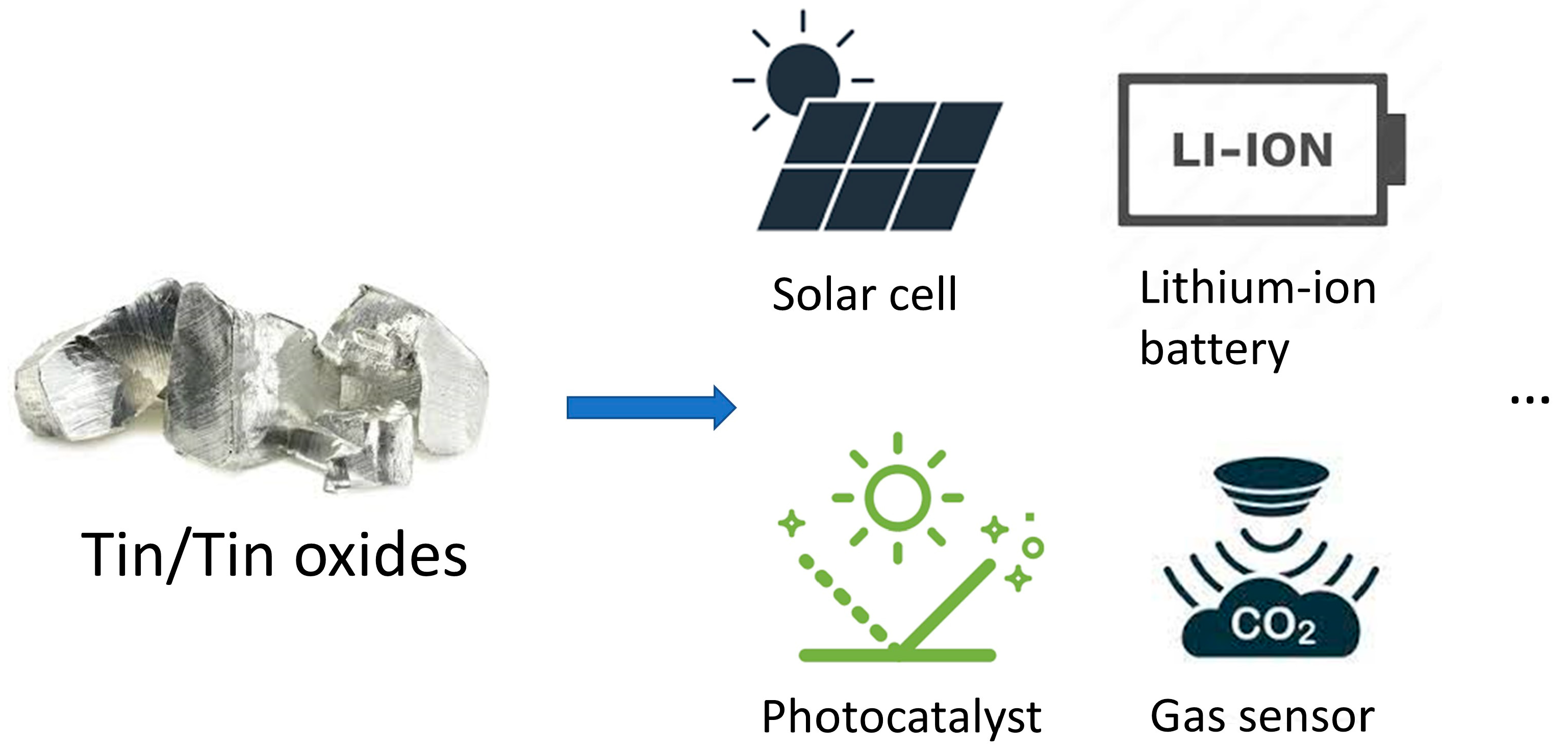
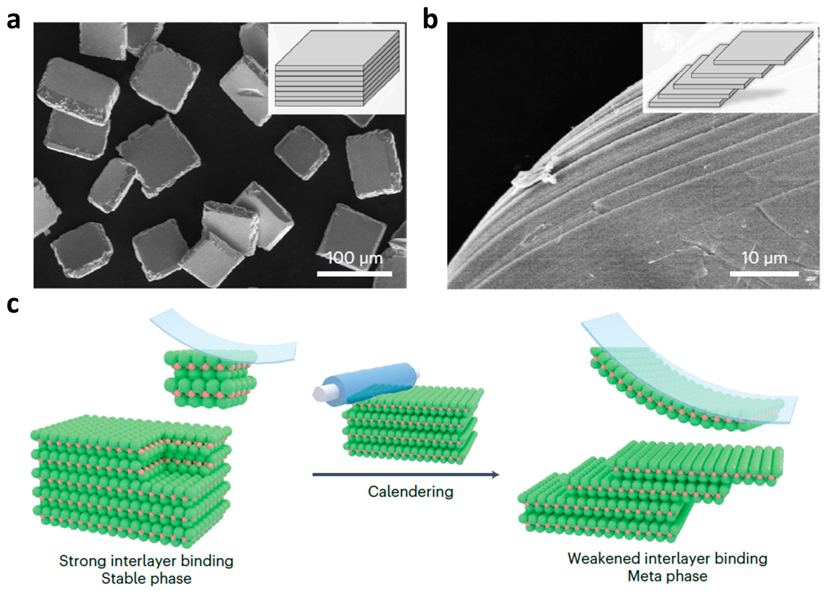
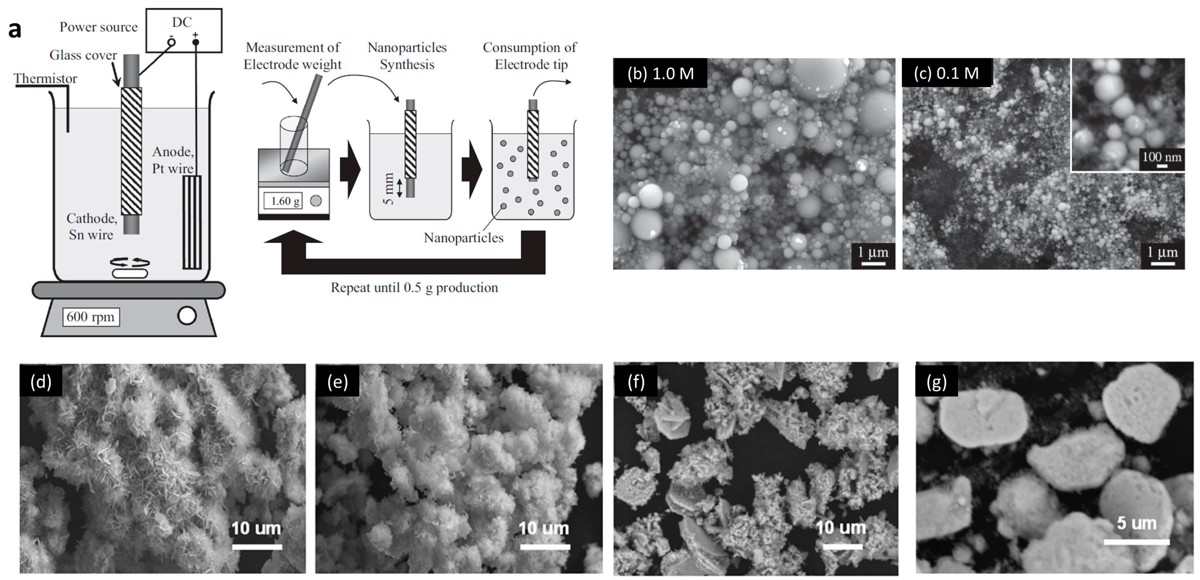
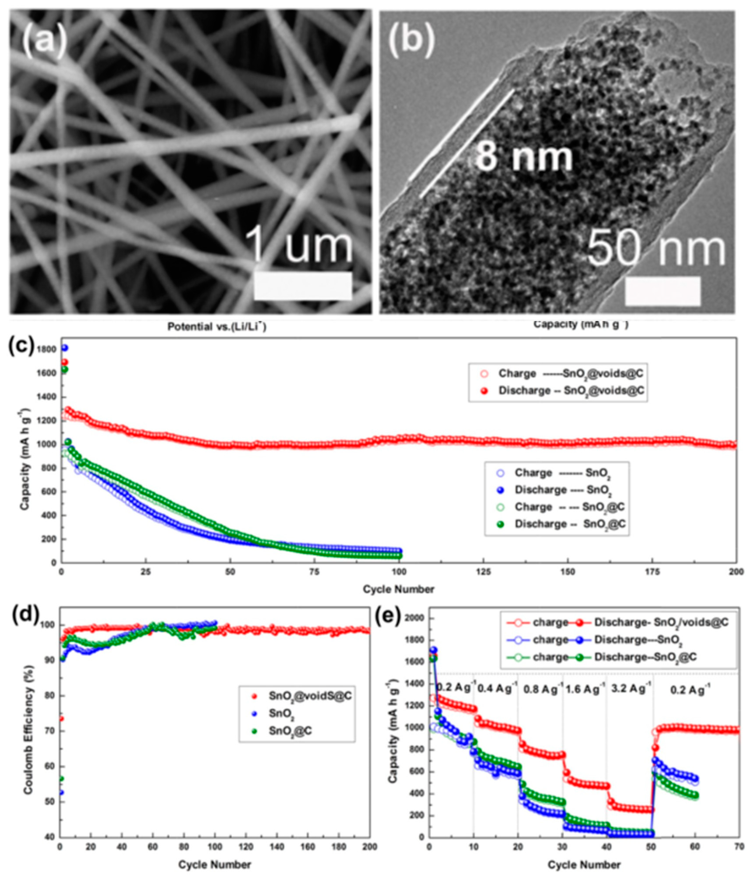
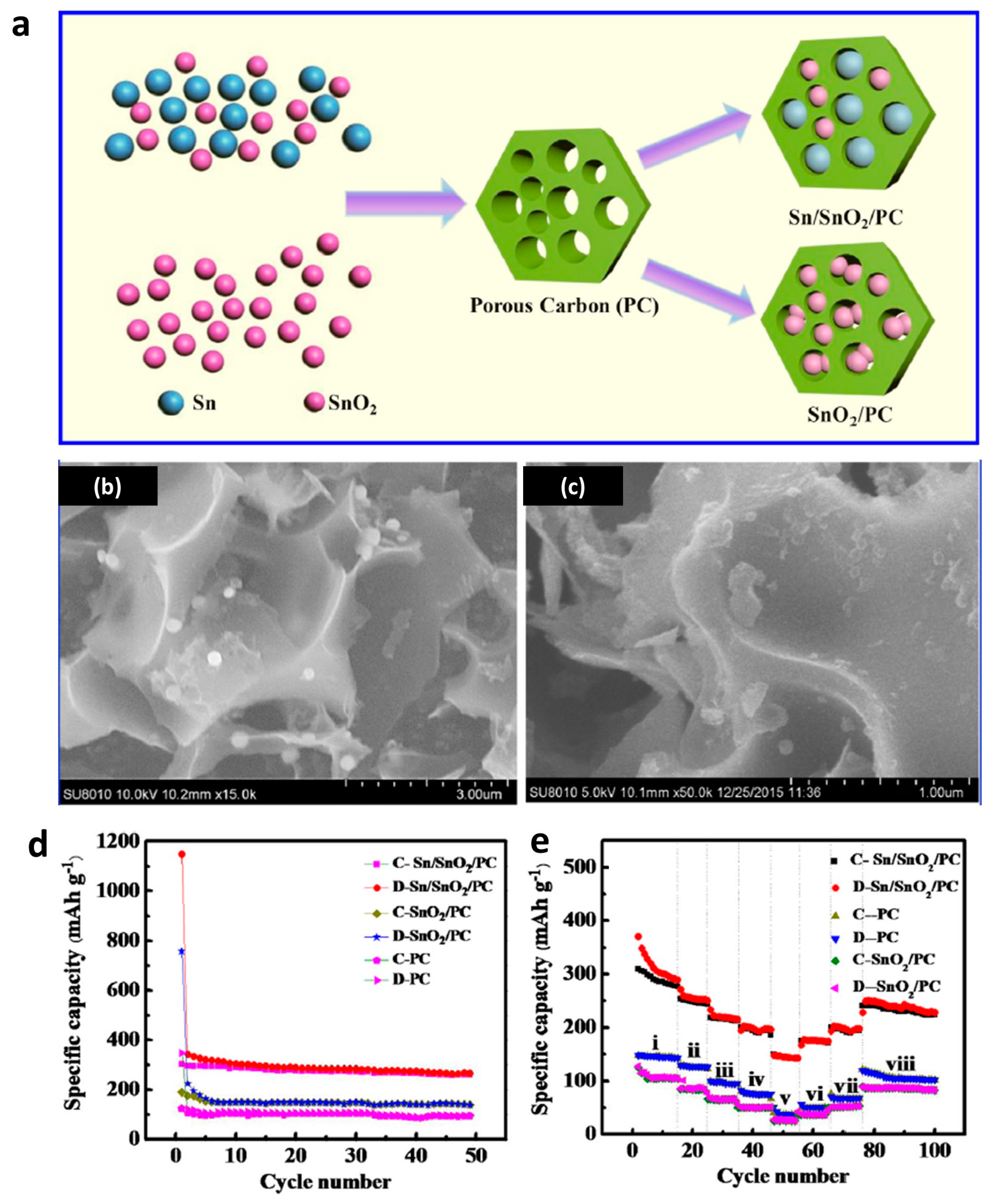
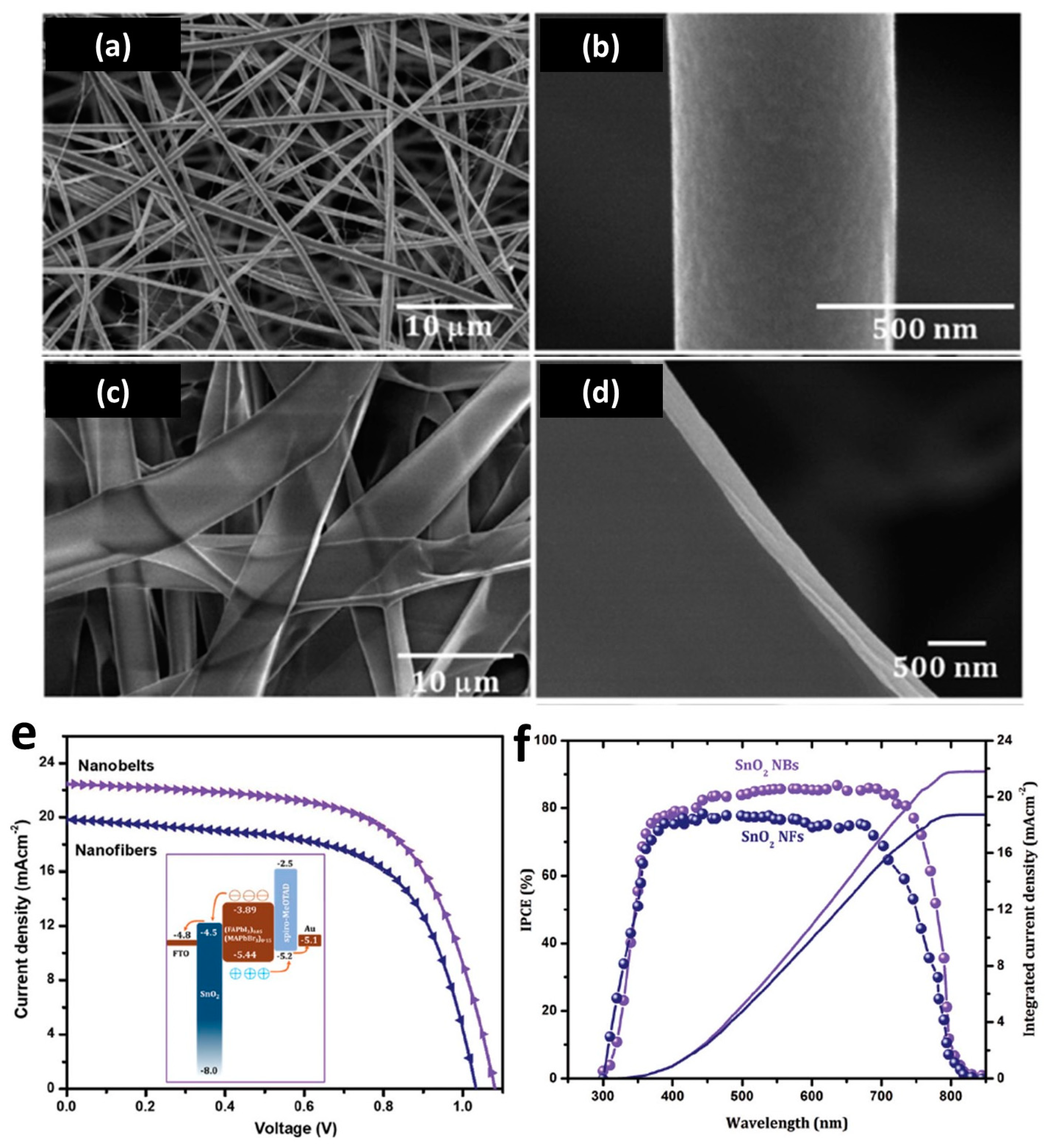
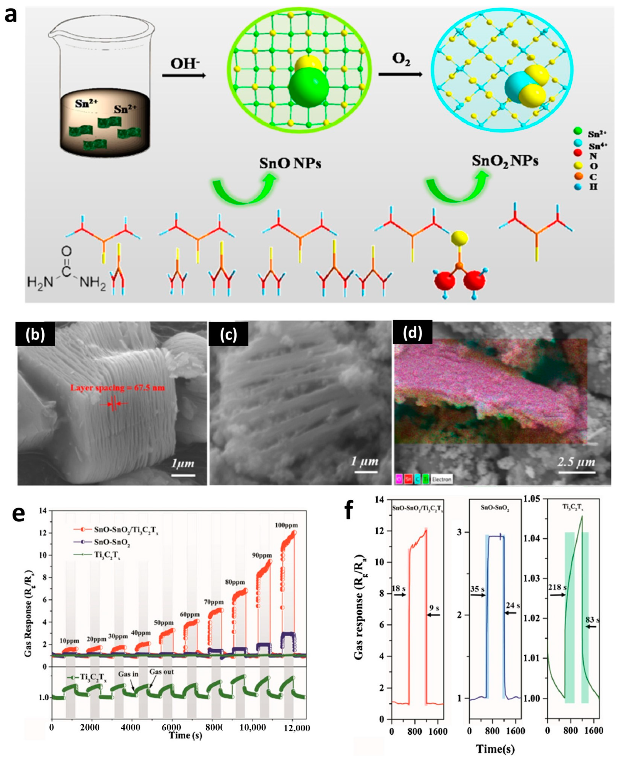
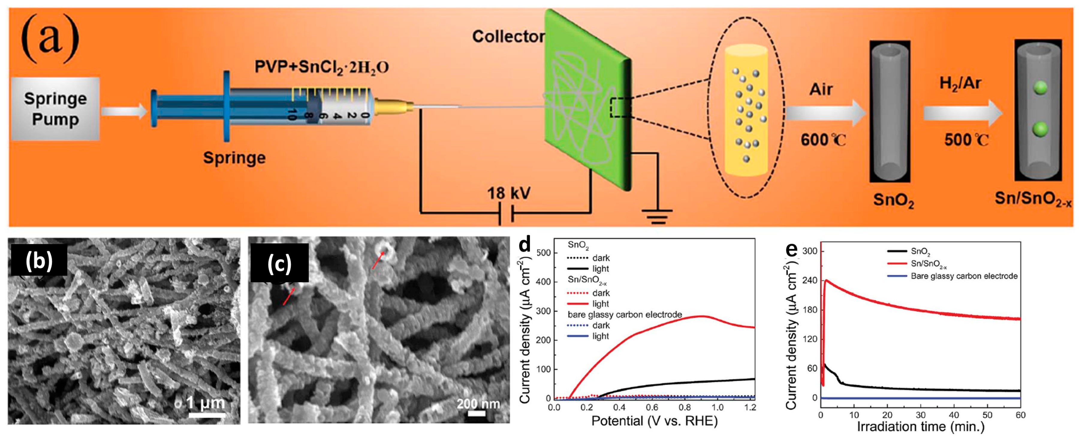
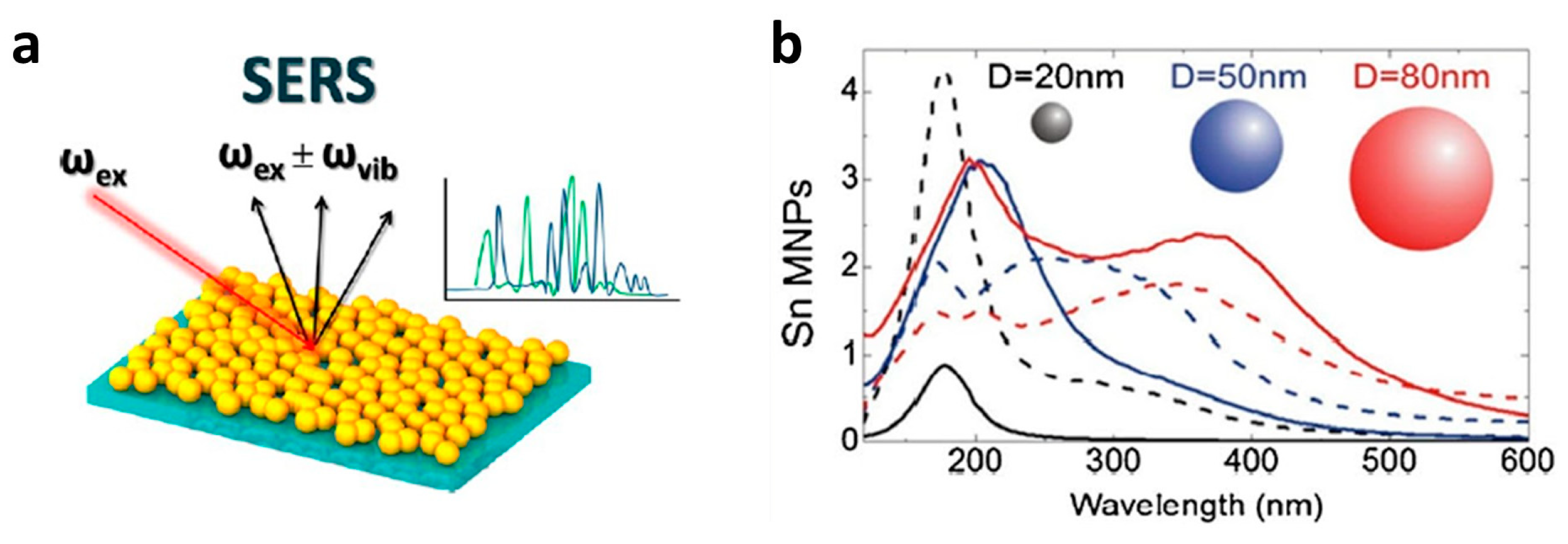
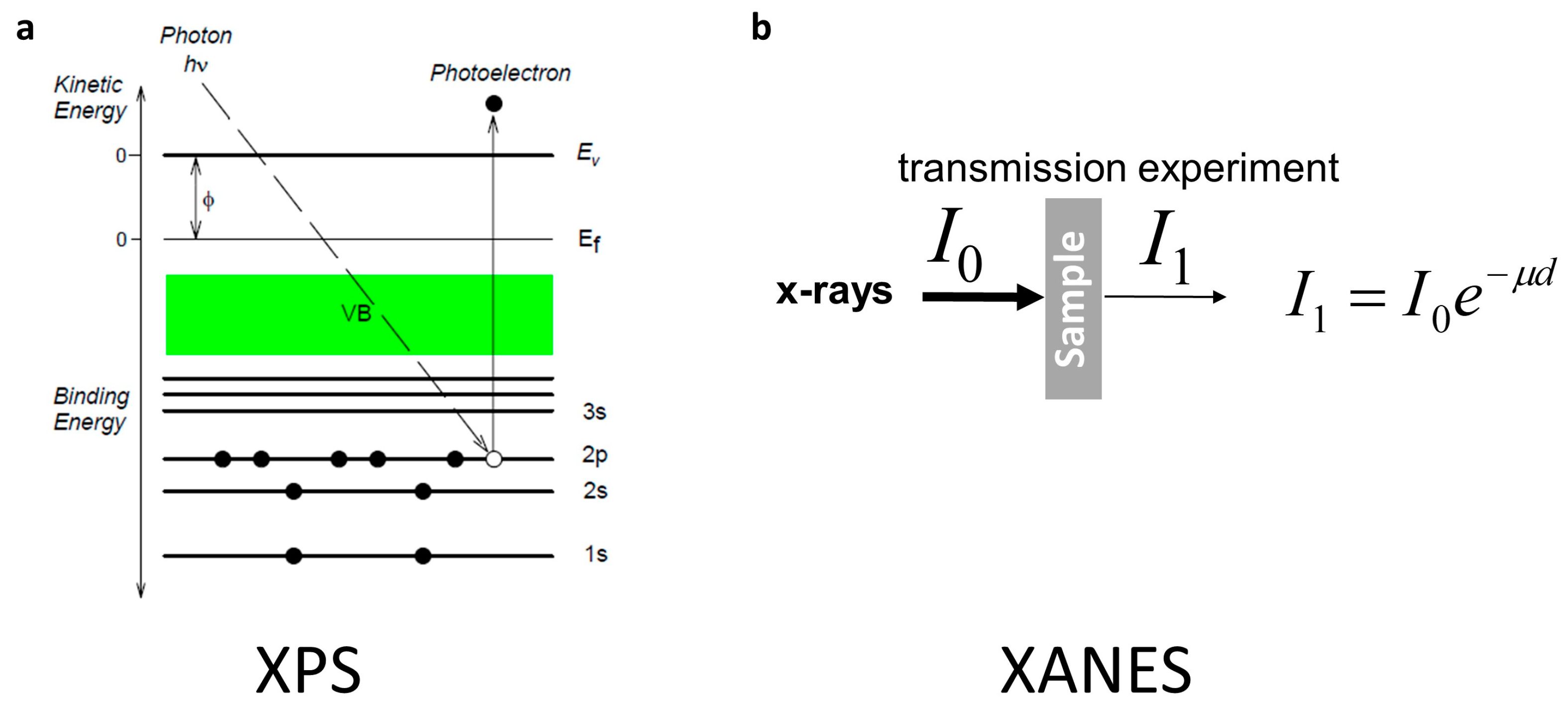
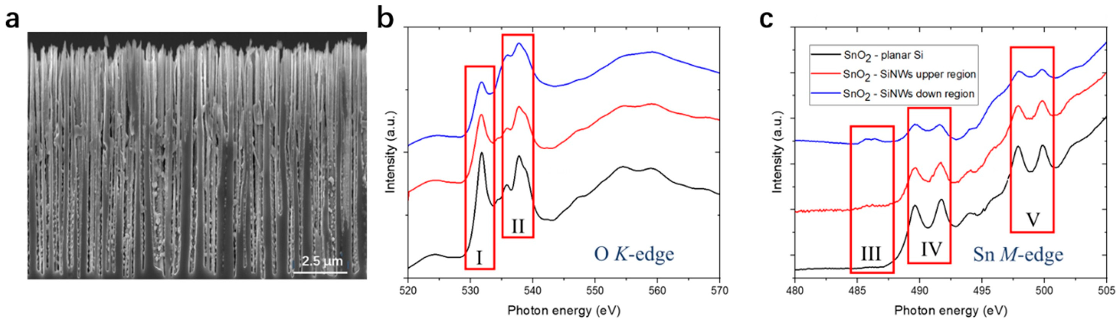
Disclaimer/Publisher’s Note: The statements, opinions and data contained in all publications are solely those of the individual author(s) and contributor(s) and not of MDPI and/or the editor(s). MDPI and/or the editor(s) disclaim responsibility for any injury to people or property resulting from any ideas, methods, instructions or products referred to in the content. |
© 2023 by the authors. Licensee MDPI, Basel, Switzerland. This article is an open access article distributed under the terms and conditions of the Creative Commons Attribution (CC BY) license (https://creativecommons.org/licenses/by/4.0/).
Share and Cite
MDPI and ACS Style
Liu, P.; Sivakov, V. Tin/Tin Oxide Nanostructures: Formation, Application, and Atomic and Electronic Structure Peculiarities. Nanomaterials 2023, 13, 2391. https://0-doi-org.brum.beds.ac.uk/10.3390/nano13172391
AMA Style
Liu P, Sivakov V. Tin/Tin Oxide Nanostructures: Formation, Application, and Atomic and Electronic Structure Peculiarities. Nanomaterials. 2023; 13(17):2391. https://0-doi-org.brum.beds.ac.uk/10.3390/nano13172391
Chicago/Turabian StyleLiu, Poting, and Vladimir Sivakov. 2023. "Tin/Tin Oxide Nanostructures: Formation, Application, and Atomic and Electronic Structure Peculiarities" Nanomaterials 13, no. 17: 2391. https://0-doi-org.brum.beds.ac.uk/10.3390/nano13172391
Note that from the first issue of 2016, this journal uses article numbers instead of page numbers. See further details here.