Development of an Immunoassay for the Detection of Copper Residues in Pork Tissues
by
Junqiu Zhang
1, 
Liwei Xu
1,
Hongtao Jiang
1,
Chuanlai Xu
1,
Wenjing Liu
2,
Ting Wu
2 and
Hua Kuang
1,* 1
International Joint Research Laboratory for Biointerface and Biodetection, State Key Lab of Food Science and Technology, School of Food Science and Technology, Jiangnan University, Wuxi 214122, China
2
Shanxi Academy of Analytical Sciences, No. 17 Beiyuan Street, Taiyuan 030012, China
*
Author to whom correspondence should be addressed.
Biosensors 2021, 11(7), 235; https://0-doi-org.brum.beds.ac.uk/10.3390/bios11070235
Submission received: 25 May 2021
/
Revised: 28 June 2021
/
Accepted: 9 July 2021
/
Published: 13 July 2021
(This article belongs to the Special Issue Nanomaterial-Based Biosensors for Biomedical Applications)
Abstract
:The presence of high concentrations of copper (Cu) residues in pork is highly concerning and therefore, this study was designed to develop a high-throughput immunoassay for the detection of such residues in edible pork tissues. The Cu content in the pork samples after digestion with HNO3 and H2O2 was measured using a monoclonal antibody (mAb) against a Cu (II)–ethylenediaminetetraacetic acid (EDTA) complex. The resulting solution was neutralized using NaOH at pH 7 and the free metal ions in the solution were chelated with EDTA for the immunoassay detection. An indirect competitive enzyme-linked immunosorbent assay (ic-ELISA) method was developed for Cu ion analysis. The half maximal inhibitory concentration of the mAb against Cu (II)–EDTA was 5.36 ng/mL, the linear detection range varied between 1.30 and 27.0 ng/mL, the limit of detection (LOD) was 0.43 μg/kg, and the limit of quantification (LOQ) was 1.42 μg/kg. The performances of the immunoassay were evaluated using fortified pig serum, liver, and pork samples and had a recovery rate of 94.53–102.24%. Importantly, the proposed immunoassay was compared with inductively coupled plasma mass spectroscopy (ICP-MS) to measure its performance. The detection correlation coefficients of the three types of samples (serum, pork, and liver) were 0.967, 0.976, and 0.983, respectively. Thirty pork samples and six pig liver samples were collected from local markets and Cu was detected with the proposed ic-ELISA. The Cu content was found to be 37.31~85.36 μg/kg in pork samples and 1.04–1.9 mg/kg in liver samples. Furthermore, we detected the Cu content in pigs with feed supplemented with tribasic copper chloride (TBCC) and copper sulfate (CS) (60, 110, and 210 mg/kg in feed). There was no significant difference in Cu accumulation in pork tissues between the TBCC and CS groups, while a remarkable Cu accumulation was found for the CS group in liver at 210 mg/kg, representing more than a two-fold higher level than seen in the TBCC group. Therefore, the proposed immunoassay was found to be robust and sensitive for the detection of Cu, providing a cost effective and practical tool for its detection in food and other complicated samples.
1. Introduction
Copper (Cu) is an essential trace element for all animals and it plays a vital role in the growth of livestock [1,2]. As an efficient and inexpensive growth promoter, the addition of high Cu concentrations has been widely used in animal feeds, including pig production, to increase growth. Tribasic Cu chloride (TBCC) and Cu sulfate (CS) have been widely used as Cu sources [3,4]. However, high Cu in feed and in tissue residues may lead to metabolic disturbances in pigs [5,6]. As early as 1981, Buntain showed that the use of high Cu as a growth promoter increased the incidence of gastric ulcers in intensive pig farming [7]. Excessive concentrations of Cu ions in vivo cause a change in the redox status of cells, leading to toxicity [8], as well as damaging DNA, proteins, and lipids [9]. Furthermore, high concentrations of Cu ions react nonspecifically with the side chains of amino acid residues, and can lead to the misfolding of proteins [10]. Uncombined Cu ions also compete with other substances for the active centers of enzymes, thus interfering with their normal function [11]. Some researchers have found that the accumulation of Cu in the human body may lead to atherosclerosis and rapid cell aging and death, posing a huge threat to public health [12,13,14]. In addition, Cu excretion from livestock has resulted in ecological concerns [15,16] and this increase in Cu in the environment can lead to soil degradation and water pollution, and can affect the growth rate of plants [17,18,19,20]. Therefore, some countries, including China, have set maximum limits to Cu supplements in fodder. According to the Announcement No. 2665 of the Ministry of Agriculture of China, the dietary Cu requirement for adult pigs is 3–6 mg/kg. Moreover, in the regional standard of safety sanitation and high-quality pork in Shanghai, China, the limit of copper is 10 mg/kg. In addition, the provisional maximum tolerable daily intake (PMTDI) for Cu determined by FAO/WHO is 0.5 mg (kg bw)−1 day−1. The average daily intake is estimated at 2–3 mg for adult persons [21]. Thus, our method can be used for actual sample detection. If the sample contains excess copper, it should be tested after proper dilution. This method can ensure the security of excessive copper in meat consumption.
Many types of qualitative and quantitative methods have been used to monitor Cu levels in food and the environment (Table 1 [22,23,24,25,26,27,28,29,30,31,32,33,34,35,36,37,38,39,40]), such as graphite furnace atomic absorption spectrometry to detect Cu levels in cattle and fish tissues [32]. Furthermore, the measurement of Cu levels in wild pig liver by flame atomic absorption spectrometry (FAAS) found that this organ is a rich source of Cu [33]. FAAS has also been used to detect Cu in pork viscera and other tissues from hybrid pigs [34] and inductively coupled plasma mass spectrometry (ICP-MS) has been used to determine the Cu content of pork and their internal organs [35]. These methods generally have the advantages of high sensitivity, but also have the disadvantages of relying on large instruments and high measurement costs.
Immunoassays such as enzyme-linked immunosorbent assays (ELISAs), however, are sensitive, fast, and cost-effective methods, which have been successfully applied to research in many fields such as the food industry, the environment, and clinical research [41,42,43,44,45]. However, it is a great challenge to develop an immunoassay for the detection of metal ions because there are no spatial antigenic epitopes with metal ions. In recent years, researchers have found a way to create specific antigenic determinants for targeting metal ions. They used chelating agents to capture metal ions and the resulting complex gave unique antigenic conformations, which could be used to screen specific antibodies, such as monoclonal antibodies (mAbs), directed to Pb (II) [46]. An immunochromatographic strip assay has also been developed with a mAb against mercury in water [47] and the development of a mAb against cadmium (Cd), as well as a highly sensitive Cu (II) enzyme-linked immunoassay, which successfully detected Cu in drinking water [48,49]. Furthermore, bifunctional chelating agents have also been used to prepare mAbs against Cu ions, which were used to establish an ELISA [50,51].
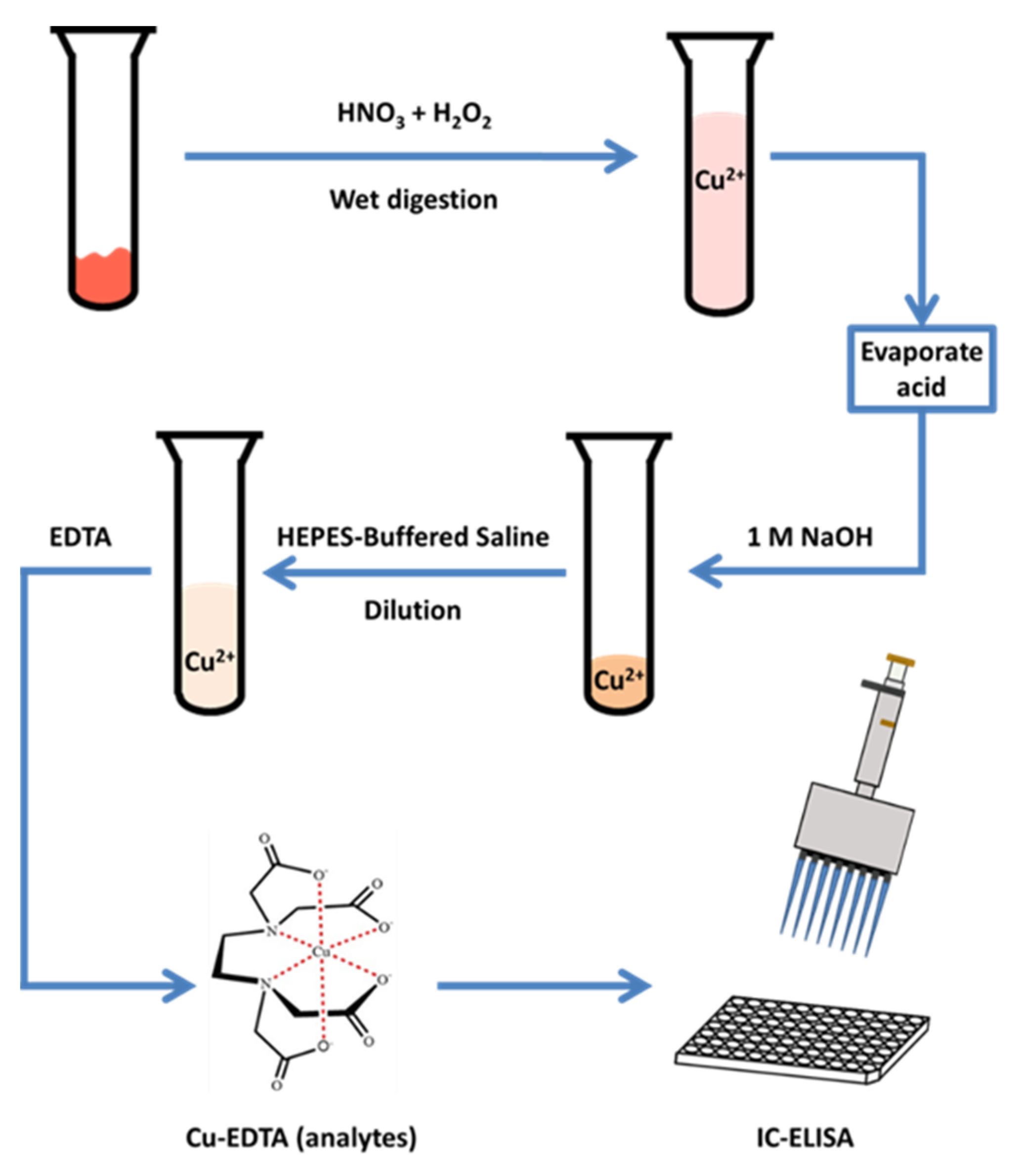
Although there exist a few examples of immunoassays for the detection of Cu, they focus on simple substrates such as water. Few immunoassays have been developed for meat, liver, and other complicated food substrates. Therefore, in this study, we developed an immunoassay for the detection of Cu in animal samples. The Cu ions in animal tissue samples were extracted and chelated with ethylenediaminetetraacetic acid (EDTA), and the resulting complex was detected with an ELISA method (Figure 1). Then, local meat and swine samples with inorganic Cu supplements were tested to determine the reliability and portability of the newly developed immunoassay.
2. Materials and Methods
2.1. Instruments and Chemicals
Inductively coupled plasma mass spectrometry (NexION 350D, PerkinElmer, Shelton, CT, USA) and a graphite-digestion device (SH230N, Hanon, Jinan, China) were employed for sample preprocessing and analysis. The spectral absorbance of microwell plates was measured with a microplate reader (Eon, BioTek, Winooski, VT, USA). All chemicals were ultrapure grade and Cu (II) (1000 μg/mL in 1% HNO3) was purchased from the National Institute of Metrology (Beijing, China). Isothiocyanobenzyl-EDTA (ITCBE) was purchased from Dojindo Laboratories (Shanghai, China). Keyhole limpet hemocyanin (KLH), bovine serum albumin (BSA), and Freund’s complete and incomplete adjuvants were purchased from Sigma-Aldrich (St. Louis, MO, USA). Goat anti-mouse IgG (Fc specific) conjugated to horseradish peroxidase (HRP) was from Jackson ImmunoResearch (West Grove, PA, USA). A monoclonal antibody exhibiting specificity for the Cu (II)–EDTA complex was from our laboratory [49]. All plasticware was soaked overnight in 3 M HCl and glassware was acid washed and rinsed thoroughly with purified water before use. Water was purified by a Millipore purification system (Bedford, MA, USA).
2.2. Buffers and Solutions
The buffers and solutions used were: (1) phosphate-buffered saline (PBS): 137 mM NaCl, 10 mM phosphate, and 3 mM KCl, pH 7.4; (2) carbonate-buffered solution (CBS): coating solution, 100 mmol/L Na2CO3–NaHCO3, pH 9.6; (3) blocking buffer: 0.1% (w/v, g/L) gelatin in PBS; (4) 2-[4-(2-hydroxyethyl)-1-piperazine] ethanesulfonic acid (HEPES, CAS:7365-45-9)-buffered saline (HBS): 10 mM HEPES, 137 mM NaCl, and 3 mM KCl; and (5) washing solution (PBST): 0.01% (v/v) Tween 20 in PBS.
2.3. Sample Pretreatment
A total of 30 pork and 6 liver samples of pig were collected from local markets, and the other pig samples (include 36 of serum, pork, and liver, respectively) were kindly donated by Professor He Pingli’s laboratory at the Chinese Agricultural University, from which weanling pigs were fortified with two mineral source supplements (TBCC and CS) through an entire 38 day experiment. Three supplemented Cu concentrations, 60, 110, and 210 mg/kg, in the diet were given to the TBCC and CS pig groups.
Pig samples (serum, pork, and liver) were prepared for the analysis of Cu concentrations, which included wet tissue weights: 0.20 g of liver, pork, and serum samples were placed into digestion tubes. Then, 3 mL of nitric acid was added at 120 °C, and the samples were allowed to digest for 40 min, and then 1.2 mL of hydrogen peroxide was added for a further 40 min. After the digestion, samples were transferred to beakers and the acid was removed using an electric hot plate. The pH was then adjusted to 7.2 with 1 mol/L NaOH, and was used as a blank control.
The ICP-MS conditions for Cu detection are shown in SI.
2.4. Ic-ELISA Method for the Determination of Cu Ions
The ic-ELISA method was applied as previously published [49]. With a pH of 7.2, optimal IC50 sensitivity and maximum absorbance signals were obtained. Furthermore, 1 mM EDTA and HEPES-KCl assay buffer containing 137 mM NaCl, 3 mM KCl, and 10 mM HEPES were used in the assay. In this study, parameters such as incubation temperature, incubation time, and plate-washing times were optimized and the specific operational procedures were as follows.
Microtiter plates (96-well) were coated with Cu (II)–ITCBE–BSA at 1.5 mg/mL in 100 μL CBS (pH 9.0) at 37 °C for 2 h. After three washes with PBS (pH 7.4, 137 mM NaCl, 3 mM KCl, and 10 mM phosphate) containing 0.05% Tween 20 (PBST), the plates were blocked with blocking buffer for 2 h at 37 °C. The plates were then washed again and air-dried at 37 °C for 15 min. Then, checkerboard assays were employed to determine the optimal concentrations of coated Cu (II)–ITCBE–BSA and Cu (II) mAb for use in competitive assays. Cu (II)–EDTA mixture was prepared (0, 0.2, 0.6, 2, 7, 25, 80, and 240 ng/mL) in HBS (50 μL/well) and mixed with diluted purified antibody (50 μL/well) in the assay plate pre-coated with Cu (II)–ITCBE–BSA. The mixture was shaken for 20 s and incubated at 37 °C for 30 min. The plate was then washed, and 100 μL of the goat anti-mouse IgG conjugated with horseradish peroxidase (GAM-HRP) diluted 1:3000 with PBST was added in each well and incubated at 37 °C for 30 min. The plate was again washed to remove excess GAM-HRP, and 100 μL of TMB substrate solution was added to each well. After a 15 min incubation at 37 °C, the reaction was stopped by the addition of 50 μL of 2 M H2SO4 and the absorbance measured at 450 nm. In the ic-ELISA format, analytes that bind the antibody cause a decrease in the amount of antibody captured by the immobilized Cu (II)–ITCBE–BSA, resulting in reduced absorbance. Standard curves were obtained by plotting absorbance versus concentration (logarithm) of the standard Cu (II) analyte concentration and fitting the data to a four-parameter logistic equation. The spiked samples including pork, liver, and serum were employed for recovery test at three levels (n = 6). All samples were detected by ic-ELISA for background content of Cu, then the suitable volume of Cu (II) solution for fortification was added as follows: 0.5, 1.0, and 2.0 mg/kg in pork, 0.75, 1.5, and 3.0 mg/kg in serum, and 5, 10, and 20 mg/kg in liver samples.
In detection of real samples, the Cu standard solutions were replaced by diluted pig samples without fortification, while the rest of the experiment was performed as described above.
3. Results and Discussion
3.1. Optimization of ic-ELISA Conditions and Correlation Analysis with ICP-MS
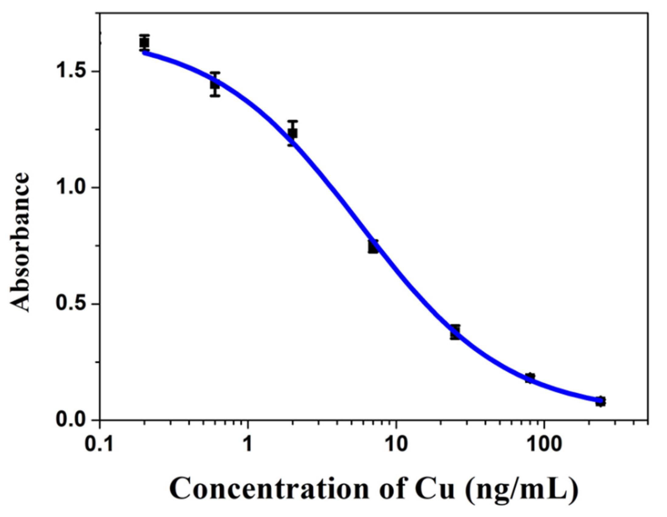
Competitive inhibition curves of the Cu (II) analyte were obtained under optimal conditions as shown (Figure 2). The IC50 for the detection of Cu was determined to be 5.36 ± 0.51 ng/mL according to the standard calibration curve (Figure 2), the linear range was from 1.30 ± 0.09 to 27.0 ± 1.8 ng/mL, the limit of detection (LOD) was 0.43 μg/kg, and the limit of quantification (LOQ) was 1.42 μg/kg.
To evaluate the practicality and accuracy of our ic-ELISA method, pig samples spiked with three concentrations of Cu (II) were analyzed, which followed the same process shown in Figure 1. As shown in Table 2, the Cu recovery from pork samples ranged from 94.53 to 102.24%, and the coefficients of variance (CV) were from 2.63 to 9.53%. The results show that the test had stable recovery and reproducibility. Furthermore, the performance of the immunoassay was evaluated by comparing it with ICP-MS detection. Three types of samples (pork, serum, and liver) were treated and analyzed. As a result, we developed a linear regression model for correlation analysis of the ELISA and ICP-MS methods (Figure 3). The correlation coefficient was 0.967 for pork, 0.976 for serum, and 0.983 for liver, indicating high reliability of this ic-ELISA method.
So far, many methods of detecting copper in liver, serum, and pork have been established. Some previously reported studies showed the LOD of Cu was 0.022 (ICP-MS) [52] and 0.5 mg/kg (ASS) [24] in liver and meat. Compared with other conventional techniques, this immunoassay has the advantage of low LOD. Moreover, the microplate of the ic-ELISA method makes the assay a high-throughput analysis, which compresses time for the detection of a large number of samples at the same time.
3.2. The Investigation of Cu Content in Retail Swine Samples
We randomly collected 30 pork and 6 liver samples from local markets and all were treated and analyzed. The Cu levels were found to be 37.60~85.36 μg/kg in pork, and 1.04~1.90 mg/kg in liver samples, and the results show much higher bioaccumulation of Cu in liver than in pork tissue. (Table 3). Cu is essential to several enzymes responsible for metabolic processes in vivo [52,53]. In addition, some research suggests that an overdose of Cu can lead to its accumulation in the liver and potentially result in health risks. Cu accumulation in liver is possibly related to the distribution of Cu-containing enzymes in animals or metabolic pathways associated with Cu [1,2].
3.3. Distributions of Cu in Pigs under Different Treatments
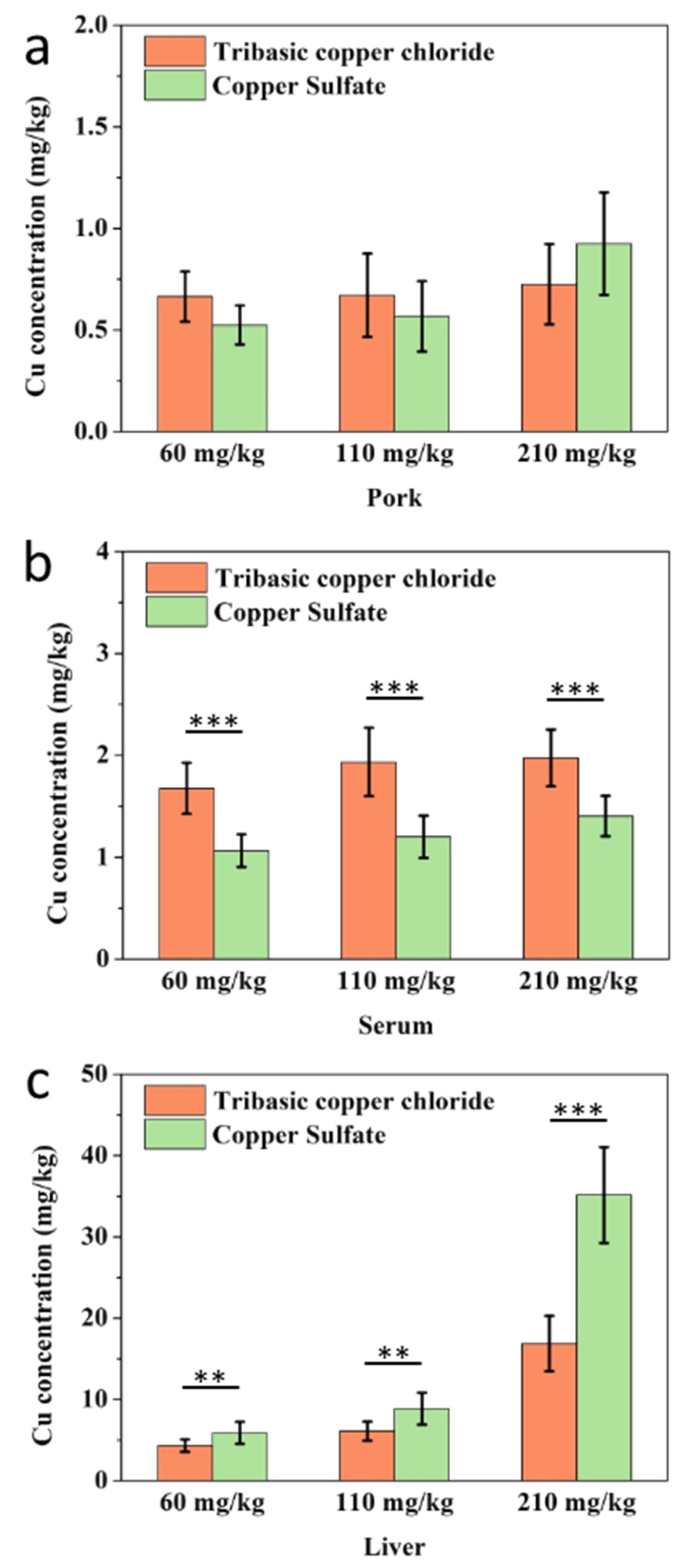
To further investigate the application of our developed immunoassay, we analyzed the Cu content in swine samples (donated sample) after supplementation with TBCC or CS from the diet. As shown in Figure 4a, no significant difference in total Cu content in pork between the two mineral types of Cu was seen. The Cu residues in pork ranged from 0.5 to 1 mg/kg, the Cu content in serum samples was found to be up to 2.38 ± 0.19 mg/kg (Table S1), and the Cu levels from the TBCC groups were approximately 1.4 times higher than the CS groups. (p < 0.001). However, there were no obvious dosage effects among the 60, 110, and 210 mg/kg fortified levels in the two Cu-type treatment groups. This may be due to Cu being absorbed into proteins in the blood and released into other tissues after reaching the bearing capacity (Figure 4b). It should be noted that a remarkable Cu accumulation was found in swine liver, which was dose dependent (Figure 4c). Furthermore, compared to the TBCC treatment group, the CS diet group led to higher residues in liver tissue. As a result, the Cu level of the CS group at 210 mg/kg group (35.06 ± 5.27 mg/kg) was more than six-fold higher than the 60 mg/kg group and four-fold greater than that of the 110 mg/kg group. which was also more than two times lower than the TBCC group at 210 mg/kg (* p < 0.001).
According to previously published data, Cu in fodder is absorbed into the blood through the intestinal tract, and mainly conjugates with plasma proteins. Cu intake by livestock is distributed in erythrocytes and plasma, and mainly manifests in the form of erythrocyte cupratin and ceruloplasmin [54,55,56,57]. When Cu overdose occurs, it is transported to the liver for storage, which results in the bioaccumulation of Cu in vivo [58,59,60]. The Cu concentrations in tissue and liquid samples indicated that the liver was a target organ and was sensitive to changes in dietary Cu content (Figure 4). Therefore, liver Cu levels could be used as a sensitive index of excessive Cu in pig products. The data show that the two inorganic Cu-sourced feed supplements underwent different metabolic pathways in animals (Supplementary Material Table S1). Therefore, an assessment of dietary Cu supplements in animals should be carried out to more fully understand its bioavailability [61,62].
4. Conclusions
In this study, we developed a rapid and high-throughput immunoassay for the detection of Cu in complicated pork, liver, and serum matrices. The result of our ELISA analysis was found to be highly consistent with other methods, such as ICP-MS. The LOD value (µg per kg sample) was far lower than the Cu residue level of pig samples (mg per kg sample). Copper analysis in local pork tissues indicated that its residues in pork was low, but its accumulation in the liver was much more serious. In pigs, we found different accumulation features for Cu depending upon its tribasic TBCC and sulfated CS forms. The latter showed a significant retention in swine liver. Therefore, it is necessary to carry out a systematic study of Cu metabolism in farmed livestock, and its environmental evaluation.
Supplementary Materials
The following are available online at https://0-www-mdpi-com.brum.beds.ac.uk/article/10.3390/bios11070235/s1, Table S1: Copper concentration in various samples of pig from two copper source feeding groups at three dosage (Mean ± SD, n = 3).
Author Contributions
C.X. and H.K. designed the experiments. J.Z., H.J., W.L. and T.W. performed the experiments. J.Z., L.X. and H.K. participated in data analysis. J.Z., L.X., H.K. and C.X. drafted the manuscript, and all authors read and approved the manuscript prior to submission.
Funding
This work is financially supported by the National Key R&D Program (2017YFC1600306), and this work was also funded by Key R&D projects of Shanxi Province (201903D311001).
Conflicts of Interest
No potential conflict of interest was reported by the authors.
References
- Ding, H.; Zhang, Q.; Xu, H.; Yu, X.; Chen, L.; Wang, Z.; Feng, J. Selection of copper and zinc dosages in pig diets based on the mutual benefit of animal growth and environmental protection. Ecotoxicol. Environ. Saf. 2021, 216, 112177. [Google Scholar] [CrossRef]
- Espinosa, C.D.; Fry, R.S.; Kocher, M.E.; Stein, H.H. Effects of copper hydroxychloride and dietary fiber on intestinal permeability, growth performance, and blood characteristics of nursery pigs. Anim. Feed. Sci. Technol. 2020, 263, 114447. [Google Scholar] [CrossRef]
- Lin, G.; Guo, Y.; Liu, B.; Wang, R.; Su, X.; Yu, D.; He, P. Optimal dietary copper requirements and relative bioavailability for weanling pigs fed either copper proteinate or tribasic copper chloride. J. Anim. Sci. Biotechnol. 2020, 11, 1189–1203. [Google Scholar] [CrossRef] [PubMed]
- Wang, J.; Zhu, X.; Guo, Y.; Wang, Z.; Zhao, B.; Yin, Y.; Liu, G. Influence of Dietary Copper on Serum Growth-Related Hormone Levels and Growth Performance of Weanling Pigs. Biol. Trace Elem. Res. 2016, 172, 134–139. [Google Scholar] [CrossRef]
- Davin, R.; Almeida, F.N.; Zhao, J.; Escobar, J.; Vazquez-Anon, M. Effects of copper source and level on growth performance and bone mineralization in pigs fed phytase-supplemented diets. J. Anim. Sci. 2016, 94, 74–75. [Google Scholar] [CrossRef]
- Gao, Y.; Yang, W.; Che, D.; Adams, S.; Yang, L. Advances in the mechanism of high copper diets in restraining pigs growth. J. Anim. Physiol. Anim. Nutr. 2020, 104, 667–678. [Google Scholar] [CrossRef] [PubMed]
- Buntain, D. Deaths in pigs on a high copper diet. Vet. Rec. 1961, 73, 707–713. [Google Scholar]
- Zhong, G.; He, Y.; Wan, F.; Wu, S.; Jiang, X.; Tang, Z.; Hu, L. Effects of Long-Term Exposure to Copper on the Keap1/Nrf2 Signaling Pathway and Msr-Related Redox Status in the Kidneys of Rats. Biol. Trace Elem. Res. 2021. [Google Scholar] [CrossRef] [PubMed]
- Husain, N.; Hasan, S.; Khan, A.A.; Mahmood, R. Copper chloride inhibits brush border membrane enzymes, alters antioxidant and metabolic status and damages DNA in rat intestine: A dose-dependent study. Environ. Sci. Pollut. Res. Int. 2021. [Google Scholar] [CrossRef]
- Niu, B.; Mackness, B.C.; Zitzewitz, J.A.; Matthews, C.R.; Gross, M.L. Trifluoroethanol Partially Unfolds G93A SOD1 Leading to Protein Aggregation: A Study by Native Mass Spectrometry and FPOP Protein Footprinting. Biochemistry 2020, 59, 3650–3659. [Google Scholar] [CrossRef]
- Khan, A.; Dobson, J.P.; Exley, C. Redox cycling of iron by A beta(42). Free. Radic. Biol. Med. 2006, 40, 557–569. [Google Scholar] [CrossRef]
- Qazmooz, H.A.; Smesam, H.N.; Mousa, R.F.; Al-Hakeim, H.K.; Maes, M. Trace element, immune and opioid biomarkers of unstable angina, increased atherogenicity and insulin resistance: Results of machine learning. J. Trace Elem. Med. Biol. 2021, 64, 126703. [Google Scholar] [CrossRef]
- Ribeiro Sant’Ana, M.A.; de Carvalho, T.C.; da Silva, I.F. Concentration of heavy metals in UHT dairy milk available in the markets of Sao Luis, Brazil, and potential health risk to children. Food Chem. 2021, 346, 128961. [Google Scholar] [CrossRef] [PubMed]
- Jo, G.; Todorov, T.I. Distribution of nutrient and toxic elements in brown and polished rice. Food Chem. 2019, 289, 299–307. [Google Scholar] [CrossRef] [PubMed]
- Perestrelo, A.P.; Miranda, G.; Goncalves, M.I.; Belino, C.; Ballesteros, R. Chronic Copper Sulfate Poisoning. Eur. J. case Rep. Intern. Med. 2021, 8, 002309. [Google Scholar]
- Ghazaryan, K.; Movsesyan, H.; Ghazaryan, N.; Watts, B.A. Copper phytoremediation potential of wild plant species growing in the mine polluted areas of Armenia. Environ. Pollut. 2019, 249, 491–501. [Google Scholar] [CrossRef]
- Luo, J.; Zhu, T.; Wang, X.; Cheng, X.; Yuan, Y.; Jin, M.; Betancor, M.B.; Tocher, D.R.; Zhou, Q. Toxicological mechanism of excessive copper supplementation: Effects on coloration, copper bioaccumulation and oxidation resistance in mud crab Scylla paramamosain. J. Hazard. Mater. 2020, 395, 122600. [Google Scholar] [CrossRef]
- Campana, O.; Simpson, S.L.; Spadaro, D.A.; Blasco, J. Sub-Lethal Effects of Copper to Benthic Invertebrates Explained by Sediment Properties and Dietary Exposure. Environ. Sci. Technol. 2012, 46, 6835–6842. [Google Scholar] [CrossRef]
- Draghici, G.A.; Dehelean, C.; Pinzaru, I.; Bordean, D.M.; Borozan, A.; Tsatsakis, A.M.; Kovatsi, L.; Nica, D. Soil copper uptake by land snails: A semi-field experiment with juvenile Cantareus aspersus snails. Environ. Toxicol. Pharmacol. 2019, 72, 103243. [Google Scholar] [CrossRef] [PubMed]
- Liao, P.; Shu, X.; Tang, M.; Tan, B.; Yin, Y. Effect of dietary copper source (inorganic vs. chelated) on immune response, mineral status, and fecal mineral excretion in nursery piglets. Food Agric. Immunol. 2018, 29, 548–563. [Google Scholar] [CrossRef] [Green Version]
- Joint FAO/WHO. Food Standards Program Codex Committee on Contaminants in Foods, Fifth Session; WHO Press: Geneva, Switzerland, 2011; Available online: https://apps.who.int/iris/handle/10665/44514 (accessed on 12 July 2021).
- Sahan, Y.; Basoglu, F.; Guecer, S. ICP-MS analysis of a series of metals (Namely: Mg, Cr, Co, Ni, Fe, Cu, Zn, Sn, Cd and Pb) in black and green olive samples from Bursa, Turkey. Food Chem. 2007, 105, 395–399. [Google Scholar] [CrossRef]
- Yenisoy-Karakas, S. Estimation of uncertainties of the method to determine the concentrations of Cd, Cu, Fe, Pb, Sn and Zn in tomato paste samples analysed by high resolution ICP-MS. Food Chem. 2012, 132, 1555–1561. [Google Scholar] [CrossRef] [PubMed]
- Papaslioti, E.M.; Parviainen, A.; Roman Alpiste, M.J.; Marchesi, C.; Garrido, C.J. Quantification of potentially toxic elements in food material by laser ablation-inductively coupled plasma-mass spectrometry (LA-ICP-MS) via pressed pellets. Food Chem. 2019, 274, 726–732. [Google Scholar] [CrossRef]
- Fu, L.; Shi, S. A novel strategy to determine the compositions of inorganic elements in fruit wines using ICP-MS/MS. Food Chem. 2019, 299, 125172. [Google Scholar] [CrossRef] [PubMed]
- Branislav, S.; Drangan, V.; Zdenka, S.; Nenad, P.; Sasa, J.; Slavoljub, S.; Milica, B.; Vesna, A. Determination of sensory properties and levels of trace elements during storage of canned meat products. J. Food Process. Preserv. 2021, 45, e15278. [Google Scholar] [CrossRef]
- Souza, S.O.; Costa, S.S.L.; Brum, B.C.T.; Santos, S.H.; Garcia, C.A.B.; Araujo, R.G.O. Determination of nutrients in sugarcane juice using slurry sampling and detection by ICP OES. Food Chem. 2019, 273, 57–63. [Google Scholar] [CrossRef]
- Limchoowong, N.; Sricharoen, P.; Techawongstien, S.; Chanthai, S. Using bio-dispersive solution of chitosan for green dispersive liquid-liquid microextraction of trace amounts of Cu(II) in edible oils prior to analysis by ICP-OES. Food Chem. 2017, 230, 398–404. [Google Scholar] [CrossRef]
- Hong-lin, L.; Qing, M.; Xin, Z.; Yu-long, Y.; Hua-rong, T. Inductively coupled plasma mass spectrometry (ICP-MS) and inductively coupled plasma optical emission spectrometer (ICP-OES)-based discrimination for the authentication of tea. Food Control 2021, 123, 107735. [Google Scholar]
- Abduljabbar, T.N.; Sharp, B.L.; Reid, H.J.; Barzegar-Befroeid, N.; Peto, T.; Lengyel, I. Determination of Zn, Cu and Fe in human patients’ serum using micro sampling ICP-MS and sample dilution. Talanta 2019, 204, 663–669. [Google Scholar] [CrossRef] [PubMed]
- Rodriguez-Solana, R.; Manuel Salgado, J.; Manuel Dominguez, J.; Cortes, S. Assessment of minerals in aged grape marc distillates by FAAS/FAES and ICP-MS. Characterization and safety evaluation. Food Control 2014, 35, 49–55. [Google Scholar] [CrossRef]
- Ferreira, K.S.; Ferreira, W.A.; Mendonca Gomes, J.M.; Correa-Junior, J.D.; Donnici, C.L.; Borba da Silva, J.B. Use of fast alkaline solubilisation to determine copper in bovine liver, fish tissues (salmon), and rolled oats by graphite furnace atomic absorption spectrometry using aqueous calibration. Microchem. J. 2016, 124, 350–355. [Google Scholar] [CrossRef]
- Kasprzyk, A.; Kilar, J.; Chwil, S.; Rudas, M. Content of Selected Macro- and Microelements in the Liver of Free-Living Wild Boars (Sus scrofa L.) from Agricultural Areas and Health Risks Associated with Consumption of Liver. Animals 2020, 10, 1519. [Google Scholar] [CrossRef] [PubMed]
- Tomovic, V.M.; Petrovic, L.S.; Tomovic, M.S.; Kevresan, Z.S.; Dzinic, N.R. Determination of mineral contents of semimembranosus muscle and liver from pure and crossbred pigs in Vojvodina (northern Serbia). Food Chem. 2011, 124, 342–348. [Google Scholar] [CrossRef]
- Nikolic, D.; Djinovic-Stojanovic, J.; Jankovic, S.; Stanisic, N.; Radovic, C.; Pezo, L.; Lausevic, M. Mineral composition and toxic element levels of muscle, liver and kidney of intensive (Swedish Landrace) and extensive (Mangulica) pigs from Serbia. Food Addit. Contam. Part A Chem. Anal. Control. Expo. Risk Assess. 2017, 34, 962–971. [Google Scholar] [CrossRef]
- Sumner, J.P.; Westerberg, N.M.; Stoddard, A.K.; Hurst, T.K.; Cramer, M.; Thompson, R.B.; Fierke, C.A.; Kopelman, R. DsRed as a highly sensitive, selective, and reversible fluorescence-based biosensor for both Cu+ and Cu2+ ions. Biosens. Bioelectron. 2006, 21, 1302–1308. [Google Scholar] [CrossRef]
- Sorenson, A.E.; Schaeffer, P.M. A new bivalent fluorescent fusion protein for differential Cu(II) and Zn(II) ion detection in aqueous solution. Anal. Chim. Acta 2020, 1101, 120–128. [Google Scholar] [CrossRef] [PubMed]
- Bagheri, N.; Mazzaracchio, V.; Cinti, S.; Colozza, N.; Di Natale, C.; Netti, P.A.; Saraji, M.; Roggero, S.; Moscone, D.; Arduini, F. Electroanalytical Sensor Based on Gold-Nanoparticle-Decorated Paper for Sensitive Detection of Copper Ions in Sweat and Serum. Anal. Chem. 2021, 93, 5225–5233. [Google Scholar] [CrossRef] [PubMed]
- Liu, K.; Chen, K.S.; Sen, D.; Yu, H.-Z. Ultrasensitive detection of total copper with an electrochemical biosensor built on the in cis coupling of hexynyl CLICK-17 DNAzyme with azido self-assembled monolayers. Electrochim. Acta 2021, 379, 138125. [Google Scholar] [CrossRef]
- Feng, H.; Fu, Q.; Du, W.; Zhu, R.; Ge, X.; Wang, C.; Li, Q.; Su, L.; Yang, H.; Song, J. Quantitative Assessment of Copper(II) in Wilson’s Disease Based on Photoacoustic Imaging and Ratiometric Surface-Enhanced Raman Scattering. Acs. Nano 2021, 15, 3402–3414. [Google Scholar] [CrossRef]
- Li, C.; Zhang, Y.; Eremin, S.A.; Yakup, O.; Yao, G.; Zhang, X. Detection of kanamycin and gentamicin residues in animal-derived food using IgY antibody based ic-ELISA and FPIA. Food Chem. 2017, 227, 48–54. [Google Scholar] [CrossRef] [PubMed] [Green Version]
- Li, H.; He, S.; Liu, G.; Li, C.; Ma, Z.; Zhang, X. Residue and dissipation kinetics of toosendanin in cabbage, tobacco and soil using IC-ELISA detection. Food Chem. 2021, 335, 127600. [Google Scholar] [CrossRef]
- Wang, W.-J.; Ling, Y.; Xu, T.; Gao, H.-B.; Sheng, W.; Li, J. Development of an indirect competitive ELISA based on polyclonal antibody for the detection of diethylstilbestrol in water samples. Chin. J. Chem. 2007, 25, 1145–1150. [Google Scholar] [CrossRef]
- Xu, Z.-L.; Deng, H.; Deng, X.-F.; Yang, J.-Y.; Jiang, Y.-M.; Zeng, D.-P.; Huang, F.; Shen, Y.-D.; Lei, H.-T.; Wang, H.; et al. Monitoring of organophosphorus pesticides in vegetables using monoclonal antibody-based direct competitive ELISA followed by HPLC-MS/MS. Food Chem. 2012, 131, 1569–1576. [Google Scholar] [CrossRef]
- Zhang, T.; Cheng, Y.; Li, Y.; Yang, J.; Liang, L.; Yang, J.; Cui, P.; Song, C.; Zhou, Y.; Zeng, M.; et al. Evaluation of the diagnostic performance and its associated factors of a commercial anti-EV-A71 IgM-capture ELISA kit in hospitalized children with clinical diagnostic HFMD. J. Clin. Virol. 2020, 130, 104582. [Google Scholar] [CrossRef] [PubMed]
- Li, S.; Wu, X.; Kuang, H.; Zhu, J.; Liu, L. Development of a fluorescent quantification strip assay for the detection of lead. Food Agric. Immunol. 2020, 31, 642–652. [Google Scholar] [CrossRef]
- Zou, S.; Cui, G.; Liu, L.; Song, S.; Kuang, H. Development of ic-ELISA and an immunochromatographic strip assay for the detection of methylmercury. Food Agric. Immunol. 2017, 28, 699–710. [Google Scholar] [CrossRef]
- Xing, Y.; Wu, X.; Liu, L.; Zhu, J.; Xu, L.; Kuang, H. Development of a fluorescent immunoassay strip for the rapid quantitative detection of cadmium in rice. Food Agric. Immunol. 2020, 31, 501–512. [Google Scholar] [CrossRef] [Green Version]
- Xing, C.; Hao, C.; Liu, L.; Xu, C.; Kuang, H. A highly sensitive enzyme-linked immunosorbent assay for copper(II) determination in drinking water. Food Agric. Immunol. 2014, 25, 432–442. [Google Scholar] [CrossRef]
- Zhu, X.; Miao, X.; Qin, X.; Zhu, X. Design of immunogens: The effect of bifunctional chelator on immunological response to chelated copper. J. Pharm. Biomed. Anal. 2019, 174, 263–269. [Google Scholar] [CrossRef]
- Zhu, X.; Wang, Q.; Shi, X.; Liu, F.; Wang, L.; Wang, H. Development and analytical validation of an Enzyme-Linked Immunosorbent Assay (ELISA) for the detection of copper in human hair and serum samples. Anal. Methods 2013, 5, 2578–2583. [Google Scholar] [CrossRef]
- Brooks, A.; Liang, X.; Zhang, Y.; Zhao, C.-X.; Roberts, M.S.; Wang, H.; Zhang, L.; Crawford, D.H.G. Liver organoid as a 3D in vitro model for drug validation and toxicity assessment. Pharmacol. Res. 2021, 169, 105608. [Google Scholar] [CrossRef] [PubMed]
- De Martin, E.; Michot, J.-M.; Rosmorduc, O.; Guettier, C.; Samuel, D. Liver toxicity as a limiting factor to the increasing use of immune checkpoint inhibitors. JHEP Rep. Innov. Hepatol. 2020, 2, 100170. [Google Scholar] [CrossRef] [PubMed]
- Bertinato, J.; Sherrard, L.; Plouffe, L.J. Decreased Erythrocyte CCS Content is a Biomarker of Copper Overload in Rats. Int. J. Mol. Sci. 2010, 11, 2624–2635. [Google Scholar] [CrossRef] [Green Version]
- Dirksen, K.; Roelen, Y.S.; van Wolferen, M.E.; Kruitwagen, H.S.; Penning, L.C.; Burgener, I.A.; Spee, B.; Fieten, H. Erythrocyte copper chaperone for superoxide dismutase and superoxide dismutase as biomarkers for hepatic copper concentrations in Labrador retrievers. Vet. J. 2016, 218, 1–6. [Google Scholar] [CrossRef] [PubMed]
- Linder, M.C. Apoceruloplasmin: Abundance, Detection, Formation, and Metabolism. Biomedicines 2021, 9, 233. [Google Scholar] [CrossRef] [PubMed]
- Ramos, D.; Mar, D.; Ishida, M.; Vargas, R.; Gaite, M.; Montgomery, A.; Linder, M.C. Mechanism of Copper Uptake from Blood Plasma Ceruloplasmin by Mammalian Cells. PLoS ONE 2016, 11, e0149516. [Google Scholar] [CrossRef] [Green Version]
- Sarkar, B.; Roberts, E.A. The puzzle posed by COMMD1, a newly discovered protein binding Cu(II). Metallomics 2011, 3, 20–27. [Google Scholar] [CrossRef]
- Manley, S.A.; Byrns, S.; Lyon, A.W.; Brown, P.; Gailer, J. Simultaneous Cu-, Fe-, and Zn-specific detection of metalloproteins contained in rabbit plasma by size-exclusion chromatography-inductively coupled plasma atomic emission spectroscopy. J. Biol. Inorg. Chem. 2009, 14, 61–74. [Google Scholar] [CrossRef]
- Blavi, L.; Sola, D.; Monteiro, A.; Perez, J.F.; Stein, H.H. Inclusion of dicopper oxide instead of copper sulfate in diets for growing-finishing pigs results in greater final body weight and bone mineralization, but reduced accumulation of copper in the liver. J. Anim. Sci. 2021, 99, skab127. [Google Scholar] [CrossRef]
- Espinosa, C.D.; Stein, H.H. Digestibility and metabolism of copper in diets for pigs and influence of dietary copper on growth performance, intestinal health, and overall immune status: A review. J. Anim. Sci. Biotechnol. 2021, 12, 1–12. [Google Scholar] [CrossRef]
- Zheng, X.; Wu, K.; Sun, P.; Zhouyang, S.; Wang, Y.; Wang, H.; Zheng, Y.; Li, Q. Effects of substrate types on the transformation of heavy metal speciation and bioavailability in an anaerobic digestion system. J. Environ. Sci. 2021, 101, 361–372. [Google Scholar] [CrossRef] [PubMed]
Figure 1.
Immunoassay procedure for copper residues in animal tissues.

Figure 2.
Standard inhibition curve for Cu ions by ic-ELISA method (n = 3).

Figure 3.
Comparison between ic-ELISA and ICP-MS for Cu residues in swine liver, serum, and pork samples (▴ Liver: y = x + 0.141, R2 = 0.983; ● serum: y = 0.673x + 0.369, R2 = 0.967; ■ pork: y = 0.645x + 0.118, R2 = 0.976).
Figure 3.
Comparison between ic-ELISA and ICP-MS for Cu residues in swine liver, serum, and pork samples (▴ Liver: y = x + 0.141, R2 = 0.983; ● serum: y = 0.673x + 0.369, R2 = 0.967; ■ pork: y = 0.645x + 0.118, R2 = 0.976).

Figure 4.
Cu concentration in pork, serum, and liver sample by ic-ELISA for (a) copper concentration in pork; (b) copper concentration in serum; (c) copper concentration in liver. (*: p < 0.05, **: p < 0.01, ***: p < 0.001). p-values represent the difference among three groups as determined by Student’s t-test.


Table 1.
Detection method of Cu content in different samples.
| Detect Method | Sample Type | LOD (μg/kg, mg/kg *) | LOQ (μg/kg, mg/kg *) | Reference |
|---|---|---|---|---|
| ICP-MS | Olive | 0.03 | 0.07 | [22] |
| Tomato paste | 0.16 | 0.53 | [23] | |
| Leaves | 9 | [24] | ||
| Fruit wines | 7.13 × 10⁻³ | 23.8 × 10⁻³ | [25] | |
| Bovine liver | 0.022 * | 0.066 * | [26] | |
| ICP-OES | Sugarcane juice | 0.04 * | 0.13 * | [27] |
| Oil | 2.1 * | 6.8 * | [28] | |
| Tea | 0.06 * | 0.2 * | [29] | |
| Human serum | 0.08 | 0.28 | [30] | |
| AAS | Grape marc distillate | 0.097 * | 0.322 * | [31] |
| Bovine liver | 1.8 * | 6.0 * | [32] | |
| Salmon | 2.0 * | 6.6 * | ||
| Rolled oats | 1.4 * | 4.6 * | ||
| Wild boar liver | 2.6 * | [33] | ||
| Pig semimembranosus muscle | 0.5 * | 0.75 * | [34] | |
| Pig liver | 0.5 * | 0.75 * | ||
| Pig longissimus thoracis et lumborum | 0.022 * | 0.066 * | [35] | |
| Pig liver | 0.022 * | 0.066 * | ||
| Pig kidney | 0.022 * | 0.066 * | ||
| Biosensor | Cu (II) stock solutions | 0.71 | [36] | |
| Aqueous solutions. | 64 | [37] | ||
| Sweat; serum | 3 | 10 | [38] | |
| Mine | 0.06 | [39] | ||
| Mice liver | 0.9* | [40] | ||
| Mice urinary | 0.9* |
LOD: Limit of detection. LOQ: Limit of quantification. * in Table 1 refer the unit of data to mg/kg.
Table 2.
Recovery test of Cu (II) concentration in pig samples (n = 6).
| Sample | Original Concentrations (mg/kg) | Fortified Cu (II) Level (mg/kg) | Detection by ELISA (Mean ± SD) (mg/kg) | Recovery (%) | CV (%) |
|---|---|---|---|---|---|
| Pork | 1.07 | 0.50 | 1.56 ± 0.14 | 99.24 | 8.35 |
| 1.07 | 1.00 | 2.10 ± 0.16 | 101.23 | 6.84 | |
| 1.07 | 2.00 | 2.90 ± 0.15 | 94.53 | 8.05 | |
| Serum | 1.49 | 0.75 | 2.23 ± 0.24 | 99.53 | 7.70 |
| 1.49 | 1.50 | 2.94 ± 0.08 | 98.46 | 2.63 | |
| 1.49 | 3.00 | 4.59 ± 0.64 | 102.24 | 7.85 | |
| Liver | 9.32 | 5.00 | 13.64 ± 0.95 | 95.26 | 6.18 |
| 9.32 | 10.00 | 18.79 ± 1.91 | 97.27 | 9.53 | |
| 9.32 | 20.00 | 28.88 ± 2.75 | 98.51 | 7.75 |
Abbreviations: standard deviation (SD).
Table 3.
Cu (II) content of pig samples from local markets (n = 3).
| Tissue | Sample | Detection Level | Sample | Detection Level | Sample | Detection Level |
|---|---|---|---|---|---|---|
| Pork | CQ1 | 60.96 ± 5.70 | CQ11 | 39.54 ± 2.57 | CQ21 | 66.55 ± 6.42 |
| (μg/kg) | CQ2 | 51.18 ± 4.06 | CQ12 | 43.72 ± 4.24 | CQ22 | 52.04 ± 3.55 |
| CQ3 | 58.34 ± 5.65 | CQ13 | 39.77 ± 3.97 | CQ23 | 49.76 ± 4.39 | |
| CQ4 | 42.47 ± 2.81 | CQ14 | 42.56 ± 0.98 | CQ24 | 67.96 ± 6.35 | |
| CQ5 | 38.07 ± 3.14 | CQ15 | 37.31 ± 3.10 | CQ25 | 85.36 ± 8.42 | |
| CQ6 | 39.35 ± 2.50 | CQ16 | 39.85 ± 2.53 | CQ26 | 54.32 ± 4.75 | |
| CQ7 | 42.88 ± 4.12 | CQ17 | 51.02 ± 4.53 | CQ27 | 55.91 ± 5.39 | |
| CQ8 | 38.35 ± 2.82 | CQ18 | 44.38 ± 4.34 | CQ28 | 50.41 ± 4.98 | |
| CQ9 | 68.12 ± 5.21 | CQ19 | 39.95 ± 3.65 | CQ29 | 66.31 ± 6.13 | |
| CQ10 | 37.60 ± 3.05 | CQ20 | 65.66 ± 6.42 | CQ30 | 38.88 ± 2.70 | |
| Liver | CQ31 | 1.30 ± 0.04 | CQ33 | 1.04 ± 0.05 | CQ34 | 1.43 ± 0.06 |
| (mg/kg) | CQ32 | 1.90 ± 0.12 | CQ34 | 1.57 ± 0.17 | CQ35 | 1.90 ± 0.08 |
Publisher’s Note: MDPI stays neutral with regard to jurisdictional claims in published maps and institutional affiliations. |
© 2021 by the authors. Licensee MDPI, Basel, Switzerland. This article is an open access article distributed under the terms and conditions of the Creative Commons Attribution (CC BY) license (https://creativecommons.org/licenses/by/4.0/).
Share and Cite
MDPI and ACS Style
Zhang, J.; Xu, L.; Jiang, H.; Xu, C.; Liu, W.; Wu, T.; Kuang, H. Development of an Immunoassay for the Detection of Copper Residues in Pork Tissues. Biosensors 2021, 11, 235. https://0-doi-org.brum.beds.ac.uk/10.3390/bios11070235
AMA Style
Zhang J, Xu L, Jiang H, Xu C, Liu W, Wu T, Kuang H. Development of an Immunoassay for the Detection of Copper Residues in Pork Tissues. Biosensors. 2021; 11(7):235. https://0-doi-org.brum.beds.ac.uk/10.3390/bios11070235
Chicago/Turabian StyleZhang, Junqiu, Liwei Xu, Hongtao Jiang, Chuanlai Xu, Wenjing Liu, Ting Wu, and Hua Kuang. 2021. "Development of an Immunoassay for the Detection of Copper Residues in Pork Tissues" Biosensors 11, no. 7: 235. https://0-doi-org.brum.beds.ac.uk/10.3390/bios11070235
Note that from the first issue of 2016, this journal uses article numbers instead of page numbers. See further details here.