
New Antibacterial Secondary Metabolites from a Marine-Derived Talaromyces sp. Strain BTBU20213036


Abstract
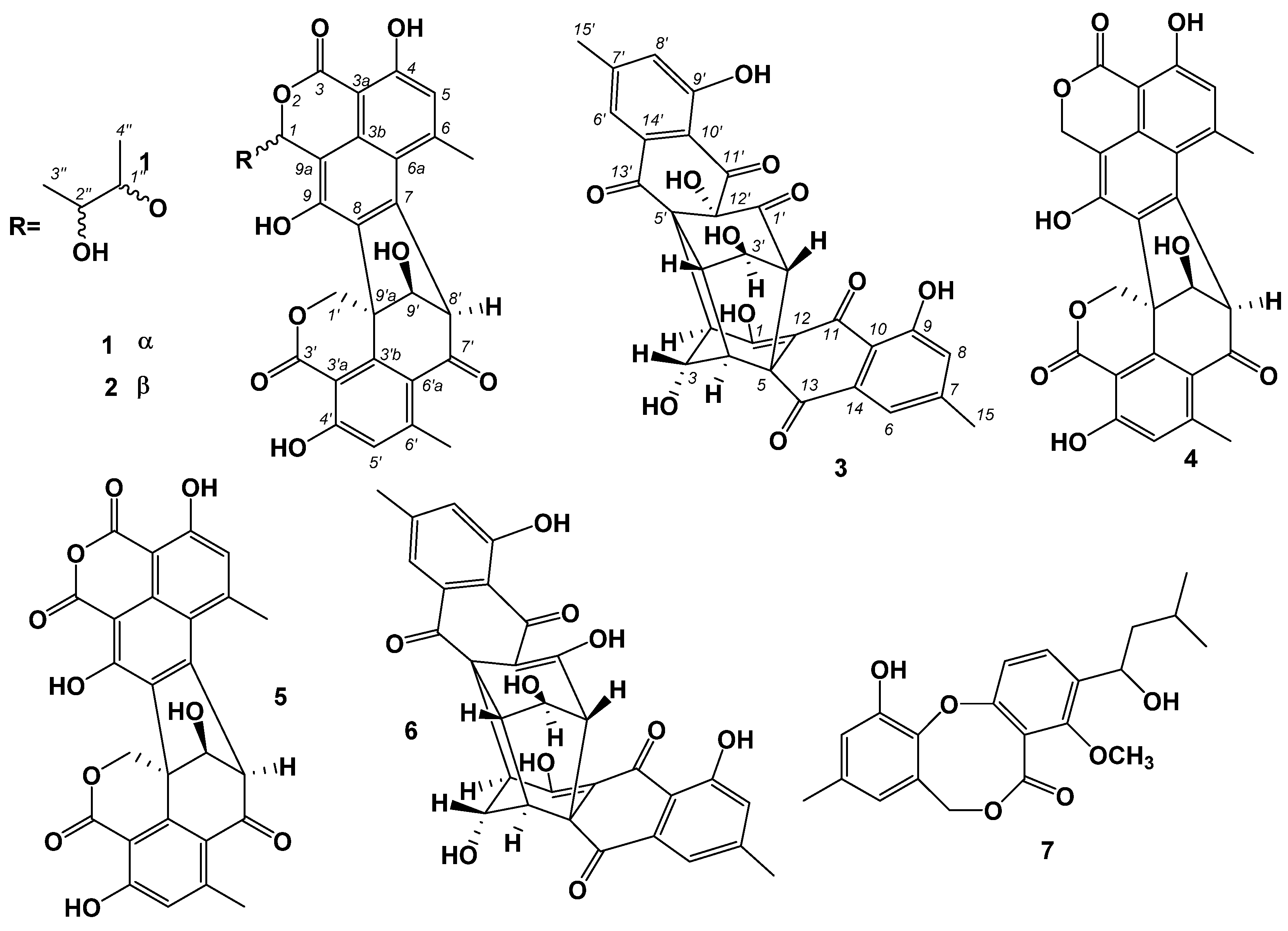
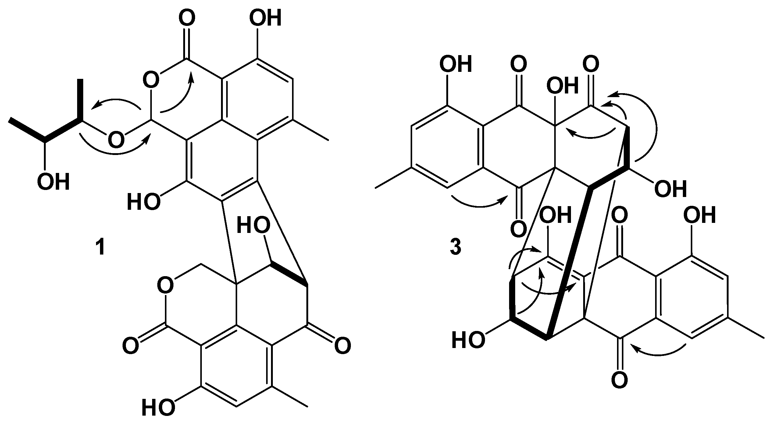
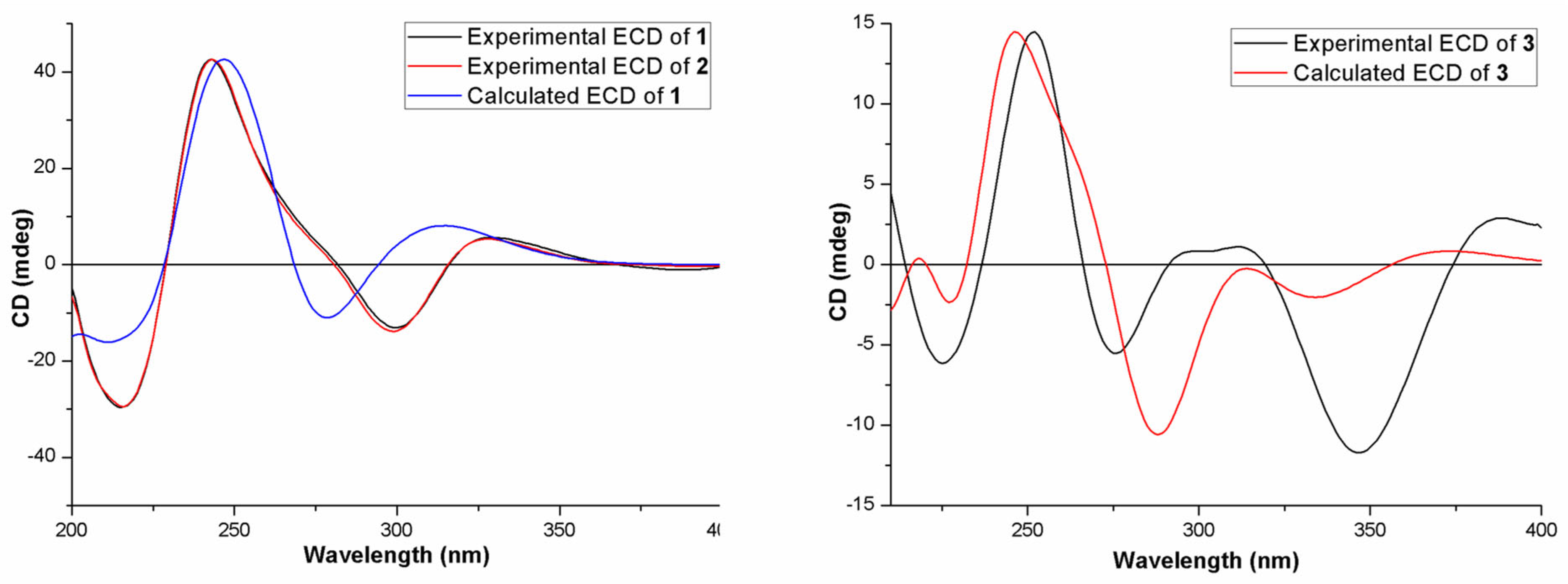
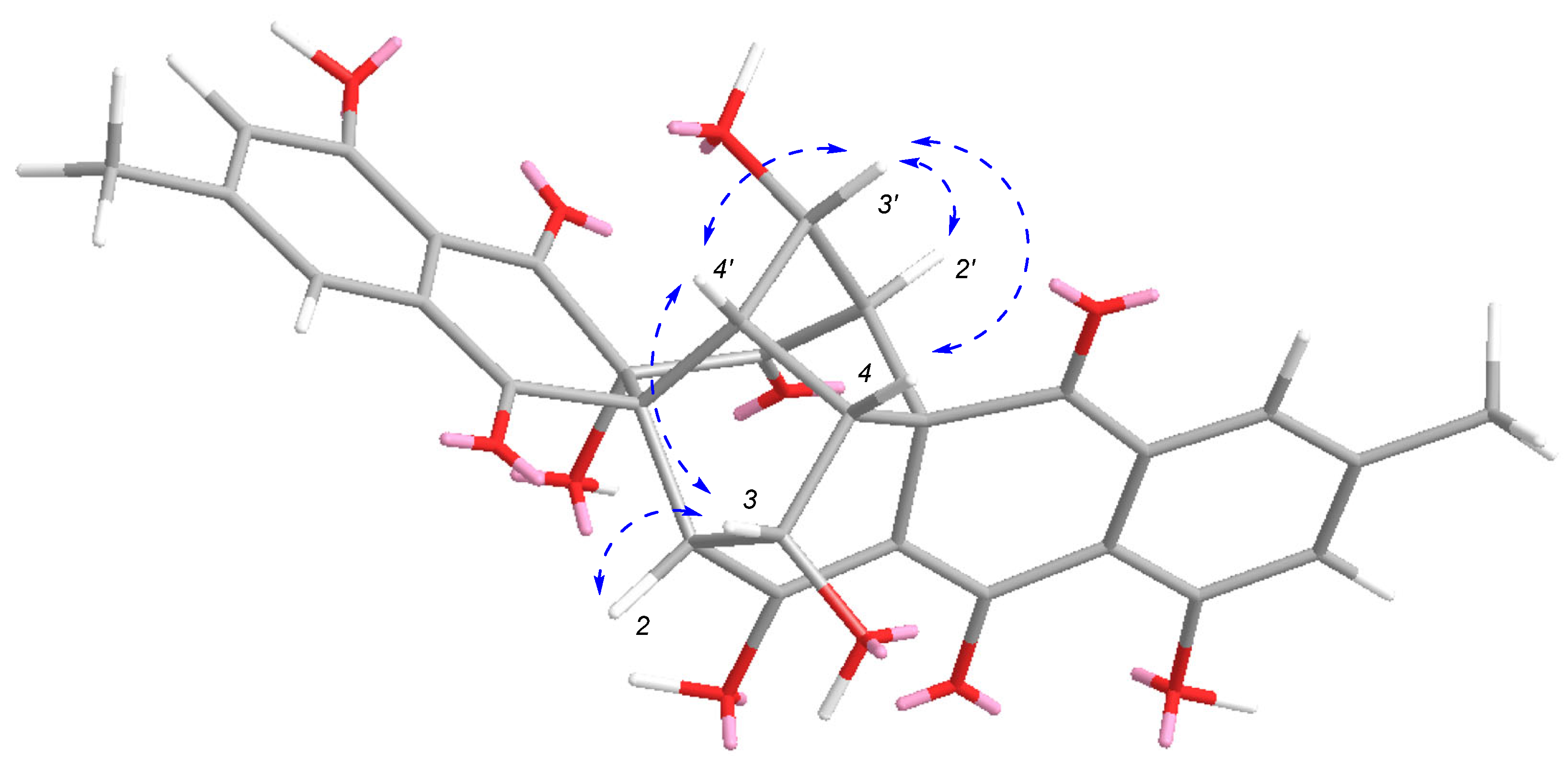
:1. Introduction
2. Results
3. Materials and Methods
3.1. General Experimental Procedures
3.2. Microbial Material
3.3. Fermentation, Extraction and Purification
3.3.1. Bacillisporin K (1)
3.3.2. Bacillisporin M (2)
3.3.3. Rugulosin D (3)
3.4. Antibacterial Activity Assays
4. Conclusions
Supplementary Materials
Author Contributions
Funding
Data Availability Statement
Conflicts of Interest
References
- Shen, B. A new golden age of natural products drug discovery. Cell 2015, 163, 1297–1300. [Google Scholar] [CrossRef] [Green Version]
- Jernigan, J.A.; Hatfield, K.M.; Wolford, H.; Nelson, R.E.; Olubajo, B.; Reddy, S.C.; McCarthy, N.; Paul, P.; McDonald, L.C.; Kallen, A.; et al. Multidrug-resistant bacterial infections in U.S. hospitalized patients, 2012–2017. N. Engl. J. Med. 2020, 382, 1309–1319. [Google Scholar] [CrossRef] [PubMed]
- Arendrup, M.C.; Patterson, T.F. Multidrug-resistant Candida: Epidemiology, molecular mechanisms, and treatment. J. Infect. Dis. 2017, 216, S445–S451. [Google Scholar] [CrossRef] [PubMed] [Green Version]
- Martínez-Vázquez, A.V.; Vázquez-Villanueva, J.; Leyva-Zapata, L.M.; Barrios-García, H.; Rivera, G.; Bocanegra-García, V. Multidrug resistance of Escherichia coli strains isolated from Bovine Feces and Carcasses in Northeast Mexico. Front. Vet. Sci. 2021, 8, 643802. [Google Scholar] [CrossRef] [PubMed]
- Hiramatsu, K.; Katayama, Y.; Matsuo, M.; Sasaki, T.; Morimoto, Y.; Sekiguchi, A.; Baba, T. Multi-drug-resistant Staphylococcus aureus and future chemotherapy. J. Infect. Chemother. 2014, 20, 593–601. [Google Scholar] [CrossRef] [Green Version]
- Ciloglu, F.U.; Caliskan, A.; Saridag, A.M.; Kilic, I.H.; Tokmakci, M.; Kahraman, M.; Aydin, O. Drug-resistant Staphylococcus aureus bacteria detection by combining surface-enhanced Raman spectroscopy (SERS) and deep learning techniques. Sci. Rep. 2021, 11, 18444. [Google Scholar] [CrossRef] [PubMed]
- Carroll, A.R.; Copp, B.R.; Davis, R.A.; Keyzers, R.A.; Prinsep, M.R. Marine natural products. Nat. Prod. Rep. 2021, 38, 362–413. [Google Scholar] [CrossRef]
- Venkatachalam, M.; Gérard, L.; Milhau, C.; Vinale, F.; Dufossé, L.; Fouillaud, M. Salinity and temperature influence growth and pigment production in the marine-derived fungal strain Talaromyces albobiverticillius 30548. Microorganisms 2019, 7, 10. [Google Scholar] [CrossRef] [Green Version]
- Mishra, R.C.; Kalra, R.; Dilawari, R.; Deshmukh, S.K.; Barrow, J.; Goel, M. Characterization of an endophytic strain Talaromyces assiutensis, CPEF04 with evaluation of production medium for extracellular red pigments having antimicrobial and anticancer properties. Front. Microbiol. 2021, 12, 665702. [Google Scholar] [CrossRef]
- Kumari, M.; Taritla, S.; Sharma, A.; Jayabaskaran, C. Antiproliferative and antioxidative bioactive compounds in extracts of marine-derived endophytic fungus Talaromyces purpureogenus. Front Microbiol. 2018, 9, 1777. [Google Scholar] [CrossRef] [Green Version]
- Guo, Z.; Shao, C.; She, Z.; Cai, X.; Liu, F.; Vrijimoed, L.L.P.; Lin, Y. 1H and 13C NMR assignments for two oxaphenalenones bacillosporin C and D from the mangrove endophytic fungus SBE-14. Magn. Reson. Chem. 2007, 45, 439–441. [Google Scholar] [CrossRef] [PubMed]
- Liang, X.; Huang, Z.H.; Shen, W.B.; Lu, X.H.; Zhang, X.X.; Ma, X.; Qi, S.H. Talaromyoxaones A and B: Unusual oxaphenalenone spirolactones as phosphatase inhibitors from the marine-derived fungus Talaromyces purpureogenus SCSIO 41517. J. Org. Chem. 2021, 86, 12831–12839. [Google Scholar] [CrossRef] [PubMed]
- Wu, B.; Ohlendorf, B.; Oesker, V.; Wiese, J.; Malien, S.; Schmaljohann, R.; Imhoff, J.F. Acetylcholinesterase inhibitors from a marine fungus Talaromyces sp strain LF458. Mar. Biotechnol. 2015, 17, 110–119. [Google Scholar] [CrossRef] [PubMed]
- Wang, M.; Yang, L.; Feng, L.; Hu, F.; Zhang, F.; Ren, J.; Qiu, Y.; Wang, Z. Verruculosins A–B, new oligophenalenone dimers from the soft coral-derived fungus Talaromyces verruculosus. Mar. Drugs 2019, 17, 516. [Google Scholar] [CrossRef] [Green Version]
- Dramae, A.; Intaraudom, C.; Bunbamrung, N.; Saortep, W.; Srichomthong, K.; Pittayakhajonwut, P. Heptacyclic oligophenalenones from the soil fungus Talaromyces bacillisporus BCC17645. Tetrahedron 2020, 76, 130980. [Google Scholar] [CrossRef]
- Huang, Z.H.; Liang, X.; Li, C.J.; Gu, Q.; Ma, X.; Qi, S.H. Talaromynoids A-I, highly oxygenated meroterpenoids from the marine-derived fungus Talaromyces purpureogenus SCSIO 41517 and their lipid accumulation inhibitory activities. J. Nat. Prod. 2021, 84, 2727–2737. [Google Scholar] [CrossRef]
- Wang, W.; Wan, X.; Liu, J.; Wang, J.; Zhu, H.; Chen, C.; Zhang, Y. Two new terpenoids from Talaromyces purpurogenus. Mar. Drugs 2018, 16, 150. [Google Scholar] [CrossRef] [Green Version]
- Liu, H.; Yan, C.; Li, C.; You, T.; She, Z. Naphthoquinone derivatives with anti-Inflammatory activity from mangrove-derived endophytic fungus Talaromyces sp. SK-S009. Molecules 2020, 25, 576. [Google Scholar] [CrossRef] [Green Version]
- Ma, M.Z.; Yi, W.W.; Qin, L.; Lian, X.Y.; Zhang, Z.Z. Talaromydien a and talaroisocoumarin A, new metabolites from the marine-sourced fungus Talaromyces sp. ZZ1616. Nat. Prod. Res. 2022, 36, 460–465. [Google Scholar] [CrossRef]
- Nicoletti, R.; Trincone, A. Bioactive compounds produced by strains of Penicillium and Talaromyces of marine origin. Mar. Drugs 2016, 14, 37. [Google Scholar] [CrossRef] [Green Version]
- Lan, D.; Wu, B. Chemistry and bioactivities of secondary metabolites from the genus Talaromyces. Chem. Biodivers. 2020, 17, e200022. [Google Scholar] [CrossRef]
- Küppers, L.; Ebrahim, W.; El-Neketi, M.; Özkaya, F.C.; Mándi, A.; Kurtán, T.; Orfali, R.S.; Müller, W.E.G.; Hartmann, R.; Lin, W.; et al. Lactones from the sponge-derived fungus Talaromyces rugulosus. Mar. Drugs. 2017, 15, 359. [Google Scholar] [CrossRef] [PubMed] [Green Version]
- Noinart, J.; Buttachon, S.; Dethoup, T.; Gales, L.; Pereira, J.A.; Urbatzka, R.; Freitas, S.; Lee, M.; Silva, A.M.S.; Pinto, M.M.M.; et al. A new ergosterol analog, a new bis-anthraquinone and anti-obesity activity of anthraquinones from the marine sponge-associated fungus Talaromyces stipitatus KUFA 0207. Mar. Drugs. 2017, 15, 139. [Google Scholar] [CrossRef] [PubMed] [Green Version]
- Song, F.; Lin, R.; Yang, N.; Jia, J.; Wei, S.; Han, J.; Li, J.; Bi, H.; Xu, X. Antibacterial secondary metabolites from marine-derived fungus Aspergillus sp. IMCASMF180035. Antibiotics 2021, 10, 377. [Google Scholar] [CrossRef] [PubMed]
- Xu, X.; Li, J.; Zhang, K.; Wei, S.; Lin, R.; Polyak, S.W.; Yang, N.; Song, F. New isocoumarin analogues from the marine-derived fungus Paraphoma sp. CUGBMF180003. Mar. Drugs 2021, 19, 313. [Google Scholar] [CrossRef]
- Han, J.; Yang, N.; Wei, S.; Jia, J.; Lin, R.; Li, J.; Bi, H.; Song, F.; Xu, X. Dimeric hexylitaconic acids from the marine-derived fungus Aspergillus welwitschiae CUGBMF180262. Nat. Prod. Res. 2022, 36, 578–585. [Google Scholar] [CrossRef]
- Chaiyosang, B.; Kanokmedhakul, K.; Sanmanoch, W.; Boonlue, S.; Hadsadee, S.; Jungsuttiwong, S.; Kanokmedhakul, S. Bioactive oxaphenalenone dimers from the fungus Talaromyces macrosporus KKU-1NK8. Fitoterapia 2019, 134, 429–434. [Google Scholar] [CrossRef]
- Yamazaki, H.; Koyama, N.; Omura, S.; Tomoda, H. New rugulosins, anti-MRSA antibiotics, produced by Penicillium radicum FKI-3765-2. Org. Lett. 2010, 12, 1572–1575. [Google Scholar] [CrossRef]
- Jeon, H.; Shim, S.H. Chemical constituents of the endophyte Penicillium sp. isolated from artemisia princeps. Chem. Nat. Compd. 2020, 56, 122–124. [Google Scholar] [CrossRef]
- Thompson, J.D.; Higgins, D.G.; Gibson, T.J. Clustal-W—Improving the sensitivity of progressive multiple sequence alignment through sequence weighting, position-specific gap penalties and weight matrix choice. Nucleic. Acids. Res. 1994, 22, 4673–4680. [Google Scholar] [CrossRef] [Green Version]
- Clinical and Laboratory Standards Institute. Methods for Dilution Antimicrobial Susceptibility Tests for Bacteria that Grow Aerobically, 7th ed.; Approved Standard; Clinical and Laboratory Standards Institute: Wayne, PA, USA, 2008. [Google Scholar]


| Position | 1 | 2 | 5 | |||
|---|---|---|---|---|---|---|
| δC | δH (J in Hz) | δC | δH (J in Hz) | δC | δH (J in Hz) | |
| 1 | 98.8 | 6.86, s | 99.5 | 6.81, s | 165.1 | |
| 3 | 168.3 | 168.1 | 164.4 | |||
| 3a | 96.4 | 96.2 | 98.4 | |||
| 3b | 130.3 | 130.2 | 134.6 | |||
| 4 | 162.5 | 162.4 | 162.5 | |||
| 5 | 118.4 | 6.96, s | 118.8 | 6.97, s | 120.1 | 7.14, s |
| 6 | 146.7 | 146.6 | 148.1 | |||
| 6a | 118.4 | 118.4 | 118.0 | |||
| 7 | 139.6 | 139.8 | 149.0 | |||
| 8 | 134.3 | 133.8 | 132.5 | |||
| 9 | 152.2 | 152.4 | 160.8 | |||
| 9a | 109.0 | 109.4 | 100.3 | |||
| 1′ | 69.9 | 5.12, d (12.0) 4.95, d (12.0) | 69.7 | 5.12, d (12.0) 5.00, d (12.0) | 69.4 | 5.18, d (12.5) 5.00, d (12.5) |
| 3′ | 167.8 | 167.9 | 167.6 | |||
| 3′a | 103.7 | 103.7 | 104.0 | |||
| 3′b | 147.6 | 147.8 | 147.1 | |||
| 4′ | 163.1 | 163.3 | 163.5 | |||
| 5′ | 119.6 | 6.83, s | 119.7 | 6.83, s | 120.1 | 6.87, s |
| 6′ | 152.2 | 152.4 | 152.5 | |||
| 6′a | 116.6 | 116.7 | 116.6 | |||
| 7′ | 192.5 | 192.5 | 191.2 | |||
| 8′ | 64.9 | 4.83, s | 64.6 | 4.87, d (1.0) | 65.5 | 4.99, s |
| 9′ | 85.1 | 4.77, br s | 85.6 | 4.78, d (5.0) | 85.2 | 4.85, s |
| 9′a | 49.5 | 49.7 | 49.4 | |||
| Me-6 | 24.4 | 2.98, s | 24.6 | 2.99, s | 24.5 | 3.06, s |
| Me-6′ | 23.2 | 2.48, s | 23.2 | 2.47, s | 23.2 | 2.48, s |
| 1″ | 78.6 | 4.12, m | 80.2 | 3.90, m | ||
| 2″ | 68.9 | 3.72, m | 71.3 | 3.61, m | ||
| 3″ | 15.6 | 0.75, d (6.5) | 18.9 | 1.14, d (6.5) | ||
| 4″ | 17.2 | 0.99, d (6.5) | 17.9 | 1.11, d (6.5) | ||
| OH-9′ | 6.24, d (3.0) | 6.31, d (3.0) | ||||
| Position | 3 | 6 [16] | ||
|---|---|---|---|---|
| δC | δH (J in Hz) | δC | δH (J in Hz) | |
| 1/1′ | 178.1/198.8 | 186.7 | ||
| 2/2′ | 55.7/63.4 | 2.73, d (5.0)/2.90, d (4.5) | 59.0 | 2.77, d (6.0) |
| 3/3′ | 70.2/69.0 | 4.27, dd (5.0, 3.0)/4.56, dd (4.5, 4.0) | 69.2 | 4.38, (dd, 6.0, 2.3) |
| 4/4′ | 48.1/44.0 | 3.46, brs/3.73, brs | 48.4 | 3.36, brs |
| 5/5′ | 53.6/63.9 | 56.3 | ||
| 6/6′ | 120.6/120.1 | 7.46, s/7.41, s | 121.2 | 7.44, d (1.2) |
| 7/7′ | 148.5/148.6 | 148.3 | ||
| 8/8′ | 124.0/123.8 | 7.24, s/7.21, s | 124.7 | 7.18, d (1.2) |
| 9/9′ | 160.9/161.0 | 160.8 | ||
| 10/10′ | 114.3/113.3 | 114.8 | ||
| 11/11′ | 184.8/192.1 | 181.7 | ||
| 12/12′ | 106.8/74.6 | 106.8 | ||
| 13/13′ | 193.0/192.8 | 194.6 | ||
| 14/14′ | 132.3/133.5 | 132.7 | ||
| 15/15′ | 21.6/21.5 | 2.44, s/2.43, s | 22.2 | 2.41, s |
| 9-OH/ | 9-OH′ | 11.71, s/11.04, s | 11.4, s | |
| Number | 1 | 2 | 3 | 4 | 5 | 6 | 7 | Control |
|---|---|---|---|---|---|---|---|---|
| C. albicans | >100 | >100 | >100 | >100 | >100 | >100 | >100 | 1 a |
| S. aureus | 12.5 | 25 | >100 | 12.5 | 6.25 | 0.195 | 100 | 1 b |
| E. coli | >100 | >100 | >100 | >100 | >100 | >100 | >100 | 1 c |
Publisher’s Note: MDPI stays neutral with regard to jurisdictional claims in published maps and institutional affiliations. |
© 2022 by the authors. Licensee MDPI, Basel, Switzerland. This article is an open access article distributed under the terms and conditions of the Creative Commons Attribution (CC BY) license (https://creativecommons.org/licenses/by/4.0/).
Share and Cite
Song, F.; Dong, Y.; Wei, S.; Zhang, X.; Zhang, K.; Xu, X. New Antibacterial Secondary Metabolites from a Marine-Derived Talaromyces sp. Strain BTBU20213036. Antibiotics 2022, 11, 222. https://0-doi-org.brum.beds.ac.uk/10.3390/antibiotics11020222
Song F, Dong Y, Wei S, Zhang X, Zhang K, Xu X. New Antibacterial Secondary Metabolites from a Marine-Derived Talaromyces sp. Strain BTBU20213036. Antibiotics. 2022; 11(2):222. https://0-doi-org.brum.beds.ac.uk/10.3390/antibiotics11020222
Chicago/Turabian StyleSong, Fuhang, Yifei Dong, Shangzhu Wei, Xinwan Zhang, Kai Zhang, and Xiuli Xu. 2022. "New Antibacterial Secondary Metabolites from a Marine-Derived Talaromyces sp. Strain BTBU20213036" Antibiotics 11, no. 2: 222. https://0-doi-org.brum.beds.ac.uk/10.3390/antibiotics11020222