Article

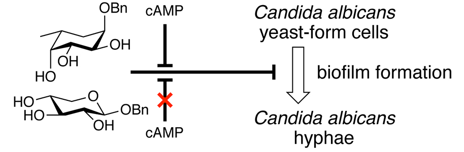
Simple Carbohydrate Derivatives Diminish the Formation of Biofilm of the Pathogenic Yeast Candida albicans

1
Department of Biology, Lund University, Sölvegatan 35, SE-223 62 Lund, Sweden
2
Centre for Analysis and Synthesis, Centre for Chemistry and Chemical Engineering, Lund University, P.O. Box 124, SE-221 00 Lund, Sweden
*
Author to whom correspondence should be addressed.
Submission received: 14 November 2019 / Revised: 4 December 2019 / Accepted: 24 December 2019 / Published: 30 December 2019
(This article belongs to the Section Novel Antimicrobial Agents)

Abstract

:
The opportunistic human fungal pathogen Candida albicans relies on cell morphological transitions to develop biofilm and invade the host. In the current study, we developed new regulatory molecules, which inhibit the morphological transition of C. albicans from yeast-form cells to cells forming hyphae. These compounds, benzyl α-l-fucopyranoside and benzyl β-d-xylopyranoside, inhibit the hyphae formation and adhesion of C. albicans to a polystyrene surface, resulting in a reduced biofilm formation. The addition of cAMP to cells treated with α-l-fucopyranoside restored the yeast-hyphae switch and the biofilm level to that of the untreated control. In the β-d-xylopyranoside treated cells, the biofilm level was only partially restored by the addition of cAMP, and these cells remained mainly as yeast-form cells.

Graphical Abstract

1. Introduction

A majority of human microbial infections involve the formation of biofilms. Biofilms are cell communities that adhere to solid surfaces. Apart from forming biofilms on host surfaces, pathogens often form biofilms on abiotic surfaces of host implants, which brings serious implications for the use of medical devices such as prostheses, joint replacements, catheters, and pacemakers. Compared to planktonic cells, cells in biofilms are more resistant to antibiotics and antifungal drugs [1,2,3,4,5,6,7], host immune responses [8,9], and can promote further pathogen dissemination, forming new biofilms, which are associated with chronic or life threatening infections [10,11,12,13]. Thus, it is important to develop new strategies for the prevention of biofilm formation, and to eliminate already formed biofilm.
Candida albicans is one of the most prevalent human opportunistic fungal pathogens and is able to form biofilm. Although it is a common part of the human microbiome [14], this yeast can, under certain conditions, cause various diseases. Mucosal infections such as vulvovaginal candidiasis occur in up to 75% of all women [15]. Furthermore, immune-compromised individuals may develop candidiasis of the oral cavity as well as systemic blood stream infections with high mortality [16]. The biofilms developed by C. albicans are highly heterogenous and made up of a dense matrix, composed of morphologically different cell types (i.e., blastospores (yeast-form cells), hyphae, and pseudo-hyphae cells surrounded by extracellular polymeric substances (EPS)) [1,17]. The basal thin biofilm layer is composed of yeast-form cells followed by a thicker and more open hyphae layer [1,18,19,20]. In addition to being a scaffold component, the hyphae development is an important virulence factor and facilitates the pathogen invasion through epithelial tissue of the host [21]. The cell wall of C. albicans hyphae and yeast-form cells differ in its composition, which affects their adhesion and recognition by the host’s immune system. For example, the hyphal glucan consists of a unique cyclic (1-3)-linked polymer backbone with long (1-6)-linked side chains [22]. Hyphae also have less mannan, compared to yeast-form cells [23]. The morphological transition from yeast-form cells to hyphae (i.e., switching) is a complex, tightly regulated process during the biofilm maturation, which is regulated by environmental conditions and quorum sensing molecules such as farnesol and tyrosol [21]. Multiple signaling pathways such as cyclic adenosine monophosphate/protein kinase A (cAMP-PKA), high-osmolarity glycerol (HOG), mitogen-activated protein kinases (MAPK), and several transcriptional factors, Efg1, Bcr1, Ndt80, Cph1, Cph2, Tec1, Nrg1, Rfg1, and Tup1 are known to regulate the morphological transition and hyphal adhesion [1,21,24,25,26]. Upon stimuli favoring hyphae, the yeast cells produce a germ tube that extends by polarized growth by the activity of membrane-bound vesicles supplying the components of the plasma membrane and cell wall to the growing hyphae [21].
Different carbohydrates have been explored for their possibilities to disturb or prevent adhesion, biofilm formation, and the extension of an already existing biofilm of C. albicans [27,28,29,30,31,32,33,34]. In addition, some natural products of plant origin were recently shown to reduce the adhesion of C. albicans and its biofilm formation by inhibiting the yeast-to-hyphae transition [35,36].
In this study, we synthesized and tested the effect of simple monosaccharides and glycosides (Chart 1) on C. albicans biofilm development on polystyrene surfaces.

2. Results

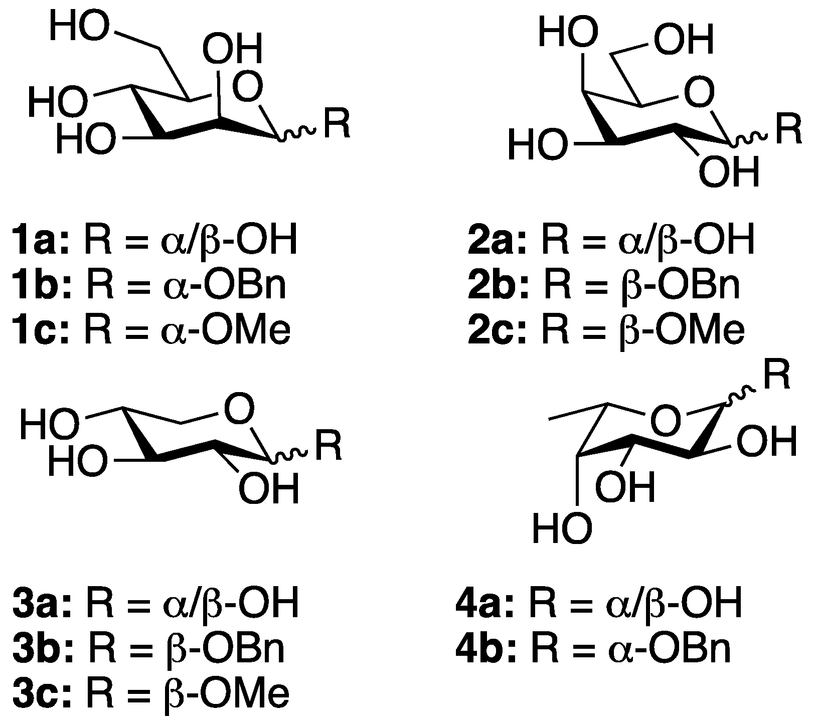
2.1. Carbohydrate Derivatives

The unprotected carbohydrates (1a, 2a, 3a, and 4a) and the methyl pyranosides (1c, 2c, and 3c) were commercially available. The synthesis of the benzylated compounds 1b [37], 2b [38], and 3b [39] was performed as described previously. 4a was benzylated at the anomeric position using benzyl alcohol and acetyl chloride [40] and deprotected to give compound 4b in a 32% yield over two steps (Scheme 1). Compound 4b was synthesized previously via dihydroxylation of the corresponding deoxy sugar [41].

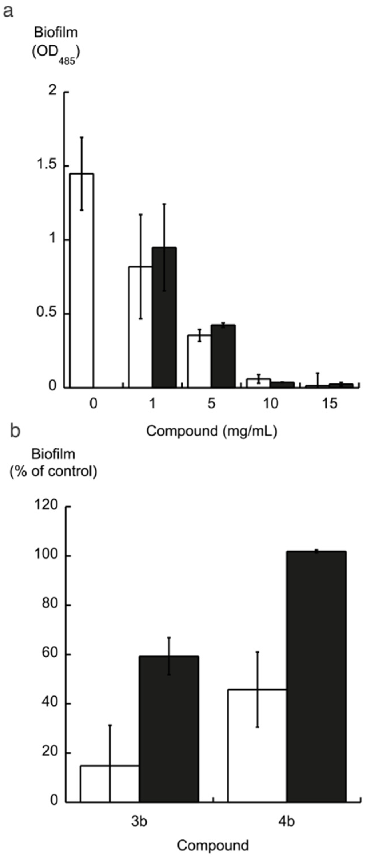
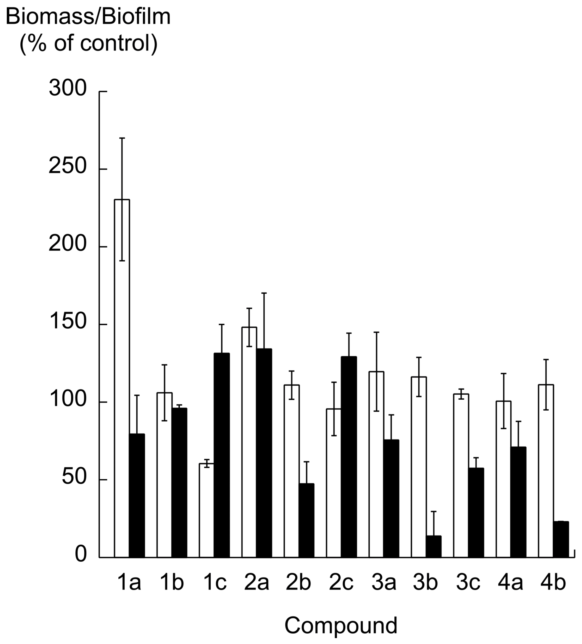
2.2. Biofilm Formation

To evaluate the effects of carbohydrate derivatives, the formation of biofilm on polystyrene surfaces was measured using liquid cultures with the Candida albicans SC5314 strain and the addition of 10 mg/mL of each compound. The formation of biomass and biofilm was measured after 24 h of incubation using the crystal violet method [42]. The data are shown in Figure 1.
During the biofilm experiments, the C. albicans biomass substantially increased only on mannose (1a) (2.3 times, Analysis of Varience (ANOVA) p = 0.0079), but not on the other carbohydrates. None of the tested compounds inhibited biomass except for 1c, which reduced the biomass formation by ~40%, ANOVA p = 0.0225 (Figure 1). The unprotected monosaccharides 1a4a and the methyl glycosides 1c3c did not show any significant effect on the formation of biofilm. In contrast, the benzyl-glycosides reduced the formation of biofilm. After 24 h of incubation, compounds 2b, 3b, and 4b reduced the biofilm 2.1, 7.1, and 4.3 times, respectively (ANOVA p = 0.0491; 0.0029 and 0.0039), compared to the untreated control (Figure 1).
Compounds 3b and 4b were selected for further analysis due to their highest inhibition effect on biofilm. The viability of biofilms of C. albicans after treatment with 3b and 4b at different concentrations was evaluated by staining the adherent cells with XTT (2, 3-bis (2-methoxy-4-nitro-5-sulfophenyl)-5-[(phenylamino)carbonyl]-2H-tetrazolium hydroxide), a colorimetric assay for the quantification of cellular viability and cytotoxicity [43]. Compounds 3b and 4b each displayed a dose-dependent inhibitory effect on biofilm viability (Figure 2a). The minimum biofilm inhibitory concentration (MBIC) was estimated to be 5 mg/mL and resulted in 3.7-times reduced biofilm (Figure 2a, ANOVA p = 0.03 for both 3b and 4b).
The addition of cAMP (regulatory molecule of major signaling pathway of biofilm development, the cAMP-PKA pathway) to the media with carbohydrates affected the biofilm development. It rescued the inhibition by the 4b compound and restored the biofilm level to that of the untreated control (Figure 2b). On the other hand, the inhibitory effect of 3b was only partially restored by cAMP treatment (Figure 2b).

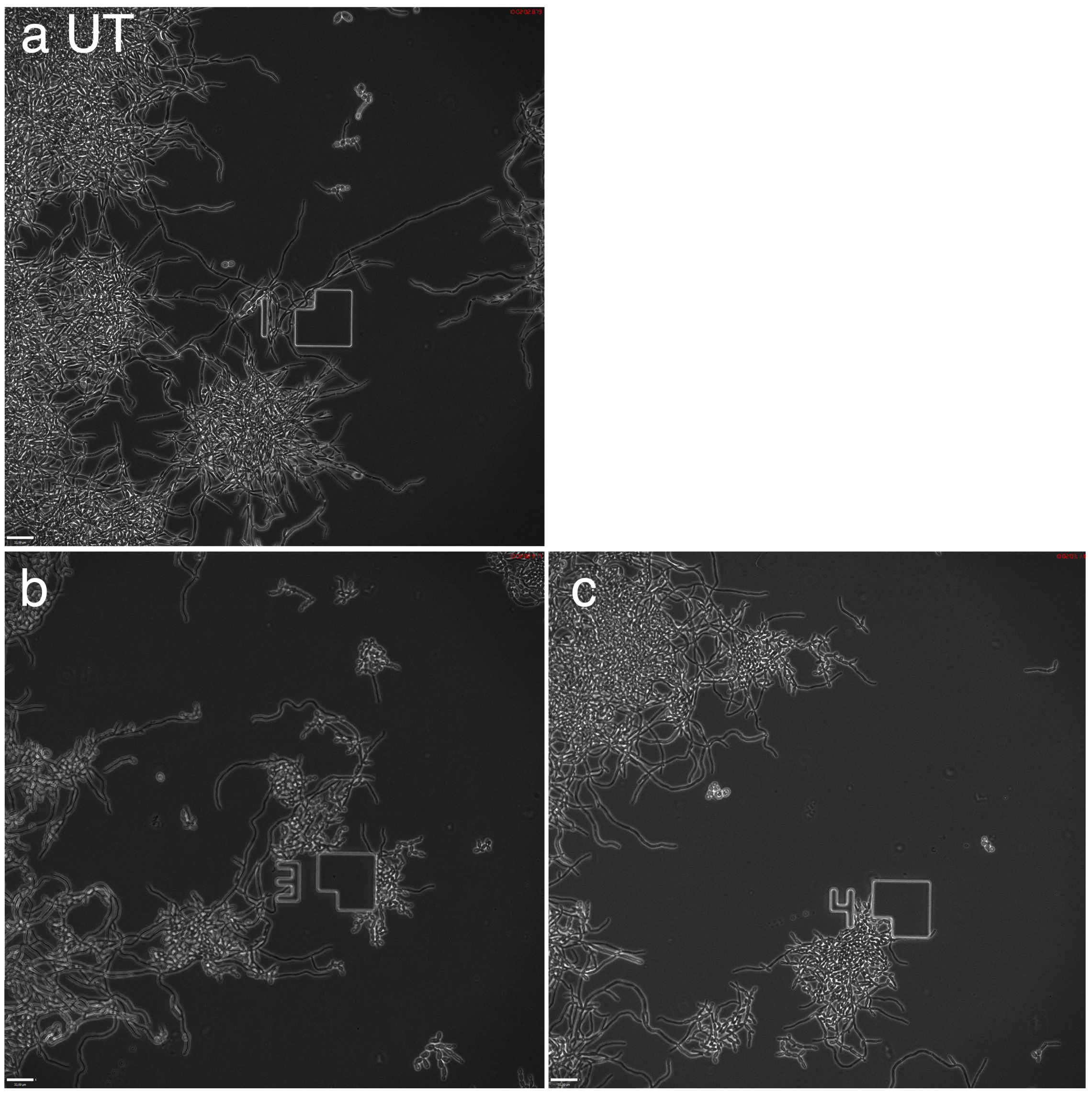
2.3. Yeast-Hyphae Transition

The formation of hyphae by C. albicans is important for biofilm formation since hyphae are more adherent than yeast cells. After the formation of hyphae, the adhesins, which are expressed mostly on hyphae, play an important role in further adhesion [9]. To monitor the C. albicans cell morphology upon carbohydrates treatment, we also studied the biofilm development using microscopy and microfluidics.
After the yeast cells were inoculated into the biofilm medium, hyphae started to form within the first hour of incubation (YNB supplemented with 0.45% ammonium sulphate, 100 mM l-proline and 0.2% glucose, pH 7.0, see Figure 3a, Supplementary Materials, Figure S1, time-lapse videos of biofilm formation over time). The addition of benzyl β-d-xylopyranoside (3b) or benzyl α-l-fucopyranoside (4b) resulted in a substantial decrease of hyphae formation, and the majority of the cells remained in yeast-form during the incubation period (Figure 3b,c, Supplementary Materials, Figures S2 and S3, time-lapse videos of biofilm formation over time). High-resolution pictures after 10 h and 18.5 h of incubation can also be found in the Supplementary Materials.
The branching of hyphae and cell size could be affected by vacuole inheritance [44,45]. However, the benzylated compounds tested did not exhibit any effect on vacuole morphology. Furthermore, the addition of cAMP to the media released the hyphae formation inhibitory effect caused by benzyl α-l-fucopyranoside (4b) (Supplementary Materials, Figure S4) and the switching of yeast-form cells to hyphae occurred effectively. In contrast, the cAMP did not rescue the benzyl β-d-xylopyranoside (3b) inhibition effect on hyphae formation, and most cells remained in yeast-form after 24 h of the treatment (Supplementary Materials, Figure S4).

3. Discussion

In the current study, we developed new carbohydrates derivatives that diminished the biofilm formation of an opportunistic pathogen Candida albicans. Carbohydrates are abundant in nature and are a part of the building blocks of living cells. Xylose and fucose are found in the fungal extracellular biofilm matrix [46]. Xylose comprises around 12% of total carbohydrates of C. albicans biofilm matrix [46]. Furthermore, l-fucose is a component of host glucan surface structures, found in mammalian cells. The C. albicans Als1 protein specifically recognizes the fucose-containing sugars of glucan of the host [47]. Thus, the addition of these carbohydrates can possibly perturb the pathogen interaction with the host or competitively bind the pathogen adhesins and prevent adhesion to abiotic surfaces. Therefore, these carbohydrates have been investigated for their ability to inhibit adhesion and biofilm formation.
In the early 1980s, the effect of both fucose and xylose was studied on C. albicans adhesion to human cells. Sobel et al. showed that both d- and l-fucose inhibited C. albicans adhesion to vaginal epithelial cells (by ~30%) [27]. Xylose inhibited the adhesion to buccal cells by 15% [28]. In our study, the addition of l-fucose and d-xylose to the biofilm media (containing the hyphae inducer l-proline) slightly decreased the biofilm formation on polystyrene. On the other hand, the benzyl-derivatives of both fucose and xylose had a significantly higher inhibitory effect on the biofilm formation of C. albicans. We found that in addition to reducing the adhesion to polystyrene, these compounds affected the C. albicans morphological transition of yeast-form cells to hyphae, which is the key property for mature biofilm [1]. Upon the addition of either benzyl α-l-fucopyranoside (4b) or benzyl β-d-xylopyranoside (3b), the cells continued to grow in yeast-like form and produced only few hyphae. On the other hand, methyl glycosides did not affect the morphological transition, indicating that the inhibitory effect is linked to the benzyl group. The morphological transition of yeast to the hyphae form is regulated by the cAMP-PKA pathway in C. albicans [1,25]. Interestingly, aromatic compounds have been reported to influence the adhesion of both Saccharomyces cerevisiae and C. albicans by inducing the expression of adhesin genes [48,49]. For S. cerevisiae, tryptophol and phenyl ethanol were reported to be a part of quorum sensing by activating the FLO11 gene expression via the cAMP-PKA pathway [48,50]. C. albicans was found to respond to the aromatic alcohol tyrosol [51], while tryptophol and phenylethanol did not induce phenotypic changes in C. albicans [50]. Farnesol is a negative regulator of quorum sensing through the Ras-cAMP-PKA pathway in both C. albicans and S. cerevisiae, [49,52] as it was found to inhibit C. albicans adhesion [53] and S. cerevisiae cell growth [54].
The addition of cAMP (a key regulatory molecule in yeast-to-hyphae transition of Ras-cAMP-PKA pathway) to the biofilm medium containing benzyl-glycosides reversed the hyphae formation inhibitory effect only of benzyl α-l-fucopyranoside (4b), but not of benzyl β-d-xylopyranoside (3b). Apart from restoring the morphological transition, the cAMP restored the adhesion level of the α-l-fucopyranoside treated sample to 50% of the control. This suggests that the effects of benzyl α-l-fucopyranoside (4b) and benzyl β-d-xylopyranoside (3b) are mediated by different mechanisms. Compound 4b likely operates through the Ras-cAMP-PKA pathway by inhibiting the pathway by downregulating the level of cAMP. Compound 3b could affect the pathway gene expression downstream of cAMP. Future studies such as C. albicans transcriptome analysis upon the exposure to the compounds tested are needed to find the gene targets.

4. Materials and Methods

4.1. General Methods, Strains, and Growth Conditions

Thin-layer chromatography (TLC) was performed on precoated TLC-glass plates with silica gel 60 F254 0.25 mm (Merck). Spots were visualized with UV light or by charring with an ethanolic anisaldehyde solution. Preparative chromatography was performed on a Biotage Isolera One flash purification system using Biotage SNAP KP-Sil silica cartridges. Nuclear magnetic resonance (NMR) spectra were recorded at ambient temperatures on a Bruker Avance II at 400 MHz (1H), and 100 MHz (13C) and assigned using 2D methods (COSY, HMQC). Chemical shifts are reported in ppm, with reference to residual solvent peaks (δH CHCl3 = 7.26 ppm and δC CDCl3 = 77.0 ppm). Coupling constant values are given in Hz. 13C-NMR spectra are proton decoupled. High-resolution mass spectra (HRMS) were recorded on Waters QTOF XEVO-G2 (ESI+).
Candida albicans SC5314 strain [55] was grown at 37 °C in complete medium YPD (0.5% yeast extract [Sigma-Aldrich, Y1625], 1% peptone [Sigma-Aldrich, 70175], 2% glucose [Sigma-Aldrich, G7021]) or minimal medium consisting of YNB (yeast nitrogen base without amino acids and ammonium sulfate, FORMEDIUMTM, CYN0505) supplemented with 0.45% ammonium sulfate (Sigma-Aldrich, A4915), 0.2% glucose (Sigma-Aldrich, G7021), and 100 mM l-proline (Sigma-Aldrich, P0380) of pH 7.0. If needed, 2% agar (Sigma-Aldrich, A5306) was used to solidify the media.
The liquid minimal medium (YNB [yeast nitrogen base without amino acids and ammonium sulfate, FORMEDIUMTM, CYN0505] supplemented with 0.45% ammonium sulfate [Sigma-Aldrich, A4915], 0.2% glucose [Sigma-Aldrich, G7021], and 100 mM l-proline [Sigma-Aldrich, P0380]) of pH 7.0 was used for the biofilm assay (biofilm medium).
Different carbohydrates (Chart 1) were added to the biofilm medium at the final concentration 0.1–15 mg/mL. The cell permeable cAMP (N6, 2′-O-Dibutyryladenosine 3′,5′-cyclic monophosphate sodium salt, Sigma-Aldrich, D0260) was added to the biofilm medium at the final concentration of 10 mM to study if the sugar inhibition effect on the biofilm could be reversed.

4.2. Biofilm Assay

Prior to the biofilm assay, yeast cultures were grown in liquid YPD medium for 24 h until the stationary phase (OD600 11–17), cells were then pelleted by centrifugation (1699 g), washed with sterile water, and cells were further inoculated into a test biofilm medium at a final concentration of 0.2 OD600/mL and incubated in 96-well flat-bottom polystyrene microtiter plates (Sigma-Aldrich, Corning® Costar® culture plates, CLS3596-50EA) for 24 or 48 h at 37 °C. At the defined time points, the biofilm developed on polystyrene was measured either by crystal violet staining as described [42,56] or by improved XTT method, where the 200 mM glucose and XTT (2, 3-bis (2-methoxy-4-nitro-5-sulfophenyl)-5-[(phenylamino)carbonyl]-2H-tetrazolium hydroxide, X4626, Sigma-Aldrich) were added to the reaction mixture [43]. The crystal violet (HT901-8FOZ, Sigma Aldrich) was added to the media at the final concentration of 0.05%. After 24 and/or 48 h of cell staining, plate wells were washed four times with 200 μL of water to remove planktonic cells, and the biofilms were then dried and dissolved in 200 μL of 96% ethanol. In the XTT assay, the planktonic cells were removed by washing twice with PBS buffer. Total biomass and crystal violet biofilm staining measurements were performed at OD560, and the formazan formed during the XTT biofilm assay was measured at OD485 with FLUOstar OPTIMA plate reader, BMG LABTECH.

4.3. Microfluidics and Microscopy

Microfluidic plates (CellASIC® ONIX, Merck Millipore, Y04D-02-5PK) were used with the ONIX Microfluidic Perfusion System and inoculated with yeast at 8 psi for 5 s, according to the manufacturer’s recommendations, and flowed at 1.5 psi with media tested (with/without carbohydrates, 15 mg/mL). The yeast growth and biofilm development were monitored over time on a fully motorized and automated inverted widefield microscope Observer Z1 (Carl Zeiss) equipped with a sCMOS camera. The phase-contrast images were taken over the time specified.

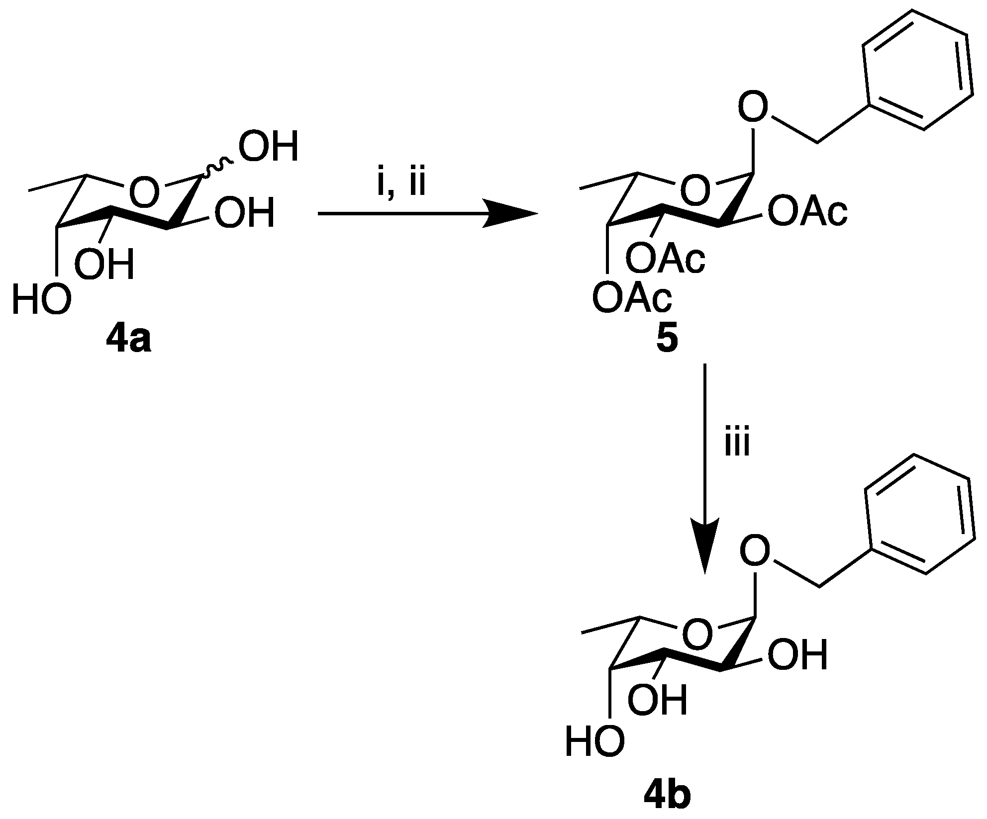
4.4. Synthesis of Benzyl (2,3,4-tri-O-acetyl) α-l-fucopyranoside (5)

Acetyl chloride (0.43 mL, 6.1 mmol) was added to a stirred solution of benzyl alcohol (8.83 mL, 85.0 mmol) and l-fucose 4a (1.0 g, 6.1 mmol). After 22 h at 50 °C, the reaction mixture was allowed to reach room temperature (r.t.) before the removal of benzyl alcohol by vacuum distillation (0.1 torr, 65 °C). The dark brown crude residue was dissolved in pyridine (9.0 mL, 0.11 mol) and acetic anhydride (9.0 mL, 0.16 mol) and stirred at r.t. After 15 h, toluene was added, and the solvent was removed under reduced pressure. Purification by column chromatography (Biotage, KP-Sil 50 g, heptane:EtOAc 95:5→88:12) gave benzyl (2,3,4-tri-O-acetyl) α-l-fucopyranoside (5, 986 mg, 56%, clear oil) and benzyl (2,3,4-tri-O-acetyl) β-l-fucopyranoside (6, 247 mg, 14%, white amorphous solid). 5: 1H-NMR (CDCl3) δ 7.37–7.28 (m, 5H) 5.42–5.38 (m, 1H) 5.30–5.29 (m, 1H) 5.15–5.12 (m, 2H) 4.71 (dAB, 1H, JAB 12.4 Hz) 4.55 (dAB, 1H, JAB 12.0 Hz) 4.17 (q, 1H, J 6.4 Hz) 2.16 (s, 3H) 2.03 (s, 3H) 1.98 (s, 3H) 1.11 (d, 3H, J 6.4 Hz); 13C-NMR (CDCl3) δ 170.7, 170.5, 170.2, 137.3, 128.6, 128.1, 127.9, 95.6, 71.3, 70.0, 68.2, 68.2, 64.7, 20.9, 20.8, 20.8, 15.9; HRMS (ESI/QTOF) m/z [M + Na] calcd. for C19H24O8Na 403.1363, found 403.1368. 6: 1H-NMR (CDCl3) δ 7.37–7.28 (m, 5H) 5.28–5.22 (m, 2H) 4.98 (dd, 1H, J 3.6, 10.4 Hz) 4.92 (dAB, 1H, JAB 12.4 Hz) 4.62 (dAB, 1H, JAB 12.4 Hz) 2.18 (s, 3H) 2.00 (s, 3H) 1.98 (s, 3H) 1.25 (d, 3H, J 6.4 Hz); 13C-NMR (CDCl3) δ 170.9, 170.4, 169.7, 137.2, 128.5, 128.0, 127.8, 99.8, 71.5, 70.7, 70.4, 69.3, 69.1, 20.9, 20.9, 20.8, 16.2; HRMS (ESI/QTOF) m/z [M + Na] calcd for C19H24O8Na 403.1363, found 403.1369.

4.5. Synthesis of Benzyl α-l-fucopyranoside (4b)

5 (0.18 g, 0.63 mmol) was dissolved in MeOH (3 mL) and 1 M NaOMe (0.15 mL, 0.15 mmol) was added. After 2 h, glacial acetic acid was added until neutral pH. The reaction mixture was co-evaporated with toluene several times. Purification by column chromatography (SiO2, CH2Cl2:MeOH 95:5) gave 4b (90 mg, 57%) as a white amorphous solid. Data analysis was according to the published data [41].

4.6. Statistical Analysis

The software package Minitab® 18.1 was used to analyze the obtained data.

5. Conclusions

We showed that benzyl β-d-xylopyranoside (3b) and benzyl α-l-fucopyranoside (4b) were both effective against biofilm and inhibited the morphological transition of C. albicans from yeast-form cells to hyphae, diminishing the biofilm. The MBIC for both β-d-xylopyranoside and α-l-fucopyranoside was estimated as 5 mg/mL. Neither the unprotected monosaccharides, nor the corresponding methyl-glycosides showed any significant effects on biofilm inhibition.
The exposure to cAMP of α-l-fucopyranoside treated cells restored yeast-hyphae switching and the biofilm level to that of the untreated control. While the biofilm level was partially restored by the addition of the cAMP, the benzyl β-d-xylopyranoside treated cells remained mainly in yeast-like form. We propose that the effects shown by these compounds are mediated by different mechanisms.

Supplementary Materials

The following are available online at https://0-www-mdpi-com.brum.beds.ac.uk/2079-6382/9/1/10/s1, Figure S1: Control, biofilm medium untreated, Figure S2: Benzyl β-d-xylopyranoside, Figure S3: Benzyl α-l-fucopyranoside, Figure S4: cAMP treatment of benzyl β-d-xylopyranoside and benzyl α-l-fucopyranoside samples. All data generated or analyzed during this study are included in this published article and its supplementary material files.

Author Contributions

Conceptualization, U.E. and S.M.; Formal analysis, O.P.I.; Funding acquisition, O.S.; Investigation, O.P.I.; Methodology, O.P.I.; Project administration, U.E. and O.P.I.; Visualization, O.P.I.; Writing—original draft, U.E., O.P.I., O.S. and S.M.; Writing—review & editing, U.E., O.P.I., O.S. and S.M. All authors have read and agreed to the published version of the manuscript.

Funding

This work was supported by grants from the Lars Hiertas Memorial Foundation, the Magnus Bergwall Foundation, Lund University, the Sten K. Johnson Foundation, and the Royal Physiographic Society in Lund. Part of this work was performed at the Lund Protein Production Platform.

Conflicts of Interest

The authors declare no conflict of interest.

References

  1. Finkel, J.S.; Mitchell, A.P. Genetic control of Candida albicans biofilm development. Nat. Rev. Microbiol. 2011, 9, 109–118. [Google Scholar] [CrossRef]
  2. Seneviratne, C.J.; Wang, Y.; Jin, L.; Abiko, Y.; Samaranayake, L.P. Proteomics of drug resistance in Candida glabrata biofilms. Proteomics 2010, 10, 1444–1454. [Google Scholar] [CrossRef]
  3. Fonseca, E.; Silva, S.; Rodrigues, C.F.; Alves, C.T.; Azeredo, J.; Henriques, M. Effects of fluconazole on Candida glabrata biofilms and its relationship with ABC transporter gene expression. Biofouling 2014, 30, 447–457. [Google Scholar] [CrossRef]
  4. Mukherjee, P.K.; Chandra, J.; Kuhn, D.M.; Ghannoum, M.A. Mechanism of fluconazole resistance in Candida albicans biofilms: Phase-specific role of efflux pumps and membrane sterols. Infect. Immun. 2003, 71, 4333–4340. [Google Scholar] [CrossRef] [Green Version]
  5. Nett, J.; Lincoln, L.; Marchillo, K.; Massey, R.; Holoyda, K.; Hoff, B.; VanHandel, M.; Andes, D. Putative role of beta-1,3 glucans in Candida albicans biofilm resistance. Antimicrob. Agents Chemother. 2007, 51, 510–520. [Google Scholar] [CrossRef] [Green Version]
  6. Nobile, C.J.; Johnson, A.D. Candida albicans Biofilms and Human Disease. Annu. Rev. Microbiol. 2015, 69, 71–92. [Google Scholar] [CrossRef] [Green Version]
  7. Pradeep Kumar, S.S.; Easwer, H.V.; Maya Nandkumar, A. Multiple drug resistant bacterial biofilms on implanted catheters—A reservoir of infection. J. Assoc. Physicians India 2013, 61, 702–707. [Google Scholar]
  8. Secor, P.R.; James, G.A.; Fleckman, P.; Olerud, J.E.; McInnerney, K.; Stewart, P.S. Staphylococcus aureus Biofilm and Planktonic cultures differentially impact gene expression, mapk phosphorylation, and cytokine production in human keratinocytes. BMC Microbiol. 2011, 11, 143. [Google Scholar] [CrossRef] [Green Version]
  9. Hofs, S.; Mogavero, S.; Hube, B. Interaction of Candida albicans with host cells: Virulence factors, host defense, escape strategies, and the microbiota. J. Microbiol. 2016, 54, 149–169. [Google Scholar] [CrossRef]
  10. Potera, C. Forging a link between biofilms and disease. Science 1999, 283, 1837–1839. [Google Scholar] [CrossRef]
  11. Donlan, R.M.; Costerton, J.W. Biofilms: Survival mechanisms of clinically relevant microorganisms. Clin. Microbiol. Rev. 2002, 15, 167–193. [Google Scholar] [CrossRef] [Green Version]
  12. Lebeaux, D.; Ghigo, J.M.; Beloin, C. Biofilm-related infections: Bridging the gap between clinical management and fundamental aspects of recalcitrance toward antibiotics. Microbiol. Mol. Biol. Rev. 2014, 78, 510–543. [Google Scholar] [CrossRef] [Green Version]
  13. Wall, G.; Montelongo-Jauregui, D.; Vidal Bonifacio, B.; Lopez-Ribot, J.L.; Uppuluri, P. Candida albicans biofilm growth and dispersal: Contributions to pathogenesis. Curr. Opin. Microbiol. 2019, 52, 1–6. [Google Scholar] [CrossRef]
  14. da Silva Dantas, A.; Lee, K.K.; Raziunaite, I.; Schaefer, K.; Wagener, J.; Yadav, B.; Gow, N.A. Cell biology of Candida albicans-host interactions. Curr. Opin. Microbiol. 2016, 34, 111–118. [Google Scholar] [CrossRef] [Green Version]
  15. Mardh, P.A.; Rodrigues, A.G.; Genc, M.; Novikova, N.; Martinez-de-Oliveira, J.; Guaschino, S. Facts and myths on recurrent vulvovaginal candidosis—A review on epidemiology, clinical manifestations, diagnosis, pathogenesis and therapy. Int. J. STD AIDS 2002, 13, 522–539. [Google Scholar] [CrossRef]
  16. Low, C.-Y.; Rotstein, C. Emerging fungal infections in immunocompromised patients. F1000 Med. Rep. 2011, 3, 14. [Google Scholar] [CrossRef] [Green Version]
  17. Chandra, J.; Kuhn, D.M.; Mukherjee, P.K.; Hoyer, L.L.; McCormick, T.; Ghannoum, M.A. Biofilm formation by the fungal pathogen Candida albicans: Development, architecture, and drug resistance. J. Bacteriol. 2001, 183, 5385–5394. [Google Scholar] [CrossRef] [Green Version]
  18. Baillie, G.S.; Douglas, L.J. Role of dimorphism in the development of Candida albicans biofilms. J. Med. Microbiol. 1999, 48, 671–679. [Google Scholar] [CrossRef]
  19. Douglas, L.J. Candida biofilms and their role in infection. Trends Microbiol. 2003, 11, 30–36. [Google Scholar] [CrossRef]
  20. Andes, D.; Nett, J.; Oschel, P.; Albrecht, R.; Marchillo, K.; Pitula, A. Development and characterization of an in vivo central venous catheter Candida albicans biofilm model. Infect. Immun. 2004, 72, 6023–6031. [Google Scholar] [CrossRef] [Green Version]
  21. Sudbery, P.E. Growth of Candida albicans hyphae. Nat. Rev. Microbiol. 2011, 9, 737–748. [Google Scholar] [CrossRef]
  22. Lowman, D.W.; Greene, R.R.; Bearden, D.W.; Kruppa, M.D.; Pottier, M.; Monteiro, M.A.; Soldatov, D.V.; Ensley, H.E.; Cheng, S.-C.; Netea, M.G.; et al. Novel Structural Features in Candida albicans Hyphal Glucan Provide a Basis for Differential Innate Immune Recognition of Hyphae Versus Yeast. J. Biol. Chem. 2014, 289, 3432–3443. [Google Scholar] [CrossRef] [Green Version]
  23. Machova, E.; Fiacanova, L.; Cizova, A.; Korcova, J. Mannoproteins from yeast and hyphal form of Candida albicans considerably differ in mannan and protein content. Carbohydr. Res. 2015, 408, 12–17. [Google Scholar] [CrossRef]
  24. Lohse, M.B.; Gulati, M.; Johnson, A.D.; Nobile, C.J. Development and regulation of single- and multi-species Candida albicans biofilms. Nat. Rev. Microbiol. 2018, 16, 19–31. [Google Scholar] [CrossRef] [Green Version]
  25. Lin, C.J.; Chen, Y.L. Conserved and Divergent Functions of the cAMP/PKA Signaling Pathway in Candida albicans and Candida tropicalis. J. Fungi 2018, 4, 68. [Google Scholar] [CrossRef] [Green Version]
  26. Biswas, S.; Van Dijck, P.; Datta, A. Environmental sensing and signal transduction pathways regulating morphopathogenic determinants of Candida albicans. Microbiol. Mol. Biol. Rev. 2007, 71, 348–376. [Google Scholar] [CrossRef] [Green Version]
  27. Sobel, J.D.; Myers, P.G.; Kaye, D.; Levison, M.E. Adherence of Candida albicans to human vaginal and buccal epithelial cells. J. Infect. Dis. 1981, 143, 76–82. [Google Scholar] [CrossRef]
  28. Sandin, R.L.; Rogers, A.L.; Patterson, R.J.; Beneke, E.S. Evidence for mannose-mediated adherence of Candida albicans to human buccal cells in vitro. Infect. Immun. 1982, 35, 79–85. [Google Scholar]
  29. Sato, M.; Ohshima, T.; Maeda, N.; Ohkubo, C. Inhibitory effect of coated mannan against the adhesion of Candida biofilms to denture base resin. Dent. Mater. J. 2013, 32, 355–360. [Google Scholar] [CrossRef] [Green Version]
  30. Gharbi, A.; Humblot, V.; Turpin, F.; Pradier, C.M.; Imbert, C.; Berjeaud, J.M. Elaboration of antibiofilm surfaces functionalized with antifungal-cyclodextrin inclusion complexes. FEMS Immunol. Med. Microbiol. 2012, 65, 257–269. [Google Scholar] [CrossRef] [Green Version]
  31. Enache, E.; Eskandari, T.; Borja, L.; Wadsworth, E.; Hoxter, B.; Calderone, R. Candida albicans adherence to a human oesophageal cell line. Microbiology 1996, 142 Pt 10, 2741–2746. [Google Scholar] [CrossRef] [Green Version]
  32. Fradin, C.; Jouault, T.; Mallet, A.; Mallet, J.M.; Camus, D.; Sinay, P.; Poulain, D. Beta-1,2-linked oligomannosides inhibit Candida albicans binding to murine macrophage. J. Leukoc. Biol. 1996, 60, 81–87. [Google Scholar] [CrossRef]
  33. Segal, E.; Kremer, I.; Dayan, D. Inhibition of adherence of Candida albicans to acrylic by a chitin derivative. Eur. J. Epidemiol. 1992, 8, 350–355. [Google Scholar] [CrossRef]
  34. Gondim, B.L.C.; Castellano, L.R.C.; de Castro, R.D.; Machado, G.; Carlo, H.L.; Valença, A.M.G.; de Carvalho, F.G. Effect of chitosan nanoparticles on the inhibition of Candida spp. biofilm on denture base surface. Arch. Oral Biol. 2018, 94, 99–107. [Google Scholar] [CrossRef]
  35. Li, Y.; Chang, W.; Zhang, M.; Ying, Z.; Lou, H. Natural product solasodine-3-O-beta-d-glucopyranoside inhibits the virulence factors of Candida albicans. FEMS Yeast Res. 2015, 15. [Google Scholar] [CrossRef] [Green Version]
  36. Vediyappan, G.; Dumontet, V.; Pelissier, F.; d’Enfert, C. Gymnemic acids inhibit hyphal growth and virulence in Candida albicans. PLoS ONE 2013, 8, e74189. [Google Scholar] [CrossRef] [Green Version]
  37. Zunk, M.; Kiefel, M.J. An efficient synthesis of selectively functionalized d-rhamnose derivatives. Tetrahedron Lett. 2011, 52, 1296–1299. [Google Scholar] [CrossRef] [Green Version]
  38. Roslund, M.U.; Aitio, O.; Wärnå, J.; Maaheimo, H.; Murzin, D.Y.; Leino, R. Acyl Group Migration and Cleavage in Selectively Protected β-d-Galactopyranosides as Studied by NMR Spectroscopy and Kinetic Calculations. J. Am. Chem. Soc. 2008, 130, 8769–8772. [Google Scholar] [CrossRef]
  39. Siegbahn, A.; Manner, S.; Persson, A.; Tykesson, E.; Holmqvist, K.; Ochocinska, A.; Roennols, J.; Sundin, A.; Mani, K.; Westergren-Thorsson, G.; et al. Rules for priming and inhibition of glycosaminoglycan biosynthesis; probing the β4GalT7 active site. Chem. Sci. 2014, 5, 3501–3508. [Google Scholar] [CrossRef] [Green Version]
  40. Bashyal, B.P.; Fleet, G.W.J.; Gough, M.J.; Smith, P.W. Synthesis of the α-Mannosidase inhibitors swainsonine [(1S, 2R, 8R, 8aR)-1,2,8-trihydroxyoctahydroindolizine] and 1,4-dideoxy-1,4-imino-d-mannitol from mannose. Tetrahedron 1987, 43, 3083–3093. [Google Scholar] [CrossRef]
  41. Wang, H.-Y.L.; O’Doherty, G.A. De novo synthesis of deoxy sugarvia a Wharton rearrangement. Chem. Commun. 2011, 47, 10251–10253. [Google Scholar] [CrossRef]
  42. Andersen, K.S.; Bojsen, R.; Sørensen, L.G.R.; Nielsen, M.W.; Lisby, M.; Folkesson, A.; Regenberg, B. Genetic basis for Saccharomyces cerevisiae biofilm in liquid medium. G3 2014, 4, 1671–1680. [Google Scholar] [CrossRef] [PubMed] [Green Version]
  43. da Silva, W.J.; Seneviratne, J.; Parahitiyawa, N.; Rosa, E.A.; Samaranayake, L.P.; Del Bel Cury, A.A. Improvement of XTT assay performance for studies involving Candida albicans biofilms. Braz. Dent. J. 2008, 19, 364–369. [Google Scholar] [CrossRef] [PubMed] [Green Version]
  44. Veses, V.; Richards, A.; Gow, N.A. Vacuole inheritance regulates cell size and branching frequency of Candida albicans hyphae. Mol. Microbiol. 2009, 71, 505–519. [Google Scholar] [CrossRef] [Green Version]
  45. Barelle, C.J.; Richard, M.L.; Gaillardin, C.; Gow, N.A.R.; Brown, A.J.P. Candida albicans VAC8 Is Required for Vacuolar Inheritance and Normal Hyphal Branching. Eukaryot. Cell 2006, 5, 359–367. [Google Scholar] [CrossRef] [Green Version]
  46. Zarnowski, R.; Westler, W.M.; Lacmbouh, G.A.; Marita, J.M.; Bothe, J.R.; Bernhardt, J.; Lounes-Hadj Sahraoui, A.; Fontaine, J.; Sanchez, H.; Hatfield, R.D.; et al. Novel entries in a fungal biofilm matrix encyclopedia. MBio 2014, 5, e01333-14. [Google Scholar] [CrossRef] [Green Version]
  47. Donohue, D.S.; Ielasi, F.S.; Goossens, K.V.; Willaert, R.G. The N-terminal part of Als1 protein from Candida albicans specifically binds fucose-containing glycans. Mol. Microbiol. 2011, 80, 1667–1679. [Google Scholar] [CrossRef]
  48. Bojsen, R.K.; Andersen, K.S.; Regenberg, B. Saccharomyces cerevisiae—A model to uncover molecular mechanisms for yeast biofilm biology. FEMS Immunol. Med. Microbiol. 2012, 65, 169–182. [Google Scholar] [CrossRef] [Green Version]
  49. Albuquerque, P.; Casadevall, A. Quorum sensing in fungi—A review. Med. Mycol. 2012, 50, 337–345. [Google Scholar] [CrossRef] [Green Version]
  50. Chen, H.; Fink, G.R. Feedback control of morphogenesis in fungi by aromatic alcohols. Genes Dev. 2006, 20, 1150–1161. [Google Scholar] [CrossRef] [Green Version]
  51. Chen, H.; Fujita, M.; Feng, Q.; Clardy, J.; Fink, G.R. Tyrosol is a quorum-sensing molecule in Candida albicans. Proc. Natl. Acad. Sci. USA 2004, 101, 5048–5052. [Google Scholar] [CrossRef] [PubMed] [Green Version]
  52. Hornby, J.M.; Jensen, E.C.; Lisec, A.D.; Tasto, J.J.; Jahnke, B.; Shoemaker, R.; Dussault, P.; Nickerson, K.W. Quorum sensing in the dimorphic fungus Candida albicans is mediated by farnesol. Appl. Environ. Microbiol. 2001, 67, 2982–2992. [Google Scholar] [CrossRef] [Green Version]
  53. Ramage, G.; Saville, S.P.; Wickes, B.L.; Lopez-Ribot, J.L. Inhibition of Candida albicans biofilm formation by farnesol, a quorum-sensing molecule. Appl. Environ. Microbiol. 2002, 68, 5459–5463. [Google Scholar] [CrossRef] [PubMed] [Green Version]
  54. Machida, K.; Tanaka, T.; Yano, Y.; Otani, S.; Taniguchi, M. Farnesol-induced growth inhibition in Saccharomyces cerevisiae by a cell cycle mechanism. Microbiology 1999, 145 Pt 2, 293–299. [Google Scholar] [CrossRef] [Green Version]
  55. Odds, F.C.; Brown, A.J.P.; Gow, N.A.R. Candida albicans genome sequence: A platform for genomics in the absence of genetics. Genome Biol. 2004, 5, 230. [Google Scholar] [CrossRef] [PubMed] [Green Version]
  56. Serrano-Fujarte, I.; Lopez-Romero, E.; Reyna-Lopez, G.E.; Martinez-Gamez, M.A.; Vega-Gonzalez, A.; Cuellar-Cruz, M. Influence of culture media on biofilm formation by Candida species and response of sessile cells to antifungals and oxidative stress. Biomed Res. Int. 2015, 2015, 783639. [Google Scholar] [CrossRef] [PubMed] [Green Version]
Chart 1. Structures of carbohydrates used in this study.
Antibiotics 09 00010 ch001
Scheme 1. (i) BnCl, AcCl, 50 °C, 22 h; (ii) Pyridine, Ac2O, r.t., 15 h, (iii) MeOH, NaOMe, r.t., 2 h.
Antibiotics 09 00010 sch001
Figure 1. The biomass and biofilm formation of C. albicans in the minimal medium supplemented with different carbohydrate compounds at 10 mg/mL. Unfilled columns indicate the biomass (both planktonic and biofilm cells). Filled columns indicate the biofilm (adherent cells) stained by crystal violet. All experiments were performed in duplicate and the error bars represent the standard deviation. The data were normalized by untreated control, which was set at 100%.
Antibiotics 09 00010 g001
Figure 2. The biofilm formation of C. albicans on polystyrene in liquid culture measured by the XTT assay. (a) The biofilm growth after 24 h at different concentrations (1–15 mg/mL) of compounds 3b (filled columns) and 4b (unfilled columns). ANOVA p = 0.1 (1 mg/mL), p = 0.03 (5 mg/mL), p = 0.02 (10 mg/mL) and p = 0.02 (15 mg/mL). (b) The biofilm growth after 24 h supplemented with 10 mg of compounds 3b and 4b, without (filled columns) or with the addition of cAMP (unfilled columns). Samples compared to the control (set to 100%) (i.e., biofilm medium without treatment). All experiments were performed in duplicate and the error bars represent the standard deviation.
Antibiotics 09 00010 g002
Figure 3. The effect of compounds 3b and 4b (panel c), on the morphological transition of C. albicans. The images were taken at the 10 h time point of incubation at 37 °C. Untreated, UT (panel a)—cells are predominantly in hyphae form; the different carbohydrates were tested at the concentration of 15 mg/mL and resulted in fewer hyphae and more predominant yeast-form cells: 3b (panel b); 4b (panel c). The scale bar is 32 µm. Number in the middle of the image corresponds to the number of microfluidic chambers used.
Antibiotics 09 00010 g003

