Polyphenols of Honeybee Origin with Applications in Dental Medicine


1
Department of Microbiology and Immunology, Faculty of Biology, University of Bucharest, 030018 Bucharest, Romania
2
Department of Science and Engineering of Oxide Materials and Nanomaterials, Faculty of Applied Chemistry and Materials Science, University Politehnica of Bucharest, 1-7 Polizu Street, 011061 Bucharest, Romania
3
Research Institute of the University of Bucharest—ICUB, University of Bucharest, 90-92 Panduri Road, 050657 Bucharest, Romania
*
Author to whom correspondence should be addressed.
Antibiotics 2020, 9(12), 856; https://0-doi-org.brum.beds.ac.uk/10.3390/antibiotics9120856
Submission received: 23 October 2020
/
Revised: 22 November 2020
/
Accepted: 27 November 2020
/
Published: 30 November 2020
(This article belongs to the Special Issue Honey Bee Products as an Alternative or Complement to Classical Antibiotics)
Abstract
:Honeybee products are a great source of polyphenols with recognized applications in dental medicine. Although their biological mechanisms in oral diseases are not fully understood, numerous in vitro, in vivo and clinical studies have reported promising results in the prevention and treatment of oral diseases. Bioactivities, such as antibacterial, antiviral, antiparasite, anticancer, anti-inflammatory and anti-oxidant properties, recommend their future study in order to develop efficient alternatives in the management of widespread oral conditions, such as dental caries and periodontitis. The most investigated mechanisms of polyphenols in oral health rely on their ability to strengthen the dental enamel, decrease the development of dental plaque formation, inhibit the progression of dental caries and development of dental pathogens and show anti-inflammatory properties. These features recommend them as useful honeybee candidates in the management of emerging oral diseases.
1. Introduction
In the context of the current crisis in managing infectious diseases, alternative antimicrobial approaches are urgently needed [1,2,3].
Alternative antibiotics of natural origin, both plant and animal-derived, have increasingly attracted the attention of researchers in the last decade [4,5,6]. Honeybee products have proved their great biomedical potential since ancient times [7,8]. Their well-known curative and infection-preventive properties have been exploited in numerous applications. However, honeybee products may have significant differences in terms of chemical, physical, and biological composition, and such aspects could impact their biomedical properties [9,10]. For example, the composition of honey depends on its source (geographical area and plant species) and the genus and species of bees that produce the honey sample [11].
In order to overcome the limitations of natural products, identification of the most useful bioactive, and their dosage represents a critical step in the design of alternative antibiotics [12].
The most investigated honeybee products for their biomedical properties are: honey, beeswax, propolis, and royal jelly [13,14,15,16]. All these products may have diverse medical effects, being used today for preventing and healing severe diseases, such as cancers and infections. Moreover, these products are used on a wide scale in traditional treatments for increasing well-being and even life expectancy due to their proven antibacterial, antiparasitic, anticancer, anti-caries, and immunomodulatory potential [17]. Honey is mainly composed of sugars and water together with minor constituents such as minerals, vitamins, amino acids, organic acids, flavonoids, and other phenolic compounds and aromatic substances [18,19]. Beeswax consists of around 300 different compounds. These are mainly represented by long-chain alkanes, acids, esters, polyesters, and hydroxyl esters [20]. Propolis has a complex composition, being composed of about 58% phenolic compounds, 24% beeswax, 8% lipids and wax, 6% flavonoids, 0.5% terpenes, 0.5% bioelements and 3% other substances [21].
Royal jelly is a complex product, consisting of water (50–60%), proteins (18%), carbohydrates (15%), lipids (3–6%), trace minerals, water-soluble vitamins, free amino acids, and many other compounds, which are less known [13,22,23,24,25].
Although most of the major compounds found in honeybee products have proven biomedical potential, polyphenols have attracted the attention of researchers, mainly due to their wide distribution among all honeybee derived products (especially honey and propolis), but also because of their complex structure and properties (i.e., antibacterial, antiviral, anti-inflammatory, antineoplastic, and anti-ulcer effects) [26].
The biomedical potential of polyphenols also extends to dental applications. These natural origin compounds have been investigated for the improvement of dental materials and engineering of dental tissues [27]. Their antimicrobial potential may be used as a powerful approach in handling the biofilm of dental plaque, oral infections, and inflammatory conditions. In addition, antioxidant activities of polyphenols seem to offer significant protection against oral cancers [28], help in the management of the periodontal disease [29] and the prevention of dental caries and strengthen the teeth [30,31].
The purpose of this paper is to present the main types of polyphenols and discuss their applications in dental medicine, empathizing their antimicrobial effect and impact in the prevention and therapy of widespread dental pathologies.
2. Polyphenols in Honeybee Products
Polyphenols, the most numerous and widespread group of biologically active compounds, are secondary metabolites with a complex, heterogeneous chemical structure [32,33]. Based on the main monomer, the phenolic ring, polyphenols can be divided into two main classes: phenolic acids (hydroxybenzoic and hydroxycinnamic acids) and flavonoids (flavones, flavanones, flavonols, flavanols, and isoflavones) [31,34,35,36]. Lignans, stilbenes, and tannins are other important classes of polyphenols with different biological functions.
The phenolic content of honey is important for its biological effects. Different types of honey have different phenolic composition, depending on several factors, such as the bee species and the type of flowers from which they collect nectar, the geographical area, and the environmental conditions [37,38].
The polyphenol composition of honeybee products is very diverse and correlated to the geographical area of their production. Along with their high therapeutic and dietary value, polyphenols are primary identifying markers for the botanical origin of honey [39]. They are responsible for the colors, aromas, and tastes of all plant-based products, including fruits, cereals, and vegetables. Thus, the pollen source significantly impacts the color, aroma, and taste of honeybee products [26].
Flavonoids and phenolic acids are the most well-represented polyphenols in honeybee-derived products. The chemical structure of the bioactive flavonoid and phenolic acids in honey and propolis are largely similar across products originated in various geographic areas but vary in their relative quantities [40,41,42]. Generally, it seems that honey of darker color contains higher amounts of bioactive compounds as compared to lighter-colored honey [43].
For example, although there are qualitative and quantitative differences in flavonoid contents reported from different studies, probably based on the extraction and detection methods used, Manuka honey, monofloral honey derived from the manuka tree (Leptospermum scoparium) with proven antimicrobial and anti-oxidant properties is considered to be a better source of flavonoids than other types of honey. Pinobanksin, pinocembrin, and chrysin are the major flavonoids in Manuka honey, and in small quantities, luteolin, quercetin, 8-methoxykaempferol, isorhamnetin, kaempferol, and galangin. 4-Hydroxybenzoic acid and syringic acid are the most abundant phenolic acids detected in this type of honey [44].
In a study from 2019, Yi Tang et al. compared the content of polyphenols (16 phenolic acids and 14 flavonoids were investigated by HPLC) from 40 samples of honey from China and other countries [45]. Gallic acid, a well-known compound for the anti-oxidant activity of honey, was the most detected phenolic acid. In the respective study, almost all honey samples were found to be a good source of gallic acid except Loquat honey and Honeysuckle honey from China, in which gallic acid was not detected. In addition, chlorogenic acid and caffeic acid, which present very good antibacterial function, were investigated. Manuka honey from New Zealand presented the highest content of these two phenolic acids, followed by Forest honey from Spain and Wildflower honey from Italy. Vanillic acid was detected in Thyme honey from Spain, but not in Manuka honey, in which was detected p-hydroxybenzoic acid. Some other phenolic acids such as p-coumaric acid and syringaldehyde were detected only in two honey samples, whereas salicylic acid, sinapic acid, and ferulic acid were not detected at all. Among the flavonoids, apigenin and chrysin were most commonly found in honey from New Zealand and Spain. Significantly high content of quercetin was detected in Orange blossom honey. Chrysin, naringenin, and galangin were also found in Spanish honey samples, but not tangeretin and hesperidin. Based on these results, the authors conclude that the geographical location influences the honey composition in flavonoids. The floral origin determines the quantity of flavonoids [46].
This variation of the content of the polyphenols was also described by Olas Beata (2020) regarding the phenolic acid profiles of 12 kinds of honey collected from various regions in Greece: a significantly higher concentration of protocatechuic acid was detected in pine and fir honey than thyme and citrus honey, whereas p-hydroxybenzoic acid was the major phenolic compound detected in thyme honey [47].
Numerous studies performed to elucidate the most common bioactive flavonoid and phenolic acid compounds found in honeybee products have been demonstrated that they are: gallic acid, caffeic acid, ellagic acid, syringic acid, cinnamic acid, coumaric acid, benzoic acid (and its derivatives), quercetin, naringenin, kaempferol, luteolin, crysin and galancin [26,48].
3. Bioactivities of Polyphenols
Polyphenols have shown numerous mechanisms of biomedical interest, which could be exploited for their applications in dental medicine. Their anti-inflammatory, antimicrobial, anti-inflammatory, and tissue healing properties are currently being investigated for cardiac, digestive, and wound healing capacity. These mechanisms are very useful in the design of prophylactic and therapeutic approaches in oral pathologies, such as periodontitis (which has an important inflammatory component), oral cancers, and even dental caries. In the section below, we present a brief review of such useful properties.
3.1. Cardio-Protective and Anti-Ulcer Properties
Many of the phenolic constituents of honey-like quercetin, caffeic acid phenethyl ester (CAPE), acacetin, kaempferol, and galangin were found to have an encouraging effect in the prevention and treatment of cardiovascular diseases. These compounds showed antithrombotic, anti-ischemic, anti-oxidant, and vasorelaxant activity. The main activities of flavonoids included improving coronary vasodilatation and decreasing the blood coagulation capacity, and the oxidation of low-density lipoproteins (LDLs) [49].
Anti-ulcer activity is also attributed to flavonoids, such as kaempherol, quercetin, hesperitin, and naringin, which includes their capacity to stimulate the level of mucosal prostaglandins, and to decrease acid secretion and prevent ulceration [50]. There are also some in vitro studies that demonstrated the antimicrobial activity of different samples of honey against Helicobacter pylori, the bacterial species responsible for gastroduodenal ulcers [51].
3.2. Antitumor Activity
Besides other effects, honey also showed anti-tumoral activity by its capacity to induce apoptosis. It was demonstrated that honey inhibited colon cancer cell proliferation. The antimetastatic effect of propolis and some phenolic compounds was observed in tumor mice models before and after the injection of tumor cells. These effects were also reported for other types of cancer cell lines, like bladder cancer. Moreover, it seems that honey facilitated the antitumor activity of some chemotherapeutic agents such as 5-fluorouracil and cyclophosphamide. Researchers investigated if the anti-tumoral activity of honey could be attributed to its phenolic compounds. Many of them such as caffeic acid, caffeic acid phenyl ester, chrysin, galangin quercetin, acacetin, kaempferol, pinocembrin, pinobanksin, and apigenin were investigated and had an anti-proliferative effect in a dose-dependent manner in different types of cancers [52]. Phenolic compounds act on carcinogenesis through the induction of cell defense systems, including detoxifying and antioxidant enzyme systems, as well as the inhibition of the anti-inflammatory and anti-cellular growth signaling pathways that culminate in cell cycle arrest and/or cellular death. Polyphenols seem to elicit important alterations in the epigenome of cancer cells, which leads to a variety of anticancer mechanisms, including the modulation of cell cycle signaling, the removal of anticancer agents, the activity of antioxidant enzymes, apoptosis, and arrest of the cell cycle [53]. Polyphenols such as lycopene, epigallocatechin-3 gallate (EGCG), and sulphoraphane downregulate several signal transduction pathways responsible for anti-angiogenetic and antimetastatic activities, being efficient against prostate cancer [54]. EGCG can be methylated through catechol-O-methyltransferase and thus inhibit the DNA methyltransferase (DNMT), which prevents hypermethylation of newly formed DNA strands, leading to a reversal process of silenced genes. Furthermore, EGCG can suppress DNMT action by activating genes silenced by tumor cells by methylation. These mechanisms were demonstrated in colorectal cancer cells and are considered to also apply in other types of cancer cells [55]. EGCG and resveratrol decrease cell proliferation and induce DNA damage in various bladder tumor cells. The main incriminated mechanism is related to the modulation of the DNMT1 gene and apoptosis activation [56].
Resveratrol and quercetin are also active against human oral cancer cells. Studies demonstrated that 10 and 100 μM resveratrol induced significant dose-dependent inhibition in growth as well as in DNA synthesis of oral squamous carcinoma cells (SCC-25). Quercetin exhibits a biphasic effect, being simulative at 1 and 10 μM, while at 100 μM inhibits cell growth and DNA synthesis. Researchers combined 50 μM of resveratrol with 10, 25, and 50 μM of quercetin and demonstrated a gradual and significant increase in the inhibitory effect of quercetin on oral cancer cell growth and DNA synthesis [57]. These reports suggest that polyphenols and especially their combinations (such as resveratrol and quercetin) are effective inhibitors of oral squamous carcinoma cell growth and proliferation and warrant further investigation as cancer chemopreventive agents.
3.3. Antidiabetic Activity
Although honey has a large content of sugars, clinical studies revealed that the use of honey reduces the postprandial glycemic response, lowering the glucose serum concentration in patients with type 1 and type 2 diabetes. Because, in patients with diabetes, oxidative stress contributes to insulin resistance, a high proportion of LDLs are oxidized, and glycation determines endothelial damage, the use of honey is appropriate, taking into account its anti-oxidant capacity and its ability to inhibit lipid oxidation [58,59].
3.4. Neurological Diseases
In the last years, increased attention to the influence of diet and lifestyle on health has occurred and been demonstrated to have an important role in delaying the onset or stopping the progression of neurodegenerative diseases and also in improving cognitive function. Numerous studies have shown that the consumption of polyphenols decreased the risk of developing dementia and improved cognitive evolution, language, and verbal memory [60].
In neurodegenerative diseases such as Alzheimer’s disease or Parkinson’s disease, increased oxidative stress appears with the accumulation of reactive oxygen species (ROS), which are neurotoxic. Neuroinflammation, which is critical for the brain host defense, also contributes to the neuronal loss in these disorders and to damage associated with cerebral ischemia. In addition, the deposition of misfolded proteins, such as beta-amyloid, occurs. Polyphenols have the potential to protect neurons against injury induced by neurotoxins and have the ability to suppress neuroinflammation and counteract the pathological accumulation of proteins [61].
3.5. Wound Healing
Many studies have shown the great potential of polyphenols for wound healing due to their antioxidant, anti-inflammatory, and antimicrobial ability. The use of honey or propolis increases cellular proliferation and autolytic debridement and stimulates the immune system to reduce edema and pain.
3.6. Anti-Oxidant Activity
Anti-oxidants are substances that neutralize the effect of oxidants (such as free radicals or ROS). The absence of anti-oxidants determines the appearance of oxidative stress that can affect many physiological activities.
The polyphenols are considered the main compounds responsible for the antioxidant activity of honey. They show free radical scavenging activity, leading to the formation of more stable and less toxic molecules. They are hydrogen donors to free radicals from one of their hydroxyl groups [62,63]. In addition, they can inhibit oxidative processes by acting as lipoxygenase inhibitors and reducing agents for metmyoglobin [64]. The most investigated polyphenols in terms of anti-oxidant activity are curcumin and resveratrol [65]. The antioxidant properties of polyphenols are mainly mediated by their ability to down-regulate the nuclear factor NF-kB, modulating crucial cell signaling pathways involved in inflammation and cancer [66]. These natural compounds increase the antioxidant capacity of oral fluids, which suggests a preventive effect against periodontal disease [67], which is inextricably linked to oxidative-reductive (redox) imbalance. Due to the chronic inflammatory process, the total antioxidant capacity is reduced in gingival crevicular fluid and saliva of patients with periodontitis. Researchers state that such a condition may predispose to oxidative damage to proteins, lipids, and DNA and may cause progressive destruction of the periodontal attachment apparatus [68]. Honeybee derived polyphenols are potent antioxidants in both in vitro and in vivo biological systems. Such compounds extracted from honey, pollen, royal jelly, and propolis showed important antioxidant and free radical scavenging activities, as revealed through the 1,1-diphenyl-2-picryl hydrazyl (DPPH), ABTS (2,2′-Azinobis-(3-Ethylbenzthiazolin-6-Sulfonic Acid), Ferric Reducing Antioxidant Power (FRAP) and Oxygen radical absorbance capacity (ORAC) assays [69,70].
3.7. Antimicrobial Effect
Chemical structure can influence the antimicrobial activity of polyphenols. It seems that acids (i.e., hydroxycinnamic, phenolic, and hydroxybenzoic) have higher antibacterial activity as compared with their derivatives and other structures [71].
The following polyphenolic classes: stilbenes, cinnamic acids, benzoic acids, flavonoids, coumarins, and naphthoquinones, were intensively studied for their antimicrobial activity [72]. The molecular weight of polyphenols is very important for their antibacterial activity. Studies have reported that polyphenol concentrations equivalent to 1 g L−1 and 4 mmol L−1 cause a significant reduction in bacteria load in various opportunistic microorganisms [72].
In a study from 2004, the authors investigated the antimicrobial effect of 10 polyphenols (epigallocatechin (EGC), epigallocatechin-3 gallate (EGCG), punicalagin, tannic acid, castalagin, prodelphinidin, geraniin, procyanidins, a tea flavin mixture of black tea and green tea polyphenols treated with loquat polyphenol oxidase) against many strains of food-borne pathogens like Staphylococcus aureus, Vibrio sp., Escherichia coli, and Salmonella. The results revealed that many of the tested polyphenols had an antimicrobial effect, but the sensitivity of bacteria depends on bacterial species and also on polyphenol structure. EGC, EGCG, castalagin, and prodelphinidin oligomers showed relatively high antibacterial activities, suggesting the importance of 3,4,5-trihydroxyphenyl groups [73].
Gallic acid is a natural phenolic compound found in numerous fruits, medicinal plants, and honeybee products. The most relevant medical effects of gallic acid are anti-oxidant, anti-inflammatory, antimicrobial, and antineoplastic properties [74]. In vitro and in vivo studies have demonstrated that this polyphenolic compound has great therapeutic activities, being applied in inflammatory, gastrointestinal, metabolic, neuropsychological, urogenital, dermal, respiratory, cardiovascular diseases, malignancies, and oral health [74].
The antimicrobial activity of gallic acid is wide and proven on bacteria, fungi, parasites, and viruses [74]. This compound inhibits key virulence mechanisms, such as motility, adherence, and biofilm formation in clinically relevant gram-positive (i.e., S. aureus, Streptococcus mutans, Listeria monocytogenes) and gram-negative (i.e., Pseudomonas aeruginosa, E. coli, Chromobacterium violaceum, and Campylobacter jejuni) bacteria [75,76,77].
Ferulic acid, another phenolic compound, was proven to have antibacterial activity. It has also been shown that ferulic acid could be useful even in the treatment of some bacterial species known to be resistant to many antibiotics and difficult to treat, such as Acinetobacter baumannii. In this regard, it was observed that ferulic acid could enhance the efficiency of quinolone-based antibiotics against A. baumannii by increasing ROS generation [78].
Another study in which both gallic acid and ferulic acid were used demonstrated their antimicrobial properties against different species (E. coli, P. aeruginosa, S. aureus, and L. monocytogenes). The use of polyphenols is associated with irreversible changes in membrane properties that determine the formation of pores in the membrane and the leakage of cellular contents [71].
Quercetin, a 3,5,7,3′,4′-pentahydroxy flavone, is one of the most studied flavonoids, showing anti-oxidant, antithrombotic, anti-tumoral, antiviral, and antimicrobial properties.
Its antimicrobial activity was proven against Bacillus subtilis, Micrococcus luteus, and Aspergillus flavus but also against some Staphylococcus resistant strains. Moreover, it was demonstrated that quercetin had antibiofilm activity, 50% of the biofilm produced by S. aureus, and S. saprophyticus vancomycin-resistant strains being inhibited even at sub-inhibitory concentrations of quercetin [79].
Flavonoids, such as the compounds 2-phenyl acetophenone and trans-chalcone, showed enhanced anti-infective properties by inhibiting bacterial drug-efflux pumps and consequential synergism with antimicrobial agents. 2-phenyl acetophenone showed broad-spectrum efflux pump inhibition activity, whilst trans-chalcone displayed potent activity against Gram-negative (E. coli) and mycobacterial (Mycobacterium smegmatis ATCC700084, Mycobacterium aurum ATCC 10437, and Mycobacterium bovis BCG Pasteur ATCC 35734) efflux pumps. These polyphenols caused a higher inhibition than known potent efflux pump inhibitors, such as verapamil and chlorpromazine [80]. 2-phenyl acetophenone also showed the potential to work additively with known antibacterial agents that affect the cell-wall and DNA replication, while trans-chalcone has the potential to work synergistically with anti-tubercular agents [80].
Coumarin derivatives have also shown antimicrobial potential, being useful against Gram-positive and Gram-negative bacteria, as well as fungi. Coumarins showed minimum inhibitory concentrations (MICs) between 500 and 1000 µg/mL in fungal Aspergillus, Penicillium, and Mucor genuses and between 250 and 750 µg/mL in yeasts, such as Candida albicans. In bacteria, the MICs were 250–1000 µg/mL in both Gram-positive (ex. Bacillus sp) and Gram-negative (ex. E. coli) species [81]. In Streptococcus species, coumarins showed MICs of 0.9 to >12.4 μM [82]. Together with their wide antimicrobial potential, coumarins may interfere with bacteria Quorum Sensing (QS) signaling, and may control key mechanisms in dental pathologies, such as biofilm formation of pathogenic bacteria [83].
Coumarins of natural origin inhibited biofilm formation via QS regulation in P. aeruginosa, E. coli, and S. aureus strains [83].
4. Polyphenols in Dental Medicine
As potent antioxidants and antimicrobial agents, polyphenols of honeybee origin can help fight oral diseases due to their anti-inflammatory and anticarcinogenic actions. Products of natural origin are still the main source of health care treatment in some patients, and together with nutrition, play an important role in combating several diseases, including oral-derived ones.
Studies have demonstrated that polyphenols may be efficiently used as: (i) anti-inflammatory agents in the therapy of periodontitis [65], (ii) anti-proliferative compounds, being investigated for the prevention and therapy of oral cancers [84], (iii) antimicrobial agents for prevention and therapy of dental caries and also dental plaque biofilm inhibition [85], and (iv) repair materials of dental sockets [86,87].
Table 1 presents the most investigated types of polyphenols present in honeybee products for the prevention and therapy of dental diseases induced by microorganisms.
4.1. Dental Caries
The World Health Organization reports that nearly 100% of adults globally present with caries, as well as 60% to 90% of school-age children [102]. As a multifactorial disease, dental caries is an interplay between oral microbiota composition, the teeth particularities, and dietary factors. Polyphenols obtained from many natural products such as honeybee derivatives, tea, propolis, cranberry, Galla chinensis, grapes, coffee, and cacao proved efficient in the prevention of dental caries. Mouthwashes containing polyphenols are able to reduce the salivary level of bacteria species, which are responsible for caries development, such as Streptococcus mutans and Lactobacilli species [88]. They could also inhibit the growth, adherence, and acid production of the acidogenic oral streptococci [89]. In vitro studies have also demonstrated that polyphenols show powerful antimicrobial activity against planktonic and biofilm-embedded E. faecalis. Polyphenolic acid extracts from honey have impressive antimicrobial properties against Streptococcus mutans and Rothia dentocariosa, showed synergistic activity with antibiotics (i.e., amoxicillin), and are currently considered efficient nutraceutical agents to recommend in the prevention and treatment of oral diseases as a valid alternative to synthetic drugs [90].
The antimicrobial mechanism of polyphenols was intensively investigated, and results revealed that chemical structure might have an impact on the antimicrobial efficiency. For example, catechins produce irreversible damage to the microbial cytoplasmic membrane, control biofilm development and suppress multiple cariogenic virulence factors, mainly those associated with carbohydrate metabolism and acid tolerance, thus promoting the control of dental caries. Studies revealed that the expression of numerous genes controlling biofilm formation and production of soluble virulence factors, such as GtfB/C/D in S. mutans and E. faecalis is suppressed [91]. In addition, catechins also inhibit the activity of salivary amylase, leading to reduced cariogenicity of starch-containing foods [92].
Polyphenols in propolis have shown an inhibitory effect against the growth and the adherence of microbial species involved in caries development, such as S. mutans, S. gordonii [93], Lactobacilli, Prevotella intermedia, Porphyromonas gingivalis, Actinomyces israelii, and Candida albicans [94]. It seems that the complex bioactive mixtures present in honey (three types were investigated: Swiss midland honey, German lowland honey, and a Manuka honey) and propolis (utilized as 50% tincture in ethanol) inhibit the growth of oral pathogens and decrease the initial attachment of S. gordonii, while inhibiting demineralization of enamel by biofilm formation. Moreover, pellicle modification with the Swiss midland honey was protective against cariogenic surface hardness loss in vitro [93]. Such studies demonstrate that the source of honeybee products may be very important in dental caries applications. Propolis applied as ethanolic extract proved to be more efficient than chlorhexidine gluconate in inhibiting the planktonic growth of bacteria involved in dental caries and was also efficient against oral microorganisms in their biofilm state [94].
The topical application of propolis twice daily reduces the incidence and severity of dental caries in rats [103]. Compounds identified in propolis that belong to flavonoid aglycones, cinnamic acid derivatives, and terpenoids groups could inhibit either glucosyltransferases and/or growth of mutants streptococci at low concentrations. Initially, flavonoid and cinnamic acid derivatives were proposed as the most efficient propolis derived polyphenols against caries-producing bacteria [104]. However, recent studies have demonstrated that apigenin (4′,5,7-trihydroxylflavone) and trans-trans farnesol (tt-farnesol, 3,7,11-trimethyl-2,6,10-dodecatrien-1-ol) may exhibit significant biological activities against dental caries. Studies aiming to reveal their antimicrobial mechanisms showed apigenin effectively inhibits some virulence-related genes, such as GtfB and C in S. mutans, while tt-farnesol reduces cell viability by disrupting membrane integrity and destabilizing the oral biofilm [85,95]. Apigenin is a potent inhibitor of glucosyltransferases, but it has virtually no antibacterial activity against mutans streptococci. However, this polyphenol isolated from propolis regulated key virulence genes in S. mutans and thus controls its impact on caries development. tt-Farnesol showed bactericidal activity against planktonic cells and biofilms of mutans streptococci [105]. Anti-caries efficiency of tt-farnesol was also shown in vivo in a rat model. This polyphenol can reduce the severity of smooth surface caries in rats due to the lipophilic moiety interaction with bacterial membranes, being efficient also in the inhibition of S. mutans biofilms [96]. The data show clearly that apigenin (0.035%, w/v) and, to a lesser extent, tt-farnesol (0.028%, w/v) exhibit a cariostatic effect on smooth-surface caries even at low concentrations. Apigenin and tt-farnesol displayed distinct biological properties. The anti-caries mechanism of apigenin may be related to its exceptional inhibitory effects on glucosyltransferase activities both in solution and adsorbed onto saliva-coated hydroxyapatite beads. In addition, Gtf B and C are inhibited more than Gtf D by higher dilutions of apigenin. It seems that the deletion of genes controlling the production of glucosyltransferases, especially Gtf B and C, resulted in a dramatic decline in the virulence of S. mutans [106]. Apigenin effectively inhibited the activity of Gtf B and C enzymes and also showed cariostatic properties in the rat model [105]. The authors of the study suggest that apigenin affects the pathogenic potential of dental plaque related to caries by reducing the synthesis of extracellular glucans because it is devoid of antibacterial effects on mutants streptococci. This compound was reported as the most effective glucosyltransferase inhibitor tested so far, including currently commercially available mouth rinses [105].
Apigenin and tt-farnesol obtained from propolis are currently investigated for the development of alternative anti-caries products. Recent studies showed that topical applications (twice a day) of mixtures containing 1 mM apigenin, 5 mM tt-farnesol, and 13 mM fluoride significantly reduce the formation of S. mutans biofilms in rats [107]. After the application of this mixture in vitro, the amount of insoluble and iodophilic polysaccharides was drastically reduced in the biofilm, and the acidogenicity of the treated-biofilms was also reduced by 0.9–1.1 units. Moreover, the percentage of S. mutans UA159 (calculated from total cultivable oral microbiota and S. mutans population) recovered from the jaws of the rats after the treatment was significantly lower, as compared to the untreated control group [107]. The incidence of smooth-surface caries was reduced to up to 65% in treatment groups as compared with the control. The combination of these novel agents with fluoride may represent a potentially useful and alternative approach to the current chemotherapeutic strategies to prevent dental caries by reducing the expression of virulence of S. mutans without necessarily suppressing the resident oral microbiota.
A novel varnish containing apigenin and tt-farnesol from propolis and chitosan adheres to the tooth surface, quickly forms a film on the tooth surface, and continuously releases bioactive polyphenols for at least one week. This varnish was tested in vitro in bovine teeth and presented a tooth surface adherence and was able to form protective films very fast. The antimicrobial activity of the new propolis-based varnish against oral pathogens was shown to be higher than chlorhexidine varnish [97]. Authors of the study state that the obtained varnish could be translated into 5%, 10%, and 15% propolis content products, suitable for clinical application in the dental caries prevention field, deserving clinical studies to confirm its in vivo activity.
Other polyphenols, such as proanthocyanins, flavonols, and myricetin, can disrupt biofilm formation, inhibit attachment of oral pathogenic bacteria, and diminish the acidogenicity caused by S. mutans [98]. Such products are very efficient in reducing the development of carious lesions on the smooth surface and are promising novel alternatives or adjunctive anti-caries chemotherapy [85].
4.2. Periodontitis
Approximately 65 million individuals (almost half of the USA population aged 30 and older) have some type of periodontal disease [108]. Microbial subgingival colonizers responsible for periodontal destruction are: Porphyromonas gingivalis, Prevotella intermedia, Fusobacterium nucleatum, Capnocytophaga sp., Lactobacillus sp., Treponema denticola and Tannerella forsythia [99,109]. Results have demonstrated that polyphenols may have numerous activities for the prevention and treatment of periodontitis, both in vitro and in vivo. Their main activities are: (i) antimicrobial effects against subgingival colonizers, (ii) anti-inflammatory; (iii) modification of periodontitis severity parameters, such as probing depth (PD), gingival index (GI), and clinical attachment level (CAL) [65].
Main polyphenolic compounds obtained from honeybee products and other sources, such as hydroxybenzoic acids, hydroxycinnamic acids, hydroxyphenyl acetic acids, flavanols, flavanones, anthocyanins, flavones, isoflavonoids, and phenolics, have all been proven to inhibit periodontal pathogens under certain conditions. However, curcumin (followed by pyrogallol, pyrocatechol, and quercetin) was the most potent inhibitor of the growth of bacterial species responsible for periodontal destruction [99].
Moreover, these polyphenols proved to selectively target pathogenic biofilm microorganisms, especially P. gingivalis, while showing an insignificant impact on normal microbiota members of the dental biofilm such as Streptococcus mitis [99].
Since inflammation is a main trigger for periodontitis, studies have also focused on the impact of polyphenols in the modulation of inflammatory processes. It has been revealed that polyphenolic mixtures have significant anti-oxidant effects and may inhibit the release of several cytokines in vitro, in cultured human gingival epithelial cells [101].
In vivo polyphenol treatment applied in animal models has been shown to decrease inflammatory markers and macroscopic damage associated with periodontal disease. Curcumin was reported to decrease the circulating levels of IL-1β, TNF-α, and IL-17 inflammatory cytokines, which are recognized triggers of inflammation and disease progression in periodontitis [110]. Polyphenols can also reduce the extent of immune cell infiltration caused by bacteria into the periodontal tissues, which could help mitigate further inflammatory damage [65,111]. These bioactive compounds also modulate the expression of osteoclast-related genes, and reduce bone loss in mice [112]. Gelam honey (Melaleuca cajuputi) extracts have been shown to have a role in alveolar bone loss in experimental periodontitis, as showed in Sprague-Dawley rats. Hamzah and coworkers showed that systemically supplemented Gelam honey has the potential of reducing osteoclast activity in this experimental periodontitis rats, even though the effect on the alveolar bone level is not clear yet, and it warrants further research [113].
To our best knowledge, the polyphenols with the highest impact in decreasing the production of cytokines and in alleviating periodontitis dental changes, such as bone loss, are: myricetin, resveratrol, and curcumin [65,112].
Propolis polyphenols also proved to efficiently modulate the hallmarks of periodontitis in clinical trials. A hydroalcoholic solution of propolis extract applied twice a week for at least two weeks determined a significant decrease in the total viable counts of anaerobic bacteria (p = 0.007), an increase in the proportion of sites with low levels (≤105 CFU/mL) of Porphyromonas gingivalis (p = 0.044), and an increase in the number of sites that are negative for bleeding on probing [114].
Moreover, propolis-based mouthwashes are very efficient in reducing plaque and gingival inflammation in clinical trials, as a recent study reveals [115]. Nine clinical trials comprising 333 human subjects were analyzed to demonstrate the efficacy of propolis mouthwashes compared with chlorhexidine, and the results clearly showed a higher efficiency of propolis on plaque and gingivitis inhibition or removal [115].
4.3. Dental Plaque
The biofilm of dental plaque is a multi-species and highly organized structure, which is mechanically and partially removed every day by personal brushing and formed again on the teeth surface. Briefly, the formation of the dental plaque biofilm follows the general biofilm formation steps: the formation of a conditioning film (acquired pellicle) on the surface of teeth and oral cavity, attachment of bacteria (early colonizers and then late colonizers), the maturation of the biofilm structure with the development of specific architectures and dissemination of cells embedded in biofilms [116].
Minutes after the tooth surface was cleaned, it rapidly gets covered with a glycoprotein layer (acquired pellicle), which is composed of salivary constituents, like albumin, lysozyme, amylase, immunoglobulin A, proline-rich proteins, and mucins. After the formation of the organic pellicle is completed, several microorganisms considered early (primary) colonizers attach to it. The most known primary colonizers of the dental plaque are Gram-positive bacteria belonging to the genus Streptococcus (Streptococcus oralis, S. mutans, S. sanguis, S. mitis, and Actinomyces (Actinomyces viscosus). After the initial colonization, the plaque grows in size by the multiplication of already attached bacteria and by the attachment of new microbial species (secondary colonizers), which contain mainly Gram-negative species, such as Fusobacterium nucleatum, Prevotella intermedia, and Capnocytophaga sp. Further maturation of the dental plaque leads to the attachment of other species (late or tertiary colonizers), which attach to the already adhered Gram-negative species. They include Porphyromonas gingivalis, Campylobacter rectus, Aggregatibacter actynomicetemcomitans, and oral spirochete (ex. Treponema denticola) [116,117]. Along with these microbial species, studies have reported many other genera and species, which are considered very important in the development of dental plaque biofilm.
Polyphenols were shown to inhibit biofilm formation on tooth models in a dose and chemical composition manner. Polyphenolic mixtures used in low concentrations (0.16–0.31 mg/mL) inhibited biofilm formation without affecting the planktonic growth of dental biofilm species: S. mutans and C. albicans. Polyphenols inhibited the total cell growth by 54% and exopolysaccharide secretion by 81% in co-species biofilms of S. mutans and C. albicans [118].
Another study reveals that polyphenols applied together with an amine fluoride, Fluorinol(®), have enhanced antiplaque activity in vitro, being evaluated on a multi-species biofilm grown on saliva-coated hydroxyapatite discs. Biofilm formation was reduced by up to 4.76 log10, and this mixture inhibited insoluble glucan synthesis by glucosyltransferases by 97.4%. Moreover, this polyphenol–fluoride mixture showed very high anti-oxidant properties, even greater than vitamin C [119].
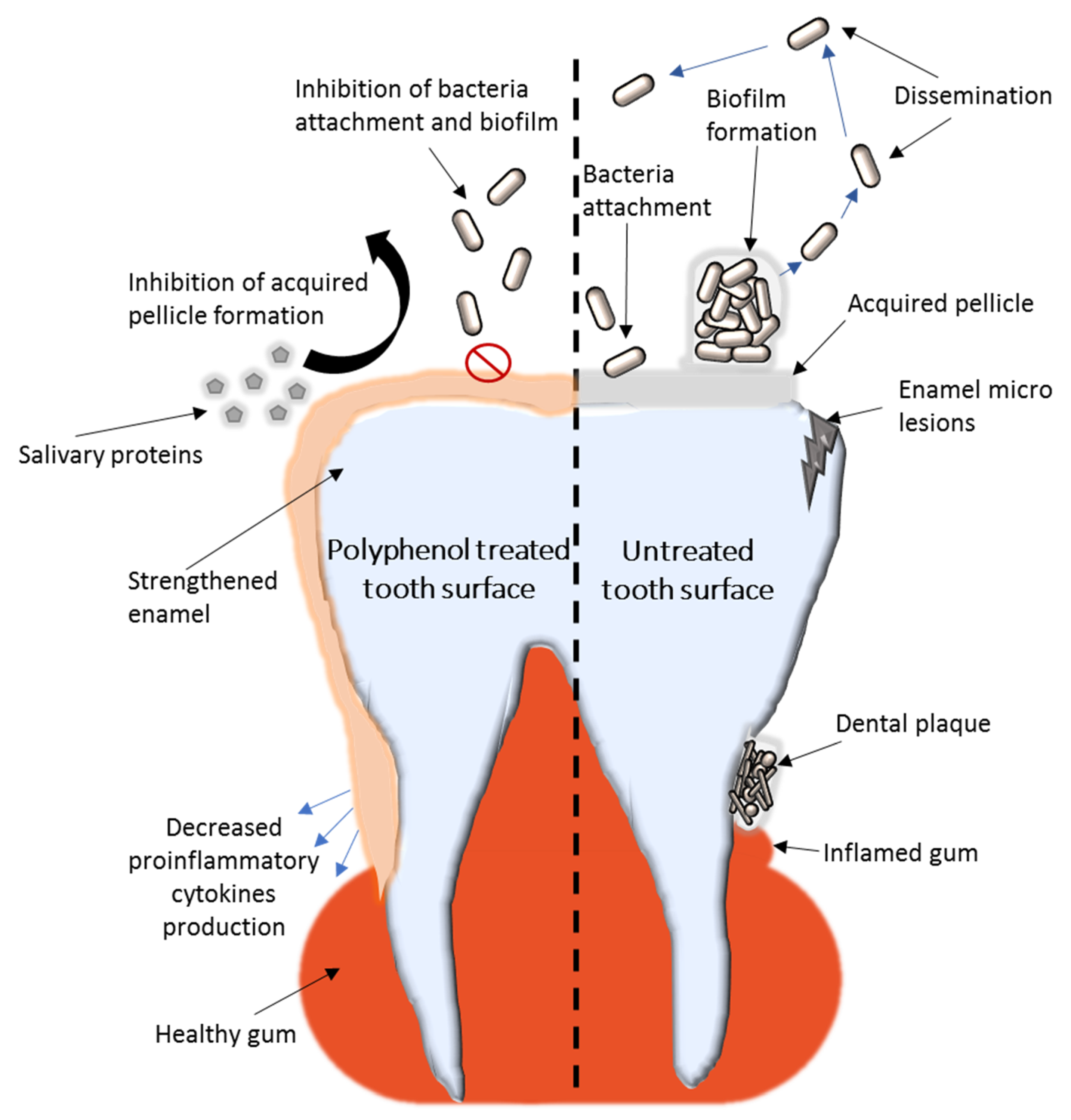
A schematic representation of the known polyphenol mechanisms to prevent dental caries and plaque formation are presented in Figure 1.
4.4. Enamel Strengthening
Despite its low pH, honey does not cause enamel erosion. Oppositely, honey and propolis have been shown to offer enamel protection by inhibiting the attachment of degradative bacteria and even strengthen the enamel surface. The main enamel protection mechanism of honeybee based products seems to be related to enamel demineralization inhibition by the biofilm [77]. Polyphenols have been investigated recently for dentin bio-modification, offering promising perspectives in dental strengthening. Hydrolysable and condensed tannins, cardol and cardanol, have been used to achieve cross-linking in the dentin matrix. After only 1 min of treatment, the best bio-modifiers were cardol and cardanol [120]. Recent studies reported that the formation of polyphenol-induced cross-links in the collagen matrix provides cohesion and makes it more resistant to degradation. Tannic acid was shown to form stable cross-links with exposed collagen fibrils, allowing them to increase the resistance against their degradation process. These compounds increase the stiffness of demineralized dentin and reduce the enzymatic degradation of collagen. The proposed mechanism is related to hydrogen bonds between the biopolymer and tannic acid [121]. It was also shown that tannic acid incorporated into polycarboxylate cement enhances the resistance of dentinal collagen to collagenase and proteolytic enzymes.
Gallic acid is able to reduce dentin fluid flow, which is the main cause of dentin hypersensitivity. The formed catechol-iron complex is deposited on the surface, creating stable cross-linked complexes in the oral aqueous environment, creating tight bridge-like connections between adjacent peritubular dentin, which resulted in less outward flow. In addition, a complex made of fluoride-tannin acid-lanthanum-apatite was also reportedly used to reduce dentinal hypersensitivity by similar mechanisms [27,122].
4.5. Oral Cancers
Honeybee-originated polyphenols have also shown enhanced anticancer properties, being efficient against initiation, proliferation, and progression of oral cancers. Their main anticancer mechanisms refer to the induction of apoptosis, cell cycle arrest, the modulation of oxidative stress, the amelioration of inflammation, the induction of mitochondrial outer membrane permeabilization, and the inhibition of angiogenesis [123].
Tualang honey showed anti-proliferative and proapoptotic effects on oral squamous cell carcinoma in vitro [124]. The antitumor activity of crude honey extracts in oral cancers seems to be related to the induction of apoptosis through caspase 3 activation [52]. Due to well-developed purification techniques, in recent years, cancer research has focused on purified polyphenols instead of crude honey. The most investigated simple polyphenols in honeybee products that have evolved as promising pharmacological agents in the treatment of cancer are caffeic acid, caffeic acid phenyl esters, chrysin, galangin, quercetin, kaempferol, acacetin, pinocembrin, pinobanksin, and apigenin [52]. Although these phenolic compounds showed significant anticancer properties in colon cancer, gastric cancer, skin cancer, fibrosarcoma, and glioma cell cancer, research regarding the mechanisms of oral cancer inhibition is widely unavailable. Due to their easy application on the oral cavity, proven anticancer impact in other cancers, and diversity of the honeybee polyphenolic compounds, these offer an impressive perspective in developing an efficient, natural, non-toxic, and personalized alternative in the therapy of oral cancers [52].
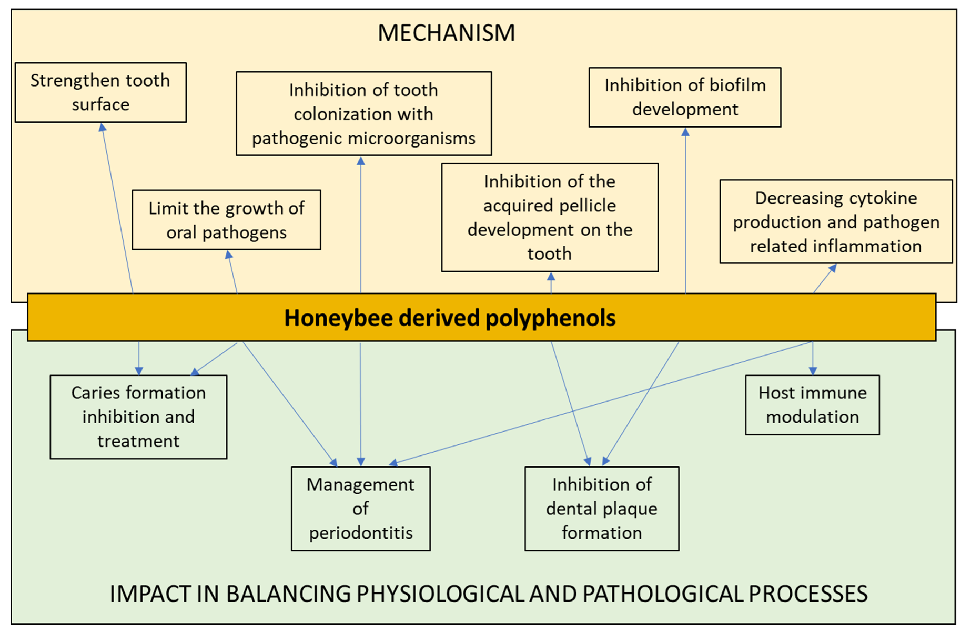
Figure 2 shows the main processes and mechanisms induced by polyphenols in balancing oral health and disease.
5. Conclusions
Natural products, such as honeybee-derived products, contain numerous bioactive compounds with impressive biomedical potential. Polyphenols are currently investigated in order to develop new alternatives in the treatment of severe health conditions, such as infections and cancer. The use of these versatile bioactive compounds in oral health is increasing. In recent years researchers have made important progress in elucidating mechanisms of polyphenols that could be exploited for dental medicine.
Their antioxidant, anti-inflammatory, and antimicrobial effects are considered the most important traits to recommend these classes of compounds in oral health. Polyphenols found in honeybee products can be easily obtained and safely utilized for daily care, as demonstrated by several in vitro, in vivo, and clinical studies. They could reduce the risk of emerging oral diseases such as dental caries and inflammatory conditions, which have a microbial component, such as periodontitis. Moreover, researchers are making constant progress in elucidating their anti-tumoral mechanisms; therefore, polyphenols offer great perspectives in treating oral cancers.
Author Contributions
C.C., L.M.D., A.M.G. and A.M.H. have participated in review writing and revision. All authors have read and agreed to the published version of the manuscript.
Funding
This study was supported by UEFISCDI, by the Grant no 271/2020 “Cold plasma for fluoride retention improvement and biofilm modulation in dental application”, project code: PN-III-P2-2.1-PED-2019-4569.
Acknowledgments
We are very grateful to our peer reviewers, which significantly helped in improving this paper.
Conflicts of Interest
The authors declare no conflict of interest.
References
- Bloom, D.E.; Cadarette, D. Infectious Disease Threats in the Twenty-First Century: Strengthening the Global Response. Front. Immunol. 2019, 10, 549. [Google Scholar] [CrossRef] [PubMed] [Green Version]
- Kenis, P.; Schol, L.G.C.; Kraaij-Dirkzwager, M.M.; Timen, A. Appropriate Governance Responses to Infectious Disease Threats: Developing Working Hypotheses. Risk Hazards Crisis Public Policy 2019, 10, 275–293. [Google Scholar] [CrossRef] [Green Version]
- Dos Santos, L.D.R.; Dos Santos, A.E.S.; Cerávolo, I.P.; Figueiredo, F.J.B.; Dias-Souza, M.V. Antibiofilm activity of black tea leaf extract, its cytotoxicity and interference on the activity of antimicrobial drugs. Biointerface Res. Appl. Chem. 2018, 8, 3565–3569. [Google Scholar]
- Genilloud, O. Natural products discovery and potential for new antibiotics. Curr. Opin. Microbiol. 2019, 51, 81–87. [Google Scholar] [CrossRef]
- Dias-Souza, M.V.; Dias, C.G.; Ferreira-Marçal, P.H. Interactions of natural products and antimicrobial drugs: Investigations of a dark matter in chemistry. Biointerface Res. Appl. Chem. 2018, 8, 3259–3264. [Google Scholar]
- Darwesh, O.M.; Barakat, K.M.; Mattar, M.Z.; Sabae, S.Z.; Hassan, S.H. Production of antimicrobial blue green pigment pyocyanin by marine Pseudomonas aeruginosa. Biointerface Res. Appl. Chem. 2019, 9, 4334–4339. [Google Scholar] [CrossRef]
- Jin, X.; Liu, M.-Y.; Zhang, D.-F.; Zhong, X.; Du, K.; Qian, P.; Gao, H.; Wei, M.-J. Natural products as a potential modulator of microglial polarization in neurodegenerative diseases. Pharmacol. Res. 2019, 145, 104253. [Google Scholar] [CrossRef] [PubMed]
- Ramsay, E.I.; Rao, S.; Madathil, L.; Hegde, S.K.; Baliga-Rao, M.P.; George, T.; Baliga, M.S. Honey in oral health and care: A mini review. J. Oral Biosci. 2019, 61, 32–36. [Google Scholar] [CrossRef] [PubMed]
- Rao, P.V.; Krishnan, K.T.; Salleh, N.; Gan, S.H. Biological and therapeutic effects of honey produced by honey bees and stingless bees: A comparative review. Rev. Bras. Farmacogn. 2016, 26, 657–664. [Google Scholar] [CrossRef] [Green Version]
- Machado De-Melo, A.A.; de Almeida-Muradian, L.B.; Sancho, M.T.; Pascual-Maté, A. Composition and properties of Apis mellifera honey: A review. J. Apic. Res. 2018, 57, 5–37. [Google Scholar] [CrossRef]
- De-Melo, A.; Almeida-Muradian, L.; Sancho, M.; Pascual Maté, A. Composition and properties of Apis mellifera honey: A review. J. Apic. Res. 2017, 1–33. [Google Scholar] [CrossRef]
- Soto-Chilaca, G.A.; Mejía-Garibay, B.; Navarro-Amador, R.; Ramírez-Corona, N.; Palou, E.; López-Malo, A. Cinnamaldehyde-loaded chitosan nanoparticles: Characterization and antimicrobial activity. Biointerface Res. Appl. Chem. 2019, 9, 4060–4065. [Google Scholar] [CrossRef]
- Adaškevičiūtė, V.; Kaškonienė, V.; Kaškonas, P.; Barčauskaitė, K.; Maruška, A. Comparison of Physicochemical Properties of Bee Pollen with Other Bee Products. Biomolecules 2019, 9, 819. [Google Scholar] [CrossRef] [Green Version]
- Tauber, J.P.; Collins, W.R.; Schwarz, R.S.; Chen, Y.; Grubbs, K.; Huang, Q.; Lopez, D.; Peterson, R.; Evans, J.D. Natural Product Medicines for Honey Bees: Perspective and Protocols. Insects 2019, 10, 356. [Google Scholar] [CrossRef] [Green Version]
- Lewkowski, O.; Mureșan, C.I.; Dobritzsch, D.; Fuszard, M.; Erler, S. The Effect of Diet on the Composition and Stability of Proteins Secreted by Honey Bees in Honey. Insects 2019, 10, 282. [Google Scholar] [CrossRef] [Green Version]
- Münstedt, K.; Männle, H. Using Bee Products for the Prevention and Treatment of Oral Mucositis Induced by Cancer Treatment. Molecules 2019, 24, 3023. [Google Scholar] [CrossRef] [PubMed] [Green Version]
- Pasupuleti, V.R.; Sammugam, L.; Ramesh, N.; Gan, S.H. Honey, Propolis, and Royal Jelly: A Comprehensive Review of Their Biological Actions and Health Benefits. Oxidative Med. Cell. Longev. 2017, 2017, 1259510. [Google Scholar] [CrossRef]
- Santos-Buelga, C.; González-Paramás, A.M. Chemical Composition of Honey. In Bee Products—Chemical and Biological Properties; Alvarez-Suarez, J.M., Ed.; Springer International Publishing: Cham, Switzerland, 2017; pp. 43–82. [Google Scholar] [CrossRef]
- Nguyen, H.T.L.; Panyoyai, N.; Kasapis, S.; Pang, E.; Mantri, N. Honey and Its Role in Relieving Multiple Facets of Atherosclerosis. Nutrients 2019, 11, 167. [Google Scholar] [CrossRef] [PubMed] [Green Version]
- Hepburn, H.R. Composition and Synthesis of Beeswax. In Honeybees and Wax: An Experimental Natural History; Hepburn, H.R., Ed.; Springer: Berlin/Heidelberg, Germany, 1986; pp. 44–56. [Google Scholar] [CrossRef]
- Kurek-Górecka, A.; Rzepecka-Stojko, A.; Górecki, M.; Stojko, J.; Sosada, M.; Świerczek-Zięba, G. Structure and Antioxidant Activity of Polyphenols Derived from Propolis. Molecules 2014, 19, 78–101. [Google Scholar] [CrossRef] [Green Version]
- Yeung, Y.T.; Argüelles, S. Chapter 4.1—Bee Products: Royal Jelly and Propolis. In Nonvitamin and Nonmineral Nutritional Supplements; Nabavi, S.M., Silva, A.S., Eds.; Academic Press: Cambridge, MA, USA, 2019; pp. 475–484. [Google Scholar] [CrossRef]
- Kunugi, H.; Mohammed Ali, A. Royal Jelly and Its Components Promote Healthy Aging and Longevity: From Animal Models to Humans. Int. J. Mol. Sci. 2019, 20, 4662. [Google Scholar] [CrossRef] [Green Version]
- Okumura, N.; Toda, T.; Ozawa, Y.; Watanabe, K.; Ikuta, T.; Tatefuji, T.; Hashimoto, K.; Shimizu, T. Royal Jelly Delays Motor Functional Impairment During Aging in Genetically Heterogeneous Male Mice. Nutrients 2018, 10, 1191. [Google Scholar] [CrossRef] [Green Version]
- Shi, Z.; Enayatullah, H.; Lv, Z.; Dai, H.; Wei, Q.; Shen, L.; Karwand, B.; Shi, F. Freeze-Dried Royal Jelly Proteins Enhanced the Testicular Development and Spermatogenesis in Pubescent Male Mice. Animals 2019, 9, 977. [Google Scholar] [CrossRef] [Green Version]
- Jibril, F.I.; Hilmi, A.B.M.; Manivannan, L. Isolation and characterization of polyphenols in natural honey for the treatment of human diseases. Bull. Natl. Res. Cent. 2019, 43, 4. [Google Scholar] [CrossRef]
- Kharouf, N.; Haikel, Y.; Ball, V. Polyphenols in Dental Applications. Bioengineering 2020, 7, 72. [Google Scholar] [CrossRef]
- Ding, Y.; Yao, H.; Yao, Y.; Fai, L.Y.; Zhang, Z. Protection of Dietary Polyphenols against Oral Cancer. Nutrients 2013, 5, 2173–2191. [Google Scholar] [CrossRef] [Green Version]
- Chatterjee, A.; Saluja, M.; Agarwal, G.; Alam, M. Green tea: A boon for periodontal and general health. J. Indian Soc. Periodontol. 2012, 16, 161–167. [Google Scholar] [CrossRef]
- Khan, H.; Sureda, A.; Belwal, T.; Çetinkaya, S.; Süntar, İ.; Tejada, S.; Devkota, H.P.; Ullah, H.; Aschner, M. Polyphenols in the treatment of autoimmune diseases. Autoimmun. Rev. 2019, 18, 647–657. [Google Scholar] [CrossRef]
- Abbasi, A.M.; Shah, M.H. Assessment of phenolic contents, essential/toxic metals and antioxidant capacity of fruits of viburnum foetens decne. Biointerface Res. Appl. Chem. 2018, 8, 3178–3186. [Google Scholar]
- Panche, A.N.; Diwan, A.D.; Chandra, S.R. Flavonoids: An overview. J. Nutr. Sci. 2016, 5, e47. [Google Scholar] [CrossRef] [Green Version]
- Kennedy, D.O. Polyphenols and the human brain: Plant “secondary metabolite” ecologic roles and endogenous signaling functions drive benefits. Adv. Nutr. 2014, 5, 515–533. [Google Scholar] [CrossRef] [Green Version]
- Durazzo, A.; Lucarini, M.; Souto, E.B.; Cicala, C.; Caiazzo, E.; Izzo, A.; Novellino, E.; Santini, A. Polyphenols: A concise overview on the chemistry, occurrence, and human health. Phytother. Res. Ptr. 2019, 33. [Google Scholar] [CrossRef] [Green Version]
- Uslu, M.E.; Mele, A.; Bayraktar, O. Evaluation of the hemostatic activity of Equisetum arvense extract: The role of varying phenolic composition and antioxidant activity due to different extraction conditions. Biointerface Res. Appl. Chem. 2019, 9, 4157–4163. [Google Scholar] [CrossRef]
- Kumar, S.; Pandey, A.K. Chemistry and Biological Activities of Flavonoids: An Overview. Sci. World J. 2013, 2013, 162750. [Google Scholar] [CrossRef] [Green Version]
- Cianciosi, D.; Forbes-Hernández, T.Y.; Afrin, S.; Gasparrini, M.; Reboredo-Rodriguez, P.; Manna, P.P.; Zhang, J.; Bravo Lamas, L.; Martínez Flórez, S.; Agudo Toyos, P.; et al. Phenolic Compounds in Honey and Their Associated Health Benefits: A Review. Molecules 2018, 23, 2322. [Google Scholar] [CrossRef] [Green Version]
- Abbas, M.; Saeed, F.; Anjum, F.M.; Afzaal, M.; Tufail, T.; Bashir, M.S.; Ishtiaq, A.; Hussain, S.; Suleria, H.A.R. Natural polyphenols: An overview. Int. J. Food Prop. 2017, 20, 1689–1699. [Google Scholar] [CrossRef] [Green Version]
- Wang, J.; Li, Q.X. Chemical composition, characterization, and differentiation of honey botanical and geographical origins. Adv. Food Nutr. Res. 2011, 62, 89–137. [Google Scholar] [CrossRef]
- Can, Z.; Yildiz, O.; Sahin, H.; Akyuz Turumtay, E.; Silici, S.; Kolayli, S. An investigation of Turkish honeys: Their physico-chemical properties, antioxidant capacities and phenolic profiles. Food Chem. 2015, 180, 133–141. [Google Scholar] [CrossRef]
- Galeotti, F.; Maccari, F.; Fachini, A.; Volpi, N. Chemical Composition and Antioxidant Activity of Propolis Prepared in Different Forms and in Different Solvents Useful for Finished Products. Foods 2018, 7, 41. [Google Scholar] [CrossRef] [Green Version]
- Duman, A.; Mogulkoc, R.; Baltaci, A.K.; Sivrikaya, A. The effect of 3′,4′-dihydroxyflavonol on plasma oxidant and antioxidant systems in testis ischemia-reperfusion injury in rats. Biointerface Res. Appl. Chem. 2018, 8, 3441–3445. [Google Scholar]
- Moniruzzaman, M.; Sulaiman, S.A.; Azlan, S.A.; Gan, S.H. Two-year variations of phenolics, flavonoids and antioxidant contents in acacia honey. Molecules 2013, 18, 14694–14710. [Google Scholar] [CrossRef] [Green Version]
- Alvarez-Suarez, J.M.; Gasparrini, M.; Forbes-Hernández, T.Y.; Mazzoni, L.; Giampieri, F. The Composition and Biological Activity of Honey: A Focus on Manuka Honey. Foods 2014, 3, 420–432. [Google Scholar] [CrossRef] [Green Version]
- Tang, G.-Y.; Zhao, C.-N.; Xu, X.-Y.; Gan, R.-Y.; Cao, S.-Y.; Liu, Q.; Shang, A.; Mao, Q.-Q.; Li, H.-B. Phytochemical Composition and Antioxidant Capacity of 30 Chinese Teas. Antioxidants 2019, 8, 180. [Google Scholar] [CrossRef] [Green Version]
- Cheung, Y.; Meenu, M.; Yu, X.; Xu, B. Phenolic acids and flavonoids profiles of commercial honey from different floral sources and geographic sources. Int. J. Food Prop. 2019, 22, 290–308. [Google Scholar] [CrossRef]
- Olas, B. Honey and Its Phenolic Compounds as an Effective Natural Medicine for Cardiovascular Diseases in Humans? Nutrients 2020, 12, 283. [Google Scholar] [CrossRef] [Green Version]
- Samarghandian, S.; Farkhondeh, T.; Samini, F. Honey and Health: A Review of Recent Clinical Research. Pharmacogn. Res. 2017, 9, 121–127. [Google Scholar] [CrossRef]
- Khalil, M.I.; Sulaiman, S.A. The potential role of honey and its polyphenols in preventing heart diseases: A review. Afr. J. Tradit. Complement. Altern. Med. 2010, 7, 315–321. [Google Scholar] [CrossRef] [Green Version]
- Uthurry, C.; Hevia, D.; Gomez-Cordoves, C. Role of honey polyphenols in health. J. Apiprod. Apimed. Sci. 2011, 3, 141–159. [Google Scholar] [CrossRef]
- Mandal, M.D.; Mandal, S. Honey: Its medicinal property and antibacterial activity. Asian Pac. J. Trop. Biomed. 2011, 1, 154–160. [Google Scholar] [CrossRef] [Green Version]
- Jaganathan, S.K.; Mandal, M. Antiproliferative Effects of Honey and of Its Polyphenols: A Review. J. Biomed. Biotechnol. 2009, 2009, 830616. [Google Scholar] [CrossRef] [Green Version]
- Briguglio, G.; Costa, C.; Pollicino, M.; Giambò, F.; Catania, S.; Fenga, C. Polyphenols in cancer prevention: New insight (review). Int. J. Funct. Nutr. 2020. [Google Scholar] [CrossRef]
- Nasir, A.; Bullo, M.M.H.; Ahmed, Z.; Imtiaz, A.; Yaqoob, E.; Jadoon, M.; Ahmed, H.; Afreen, A.; Yaqoob, S. Nutrigenomics: Epigenetics and cancer prevention: A comprehensive review. Crit. Rev. Food Sci. Nutr. 2020, 60, 1375–1387. [Google Scholar] [CrossRef]
- Zhao, Y.; Zhang, X. Interactions of tea polyphenols with intestinal microbiota and their implication for anti-obesity. J. Sci. Food Agric. 2020, 100, 897–903. [Google Scholar] [CrossRef]
- Lee, H.Y.; Chen, Y.J.; Chang, W.A.; Li, W.M.; Ke, H.L.; Wu, W.J.; Kuo, P.L. Effects of Epigallocatechin Gallate (EGCG) on Urinary Bladder Urothelial Carcinoma-Next-Generation Sequencing and Bioinformatics Approaches. Medicina 2019, 55, 768. [Google Scholar] [CrossRef] [Green Version]
- ElAttar, T.M.; Virji, A.S. Modulating effect of resveratrol and quercetin on oral cancer cell growth and proliferation. AntiCancer Drugs 1999, 10, 187–193. [Google Scholar] [CrossRef]
- Kim, J.S.; Saengsirisuwan, V.; Sloniger, J.A.; Teachey, M.K.; Henriksen, E.J. Oxidant stress and skeletal muscle glucose transport: Roles of insulin signaling and p38 MAPK. Free Radic. Biol. Med. 2006, 41, 818–824. [Google Scholar] [CrossRef]
- Rahimi, R.; Nikfar, S.; Larijani, B.; Abdollahi, M. A review on the role of antioxidants in the management of diabetes and its complications. Biomed. Pharmacother. 2005, 59, 365–373. [Google Scholar] [CrossRef]
- Vauzour, D. Dietary polyphenols as modulators of brain functions: Biological actions and molecular mechanisms underpinning their beneficial effects. Oxidative Med. Cell. Longev. 2012, 2012, 914273. [Google Scholar] [CrossRef] [PubMed] [Green Version]
- Syed-Badrul, S.-N.; Isa, N.-M.; Mohamed, S.Z.; Qodriyah, H.M.S. Natural Polyphenols in the Treatment of Alzheimer’s Disease. Curr. Drug Targets 2018, 19, 927–937. [Google Scholar] [CrossRef]
- Martinotti, S.; Ranzato, E. Propolis: A new frontier for wound healing? Burn. Trauma 2015, 3, 9. [Google Scholar] [CrossRef] [PubMed] [Green Version]
- Schramm, D.D.; Karim, M.; Schrader, H.R.; Holt, R.R.; Cardetti, M.; Keen, C.L. Honey with High Levels of Antioxidants Can Provide Protection to Healthy Human Subjects. J. Agric. Food Chem. 2003, 51, 1732–1735. [Google Scholar] [CrossRef]
- Papuc, C.; Goran, G.V.; Predescu, C.N.; Nicorescu, V.; Stefan, G. Plant Polyphenols as Antioxidant and Antibacterial Agents for Shelf-Life Extension of Meat and Meat Products: Classification, Structures, Sources, and Action Mechanisms. Compreshensive Rev. Food Sci. Food Saf. 2017, 16, 1243–1268. [Google Scholar] [CrossRef] [Green Version]
- Basu, A.; Masek, E.; Ebersole, J.L. Dietary Polyphenols and Periodontitis-A Mini-Review of Literature. Molecules 2018, 23, 1786. [Google Scholar] [CrossRef] [Green Version]
- Zhang, H.; Tsao, R. Dietary polyphenols, oxidative stress and antioxidant and anti-inflammatory effects. Curr. Opin. Food Sci. 2016, 8, 33–42. [Google Scholar] [CrossRef]
- Petti, S.; Scully, C. Polyphenols, oral health and disease: A review. J. Dent. 2009, 37, 413–423. [Google Scholar] [CrossRef]
- Toczewska, J.; Maciejczyk, M.; Konopka, T.; Zalewska, A. Total Oxidant and Antioxidant Capacity of Gingival Crevicular Fluid and Saliva in Patients with Periodontitis: Review and Clinical Study. Antioxidants 2020, 9, 450. [Google Scholar] [CrossRef]
- Özkök, D.; Silici, S. Antioxidant activities of honeybee products and their mixtures. Food Sci. Biotechnol. 2017, 26, 201–206. [Google Scholar] [CrossRef]
- Kocot, J.; Kiełczykowska, M.; Luchowska-Kocot, D.; Kurzepa, J.; Musik, I. Antioxidant Potential of Propolis, Bee Pollen, and Royal Jelly: Possible Medical Application. Oxidative Med. Cell. Longev. 2018, 2018, 7074209. [Google Scholar] [CrossRef]
- Borges, A.; Ferreira, C.; Saavedra, M.J.; Simões, M. Antibacterial activity and mode of action of ferulic and gallic acids against pathogenic bacteria. Microb. Drug Resist. 2013, 19, 256–265. [Google Scholar] [CrossRef] [PubMed]
- Bouarab-Chibane, L.; Forquet, V.; Lantéri, P.; Clément, Y.; Léonard-Akkari, L.; Oulahal, N.; Degraeve, P.; Bordes, C. Antibacterial Properties of Polyphenols: Characterization and QSAR (Quantitative Structure-Activity Relationship) Models. Front. Microbiol. 2019, 10, 829. [Google Scholar] [CrossRef]
- Taguri, T.; Tanaka, T.; Kouno, I. Antimicrobial activity of 10 different plant polyphenols against bacteria causing food-borne disease. Biol. Pharm. Bull. 2004, 27, 1965–1969. [Google Scholar] [CrossRef] [Green Version]
- Kahkeshani, N.; Farzaei, F.; Fotouhi, M.; Alavi, S.S.; Bahramsoltani, R.; Naseri, R.; Momtaz, S.; Abbasabadi, Z.; Rahimi, R.; Farzaei, M.H.; et al. Pharmacological effects of gallic acid in health and diseases: A mechanistic review. Iran. J. Basic Med. Sci. 2019, 22, 225–237. [Google Scholar] [CrossRef]
- Shao, D.; Li, J.; Li, J.; Tang, R.; Liu, L.; Shi, J.; Huang, Q.; Yang, H. Inhibition of Gallic Acid on the Growth and Biofilm Formation of Escherichia coli and Streptococcus mutans. J. Food Sci. 2015, 80, M1299–M1305. [Google Scholar] [CrossRef]
- Borges, A.; Saavedra, M.J.; Simões, M. The activity of ferulic and gallic acids in biofilm prevention and control of pathogenic bacteria. Biofouling 2012, 28, 755–767. [Google Scholar] [CrossRef] [PubMed]
- Kang, M.S.; Oh, J.S.; Kang, I.C.; Hong, S.J.; Choi, C.H. Inhibitory effect of methyl gallate and gallic acid on oral bacteria. J. Microbiol. 2008, 46, 744–750. [Google Scholar] [CrossRef] [PubMed]
- Ibitoye, O.B.; Ajiboye, T.O. Ferulic acid potentiates the antibacterial activity of quinolone-based antibiotics against Acinetobacter baumannii. Microb. Pathog. 2019, 126, 393–398. [Google Scholar] [CrossRef]
- Dias da Costa Júnior, S.; de Oliveira Santos, J.V.; de Almeida Campos, L.A.; Araújo Pereira, M.; Santos Magalhães, N.S.; Macário Ferro Cavalcanti, I. Antibacterial and antibiofilm activities of quercetin against clinical isolates of Staphyloccocus aureus and Staphylococcus saprophyticus with resistance profile. Int. J. Environ. Agric. Biotechnol. 2018, 3, 1948–1958. [Google Scholar] [CrossRef] [Green Version]
- Hellewell, L.; Bhakta, S. Chalcones, stilbenes and ketones have anti-infective properties via inhibition of bacterial drug-efflux and consequential synergism with antimicrobial agents. Access Microbiol. 2020, 2, acmi000105. [Google Scholar] [CrossRef]
- Khan, M.S.; Agrawal, R.; Ubaidullah, M.; Hassan, M.I.; Tarannum, N. Design, synthesis and validation of antimicrobial coumarin derivatives: An efficient green approach. Heliyon 2019, 5, e02615. [Google Scholar] [CrossRef] [Green Version]
- Kayser, O.; Kolodziej, H. Antibacterial activity of simple coumarins: Structural requirements for biological activity. Z. Nat. CJ. Biosci. 1999, 54, 169–174. [Google Scholar] [CrossRef]
- Reen, F.J.; Gutiérrez-Barranquero, J.A.; Parages, M.L.; O’Gara, F. Coumarin: A novel player in microbial quorum sensing and biofilm formation inhibition. Appl. Microbiol. Biotechnol. 2018, 102, 2063–2073. [Google Scholar] [CrossRef] [Green Version]
- Zhou, Y.; Zheng, J.; Li, Y.; Xu, D.-P.; Li, S.; Chen, Y.-M.; Li, H.-B. Natural Polyphenols for Prevention and Treatment of Cancer. Nutrients 2016, 8, 515. [Google Scholar] [CrossRef]
- Kuang, X.; Chen, V.; Xu, X. Novel Approaches to the Control of Oral Microbial Biofilms. Biomed. Res. Int. 2018, 2018, 6498932. [Google Scholar] [CrossRef] [Green Version]
- Sankari, S.L.; Babu, N.A.; Rani, V.; Priyadharsini, C.; Masthan, K.M.K. Flavonoids—Clinical effects and applications in dentistry: A review. J. Pharm. Bioallied Sci. 2014, 6, S26–S29. [Google Scholar] [CrossRef] [PubMed]
- Sharifi, S.; Mokhtarpour, M.; Jahangiri, A.; Dehghanzadeh, S.; Maleki-Dizaj, S.; Shahi, S. Hydroxyapatite nanofibers as beneficial nanomaterial in dental sciences. Biointerface Res. Appl. Chem. 2018, 8, 3695–3699. [Google Scholar]
- Ferrazzano, G.F.; Roberto, L.; Amato, I.; Cantile, T.; Sangianantoni, G.; Ingenito, A. Antimicrobial properties of green tea extract against cariogenic microflora: An in vivo study. J. Med. Food 2011, 14, 907–911. [Google Scholar] [CrossRef] [PubMed]
- Hirasawa, M.; Takada, K.; Otake, S. Inhibition of acid production in dental plaque bacteria by green tea catechins. Caries Res. 2006, 40, 265–270. [Google Scholar] [CrossRef] [PubMed]
- Sateriale, D.; Facchiano, S.; Colicchio, R.; Pagliuca, C.; Varricchio, E.; Paolucci, M.; Volpe, M.G.; Salvatore, P.; Pagliarulo, C. In vitro Synergy of Polyphenolic Extracts From Honey, Myrtle and Pomegranate Against Oral Pathogens, S. mutans and R. dentocariosa. Front. Microbiol. 2020, 11, 1465. [Google Scholar] [CrossRef] [PubMed]
- Xu, X.; Zhou, X.D.; Wu, C.D. Tea catechin epigallocatechin gallate inhibits Streptococcus mutans biofilm formation by suppressing gtf genes. Arch. Oral Biol. 2012, 57, 678–683. [Google Scholar] [CrossRef] [PubMed]
- Hara, K.; Ohara, M.; Hayashi, I.; Hino, T.; Nishimura, R.; Iwasaki, Y.; Ogawa, T.; Ohyama, Y.; Sugiyama, M.; Amano, H. The green tea polyphenol (-)-epigallocatechin gallate precipitates salivary proteins including alpha-amylase: Biochemical implications for oral health. Eur. J. Oral Sci. 2012, 120, 132–139. [Google Scholar] [CrossRef]
- Habluetzel, A.; Schmid, C.; Carvalho, T.S.; Lussi, A.; Eick, S. Impact of honey on dental erosion and adhesion of early bacterial colonizers. Sci. Rep. 2018, 8, 10936. [Google Scholar] [CrossRef] [PubMed]
- Akca, A.E.; Akca, G.; Topçu, F.T.; Macit, E.; Pikdöken, L.; Özgen, I. The Comparative Evaluation of the Antimicrobial Effect of Propolis with Chlorhexidine against Oral Pathogens: An In Vitro Study. Biomed. Res. Int. 2016, 2016, 3627463. [Google Scholar] [CrossRef] [Green Version]
- Jeon, J.G.; Pandit, S.; Xiao, J.; Gregoire, S.; Falsetta, M.L.; Klein, M.I.; Koo, H. Influences of trans-trans farnesol, a membrane-targeting sesquiterpenoid, on Streptococcus mutans physiology and survival within mixed-species oral biofilms. Int. J. Oral Sci. 2011, 3, 98–106. [Google Scholar] [CrossRef]
- Koo, H.; Jeon, J.G. Naturally occurring molecules as alternative therapeutic agents against cariogenic biofilms. Adv. Dent. Res. 2009, 21, 63–68. [Google Scholar] [CrossRef]
- Franca, J.R.; De Luca, M.P.; Ribeiro, T.G.; Castilho, R.O.; Moreira, A.N.; Santos, V.R.; Faraco, A.A. Propolis--based chitosan varnish: Drug delivery, controlled release and antimicrobial activity against oral pathogen bacteria. BMC Complementary Altern. Med. 2014, 14, 478. [Google Scholar] [CrossRef] [Green Version]
- Koo, H.; Duarte, S.; Murata, R.M.; Scott-Anne, K.; Gregoire, S.; Watson, G.E.; Singh, A.P.; Vorsa, N. Influence of cranberry proanthocyanidins on formation of biofilms by Streptococcus mutans on saliva-coated apatitic surface and on dental caries development in vivo. Caries Res. 2010, 44, 116–126. [Google Scholar] [CrossRef] [Green Version]
- Ben Lagha, A.; Dudonné, S.; Desjardins, Y.; Grenier, D. Wild Blueberry (Vaccinium angustifolium Ait.) Polyphenols Target Fusobacterium nucleatum and the Host Inflammatory Response: Potential Innovative Molecules for Treating Periodontal Diseases. J. Agric. Food Chem. 2015, 63, 6999–7008. [Google Scholar] [CrossRef]
- De Assis, J.S.; Lima, R.A.; Marques Lima, J.P.; Azevedo Rodrigues, L.K.; Santiago, S.L. Effect of epigallocatechin-3-gallate application for remaining carious dentin disinfection. J. Conserv. Dent. 2015, 18, 51–55. [Google Scholar] [CrossRef] [Green Version]
- De Oliveira Caleare, A.; Hensel, A.; Mello, J.C.P.; Pinha, A.B.; Panizzon, G.P.; Lechtenberg, M.; Petereit, F.; Nakamura, C.V. Flavan-3-ols and proanthocyanidins from Limonium brasiliense inhibit the adhesion of Porphyromonas gingivalis to epithelial host cells by interaction with gingipains. Fitoterapia 2017, 118, 87–93. [Google Scholar] [CrossRef]
- Available online: https://www.who.int/oral_health/disease_burden/global/en/ (accessed on 30 September 2020).
- Koo, H.; Rosalen, P.L.; Cury, J.A.; Park, Y.K.; Ikegaki, M.; Sattler, A. Effect of Apis mellifera propolis from two Brazilian regions on caries development in desalivated rats. Caries Res. 1999, 33, 393–400. [Google Scholar] [CrossRef]
- Burdock, G.A. Review of the biological properties and toxicity of bee propolis (propolis). Food Chem. Toxicol. Int. J. Publ. Br. Ind. Biol. Res. Assoc. 1998, 36, 347–363. [Google Scholar] [CrossRef]
- Koo, H.; Pearson, S.K.; Scott-Anne, K.; Abranches, J.; Cury, J.A.; Rosalen, P.L.; Park, Y.K.; Marquis, R.E.; Bowen, W.H. Effects of apigenin and tt-farnesol on glucosyltransferase activity, biofilm viability and caries development in rats. Oral Microbiol. Immunol. 2002, 17, 337–343. [Google Scholar] [CrossRef] [PubMed]
- Yamashita, Y.; Bowen, W.H.; Burne, R.A.; Kuramitsu, H.K. Role of the Streptococcus mutans gtf genes in caries induction in the specific-pathogen-free rat model. Infect. Immun. 1993, 61, 3811–3817. [Google Scholar] [CrossRef] [PubMed] [Green Version]
- Koo, H.; Schobel, B.; Scott-Anne, K.; Watson, G.; Bowen, W.H.; Cury, J.A.; Rosalen, P.L.; Park, Y.K. Apigenin and tt-farnesol with fluoride effects on S. mutans biofilms and dental caries. J. Dent. Res. 2005, 84, 1016–1020. [Google Scholar] [CrossRef] [PubMed]
- Periodontal Disease. Available online: https://www.cdc.gov/oralhealth/conditions/periodontal-disease.html (accessed on 30 September 2020).
- Bodet, C.; Piché, M.; Chandad, F.; Grenier, D. Inhibition of periodontopathogen-derived proteolytic enzymes by a high-molecular-weight fraction isolated from cranberry. J. Antimicrob. Chemother. 2006, 57, 685–690. [Google Scholar] [CrossRef]
- Corrêa, M.G.; Pires, P.R.; Ribeiro, F.V.; Pimentel, S.Z.; Casarin, R.C.; Cirano, F.R.; Tenenbaum, H.T.; Casati, M.Z. Systemic treatment with resveratrol and/or curcumin reduces the progression of experimental periodontitis in rats. J. Periodontal Res. 2017, 52, 201–209. [Google Scholar] [CrossRef]
- Yoshinaga, Y.; Ukai, T.; Nakatsu, S.; Kuramoto, A.; Nagano, F.; Yoshinaga, M.; Montenegro, J.L.; Shiraishi, C.; Hara, Y. Green tea extract inhibits the onset of periodontal destruction in rat experimental periodontitis. J. Periodontal Res. 2014, 49, 652–659. [Google Scholar] [CrossRef]
- Li, H.; Wang, Q.; Ding, Y.; Bao, C.; Li, W. Mangiferin ameliorates Porphyromonas gingivalis-induced experimental periodontitis by inhibiting phosphorylation of nuclear factor-κB and Janus kinase 1-signal transducer and activator of transcription signaling pathways. J. Periodontal Res. 2017, 52, 1–7. [Google Scholar] [CrossRef]
- Hamzah, N.; Aziz, S.; Fauzi, A.-R.; Mohd Yusof, Y.; Razali, M.; Ibrahim, N.; Baharin, B. Effects of Gelam Honey (Melaleuca cajuputi) on Alveolar Bone Loss in Experimental Periodontitis. J. Dent. Surg. 2014, 2014. [Google Scholar] [CrossRef]
- Coutinho, A. Honeybee propolis extract in periodontal treatment: A clinical and microbiological study of propolis in periodontal treatment. Indian J. Dent. Res. Off. Publ. Indian Soc. Dent. Res. 2012, 23, 294. [Google Scholar] [CrossRef]
- Halboub, E.; Al-Maweri, S.A.; Al-Wesabi, M.; Al-Kamel, A.; Shamala, A.; Al-Sharani, A.; Koppolu, P. Efficacy of propolis-based mouthwashes on dental plaque and gingival inflammation: A systematic review. BMC Oral Health 2020, 20, 198. [Google Scholar] [CrossRef]
- Yu, O.Y.; Zhao, I.S.; Mei, M.L.; Lo, E.C.-M.; Chu, C.-H. Dental Biofilm and Laboratory Microbial Culture Models for Cariology Research. Dent. J. 2017, 5, 21. [Google Scholar] [CrossRef] [Green Version]
- Dental Plaque and Plaque as Biofilm. Available online: https://periobasics.com/dental-plaque/ (accessed on 30 September 2020).
- Farkash, Y.; Feldman, M.; Ginsburg, I.; Steinberg, D.; Shalish, M. Polyphenols Inhibit Candida albicans and Streptococcus mutans Biofilm Formation. Dent. J. 2019, 7, 42. [Google Scholar] [CrossRef] [PubMed] [Green Version]
- Furiga, A.; Roques, C.; Badet, C. Preventive effects of an original combination of grape seed polyphenols with amine fluoride on dental biofilm formation and oxidative damage by oral bacteria. J. Appl. Microbiol. 2014, 116, 761–771. [Google Scholar] [CrossRef] [PubMed] [Green Version]
- Moreira, M.A.; Souza, N.O.; Sousa, R.S.; Freitas, D.Q.; Lemos, M.V.; De Paula, D.M.; Maia, F.J.N.; Lomonaco, D.; Mazzetto, S.E.; Feitosa, V.P. Efficacy of new natural biomodification agents from Anacardiaceae extracts on dentin collagen cross-linking. Dent. Mater. Off. Publ. Acad. Dent. Mater. 2017, 33, 1103–1109. [Google Scholar] [CrossRef]
- Bedran-Russo, A.K.B.; Yoo, K.J.; Ema, K.C.; Pashley, D.H. Mechanical properties of tannic-acid-treated dentin matrix. J. Dent. Res. 2009, 88, 807–811. [Google Scholar] [CrossRef] [Green Version]
- Oh, S.; Gu, Y.; Perinpanayagam, H.; Yoo, Y.-J.; Lee, Y.; Kim, R.K.; Chang, S.W.; Lee, J.; Zhu, Q.; Kum, K.Y. Dentinal tubule sealing effects of 532-nm diode-pumped solid-state laser, gallic acid/Fe3+ complex, and three commercial dentin desensitizers. Lasers Med. Sci. 2018, 33, 1237–1244. [Google Scholar] [CrossRef]
- Orsolić, N.; Knezević, A.; Sver, L.; Terzić, S.; Hackenberger, B.K.; Basić, I. Influence of honey bee products on transplantable murine tumours. Vet. Comp. Oncol. 2003, 1, 216–226. [Google Scholar] [CrossRef]
- Ghashm, A.A.; Othman, N.H.; Khattak, M.N.; Ismail, N.M.; Saini, R. Antiproliferative effect of Tualang honey on oral squamous cell carcinoma and osteosarcoma cell lines. BMC Complementary Altern. Med. 2010, 10, 49. [Google Scholar] [CrossRef] [Green Version]
Figure 1.
Representation of the known mechanisms of honeybee-derived polyphenols to inhibit dental plaque biofilm formation and dental caries.
Figure 1.
Representation of the known mechanisms of honeybee-derived polyphenols to inhibit dental plaque biofilm formation and dental caries.

Figure 2.
The main mechanisms of honeybee polyphenols useful in oral medicine and their consequence in balancing oral health and microbial-related disease. The upper part of the scheme represents the known mechanisms of polyphenols, which can be exploited in balancing oral health and disease (possible consequences are represented in the lower part of the scheme). Widespread diseases, such as dental caries could be prevented or treated by the application of polyphenol-based products, which strengthen the tooth surface and limit the growth of oral pathogens. These compounds may have an impact on the management of oral multifactorial conditions such as periodontitis by exploiting their ability to inhibit the attachment, growth, and biofilm formation of pathogenic bacteria and also by modulating inflammation at the affected site.


Table 1.
List of the main polyphenols investigated for their impact in the prevention and therapy of oral dental pathologies containing a microbial component.
Table 1.
List of the main polyphenols investigated for their impact in the prevention and therapy of oral dental pathologies containing a microbial component.
| Oral Pathology | Type of Polyphenols | Antimicrobial Mechanism | Microbial Pathogen | Reference |
|---|---|---|---|---|
| Dental caries | mixture of polyphenols | inhibition of bacterial growth | Streptococcus mutans and Lactobacilli species | [88] |
| inhibition of bacterial growth, adherence, and acid production | acidogenic oral streptococci | [89] | ||
| polyphenolic acid extracts form honey | growth inhibition | Streptococcus mutans and Rothia dentocariosa | [90] | |
| catechins | suppression of GtfB/C/D genes (responsible for biofilm formation and production of soluble virulence factors) | S. mutans and Enterococcus faecalis | [91] | |
| catechins | inhibit the activity of salivary amylase and bacterial attachment | inhibits adherence of dental colonizers | [92] | |
| Mixture of polyphenols from propolis | inhibition of bacterial growth and adherence | S. mutans and S. gordonii | [93] | |
| Lactobacilli, Prevotella intermedia, Porphyromonas gingivalis, Actinomyces israelii, and Candida albicans | [94] | |||
| apigenin | inhibit some virulence-related genes, such as GtfB and C | S. mutans | [85,95] | |
| trans-trans farnesol | reduce cell viability | |||
| trans-trans farnesol | inhibition of biofilms | S. mutans | [96] | |
| apigenin and tt-farnesol from propolis | inhibit bacterial growth and biofilm | Biofilm oral pathogens (bulk) | [97] | |
| proanthocyanins, flavonols, and myricetin | disrupt biofilm formation, inhibit attachment, and diminishes the acidogenicity | S. mutans | [98] | |
| Periodontitis | curcumin (followed by pyrogallol, pyrocatechol, and quercetin) | inhibition of growth | P. gingivalis | [99] |
| epigallocatechin-3-gallate | growth inhibition Inhibition of cytokine production in a host | S. mutans | [100,101] |
Publisher’s Note: MDPI stays neutral with regard to jurisdictional claims in published maps and institutional affiliations. |
© 2020 by the authors. Licensee MDPI, Basel, Switzerland. This article is an open access article distributed under the terms and conditions of the Creative Commons Attribution (CC BY) license (http://creativecommons.org/licenses/by/4.0/).
Share and Cite
MDPI and ACS Style
Curuțiu, C.; Dițu, L.M.; Grumezescu, A.M.; Holban, A.M. Polyphenols of Honeybee Origin with Applications in Dental Medicine. Antibiotics 2020, 9, 856. https://0-doi-org.brum.beds.ac.uk/10.3390/antibiotics9120856
AMA Style
Curuțiu C, Dițu LM, Grumezescu AM, Holban AM. Polyphenols of Honeybee Origin with Applications in Dental Medicine. Antibiotics. 2020; 9(12):856. https://0-doi-org.brum.beds.ac.uk/10.3390/antibiotics9120856
Chicago/Turabian StyleCuruțiu, Carmen, Lia Mara Dițu, Alexandru Mihai Grumezescu, and Alina Maria Holban. 2020. "Polyphenols of Honeybee Origin with Applications in Dental Medicine" Antibiotics 9, no. 12: 856. https://0-doi-org.brum.beds.ac.uk/10.3390/antibiotics9120856
Note that from the first issue of 2016, this journal uses article numbers instead of page numbers. See further details here.