Article

Evaluation of the Antifungal Activity of Bacillus amyloliquefaciens and B. velezensis and Characterization of the Bioactive Secondary Metabolites Produced against Plant Pathogenic Fungi

by
Shereen A. Soliman
1,
Mona M. Khaleil
1,2 and
Rabab A. Metwally
1,*
1
Botany and Microbiology Department, Faculty of Science, Zagazig University, Zagazig 44519, Egypt
2
Biology Department, Faculty of Science, Taibah University, Yanbu El-Bahr 46429, Saudi Arabia
*
Author to whom correspondence should be addressed.
Submission received: 5 September 2022 / Revised: 17 September 2022 / Accepted: 20 September 2022 / Published: 23 September 2022
(This article belongs to the Collection Plant Growth-Promoting Bacteria: Mechanisms and Applications)

Abstract

:

Simple Summary

Plant pathogenic fungi cause serious damage in agriculture, resulting in major losses in the yield and the quality of different economic crops. Chemical fungicides are dangerous to human health and the environment. They have many harmful side effects on non-target organisms. Moreover, their residues have been found in human food. Endophytic bacteria could be a valuable and safe alternative method for the biological control of phytopathogenic fungus. The endophyte Bacillus amyloliquefaciens provides a strong prospect as a biocontrol agent against Alternaria sp. on pepper plants.

Abstract

Endophytic bacteria are plant-beneficial bacteria with a broad host range. They provide numerous benefits to their hosts, helping them tolerate several biotic and abiotic stresses. An interest has recently been developed in endophytic bacteria which are producing bioactive compounds that contribute to the biological control of various phytopathogens. This research paper aimed to investigate the potentiality of new local strains of endophytic bacteria such as Bacillus amyloliquefaciens and B. velezensis and the production of several antimicrobial metabolites associated with the biocontrol of Alternaria sp., which cause serious diseases and affect important vegetable crops in Egypt. Twenty-five endophytic bacteria isolates were obtained from different plants cultivated in El-Sharkia Governorate, Egypt. Dual culture technique was used to evaluate the bacterial isolates’ antagonistic potentiality against Alternaria sp. and Helminthosporium sp. The most active bacterial isolates obtained were selected for further screening. The antifungal activity of the most active endophytic bacterial isolate was assessed in vivo on pepper seedlings as a biocontrol agent against Alternaria sp. A significant antifungal activity was recorded with isolates C1 and T5 against Alternaria sp. and Helminthosporium sp. The bacterial endophyte discs of C1 and T5 showed the highest inhibitory effect against Alternaria sp. at 4.7 and 3.1 cm, respectively, and Helminthosporium sp. at 3.9 and 4.0 cm, respectively. The most active endophytic isolates C1 and T5 were identified and the 16S rRNA sequence was submitted to the NCBI GenBank database with accession numbers: MZ945930 and MZ945929 for Bacillus amyloliquefaciens and Bacillus velezensis, respectively. The deformity of pathogenic fungal mycelia of Alternaria sp. and Helminthosporium sp. was studied under the biotic stress of bacteria. The culture filtrates of B. amyloliquefaciens and B. velezensis were extracted with different solvents, and the results indicated that hexane was the most efficient. Gas Chromatography-Mass Spectrometry revealed that Bis (2-ethylhexyl) phthalate, Bis (2-ethylhexyl) ester, and N,N-Dimethyldodecylamine were major constituents of the endophytic crude extracts obtained from B. amyloliquefaciens and B. velezensis. The in vivo results showed that Alternaria sp. infection caused the highest disease incidence, leading to a high reduction in plant height and in the fresh and dry weights of pepper plants. With B. amyloliquefaciens application, DI significantly diminished compared to Alternaria sp. infected pepper plants, resulting in an increase in their morphological parameters. Our findings allow for a reduction of chemical pesticide use and the control of some important plant diseases.

1. Introduction

Fungal diseases are a massive threat to crop yields and global food security. They destroy one-third of all food crops each year, causing economic losses and affecting global poverty levels [1,2]. Many of these fungi survive in the soil for extended periods as resting structures. Chemical fungicides have been used to control and avoid pathogenic fungi. However, in addition to the high cost, using these chemical compounds has limited efficacy and a considerable negative influence on non-target species and human health in the environment [3]. The development of safe alternatives to traditional fungicides has been prompted by researchers who are concerned about their effects on the environment and humans.
The biological methods of controlling diseases that affect plants is an eco-friendly alternative, especially applicable when there is pesticide resistance and the need for environmental protection toward sustainable agricultural methods [4,5]. Microbial antagonists have unique properties that inhibit fungal infection growth through direct and indirect processes [6,7]. The direct impact is mainly owing to the biocontrol agent’s antagonistic behavior against the pathogen due to competition, parasitism, antibiosis, and the production of extracellular digestive enzymes. In contrast, plant defense mechanisms are triggered in response to various pests and diseases, which has an indirect effect [7,8,9].
In pathological research, an endophytic bacterium suppressing plant diseases has received much attention [10]. Endophytes spend at least a part of their life cycle within the host plant and form a symbiotic relationship with it, making them highly effective biocontrol agents [11]. Endophytes live and survive within the stems, roots, and leaves of plants without causing disease symptoms [12,13]. Endophytic bacteria and their associations with their hosts have been studied to determine their ecological functions and assess their biotechnology potential [12]. Endophytes have been found to be an essential tool for improving crop performance compared to other biological agents since they colonize host tissue [14]. They can compete for nutrients as a colonizer of the roots and compete for space for their proliferation, resulting in the inhibition of pathogens. Furthermore, they do not pollute the environment [15].
Endophytic bacteria can be isolated from almost all plant species [12,16]. Plants’ pathogenic fungi, such as Fusarium oxysporum, Sclerotium rolfsii and Rhizoctonia solani, are inhibited by endophytic bacteria [17]. Amaresan et al. [16] found that endophytic bacteria that were isolated from Capsicum annuum have an antagonistic effect against several phytopathogens. Also, infection by fungal pathogens results in stronger reductions in plant biomass and survival compared to uninfected plants [12,18,19]. Bacillus was mentioned in numerous research papers due to its prevalence in many plants, its antibacterial properties [20,21,22], and its ability to produce endospores that are UV, pH, temperature, and salinity resistant [9]. This genus has become an attractive agent for commercial use in modern farming systems [13,23,24,25]. As a result, the current study aimed to screen and analyze new local strains of endophytic bacteria isolates for their ability to inhibit various phytopathogenic fungi, which cause serious diseases affecting important vegetable crops in Egypt, and this supports a reduction of chemical pesticides’ use. Furthermore, the antifungal activity of the most active endophytic bacterial isolate was assessed in vivo on pepper (Capsicum annuum) as a biocontrol agent against Alternaria sp.

2. Materials and Methods

2.1. Plant Materials Collection

Several crop plants were randomly collected from various locations in Minia Al-Qamh soils, El-Sharkia Governorate, Egypt, including leaves, roots, and stems from Solanum melongena, S. lycopersicum, Allium cepa, Coriandrum sativum, Pisum sativum, Portulaca oleracea, and Brassica oleracea (Table 1), and placed in polypropylene bags for transport to the laboratory.

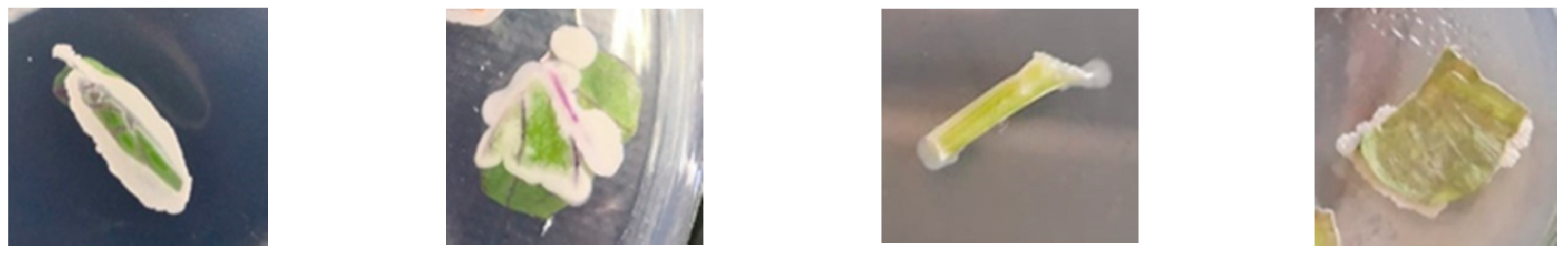
2.2. Plant Segments Sterilization and Endophytes Isolation

Endophytes were isolated from different plant parts using the [26] method. Healthy samples of different plants were used. Segments were cut from the stems (1 cm), leaves (1.5 × 1 cm), and roots (1 cm) of each plant. They were washed in running water, sterilized for 10 min with sodium hypochlorite (NaOCl, 5%), rinsed three times with sterile distilled water, and dried on a sterilized filter paper. Sterilized segments were put into a nine cm Petri plate containing potato dextrose agar (PDA) medium and incubated at 30 °C. After two days, bacterial colonies were picked out [27] and were checked by successive subcultures on the agar medium. The purified bacteria were then stored at −20 °C in a nutrient broth of 20% glycerol.

2.3. Isolation of Pathogenic Fungi

Pathogenic fungi were isolated and randomly selected from symptomatic leaves collected from El-Sharkia Governorate, Egypt. The leaves were rinsed with tap water before being soaked in 5% sodium hypochlorite (NaOCl) for 10 min, rinsed three times in sterile distilled water, and dried on sterile filter paper. Sterilized segments were placed on a 9 cm Petri dish containing potato dextrose (PDA) medium with rose bengal 30 mg/L and 250 mg/L streptomycin. Plates were incubated for 10 days at 30 °C. Fungal colonies were purified [28] and identified according to culture and microscopic characteristics [29].

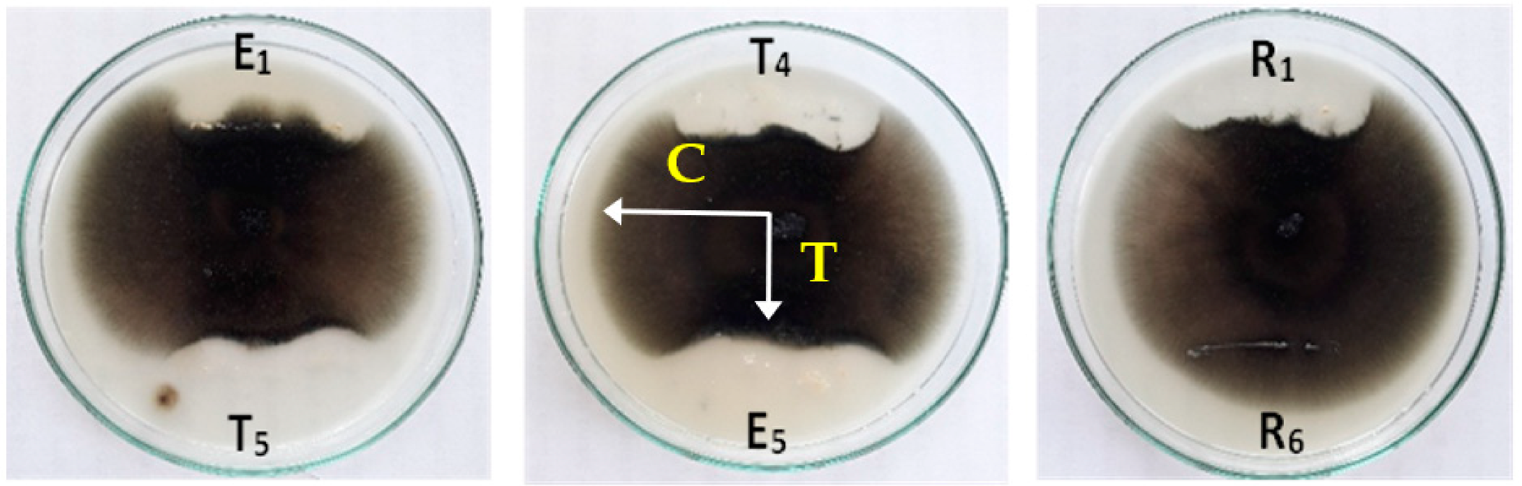
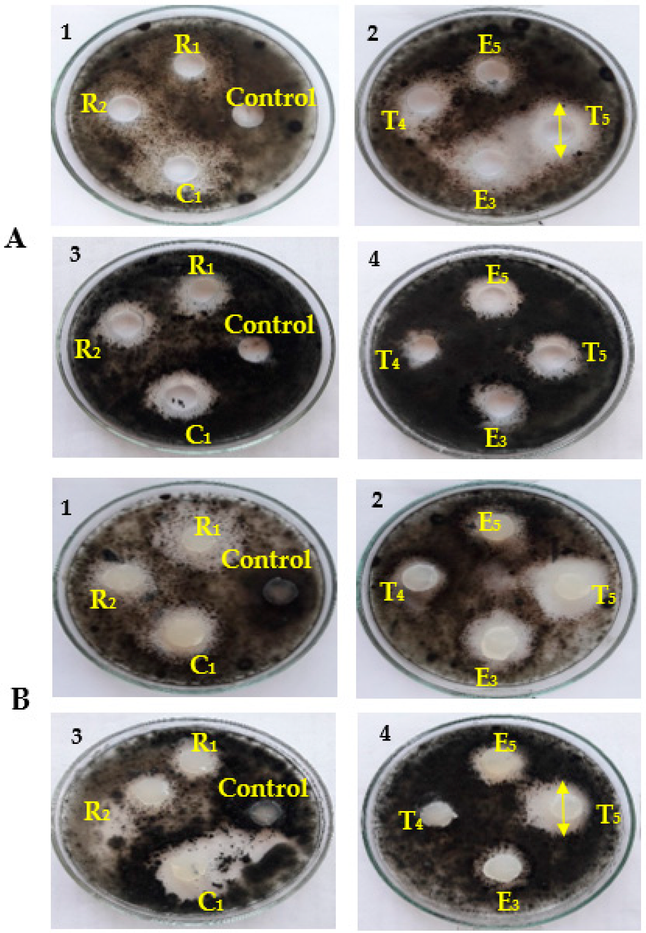
2.4. Primary Screening of the Antagonist Activity of Bacterial Isolates in Vitro

The endophytic bacterial isolates were tested for antagonistic activity against Alternaria sp. and Helminthosporium sp. using a dual culture technique. A ten-millimeter disc of a seven-day fungal pathogen culture was inoculated in the middle of Petri plates. The bacterial endophyte was streaked on the opposite side of the agar (PDA) plates. The plates inoculated with the pathogenic fungal disc were considered as the controls [30], and the plates were incubated at 28 ± 2 °C. The inhibition zone indicated the antagonistic properties of endophytic bacteria after seven days. Four replicates were measured for each isolate, and the experiment was performed twice to ensure accuracy. The following formula was used to calculate the percentage of radial growth inhibition relative to the control [31].
Percent of Inhibition % (I) = C − T/C × 100
where C-radial growth is the control and T-radial growth is the treatment.
The width of the inhibition zone was evaluated as + for 2–5 mm; ++ for 5–10 mm; and +++ for > 10 mm [32].

2.5. Evaluation of the Antifungal Activity of Endophytic Bacteria

PDA (20 mL) was inoculated with one mL of fungal spore suspension of Alternaria sp. and Helminthosporium sp. separately poured into a Petri dish of 90 mm in diameter. The plates were allowed to solidify and were then seeded with: (i) cell-free culture (150 µL) obtained by the cultivation of bacterial isolates in nutrient broth for 48 h and 150 rpm, and a millipore filter (0.45 μm) was used to filter-sterilize the culture supernatant using the well-diffusion method [33]; and (ii) bacterial discs (15 mm) from the edge of the active growing cultures of seven endophytic bacterial isolates at 48 h of age each. The plates were then left for 2 h in a refrigerator, after which they were incubated for five days at 28 °C. The inhibition zones were measured at the end of the incubation period. The most bioactive endophytic bacteria were selected for further investigations.

2.6. Morphological and Biochemical Characteristics of the Antagonistic Bacteria

The antagonistic bacteria were grown on nutrient agar for 24 h. The Gram stain technique was determined according to standard microbiological procedures [34]. Bergey’s Manual of Determinative Bacteriology was used to determine bacterial isolates’ physiological and biochemical features [35].

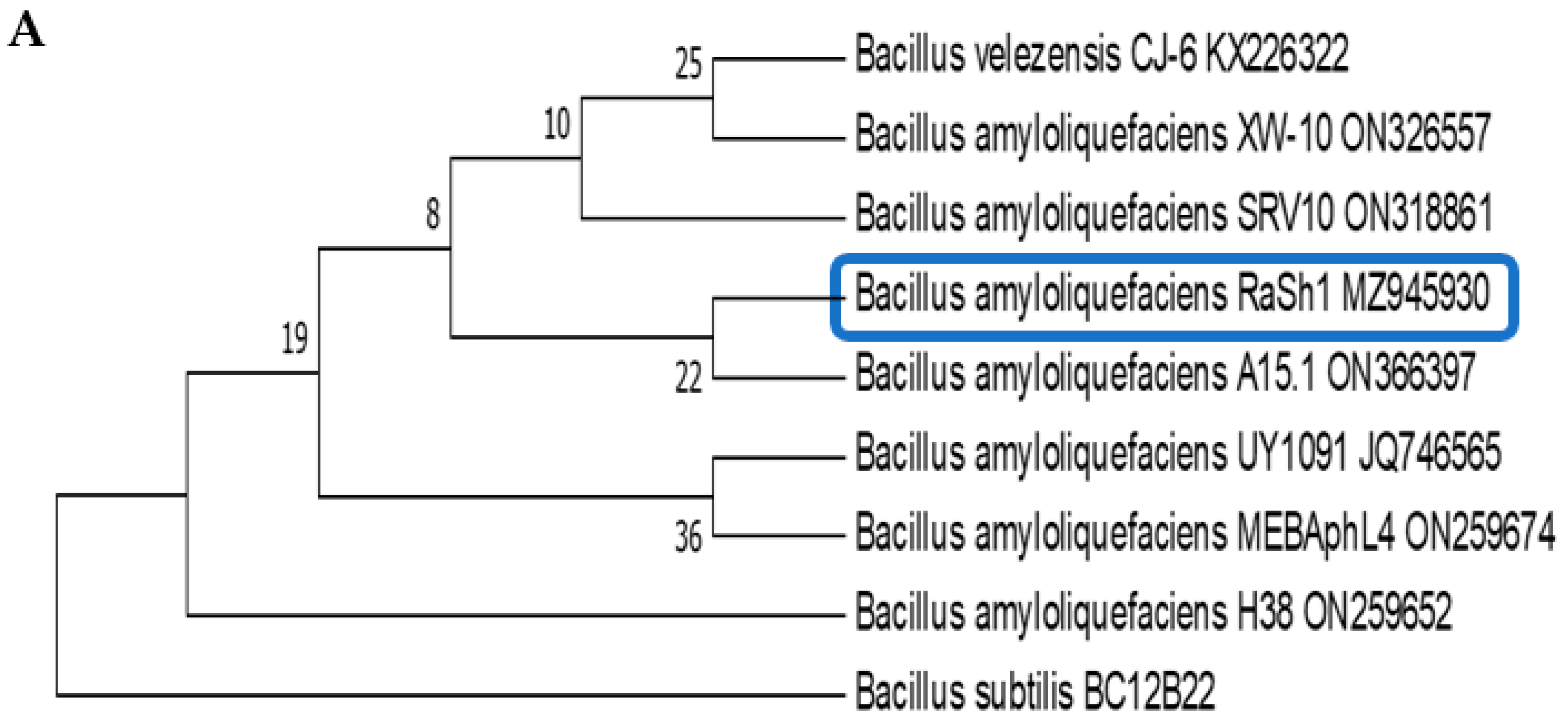
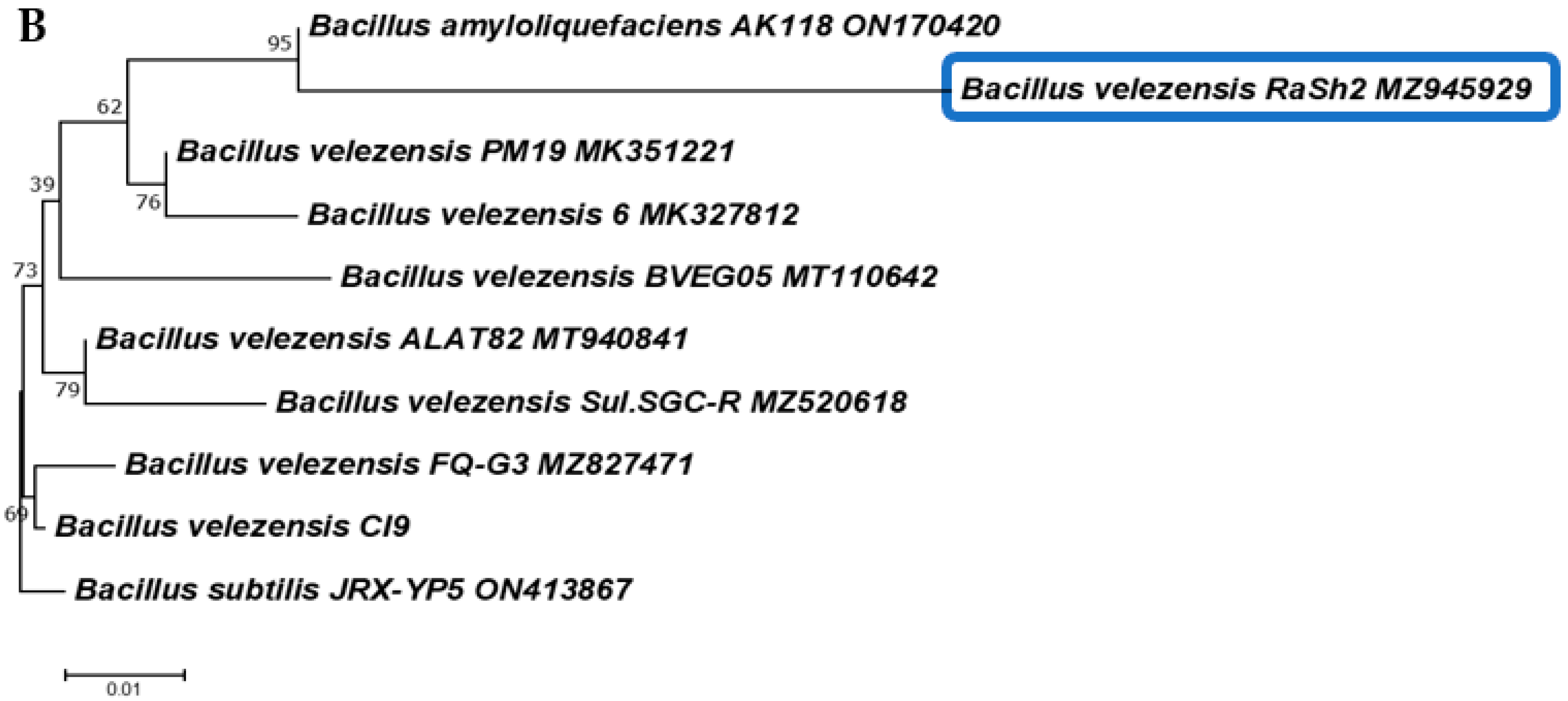
2.7. Molecular Characterization of Bacterial Isolates by Partial Sequencing of 16S rDNA

The DNA of the most active isolates was extracted using standard bacterial procedures [36]. PCR was used to preferentially amplify the 16S rRNA gene from genomic DNA using the universal forward primer (F1) 5′ AGAGTTTGATCCTGGCTCAG 3′ and the reverse primer (R1) 5′ GGTTACCTTGTTAC GACTT 3′ according to [37]. The 16S rRNA gene of the bacterial isolates was aligned with the standard reference sequences obtained from GenBank, NCBI, using BLAST (http://blast.ncbi.nlm.nih.gov/) (accessed on 1 September 2021).
A phylogenetic tree was created using MEGA 6.0 [38]. The sequence was finally submitted to GenBank, and an accession number was obtained.

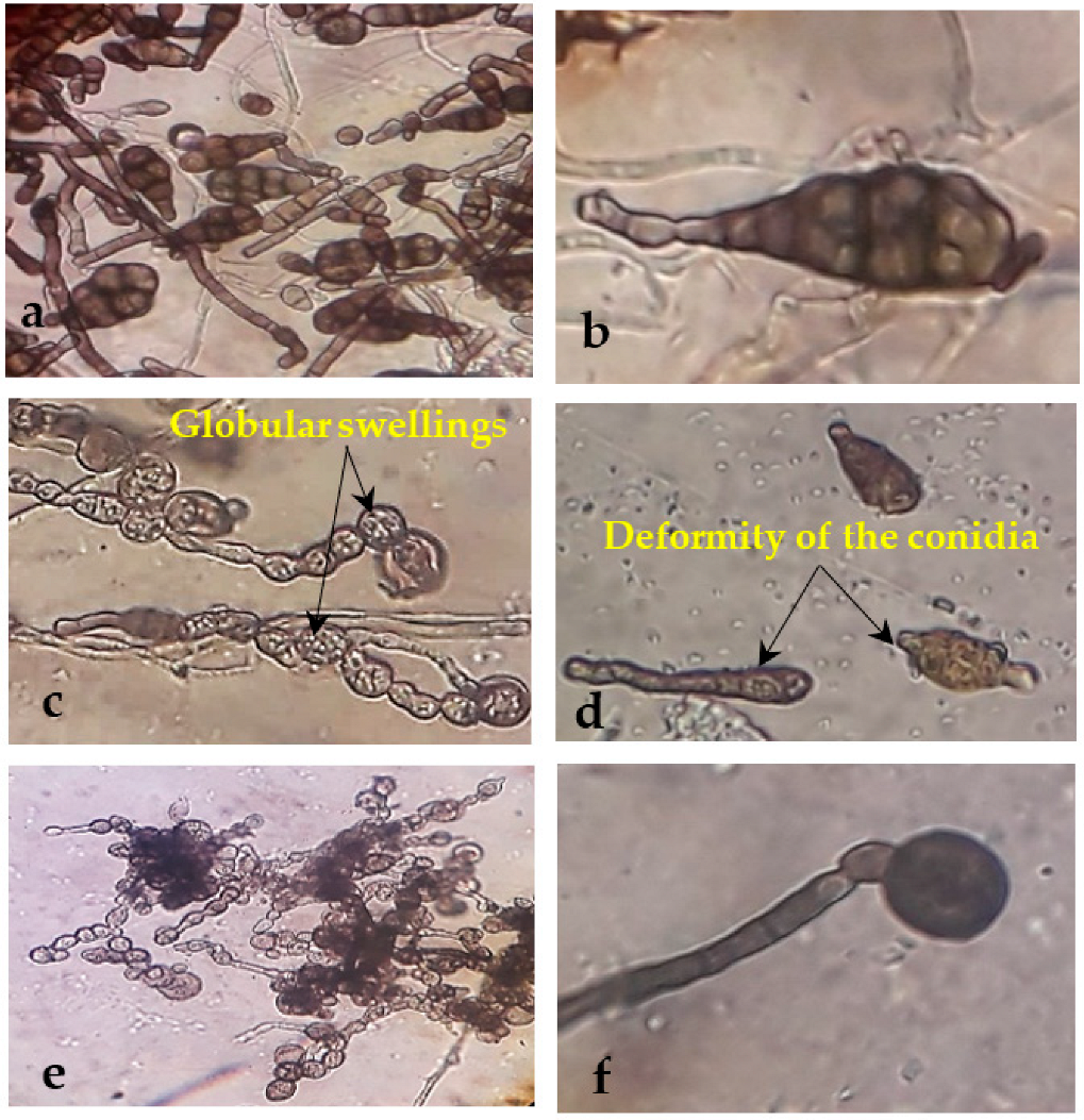
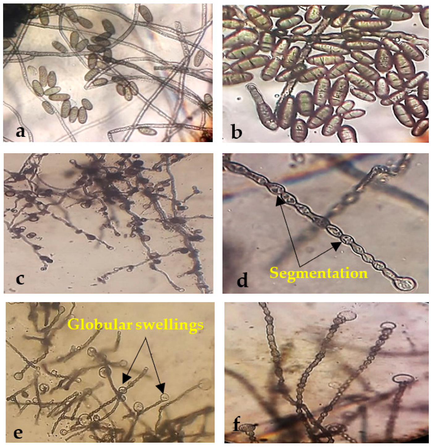
2.8. Morphological Abnormalities in the Alternaria sp. and Helminthosporium sp. Hyphae due to the Antagonistic Effects of Endophytic Bacterial

The morphological deformation caused by the most bioactive endophytic bacterial strains on the mycelia of each pathogenic fungus (Alternaria sp. and Helminthosporium sp.) on PDA plates was examined. The hyphal strands from the confrontation lines at the end of the fungal colony were extracted and compared to the control plates under a light microscope (Leitz WETZLAR, Wetzlar, Germany) for anomalies [39].

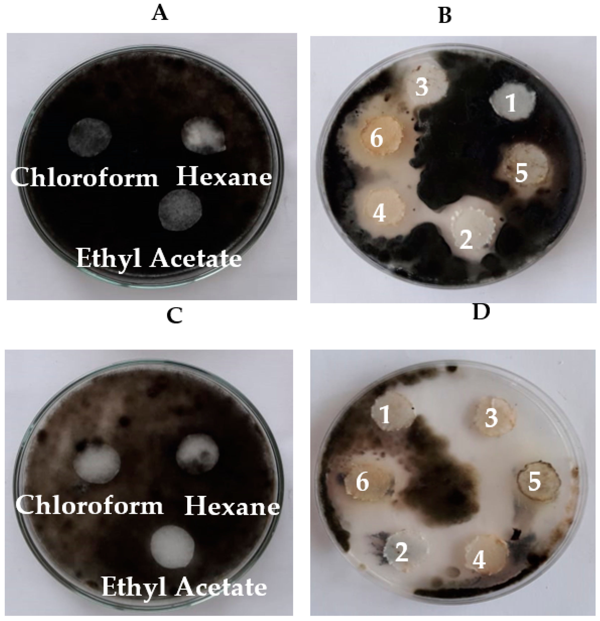
2.9. Preparation of Antifungal Bacterial Crude Extracts using Different Solvents

The two selected endophytic bacterial strains were cultured by placing agar blocks of actively growing pure culture (10 mm in diameter) in three Erlenmeyer flasks (1 L), each containing 300 mL of sterile nutrient broth for each endophytic bacterial strain, and incubated at 32 ± 2 °C for 24 h with continuous shaking at 150 rpm/min. After the incubation period, stationary growth cultures were centrifuged at 4000× g for 30 min at 4 °C. Each bacterial strain’s cell-free filtrates were extracted with an equal volume of ethyl acetate, chloroform: methanol (2:1 v/v), and hexane separately in a separating funnel by shaking vigorously for 15 min. The mixtures were allowed to settle until two different layers appeared: the upper solvent and the lower aqueous layer. The extraction was repeated three times [40]. The crude extracts were tested for their biological activity (100 µL) using the filter paper diffusion method against both. Alternaria sp. and Helminthosporium sp. were put separately on PDA media and the solvents were used as a control. Gas Chromatography and Mass Spectrometry (GC-MS) were used to analyze the crude extracts.

2.10. Gas Chromatography and Mass Spectrometry (GC–MS)

The bioactive components in the crude extracts of two endophytic bacterial strains were identified by GC-MS [41]. The crude extracts were analyzed by GC–MS using a (Thermo Scientific TRACE 1310 Milan, Italy) Gas Chromatograph attached with an ISQ LT single quadrupole Mass Spectrometer detector fitted with DB5-MS, 30 m and 0.25 mm ID (J&W Scientific) in the Al-Azhar University’s Regional Center for Mycology and Biotechnology, Cairo, Egypt. The instrument’s temperature was initially set to 40 °C and sustained for 3 min. The temperature was raised to 280 °C at a rate of 5 °C/min at the end of this period and maintained for 5 min. Then, it was increased to 290 °C at a rate of 7.5 °C/min and kept for 1 min. The injection port temperature was kept at 200 °C, while the helium flow rate was held for 1 mL/min. An ionization voltage of 70 eV was used. The mass spectra of the extracts were compared to data from WILEY and NIST to identify the bioactive chemicals present in MASS SPECTRAL DATABASE libraries [42].

2.11. In Vivo Evaluation of B. amyloliquefaciens Effects against Alternaria sp.-Infected Pepper Plants under Greenhouse Conditions

The antifungal activity of B. amyloliquefaciens against Alternaria sp. was evaluated in a pot experiment in a greenhouse of the Botany and Microbiology Department, Faculty of Science, Zagazig University, with a temperature range of 23–30 °C and a relative humidity of 60–85% in a completely randomized design.

2.11.1. B. amyloliquefaciens Inoculum Preparation

On a rotary shaker (180 rpm), B. amyloliquefaciens was grown at 30 °C for 48 h in nutrient broth. The bacterial suspension was obtained containing 107 CFU/mL.

2.11.2. Pot Experiment for Cultivation of Pepper Seedlings

Plastic bags (12 cm diameter and 22 cm high) containing sterile field clay soil (2 kg/ag) were collected from an agricultural field in Minia Al-Qamh, El-Sharkia Governorate. Experimental treatments were applied to 45–50 days old pepper seedlings procured from the local market. Initially, bacterial suspension, according to Widnyana and Javandira [43], was used to soak the roots of pepper seedlings for 4 h before transplantation; one seedling was transplanted per bag. Soils were drenched with 300 mL of the prepared inoculum or equivalent tap water. Two days after transplanting, the inoculation with the fungal pathogen Alternaria sp. was conducted by pipetting individual droplets of fungal suspension (105 cfu/mL) on the surface of healthy leaves after gently removing the leaf wax of control and infected pepper leaves using a brush. Also, pepper plants pipetted with tap water droplets were used as control plants. After pathogen inoculation, the inoculated plants were kept under polyethylene bags for 24 h to ensure the infection process and maintain high humidity conditions. Then, they were exposed to greenhouse conditions. There were ten replicates (n = 10) for each particular treatment. Four weeks after inoculation, disease symptoms were recorded.

2.11.3. Determination of Morphological Parameters

After four weeks of B. amyloliquefaciens application, pepper plants from the Alternaria sp. infected and non-infected treatments were uprooted and washed with tap water. The total heights of the pepper plants were measured. The total fresh weights (TFW) of the pepper plants for each treatment were taken, and then placed in the oven at 70 °C for two days. Their total dry weight (TDW) was recorded.

2.11.4. Assessment of Disease Incidence (DI)

The incidence of the disease for the pepper plants that were only infected with Alternaria sp. and for the Alternaria sp. pepper plants infected and treated with B. amyloliquefaciens, was determined by the following formula:
Disease   Incidence   DI   % = Number   of   infected   plants Total   number   of   plants × 100  

2.12. Statistical Analysis

One-way ANOVA was used to analyze the data. To compare the means of the treatments, Duncan’s multiple range test at p < 0.05 was used. The software package statistics 10.1 was utilized for statistical analysis.

3. Results

In the present study, twenty-five isolates of endophytic bacteria were isolated from different plants collected from El-Sharkia Governorate, Egypt, as recorded in (Table 1) and shown in (Figure 1). From the leaves, stem, and roots of Solanum melongena, five isolates (E1, E2, E3, E4, and E5) were identified, while four isolates (T1, T2, T4, and T5) were isolated from S. lycopersicum leaves. On the other hand, only one isolate (C1) was isolated from Brassica oleracea. These isolates were examined to see whether they had high anti-Alternaria sp. and anti-Helminthosporium sp. effects using the dual culture technique (Figure 2). Compared to the control, the inhibitory zone of radial growth revealed endophytic bacteria’s antagonistic activity. The width of the inhibition zone between the pathogen and the antagonist was calculated as: + for 2–5 mm; ++ for 5–10 mm; and +++ for > 10 mm. The most active isolates were T5 and C1 against both Alternaria sp. and Helminthosporium sp. As shown in Table 1, R1 and R2 had moderate inhibition zones with both pathogens. In contrast, R3 and R6 did not have any inhibitory effects on either of the fungal pathogens.

3.1. Antifungal Activity of Cell-Free Culture and Discs of Endophytic Bacteria

The results in Table 2 and Figure 3 indicate that the cell-free culture was effective (150 μL) or that the discs (15 mm) of endophytic bacteria isolated from P. oleracea (isolates R1 and R2), S. lycopersicum (isolates T4 and T5), S. melongena (isolates E3 and E5), and B. oleracea (isolate C1) had exhibited inhibitory activities against Alternaria sp. and Helminthosporium sp. Moreover, the bacterial endophyte discs of C1 and T5 isolated from the leaves of B. oleracea and S. lycopersicum, respectively, showed the highest inhibitory effects against Alternaria sp. (4.7 ± 0.252 and 3.1 ± 0.164 cm, respectively) and Helminthosporium sp. (3.9 ± 0.329 and 4.0 ± 0.212 cm, respectively) as compared to the other isolates.

3.2. Identification of Endophytic Bacterial Isolates

The morphological and biochemical characteristics of the antagonistic bacteria are listed in Table 3. Positive biochemical results involve catalase, oxidase, and the hydrolysis of gelatin and starch. Negative results include the indole test, hydrogen sulfide, the methyl red test, and urease.
The most active endophytic bacterial isolates were selected and verified using the 16S rDNA gene sequence. The obtained partial sequence of the 16S rDNA gene was deposited in the GenBank database under accession numbers; MZ945930 and MZ945929, respectively, as shown in Figure 4.

3.3. Morphological Changes under the Light Microscope

The treatment with B. amyloliquefaciens and B. velezensis caused abnormal mycelial growth and significant morphological changes in Alternaria sp. and Helminthosporium sp., primarily manifesting as contraction, collapse, deformation, deformity of the conidium, and globular swellings at the tips of hyphal strands (Figure 5 and Figure 6c–f). In contrast, the mycelia of the control group were straight and well developed (Figure 5 and Figure 6a,b).

3.4. Bioassay and Biological Activity of the Crude Extracts of Endophytic Bacterial Strains

The biological activity of the crude extracts of endophytic bacterial strains (B. amyloliquefaciens and B. velezensis) was investigated using the filter paper diffusion method against both Alternaria sp. and Helminthosporium sp. Our results showed that all solvents (control) had no inhibitory effects on Alternaria sp. and Helminthosporium sp. as seen in Figure 7A,C. Figure 7B,D, shows that the solvent extracts of B. amyloliquefaciens and B. velezensis had inhibitory effects on both fungal pathogens.

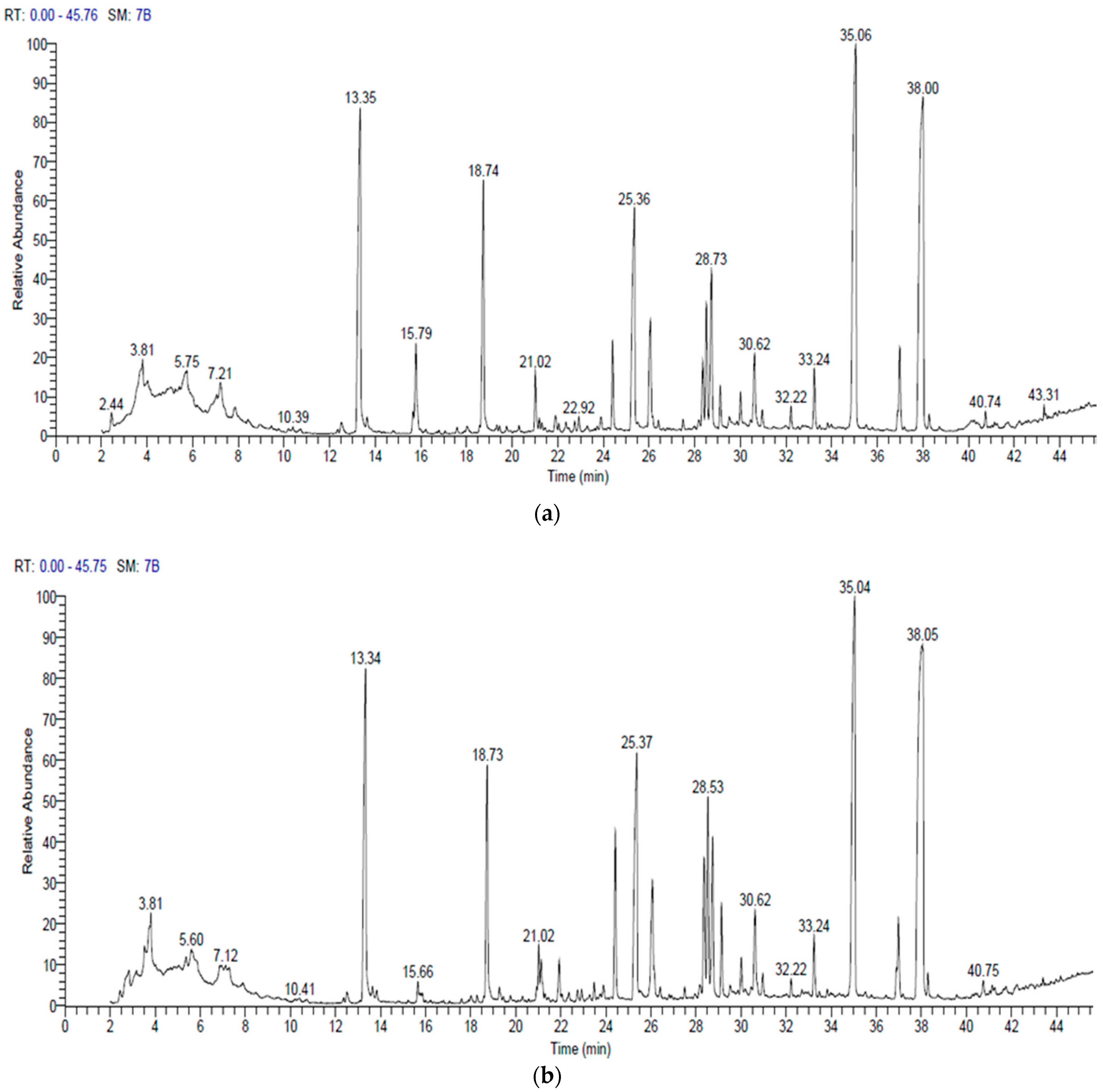
3.5. Gas Chromatography and Mass Spectrometry (GC–MS)

The bioactive components of B. amyloliquefaciens and B. velezensis were analyzed using GC-MS chromatography (Figure 8 and Table 4 and Table 5). The detected compounds’ names, molecular weights, molecular formulas, retention times, and quantities are listed (Table 4 and Table 5). There were substantial peaks in the cell-free extracts of the two bacterial strains among these bioactive chemicals, implying that they play a significant role in antibacterial and antifungal activity. These chemicals include: Bis (2-ethylhexyl) phthalate, followed by Bis (2-ethylhexyl) ester, N,N-Dimethyldodecylamine (Tertiary amine), Dibutyl phthalate, Methyl palmitate, and Ethyl hexadecanoate (Figure 8 and Table 4 and Table 5).

3.6. In Vivo Evaluation of B. amyloliquefaciens Effects against Alternaria sp. Infected Pepper Plants

The most active bacterial endophyte in our study (B. amyloliquefaciens) was selected to act as a biocontrol agent via a greenhouse experiment. Alternaria sp. was inoculated into pepper plants either in the presence or absence of B. amyloliquefaciens. The positive effect of the application of B. amyloliquefaciens on pepper TFW, TDW, and plant heights were confirmed (Table 6). The morphological changes between the different treatments are illustrated in Figure 9. Generally, the assessed growth parameters were significantly reduced in pepper plants infected with Alternaria sp. compared with the healthy control ones. However, these growth traits significantly increased with B. amyloliquefaciens, regardless of whether the plants were infected or not. In non-infected pepper leaves, the application of B. amyloliquefaciens significantly improved TFW (8.36 g/plant), TDW (1.4299 g/plant), and plant height (28 cm/plant). Also, Alternaria sp. exhibited the highest DI (80%) in the control plants, while with B. amyloliquefaciens inoculation, DI was greatly reduced in Alternaria sp. infected pepper plants (40%), as seen in Table 6. B. amyloliquefaciens reduced the disease symptoms; therefore, B. amyloliquefaciens exhibited strong antagonism toward Alternaria sp. infection and improved the growth of the infected pepper plants.

4. Discussion

The increased usage of chemical compounds to maintain healthy crops and high productivity has detrimental consequences for nature, animals, and humans [3,44]. Endophytes have a higher antagonistic potential against plant disease than microorganisms isolated from the rhizosphere or soil because they exist in a stable environment inside the plant [11] and can be found in various host plants [20,22,45]. Endophytes are implicated in the control of plant disease, development of plant tolerance, plant growth promotion, nitrogen fixation, synthesis of novel bioactive compounds, and detoxification of toxic pesticides [46,47]. Moreover, they produce secondary metabolites of biotechnological interest with a pharmaceutical application [48]. Bacterial endophytes vary among organs, tissues, soil, and plants [13].
Some studies consider bacterial endophytes as potential biocontrol agents for various hazardous fungi [6,7]. Selim et al. [17] and Riera et al. [49] revealed that Streptomyces, Pseudomonas, Bacillus, and Agrobacterium have long been the most important bacteria genera for the production of active antimicrobial substances. Massawe et al. [23] isolated and characterized Bacillus strains with volatile organic compounds (VOCs) acting against Sclerotinia sclerotiorum. Earlier investigations documented that antibiotics, such as mycosubtilins, iturins, and bacillomycins, are active metabolites with antimicrobial activities produced by B. subtilis [50,51,52].
Bacillus spp. can be used to develop effective microbial biopesticides in the form of biological control agents [4]. Olanrewaju et al. [53] reported that Bacillus sp. forms beneficial relationships with plants directly or indirectly. B. velezensis and B. amyloliquefaciens are Gram-positive bacteria that have been used to promote the growth of numerous plants directly or indirectly as they are efficient in plant colonization and commercialized around the world [13,23,54]. The antifungal mechanisms of B. velezensis and B. amyloliquefaciens are the same, whether through direct antibiosis or plant-mediated induced disease resistance [55,56,57]. As secondary metabolites, B. velezensis and B. amyloliquefaciens produce several antimicrobial compounds against various phytopathogens [58]. Some fungal pathogens, such as Helicobasidium purpureum, F. oxysporum, and Rhizoctonia solani, are inhibited by the B. velezensis strain FKM10. [59,60]. B. velezensis can also cause the development of systemic resistance in plants [59]. In some experiments, B. velezensis was found to produce several metabolites related to disease resistance, including NH3, antimicrobial proteins, polyketides, and siderophores [55,58,60,61].
The optical microscopic examination of Alternaria sp. and Helminthosporium sp. revealed that treatment with B. amyloliquefaciens and B. velezensis caused abnormal mycelial growth. These anomalies showed a problem with fungal cell wall formation [54]. Zhao et al. [62] noticed abnormal morphological changes in the fungal mycelia of F. oxysporum, Magnaporthe grisea, and Alternaria sp. when interacting with endophytes. The B. velezensis strain FKM10 destroyed the cell wall and cell membrane upon interacting with F. verticillioides [54]. Furthermore, cyclic lipopeptides produced by B. velezensis LM2303 affected the cell membrane permeability of F. graminearumon [63]. Moon et al. [64] investigated the potential of B. velezensis CE 100 in mitigating phytophthora root rot, which suppressed mycelial growth, causing hyphae to enlarge and distort.
The bioactive metabolites of B. amyloliquefaciens and B. velezensis were analyzed using GC-MS chromatography. The antifungal actions of these extracts could be related to various chemical classes, including esters, fatty acids, aldehydes, tertiary amines, alkaloids, and ketones. Among these bioactive compounds, the two bacterial strains had significant peaks in cell-free extracts, indicating that they played a substantial role in antibacterial and antifungal activities. These compounds include: Bis (2-ethylhexyl) phthalate followed by Bis (2-ethylhexyl) ester, N, N-Dimethyldodecylamine (Tertiary amine), Dibutyl phthalate, Methyl palmitate, and Ethyl hexadecanoate. Phthalates have antimicrobial and antifungal activities [65,66,67,68,69]. Al-Bari et al. [70] reported that the Bis (2-ethylhexyl) antimicrobial activity of phthalate was shown against Gram-positive bacteria and several harmful fungi. Kanjana et al. [71] reported that the Bis (2-ethyl hexyl) phthalate had antifungal, antimicrobial, and antioxidant activities. Furthermore, both bacterial extracts contained dibutyl phthalate, which had antimicrobial activity against unicellular and filamentous fungi [72,73].
The high percentage of Bis (2-ethylhexyl) ester in both bacterial extracts had an antifungal activity. Mohamad et al. [9] suggested that the Bacillus atrophaeus strain XEGI50 species was a promising candidate as a biocontrol agent. The GC-MS analysis of cell-free extracts showed that numerous compounds had antimicrobial activity, including Bis (2-ethylhexyl) phthalate and Bis(2-ethylhexyl) ester. The antimicrobial activity of N, N-Dimethyldodecylamine, N-Methyl-N-benzyltetradecanamine, and N, N-Dimethyltetradecylamine (tertiary amine) against bacteria, yeasts, fungi, and enveloped viruses has been reported. [74,75]. Massawe et al. [23] reported the biocontrol activity of N, N-dimethyl-dodecyl amine against Sclerotinia sclerotiorum. The antimicrobial activity of these amine oxides was related to their interaction with biological membranes; the permeability of cellular membranes changed, and membrane-dependent activities were inhibited, resulting in cell death. Furthermore, the amine oxides caused K+ leakage from cells and lysis of osmotically stabilized protoplasts, which inhibited glycolysis [74,75].
Methyl palmitate (fatty acid methyl esters) detected in both bacterial extracts exhibited antibacterial and antifungal activities, damaging microbial cellular membranes [76]. Chandrasekaran et al. [77] argued that fatty acid methyl ester extract showed moderate antifungal activity against two Aspergillus spp. Moreover, ethyl hexadecanoate had antimicrobial, antioxidant, and pesticidal activities [78]. The least observed in either of the bacterial extracts were octyl hexadecanoate, diethyl phthalate, 2-phenyltridecane, methyl 10-methyl undecanoate, 5-octadecene, octadecane, and methyl tetradecanoate that had antimicrobial, antioxidant, and anticancer activities [71,78,79].
Some Bacillus strains produce volatile organic compounds (VOCs) that may act as antifungal agents against various soil-borne diseases and limit fungal growth [23,48]. These VOCs can diffuse among soil particles and spread far from where they were applied, inhibiting pathogens without coming into direct contact with them [80,81]. For example, the VOCs produced by B. amyloliquefaciens may inhibit F. oxysporum mycelial development and spore germination [21,82]. Gao et al. [20] and Jiang et al. [83] reported that different strains of B. velezensis suppressed the growth of B. cinerea by different numbers of VOCs. Also, Reda et al. [84] indicated that the B. amyloliquefaciens S5I4 strain produced bioactive antimicrobial compounds.
The enhancing capacity of B. amyloliquefaciens for pepper plant growth is in coherence with Shahzad et al. [85] and Rashad et al. [19], who documented that B. amyloliquefaciens RWL-1 and GGA inoculation significantly enhanced the growth traits of tomato and garlic plants under both diseased and non-diseased conditions of F. oxysporum and S. cepivorum. The extensively distinguished mechanisms for plant growth promotion caused by B. amyloliquefaciens are through phytohormones, providing the essential nutrients, N2 fixation, and phosphate solubilization [18]. In addition, endophytic bacteria may promote plant growth through biofertilization [19]. Shahzad et al. [85] discovered that the endophytes’ capability to produce secondary metabolites provided additional support to plants and increased plant development, increasing their resilience to biotic and abiotic challenges. These mechanisms can contribute to the plant growth-promoting potential of B. amyloliquefaciens on pepper plants.

5. Conclusions

Controlling plant pathogen diseases in a safe, effective, and alternative manner has become increasingly crucial for improving the quality of agricultural products. Compared to chemical control, biological control using antagonistic microorganisms, such as bacteria, is a long-term approach to inhibiting plant pathogens. The novelty of this work is the isolation of new local strains of endophytic bacteria and the production of several antimicrobial metabolites associated with the biocontrol of Alternaria, which can cause serious diseases to important vegetable crops in Egypt. Bacillus species, as biocontrol agents, could inhibit potential plant pathogens. The B. amyloliquefaciens MZ945930 and B. velezensis MZ945929 strains in this study, shared the same antifungal mechanisms by direct antibiosis against Alternaria sp. and Helminthosporium sp. Moreover, suppressive effects were associated with a variety of secondary metabolite secretions. The resulting bacterial crude extracts from both bacterial strains were promising as they have shown the highest antifungal activities. Also, the in vivo results emphasized the significance of the effect of B. amyloliquefaciens on pepper growth under both the control and diseased conditions caused by Alternaria sp. Therefore, the present study encourages the use of these bacterial strains as biocontrol agents in agriculture.

Author Contributions

Conceptualization: S.A.S. and R.A.M.; Methodology: R.A.M., S.A.S. and M.M.K.; Formal analysis and investigation: S.A.S. and R.A.M.; Writing: S.A.S. and R.A.M.; Review and editing: R.A.M., S.A.S. and M.M.K. All authors have read and agreed to the published version of the manuscript.

Funding

This research received no external funding.

Institutional Review Board Statement

Not applicable.

Informed Consent Statement

Not applicable.

Data Availability Statement

The relevant datasets supporting the results of this article are included within the article and the [GenBank NCBI] at: https://0-www-ncbi-nlm-nih-gov.brum.beds.ac.uk/nuccore/MZ945930.1/ (accessed on 1 September 2021).

Conflicts of Interest

The authors declare no conflict of interest.

References

  1. Fisher, M.C.; Henk, D.A.; Briggs, C.J.; Brownstein, J.S.; Madoff, L.C.; McCraw, S.L.; Gurr, S.J. Emerging fungal threats to animal plant ecosystem, health. Nature 2012, 484, 186–194. [Google Scholar] [CrossRef]
  2. Almeida, F.; Rodrigues, M.L.; Coelho, C. The still underestimated problem of fungal diseases worldwide. Front. Microbiol. 2019, 10, 214. [Google Scholar] [CrossRef] [PubMed]
  3. Dadrasnia, M.; Usman, M.; Omar, R.; Ismail, S.; Abdullah, R. Potential use of Bacillus genus to control of bananas diseases: Approaches toward high yield production and sustainable management. J. King Saud Univ. 2020, 32, 2336–2342. [Google Scholar] [CrossRef]
  4. Reda, F.M.; Hassanein, W.A.; Sherief, E.A.H.; Moabed, S. Toxicological studies and field applications of a new Bacillus thuringiensis isolate (Bt1) and two chemical pesticides on Spodoptera littoralis (Boisd.) (Lepidoptera: Noctuidae). Egypt. J. Biol. Pest Control 2016, 26, 229–236. [Google Scholar]
  5. Deguine, J.P.; Aubertot, J.N.; Flor, R.J.; Lescourret, F.; Wyckhuys, K.A.G.; Ratnadass, A. Integrated pest management: Good intentions, hard realities. A review. Agron. Sustain. Dev. 2021, 41, 38. [Google Scholar] [CrossRef]
  6. Rivera-Méndez, W.; Obregón, M.; Morán-Diez, M.E.; Hermosa, R.; Monte, E. Trichoderma asperellum biocontrol activity and induction of systemic defenses against Sclerotium cepivorum in onion plants under tropical climate conditions. Biol. Control 2020, 141, 104145. [Google Scholar] [CrossRef]
  7. Al-Nadabi, H.H.; Al-Buraiki, N.S.; Al-Nabhani, A.A.; Maharachchikumbura, S.N.; Velazhahan, R.; Al-Sadi, A.M. In vitro antifungal activity of endophytic bacteria isolated from date palm (Phoenix doctylifera L.) against fungal pathogens causing leaf spot of date palm. Egypt. J. Biol. Pest Control 2021, 31, 65. [Google Scholar] [CrossRef]
  8. Heydari, A.; Pessarakli, M. A review on biological control of fungal plant pathogens using microbial antagonists. J. Biol. Sci. 2010, 10, 273–290. [Google Scholar] [CrossRef]
  9. Mohamad, O.; Li, L.; Ma, J.; Hatab, S.; Xu, L.; Guo, J.W.; Rasulov, B.A.; Liu, Y.; Hedlund, B.P.; Li, W.J. Evaluation of the antimicrobial activity of endophytic bacterial populations from Chinese traditional medicinal plant licorice and characterization of the bioactive secondary metabolites produced by Bacillus atrophaeus against Verticillium dahliae. Front. Microbiol. 2018, 9, 924. [Google Scholar] [CrossRef] [PubMed]
  10. Zhang, L.; Tao, Y.; Zhao, S.; Yin, X.; Chen, J.; Wang, M.; Cai, Y.; Niu, Q. A novel peroxiredoxin from the antagonistic endophytic Bacterium Enterobacter Sp. V1 contributes to cotton resistance against Verticillium Dahliae. Plant Soil 2020, 454, 395–409. [Google Scholar] [CrossRef]
  11. Kaul, S.; Sharma, T.K.; Dhar, M. “Omics” Tools for better understanding the plant-endophyte interactions. Front. Plant Sci. 2016, 7, 955. [Google Scholar] [CrossRef] [Green Version]
  12. Hazarika, D.J.; Goswami, G.; Gautom, T.; Parveen, A.; Das, P.; Barooah, M.; Boro, R.C. Lipopeptide mediated biocontrol activity of endophytic Bacillus subtilis against fungal phytopathogens. BMC Microbiol. 2019, 19, 71. [Google Scholar] [CrossRef] [PubMed]
  13. Sharma, A.; Kaushik, N.; Sharma, A.; Bajaj, A.; Rasane, M.; Shouche, Y.S.; Marzouk, T.; Djébali, N. Screening of tomato seed bacterial endophytes for antifungal activity reveals lipopeptide producing Bacillus siamensis Strain NKIT9 as a potential bio-control agent. Front. Microbiol. 2021, 12, 609482. [Google Scholar] [CrossRef] [PubMed]
  14. Fadiji, A.E.; Babalola, O.O. Exploring the potentialities of beneficial endophytes for improved plant growth. Saudi J. Biol. Sci. 2020, 27, 3622–3633. [Google Scholar] [CrossRef]
  15. Bhattacharjee, R.; Dey, U. An overview of fungal and bacterial biopesticides to control plant pathogens/diseases. Afr. J. Microbiol. Res. 2014, 8, 1749–1762. [Google Scholar]
  16. Amaresan, N.; Jayakumar, V.; Krishna, K.; Thajuddin, N. Endophytic bacteria from tomato and chilli, their diversity and antagonistic potential against Ralstonia solanacearum. Arch. Phytopathol. Plant Prot. 2011, 45, 344–355. [Google Scholar] [CrossRef]
  17. Selim, H.M.M.; Gomaa, N.M.; Essa, A.M.M. Antagonistic Effect of the Endophytic Bacteria and against Some Phytopathogens. Egypt. J. Bot. 2016, 56, 613–626. [Google Scholar]
  18. Kazerooni, E.A.; Maharachchikumbura, S.S.N.; Al-Sadi, A.M.; Kang, S.-M.; Yun, B.-W.; Lee, I.-J. Biocontrol Potential of Bacillus amyloliquefaciens against Botrytis pelargonii and Alternaria alternata on Capsicum annuum. J. Fungi 2021, 7, 472. [Google Scholar] [CrossRef]
  19. Rashad, Y.M.; Abbas, M.A.; Soliman, H.M.; Abdel-Fattah, G.G.; Abdel-Fattah, G.M. Synergy between endophytic Bacillus amyloliquefaciens GGA and arbuscular mycorrhizal fungi induces plant defense responses against white rot of garlic and improves host plant growth. Phytopathol. Mediterr. 2020, 59, 169–186. [Google Scholar] [CrossRef]
  20. Gao, Z.; Zhang, B.; Liu, H.; Han, J.; Zhang, Y. Identification of endophytic Bacillus velezensis ZSY-1 strain and antifungal activity of its volatile compounds against Alternaria solani and Botrytis cinerea. Biol. Control 2017, 105, 27–39. [Google Scholar] [CrossRef]
  21. Wu, Y.; Zhou, J.; Li, C.; Ma, Y. Antifungal and plant growth promotion activity of volatile organic compounds produced by Bacillus amyloliquefaciens. Microbiol. Open 2019, 8, e813. [Google Scholar] [CrossRef]
  22. Kumar, A.; Kumar, S.P.J.; Chintagunta, A.D.; Agarwal, D.K.; Pal, G.; Singh, A.N.; Simal-Gandara, J. Biocontrol potential of Pseudomonas stutzeri endophyte from Withania somnifera (Ashwagandha) seed extract against pathogenic Fusarium oxysporum and Rhizoctonia solani. Arch. Phytopathol. Plant Prot. 2021, 55, 1–18. [Google Scholar] [CrossRef]
  23. Massawe, V.C.; Hanif, A.; Farzand, A.; Mburu, D.K.; Ochola, S.O.; Wu, L.; Samad Tahir, H.A.; Gu, Q.; Wu, H.; Gao, X. Volatile compounds of endophytic Bacillus spp. have biocontrol activity against Sclerotinia sclerotiorum. Phytopathology 2018, 108, 1373–1385. [Google Scholar] [CrossRef] [PubMed] [Green Version]
  24. Reda, F.M.; Hassanein, W.A.; Moabed, S. Potential exploitation of Bacillus flexus biofilm against the cowpea weevil, Callosobruchus maculatus (F.) (Coleoptera: Bruchidae). Egypt. J. Biol. Pest Control 2020, 30, 18. [Google Scholar] [CrossRef]
  25. Hassanein, W.A.; Reda, F.M.; Moabed, S.; El Shafiey, S.N. Insecticidal impacts of Bacillus flexus S13 biofilm ‘extracellular matrix’ on cowpea weevil, Callosobruchus maculatus. Biocatal. Agric. Biotechnol. 2021, 31, 101898. [Google Scholar] [CrossRef]
  26. Huang, W.Y.; Cai, Y.Z.; Hyde, K.D.; Corke, H.; Sun, M. Endophytic fungi from Nerium oleander L (Apocynaceae): Main constituents and antioxidant activity. World J. Microbiol. Biotechnol. 2007, 23, 1253–1263. [Google Scholar] [CrossRef]
  27. Al-Hussini, H.S.; Al-Rawahi, A.Y.; Al-Marhoon, A.A.; Al-Abri, S.A.; Al-Mahmooli, I.H.; Al-Sadi, A.M.; Velazhahan, R. Biological control of damping-off of tomato caused by Pythium aphanidermatum by using native antagonistic rhizobacteria isolated from Omani soil. J. Plant Pathol. 2019, 101, 315–322. [Google Scholar] [CrossRef]
  28. Choi, Y.W.; Hyde, K.D.; Ho, W.H. Single spore isolation of fungi. Fungal Divers. 1999, 3, 29–38. [Google Scholar]
  29. Ellis, M.B. Dematiaceus Hyphomycetes; Commonwealth Mycological Institute: Surrey, UK, 1971; 608p. [Google Scholar]
  30. Archana, T.; Rajendran, L.; Manoranjitham, S.K. Culture-dependent analysis of seed bacterial endophyte, Pseudomonas spp. EGN 1 against the stem rot disease (Sclerotium rolfsii Sacc.) in groundnut. Egypt. J. Biol. Pest Control 2020, 30, 119. [Google Scholar] [CrossRef]
  31. Pandey, D.K.; Tripathi, N.N.; Tripathi, R.D. Fungitoxic and phytotoxic properties of essential oil of Hyptis suaveolens. Zeitschrift fuer Pflanzenkrankheiten und Pflanzenschutz 1982, 89, 344–349. [Google Scholar]
  32. Paul, N.C.; Kim, W.K.; Woo, S.K.; Park, M.S.; Yu, S.H. The fungal endophytes in roots of Aralia species and their antifungal activity. Plant Pathol. J. 2007, 23, 287–294. [Google Scholar] [CrossRef]
  33. O’Connor, E.B.; O’Riordan, B.; Morgan, S.M.; Whelton, H.; O’Mullane, D.M.; Ross, R.P. A lacticin 3147 enriched food ingredient reduces Streptococcus mutans isolated from the human oral cavity in saliva. J. Appl. Microbiol. 2006, 100, 1251–1260. [Google Scholar] [CrossRef] [PubMed]
  34. Gerhardt, P.; Murray, R.G.E.; Wood, W.A.; Krieg, N.R. Methods for General and Molecular Bacteriology; ASM: Washington, DC, USA, 1994. [Google Scholar]
  35. Bergey, D.H.; Holt, J.G. Bergey’s Manual of Determinative Bacteriology, 9th ed.; Bergey’s Manual of Systematic Bacteriology; Williams & Wilkins Co.: Baltimore, MD, USA, 1994. [Google Scholar]
  36. Sambrook, J.; Russell, D.W. Molecular Cloning: A Laboratory Manual; Cold Spring Harbor Laboratory: New York, NY, USA, 2001. [Google Scholar]
  37. Srivastava, S.; Singh, V.; Kumar, V.; Verma, P.C.; Srivastava, R.; Basu, V.; Gupta, V.; Rawat, A.K. Identification of regulatory elements in 16S rRNA gene of Acinetobacter species isolated from water sample. Bioinformation 2008, 3, 173–176. [Google Scholar] [CrossRef] [PubMed]
  38. Tamura, K.; Stecher, G.; Peterson, D.; Filipski, A.; Kumar, S. MEGA6: Molecular evolutionary genetics analysis version 6.0. Mol. Biol. Evol. 2013, 30, 2725–2729. [Google Scholar] [CrossRef] [PubMed]
  39. Sariah, M. Potential of Bacillus spp. as biocontrol agent for anthracnose fruit rot of chili malays. Appl. Biol. 1994, 23, 1193–1201. [Google Scholar]
  40. Muzzamal, H.; Sarwar, R.; Sajid, I.; Hasnain, S. Isolation, identification and screening of endophytic bacteria antagonistic to biofilm formers. Pak. J. Zool. 2012, 44, 249–258. [Google Scholar]
  41. Nandhini, U.; Kumari, L.; Sangareshwari, S. Gas Chromatography-Mass Spectrometry analysis of bioactive constituents from the marine Streptomyces. Asian J. Pharm. Clin. Res. 2015, 8, 244–246. [Google Scholar]
  42. Senthilkumar, N.; Murugesan, S.; Babu, D.S.; Rajeshkannan, C. GC-MS analysis of the extract of endophytic fungus, Phomopsis sp. isolated from tropical tree species of India, Tectona grandis L. Int. J. Innov. Res. Sci. Eng. Technol. 2014, 3, 10176–10179. [Google Scholar]
  43. Widnyana, I.K.; Javandira, C. Activities Pseudomonas spp. and Bacillus sp. to stimulate germination and seedling growth of tomato plants. Agric. Agric. Sci. Procedia 2016, 9, 419–423. [Google Scholar] [CrossRef] [Green Version]
  44. Anjugam, M.; Bharathidasan, R.; Shijila Rani, A.S.; Ambikapathy, V. Evaluation of antimicrobial activities of endophytic fungal metabolites against clinical importance microbes. J. Pharmacogn. Phytochem. 2019, 8, 1004–1007. [Google Scholar]
  45. Adeleke, B.S.; Babalola, O.O. Pharmacological Potential of Fungal Endophytes Associated with Medicinal Plants: A Review. J. Fungi 2021, 7, 147. [Google Scholar] [CrossRef] [PubMed]
  46. Al-Badri, B.A.; Al-Maawali, S.S.; AlBalushi, Z.M.; Al-Mahmooli, I.H.; Al-Sadi, A.M.; Velazhahan, R. Cyanide degradation and antagonistic potential of endophytic Bacillus subtilis strain BEB1 from Bougainvillea spectabilis Willd. All Life 2020, 13, 92–98. [Google Scholar] [CrossRef]
  47. Haruna, A.; Yahaya, S.M. Recent advances in the chemistry of bioactive compounds from plants and soil microbes: A Review. Chem. Africa 2021, 8, 231–345. [Google Scholar] [CrossRef]
  48. Balderas-Ruíz, K.A.; Bustos, P.; Santamaria, R.I.; González, V.; Cristiano-Fajardo, S.A.; Barrera-Ortíz, S.; Mezo-Villalobos, M.; Aranda-Ocampo, S.; Guevara-García, A.A.; Galindo, E.; et al. Bacillus velezensis 83 a bacterial strain from mango phyllosphere, useful for biological control and plant growth promotion. AMB Express 2020, 10, 163. [Google Scholar] [CrossRef]
  49. Riera, N.; Handique, U.; Zhang, Y.; Dewdney, M.M.; Wang, N. Characterization of Antimicrobial-Producing Beneficial Bacteria Isolated from Huanglongbing Escape Citrus Trees. Front. Microbiol. 2017, 8, 2415. [Google Scholar] [CrossRef]
  50. Ali, S.; Hameed, S.; Imran, A.; Iqbal, M.; Lazarovits, G. Genetic, physiological and biochemical characterization of Bacillus sp. strain RMB7 exhibiting plant growth promoting and broad-spectrum antifungal activities. Microb. Cell Factories 2014, 13, 144. [Google Scholar] [CrossRef]
  51. Dunlap, C.A.; Bowman, M.J.; Rooney, A.P. Iturinic lipopeptide diversity in the Bacillus subtilis species group—Important antifungals for plant disease biocontrol applications. Front. Microbiol. 2019, 10, 1794. [Google Scholar] [CrossRef] [PubMed]
  52. Mülner, P.; Schwarz, E.; Dietel, K.; Junge, H.; Herfort, S.; Weydmann, M.; Lasch, P.; Cernava, T.; Berg, G.; Vater, J. Profiling for bioactive peptides and volatiles of plant growth promoting strains of the Bacillus subtilis complex of industrial relevance. Front. Microbiol. 2020, 11, 1432. [Google Scholar] [CrossRef]
  53. Olanrewaju, O.S.; Glick, B.R.; Babalola, O.O. Mechanisms of action of plant growth promoting bacteria. World J. Microbiol. Biotechnol. 2017, 33, 197. [Google Scholar] [CrossRef]
  54. Wang, C.; Zhao, D.; Qi, G.; Mao, Z.; Hu, X.; Du, B.; Liu, K.; Ding, Y. Effects of Bacillus velezensis FKM10 for Promoting the Growth of Malus hupehensis Rehd. and Inhibiting Fusarium verticillioides. Front. Microbiol. 2020, 10, 2889. [Google Scholar] [CrossRef]
  55. Kim, S.Y.; Song, H.; Sang, M.K.; Weon, H.Y.; Song, J. The complete genome sequence of Bacillus velezensis strain GH1-13 reveals agriculturally beneficial properties and a unique plasmid. J. Biotechnol. 2017, 259, 221–227. [Google Scholar] [CrossRef] [PubMed]
  56. Harwood, C.R.; Mouillon, J.M.; Pohl, S.; Arnau, J. Secondary metabolite production and the safety of industrially important members of the Bacillus subtilis group. FEMS Microbiol. Rev. 2018, 42, 721–738. [Google Scholar] [CrossRef]
  57. Gorai, P.S.; Ghosh, R.; Konra, S.; Mandal, N.C. Biological control of early blight disease of potato caused by Alternaria sp. EBP3 by an endophytic bacterial strain Bacillus velezensis SEB1. Biol. Control 2021, 156, 104551. [Google Scholar] [CrossRef]
  58. Dunlap, C.; Kim, S.J.; Kwon, S.W.; Rooney, A. Bacillus velezensis is not a later heterotypic synonym of Bacillus amyloliquefaciens, Bacillus methylotrophicus, Bacillus amyloliquefaciens subsp. plantarum and ‘Bacillus oryzicola’ are later heterotypic synonyms of of Bacillus velezensis based on phylogenomics. Int. J. Syst. Evol. Microbiol. 2016, 66, 1212–1217. [Google Scholar] [PubMed]
  59. Xu, T.; Zhu, T.; Li, S. β-1,3-1,4-glucanase gene from Bacillus velezensis ZJ20 exerts antifungal effect on plant pathogenic fungi. World J. Microbiol. Biotechnol. 2016, 32, 26. [Google Scholar] [CrossRef] [PubMed]
  60. Cao, Y.; Pi, H.; Chandrangsu, P.; Li, Y.; Wang, Y.; Zhou, H.; Xiong, H.; Helmann, J.D.; Cai, Y. Antagonism of two plant-growth promoting Bacillus velezensis isolates against Ralstonia solanacearum and Fusarium oxysporum. Sci. Rep. 2018, 8, 4360. [Google Scholar] [CrossRef]
  61. Rabbee, M.F.; Ali, M.; Choi, J.; Hwang, B.S.; Jeong, S.C.; Baek, K.H. Bacillus velezensis: A valuable member of bioactive molecules within plant microbiomes. Molecules 2019, 24, 1046. [Google Scholar] [CrossRef] [PubMed]
  62. Zhao, L.; Xu, Y.; Lai, X.H.; Shan, C.; Deng, Z.; Ji, Y. Screening and characterization of endophytic Bacillus and Paenibacillus strains from medicinal plant Lonicera japonica for use as potential plant growth promoters. Braz. J. Microbiol. 2015, 46, 977–989. [Google Scholar] [CrossRef]
  63. Chen, L.; Heng, J.; Qin, S.; Bian, K. A comprehensive understanding of the biocontrol potential of Bacillus velezensis LM2303 against Fusarium head blight. PLoS ONE 2018, 13, e0198560. [Google Scholar] [CrossRef]
  64. Moon, J.H.; Won, S.J.; Maung, C.E.H.; Choi, J.H.; Choi, S.I.; Ajuna, H.B.; Ahn, Y.S. Bacillus velezensis CE 100 inhibits root rot diseases (Phytophthora spp.) and promotes growth of Japanese cypress (Chamaecyparis obtuse Endlicher) seedlings. Microorganisms 2021, 9, 821. [Google Scholar] [CrossRef]
  65. Habib, M.R.; Karim, M.R. Antimicrobial and cytotoxic activity of di-(2-ethylhexyl) phthalate and anhydrosophoradiol-3-acetate isolated from Calotropis gigantea (Linn.) flower. Mycobiology 2009, 37, 31–36. [Google Scholar] [CrossRef] [PubMed]
  66. Smaoui, S.; Mellouli, L.; Lebrihi, A.; Coppel, Y.; Fguira, L.F.B.; Mathieu, F. Purification and structure elucidation of three naturally bioactive molecules from the new terrestrial Streptomyces sp. TN17 strain. Nat. Prod. Res. 2011, 25, 806–814. [Google Scholar] [CrossRef] [PubMed]
  67. El-Sayed, M.H. Di-(2-ethylhexyl) phthalate, a major bioactive metabolite with antimicrobial and cytotoxic activity isolated from the culture filtrate of newly isolated soil streptomyces (Streptomyces mirabilis Strain NSQu-25). World Appl. Sci. J. 2012, 20, 1202–1212. [Google Scholar]
  68. Sharma, D.; Pramanik, A.; Agrawal, P.K. Evaluation of bioactive secondary metabolites from endophytic fungus Pestalotiopsis neglecta BAB-5510 isolated from leaves of Cupressus torulosa D.Don. Don. 3 Biotech 2016, 6, 210. [Google Scholar] [CrossRef] [PubMed]
  69. Huang, L.; Zhu, X.; Zhou, S.; Cheng, Z.; Shi, K.; Zhang, C.; Shao, H. Phthalic acid esters: Natural sources and biological activities. Toxins 2021, 13, 495. [Google Scholar] [CrossRef]
  70. Al-Bari, M.A.A.; Sayeed, M.A.; Rahman, M.S.; Mossadik, M.A. Characterization and antimicrobial activities of a phthalic acid derivative produced by Streptomyces bangladeshiensis—A novel species in Bangladesh. Res. J. Med. Sci. 2006, 1, 77–81. [Google Scholar]
  71. Kanjana, M.; Kanimozhi, G.; Udayakumar, R.; Panneerselvam, A. GCMS analysis of bioactive compounds of endophytic fungi Chaetomium globosum, Cladosporium tenuissimum and Penicillium janthinellum. J. Biomed. Pharm. Sci. 2019, 2, 123. [Google Scholar]
  72. Roy, R.N.; Laskar, S.; Sen, S.K. Dibutyl phthalate, the bioactive compound produced by Streptomyces albidoflavus 321.2. Microbiol. Res. 2006, 161, 121–126. [Google Scholar] [CrossRef]
  73. Mohamad, O.; Ma, J.B.; Liu, Y.H.; Zhang, D.; Hua, S.; Bhute, S.; Hedlund, B.P.; Li, W.J.; Li, L. Beneficial endophytic bacterial populations associated with medicinal plant Thymus vulgaris alleviate salt stress and confer resistance to Fusarium oxysporum. Front. Plant Sci. 2020, 11, 47. [Google Scholar] [CrossRef]
  74. Birnie, C.; Malamud, D.; Schnaare, R. Antimicrobial evaluation of N-Alkyl Betaines and N-Alkyl-N,N-Dimethylamine oxides with variations in chain length. Antimicrob. Agents Chemother. 2000, 44, 2514–2517. [Google Scholar] [CrossRef] [PubMed]
  75. Martins, C.; Correia, V.G.; Aguiar-Ricardo, A.; Cunha, Â.; Moutinho, M.G.M. Antimicrobial activity of new green-functionalized oxazoline-based oligomers against clinical isolates. Springer Plus 2015, 4, 382. [Google Scholar] [CrossRef] [PubMed]
  76. Shobier, A.H.S.; Abdel Ghani, S.A.; Barakat, K.M. GC/MS spectroscopic approach and antifungal potential of bioactive extracts produced by marine macroalgae. Egypt. J. Aquat. Res. 2016, 42, 289–299. [Google Scholar] [CrossRef]
  77. Chandrasekaran, M.; Senthilkumar, A.; Venkatesalu, V. Antibacterial and antifungal efficacy of fatty acid methyl esters from the leaves of Sesuvium portulacastrum L. Eur. Rev. Med. Pharmacol. Sci. 2011, 15, 775–780. [Google Scholar] [PubMed]
  78. Mensah-Agyei, G.O.; Ayeni, K.I.; Ezeamagu, C.O. GC-MS analysis of bioactive compounds and evaluation of antimicrobial activity of the extracts of Daedalea elegans: A Nigerian mushroom. Afr. J. Microbiol. Res. 2020, 14, 204–210. [Google Scholar]
  79. Anwar, P.Z.; Sezhian, U.G.; Narasingam, A. Bioactive compound analysis and bioactivities of endophytic bacteria from Cissus quadrangularis. Int. J. Pharm. Sci. Res. 2020, 11, 5553–5560. [Google Scholar]
  80. Minerdi, D.; Bossi, S.; Gullino, M.L.; Garibaldi, A. Volatile organic compounds: A potential direct long-distance mechanism for antagonistic action of Fusarium oxysporum strain MSA 35. Environ. Microbiol. 2009, 11, 844–854. [Google Scholar] [CrossRef] [PubMed]
  81. Tilocca, B.; Cao, A.; Migheli, Q. Scent of a killer: Microbial volatilome and its role in the biological control of plant pathogens. Front. Microbiol. 2020, 11, 41. [Google Scholar] [CrossRef] [PubMed]
  82. Yuan, J.; Raza, W.; Shen, Q.; Huang, Q. Antifungal activity of Bacillus amyloliquefaciens NJN6 volatile compounds against Fusarium oxysporum f. sp. cubense. Appl. Environ. Microbiol. 2012, 78, 5942–5944. [Google Scholar] [CrossRef] [PubMed]
  83. Jiang, C.H.; Liao, M.J.; Wang, H.K.; Zheng, M.Z.; Xu, J.J.; Guo, J.H. Bacillus velezensis, a potential and efficient biocontrol agent in control of pepper gray mold caused by Botrytis cinerea. Biol. Control 2018, 126, 147–157. [Google Scholar] [CrossRef]
  84. Reda, F.M.; Shafi, S.A.; Ismail, M. Efficient inhibition of some multi-drug resistant pathogenic bacteria by bioactive metabolites from Bacillus amyloliquefaciens S5I4 isolated from archaeological soil in Egypt. Appl Biochem. Microbiol. 2016, 52, 593–601. [Google Scholar] [CrossRef]
  85. Shahzad, R.; Khan, A.L.; Bilal, S.; Asaf, S.; Lee, I. Plant growth-promoting endophytic bacteria versus pathogenic infections: An example of Bacillus amyloliquefaciens RWL-1 and Fusarium oxysporum f. sp. lycopersici in tomato. PeerJ 2017, 5, e3107. [Google Scholar] [CrossRef] [PubMed] [Green Version]
Figure 1. Endophytic bacteria isolated from different plant samples.
Biology 11 01390 g001
Figure 2. Dual culture plate method showing inhibition of radial growth of Helminthosporium sp. by different isolates of endophytic bacteria, where C is the growth of fungus toward the control side of the Petri dish and T is the outward growth of the fungus in the direction of the antagonistic bacteria.
Biology 11 01390 g002
Figure 3. Inhibitory effects of selected endophytic bacterial isolates (E3, E5, R1, R2, T4, T5, and C1) against Alternaria sp. and Helminthosporium sp. using the well diffusion method (A) and the disc diffusion method (B).
Biology 11 01390 g003
Figure 4. Phylogenetic analysis of B. amyloliquefaciens RaSh1 (MZ945930) (A), and B. velezensis RaSh2 (MZ945929) (B), showing their relationship with the ITS sequences of closely related Bacillus strains retrieved from the NCBI GenBank database. The percentage of replicate trees in which the associated taxa clustered together in the bootstrap test (1000 replicates) is shown next to the respective branches. The evolutionary distances were computed using the Maximum Composite Likelihood method and are in the units of the number of base substitutions per site. Evolutionary analyses were conducted in MEGA7 program. Bacillus subtilis BC12B22 is used as an outgroup for B. amyloliquefaciens RaSh1, and Bacillus subtilis JRX-YP5 ON413867 is used as an outgroup for B. velezensis RaSh2.
Biology 11 01390 g004aBiology 11 01390 g004b
Figure 5. Morphological abnormalities in the mycelia of Alternaria sp. upon interaction with endophytic bacteria. Images (a,b) show the untreated (control) Alternaria sp. mycelia and spores. (c,d) show the swelling and deformity of Alternaria sp. mycelia and spores treated with B. velezensis. (e,f) were representatives of the segmentation and deformation of Alternaria sp. mycelia and spores treated with B. amyloliquefaciens.
Biology 11 01390 g005
Figure 6. Morphological abnormalities in the mycelia of Helminthosporium sp. upon interaction with endophytic bacteria. Images (a,b) are the untreated (control) Helminthosporium sp. mycelia and spores. (c,d) show the swelling and deformity of Helminthosporium sp. mycelia treated with B. velezensis. (e,f) were representatives of swelling, segmentation, and deformation of Helminthosporium sp. mycelia treated with B. amyloliquefaciens.
Biology 11 01390 g006
Figure 7. Bioassay activity using the filter paper diffusion method of the antifungal compounds extracted by different types of solvents from B. amyloliquefaciens and B. velezensis using Alternaria sp. and Helminthosporium sp. as test microorganisms. (A,C) represent the effect of different solvents (50 μL) on Helminthosporium sp. and Alternaria sp., respectively (control). (B,D) represent the effect of solvent extracts of B. amyloliquefaciens and B. velezensis (50 μL) on Helminthosporium sp. and Alternaria sp., respectively. (1) B. velezensis methanol Chloroform extract (2) B. amyloliquefaciens ethyl acetate extract (3) B. velezensis ethyl acetate extract (4) B. amyloliquefaciens methanol and chloroform extract. Hexane extracts of B. velezensis (5) and B. amyloliquefaciens (6).
Biology 11 01390 g007
Figure 8. GC-MS chromatogram of bioactive compounds in hexane extracts of endophytic B. amyloliquefaciens (a) and B. velezensis (b).
Biology 11 01390 g008
Figure 9. In vivo antagonistic activity of B. amyloliquefaciens against Alternaria sp. infection of pepper plants.
Biology 11 01390 g009
Table 1. Endophytic bacterial isolates from various crop plants were isolated and tested for inhibitory activity against Alternaria sp. and Helminthosporium sp. using a dual culture technique.
PlantsOrganIsolate No.Antagonistic Activity
Alternaria sp.Helminthosporium sp.
Solanum melongenaLeaf, stem and RootE1++
E2+
E3++++
E4++
E5+++
Allium cepaRoot and leafO1++
O2++
Portulaca oleracealeafR1++++
R2++++
R3
R4++
R5+++
R6
R7+++
Coriandrum sativumleafK1++
Pisum sativumLeaf and stemP1++
P2++
P3++
P4++
P5++
Solanum lycopersicumleafT1++
T2++
T4+++
T5++++++
Brassica oleracealeafC1+++++
Data are based on four replicates of each experiment. + represents 2–5 mm wide zone; ++ represents 5–10 mm wide zone and +++ represents > 10 mm wide zone.
Table 2. Antagonistic activity of selected endophytic bacterial isolates against Helminthosporium sp. and Alternaria sp. in vitro.
Isolate NumberInhibition of Mycelial Growth (cm)
Alternaria sp.Helminthosporium sp.
Bacterial Filtrate (Cell-Free Culture) (150 μL)Bacterial Disc
(15 mm)
Bacterial Filtrate
(Cell-Free Culture)
(150 μL)
Bacterial Disc
(15 mm)
E33.8 ± 0.201 a2.6 ± 0.137 c2.5 ± 0.132 a2.5 ± 0.132 cd
E53 ± 0.159 b2.7 ± 0.143 bc2.7 ± 0.143 a2.37 ± 0.125 cd
R13.6 ± 0.191 a3.1 ± 0.164 b2.7 ± 0.143 a2.25 ± 0.119 d
R24 ± 0.212 a3 ± 0.158 bc2.5 ± 0.132 a2.8 ± 0.148 c
T43.8 ± 0.201 a2.1 ± 0.111 d1.8 ± 0.095 b0.9 ± 0.064 e
T53.6 ± 0.191 a4.0 ± 0.212 a2.6 ± 0.138 a3.9 ± 0.329 b
C14.2 ± 0.222 a3.1 ± 0.164 b2.8 ± 0.148 a4.77 ± 0.252 a
E3, E5, R1, R2, T4, T5, and C1 are selected endophytic bacterial cultures isolated from different plant species. Data are the mean of 3 replicates ± standard error. Different letters in the same column denote significant difference at the p < 0.05 level by Duncan’s new multiple range test.
Table 3. Morphological and biochemical tests of as Bacillus amyloliquefaciens and Bacillus velezensis.
CharacteristicsB. velezensisB. amyloliquefaciens
ShapeRodRod
Gram stain++
Spore formation++
Oxidase++
Indole test--
Hydrogen sulphide--
Catalase reaction++
Methyl red test--
Nitrate (reduction)++
Voges-proskauer test++
Urease--
Hydrolyzed Starch++
Gelatin liquefaction++
Growth in 10% NaCl-+
Table 4. List of identified bioactive compounds of endophytic B. amyloliquefaciens extract through GC-MS analysis.
No.Compound Name and ClassMolecular FormulaMWArea%RT (min)Base Peak (100%)
16,6-Dimethyl-1,3-heptadien-5-ol (Alchols)C9H16O1400.462.4457.0
2Benzaldehyde, 3-benzyloxy-2-fluoro-4-methoxy benzaldehyde (Aldehyde)C15H13FO32602.913.6991.0
3Chloromethyl benzene (Halobenzene)C7H7Cl1260.733.8091.0
4decyloxy anime (Amines)C10H23NO1730.985.7543.0
5Naphthalene, 1,2,3,4-tetrahydro-5-methyl-(poly nuclear aromatic cpds)C11H141460.617.22131.0
61,3,5-Triazine-2,4-diamine, 6-chloro-N-ethyl- (Heterocyclic cpds)C5H8ClN51730.347.8443.0
7N,N-Dimethyldodecylamine (Tertiary amine)CH3(CH2)11N(CH3)221311.8413.3458.0
85-Octadecene (Alkene)C18H362520.2115.6555.0
9Cetene (Alkene)C16H322240.2115.6541.0
10Diethyl phthalate (Esters)C12H14O42222.0515.79149.0
11N,N-Dimethyltetradecylamine (Tertiary amine)C16H35N2416.5718.7458.0
121-Docosene (Alkene)C22H443081.3821.0255.0
139-Nonadecene (Alkene)C19H382661.3821.0241.0
149-Eicosene, (E)- (Alkene)C20H402801.3821.0257.0
15Octadecane (Alkane)CH3(CH2)16CH32540.1921.1957.0
164-Phenyleicosane (Alkyl benzene)C26H463580.2822.3691.0
17Methyl palmitate (fatty ester)C17H34O22702.1124.4074.0
18Dibutyl phthalate (Esters)C16H22O42788.1025.35149.0
19Ethyl hexadecanoate (fatty esters)C18H36O22843.2526.0688.0
20Methyl linoleate (un-saturated fatty ester)C19H34O22941.4728.3567.0
21Methyl 11-Octadecenoate (unsaturated fatty ester) C19H36O22962.8628.5155.0
223-(N-Benzyl-N-methylamino)-1,2-propanediol (Amino alchol)C11H17NO21954.0928.7391.0
23Methyl stearate (Sat. fatty acids)C19H38O22980.9429.1274.0
241,3,5(10)-Oestratrien-17α-ol (Chlosterol)C18H24O2560.3629.5243.0
25Ethyl oleate (un-sat. fatty acids)C20H38O23100.7430.0155.0
26Ethyl Octadecanoate (Sat.fatty esters)C20H40O23121.9130.6288.0
27N-Methyl-N-benzyltetradecanamine (Tertiary amine)C22H39N3171.5033.24134.0
281-Phenylacetone (ketone)C9H10O21341.5033.2443.00
29Bis (2-ethylhexyl) ester (Esters)C22H42O437018.5935.06129.0
30Octyl hexadecanoate (Sat. fatty acids)C24H48O23682.3336.99257.0
31Bis(2-ethylhexyl) phthalate (Esters)C24H38O439020.3638.00149.0
RT: Retention time; MW: Molecular weight.
Table 5. List of identified bioactive compounds of endophytic B. velezensis extract by GC-MS analysis.
No.Compound Name and ClassMolecular FormulaMWArea
%
RT
(min)
Base Peak (100%)
14-isopropenyl-1-methylcyclohexene (R(+) Limonene)C10H161361.422.8468.0
2Perilla alcohol (Alkaloids)C10H16O1520.403.1741.0
3Benzaldehyde, 3-benzyloxy-2-fluoro-4-methoxy- (Aldehyde)C15H13FO32600.593.5391.0
4Chloromethyl benzene (Halobenzene)C7H7Cl1260.323.8191.0
5Dodec-1-ene (Alkane)C12H241680.255.3843.0
65-Isopropenyl-2-methyl-2-cyclohexen-1-one (alkaloids)C10H16O1500.366.8682.0
71,3,5-Triazine-2,4-diamine, 6-chloro-N-ethyl-(Heterocyclic cpds)C5H8ClN51730.707.2943.0
84,4,6-Trimethyl-6-phenyl-1,3-oxazinane-2-thione (Heterocycliccpds)C13H17NOS2350.347.29118.0
9N,N-Dimethyldodecylamine (Tertiary amine)CH3(CH2)11N(CH3)22139.0813.3458.0
10Methyl 10-methylundecanoate (saturated Fatty ester)C13H26O22140.1713.8374.0
11Diethyl phthalate (Esters)C12H14O42220.1715.77149.0
12Methyl tetradecanoate (Sat. fatty acids)C15H30O22420.2919.2774.0
13N,N-Dimethyltetradecylamine (Tertiary amine)C16H35N2415.2118.7358.0
14Methyl 12-methyltetradecanoate (Sat. fatty acids)C16H32O22561.1020.9274.0
159-Eicosene, (E)-(Alkene)C20H402801.0621.0257.0
16Methyl 9-oxodecanoate (Esters)C11H20O32000.8721.9343.0
17Methyl 14-methylpentadecanoate (Sat. fatty esters)C17H34O22703.8823.4874.0
18Dibutyl phthalate (Esters)C16H22O42788.1825.37149.0
19Ethyl hexadecanoate (Fatty esters)C18H36O22843.2426.0688.0
20Methyl linoleate (un-saturated fatty ester)C19H34O22942.8528.3667.0
21Methyl elaidate (un-saturated fatty ester)C19H36O22964.0328.5355.0
22Methyl stearate (Sat. fatty acids)C19H38O22981.8729.1374.0
23Ethyl 9-octadecenoate (unsat. fatty ester)C20H38O23100.6830.0155.0
24Ethyl Octadecenoate (Sat. fatty ester)C20H20O23121.8830.6288.0
25N-Methyl-N-benzyltetradecanamine (Tertiary amine)C22H39N3171.3033.24134.0
261-Phenyl acetone (ketone)C9H10O1341.3033.2443.0
27Bis (2-ethylhexyl) ester (Esters)C22H42O437014.9835.04129.0
28Octyl hexadecanoate (Sat. fatty acids)C24H48O23682.1536.98257.0
29Bis(2-ethylhexyl) phthalate (Esters)C24H38O439024.3938.04149.0
30Octadecanoic acid (Sat. fatty acids)C18H36O22840.3640.7543.0
31Dinonyl phthalate (Esters)C26H42O44180.1741.14149.0
RT: Retention time; MW: Molecular weight.
Table 6. The effect of B. amyloliquefaciens on pepper growth promoting traits and disease incidence (DI) (%) during Alternaria sp. invasion.
TreatmentsPlant Height (cm/Plant)TFW
(g/Plant)
TDW
(g/Plant)
Disease Incidence
DI (%)
Control26 ± 0.687 ab7.73 ± 0.205 b1.339 ± 0.035 a0 ± 0.0 c
Alternaria sp.20.8 ± 0.55 c3.81 ± 0.101 d0.668 ± 0.018 c80 ± 2.07 a
B. amyloliquefaciens28 ± 0.74 a8.36 ± 0.221 a1.4299 ± 0.038 a0 ± 0.0 c
Alternaria sp. + B. amyloliquefaciens24.5 ± 0.648 b5.88 ± 0.156 c0.8024 ± 0.021 b40 ± 1.096 b
The values are the means of 10 replicates ± standard error (n = 10). The same letter within each column indicates no significant difference between the treatments (p ≤ 0.05) as determined by Duncan’s multiple range test.
Publisher’s Note: MDPI stays neutral with regard to jurisdictional claims in published maps and institutional affiliations.

