Article

Comparative Genomics Insights into a Novel Biocontrol Agent Paenibacillus peoriae Strain ZF390 against Bacterial Soft Rot

Institute of Vegetables and Flowers, Chinese Academy of Agricultural Sciences, Beijing 100081, China
*
Authors to whom correspondence should be addressed.
Submission received: 22 June 2022 / Revised: 27 July 2022 / Accepted: 3 August 2022 / Published: 4 August 2022
(This article belongs to the Collection Plant Growth-Promoting Bacteria: Mechanisms and Applications)

Abstract

:

Simple Summary

Bacterial soft rot, attributed to Pectobacterium brasiliense infection, has caused destructive impacts and colossal economic losses to China’s agricultural industry. Chemical control, which was ubiquitously used, cannot manage this disease as expected, so biocontrol has been followed with interest to date. In this study, we found a Paenibacillus peoriae strain ZF390 that had a potent control efficiency over cucumber plants against Pectobacterium brasiliense, and the comparative genomic analysis revealed biocontrol mechanisms might be involved in the strain ZF390.

Abstract

Bacterial soft rot, caused by Pectobacterium brasiliense, can infect several economically important horticultural crops. However, the management strategies available to control this disease are limited. Plant-growth-promoting rhizobacteria (PGPR) have been considered to be promising biocontrol agents. With the aim of obtaining a strain suitable for agricultural applications, 161 strains were isolated from the rhizosphere soil of healthy cucumber plants and screened through plate bioassays and greenhouse tests. Paenibacillus peoriae ZF390 exhibited an eminent control effect against soft rot disease and a broad antagonistic activity spectrum in vitro. Moreover, ZF390 showed good activities of cellulase, protease, and phosphatase and a tolerance of heavy metal. Whole-genome sequencing was performed and annotated to explore the underlying biocontrol mechanisms. Strain ZF390 consists of one 6,193,667 bp circular chromosome and three plasmids. Comparative genome analysis revealed that ZF390 involves ten gene clusters responsible for secondary metabolite antibiotic synthesis, matching its excellent biocontrol activity. Plenty of genes related to plant growth promotion, biofilm formation, and induced systemic resistance were mined to reveal the biocontrol mechanisms that might consist in strain ZF390. Overall, these findings suggest that strain ZF390 could be a potential biocontrol agent in bacterial-soft-rot management, as well as a source of antimicrobial mechanisms for further exploitation.

1. Introduction

Pectobacterium brasiliense (Pbr), considered one of the most virulent species among the Pectobacteriaceae, has a wide host range and causes soft rot on miscellaneous growing plants or harvested crops, including important food crops and ornamentals [1,2]. In addition to blackleg disease on potatoes, Pbr causes a devastating soft rot of cucumber in China, with an incidence that varies from 15 to 50% in different fields, causing 20 to 30% yield losses in five provinces during 2014–2015 [2,3,4]. The deficiency of the effective resistance present in commercial cultivars and practical chemical control agents makes the management of soft rot Pectobacteriaceae (SRP) challenging.
In recent decades, biocontrol strategies, which are more novel, cost-effective, and environmentally and human-safe than other control strategies, have become promising tools to reduce losses of vegetables [2,5]. Similar to Bacillus, the Paenibacillus species can be applied in agriculture as phytostimulators, biofertilizers, and bioagents. Paenibacillus strains have exhibited their biocontrol properties, including plant colonization, pathogen inhibition, plant-growth promotion abilities, and the activation of induced systemic resistance [6,7]. Previous studies revealed potential Paenibacillus biocontrol agents to protect plants from pathogens such as Phytophthora tropicalis, P. parasitica, Fusarium oxysporum, and Xanthomonas oryzae [8,9,10,11]. Nonetheless, the available resources and genetically explored genome sequences of Paenibacillus to date are far fewer than those of Bacillus [12,13]. Thus, Paenibacillus has attracted considerable attention, owing to its potential antagonistic microbial properties and the relative scarcity of reported biocontrol strains [14,15].
Furthermore, motility toward plant roots and biofilm formation on the root surface are crucial for the colonization of plant roots and biocontrol efficacy against plant pathogens [16,17,18]. The production of active substances is an important indicator for assessing the biocontrol efficacy of a beneficial strain [19,20], including lipopeptide antibiotics, bacteriocins, and antibacterial proteins [21]. In addition, key genes are responsible for the production of antibacterial agents and the volatile organic compounds, indole acetic acid synthesis, siderophore secretion, quorum sensing, and biosynthesis of two-component systems [22]. Currently, comparative genomics has been recognized as an important tool for identifying and understanding major biological control mechanisms and key functional genes in related organisms, providing useful inferences for their related biocontrol mechanisms [22,23].
In this study, Paenibacillus peoriae ZF390 was obtained from the healthy rhizosphere soil of cucumber plantations with a high incidence of cucumber soft rot [24]. Phylogenetic, ANI, and in silico DDH analyses were performed to define the taxonomic position of ZF390 and its relationship with other P. peoriae strains. Through comparative genomics and functional gene analysis, it was found that strain ZF390 has 10 secondary metabolite synthesis gene clusters and key genes related to various mechanisms, such as plant growth promotion, disease prevention, and environmental adaptation. This study provides technical support and a theoretical basis for the study of the biocontrol mechanism of strain ZF390.

2. Materials and Methods

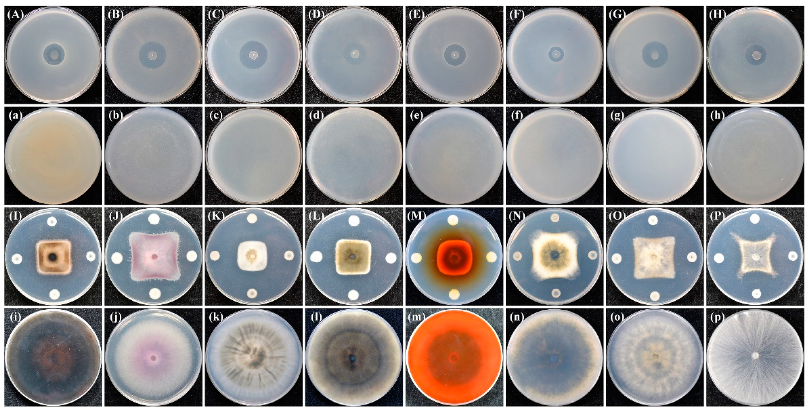
2.1. Bacterial Isolation, Growth Conditions, Antagonism Assays, and Biocontrol Assays

Strain ZF390 was derived from the healthy rhizosphere soil of cucumber plantations in Harbin, China, in August 2019. According to a previous study [24], 10 g of the soil sample was subjected to 10,000-fold dilution with sterile water, plated on LB plates, and incubated at 28 °C for 48 h. Bacillus-like and Paenibacillus-like strains were retained for further investigations. All the strains were purified on LB plates and cultivated in LB broth at 28 °C with shaking for 24 h.
The antagonistic activity of selected microorganisms against Pbr was estimated through plate bioassays. The suspensions of all the isolated strains were diluted to an OD600 = 0.6 with sterile LB broth. Then, 5 μL of the suspensions were inoculated in the center of PDA plates and cultivated for 24 h, respectively. A total of 3 mL of chloroform was added to each upside-down dish for fumigation to inactivate the bacteria colony. Subsequently, 100 μL of the suspension of Pbr was equably added into 5 mL WA medium, and the mixture was poured on top of the PDA plates. If the isolated strain possessed the ability to against Pbr, the plate would change from white to transparent. The suppressive activity test of strain ZF390 against different kinds of phytopathogenic bacteria was similar to the aforesaid method. The transparent aperture appeared on the plate if the strain ZF390 had the capacity to defend against the pathogen. In addition, the inhibitory effect of strain ZF390 against various plant pathogenic fungi was assessed through plate assays. The fungus dishes with diameters of 5 mm were inoculated on the center of PDA plates, and 5 μL of ZF390 suspension was diluted to an OD600 = 0.6 with sterile LB broth that was inoculated at the spots 3.5 cm away from the dishes. Fungal hyphae were inhibited after the plates were cultured at 28 °C for 7–10 d if strain ZF390 could defend against the phytopathogenic fungi.
The control efficacy of antagonistic strains against cucumber bacterial soft rot disease was assessed by greenhouse experiments with potted cucumbers. Each treatment contained five healthy cucumber seedings sprayed with 200 mL suspension of Pbr diluted to an OD600 = 0.8 with sterile NB broth. The greenhouse was kept at 28 °C and 75% relative humidity for 24 h. Whereafter, 3 mL antagonistic strain suspension was subjected to 100-fold dilution with sterile water and spray inoculated on the cucumber seedings. The chemical pesticide group was treated with 300 mL of 3% Zhongshengmycin wettable powder 650-fold dilution, and the 300 mL sterile water served as a control group. The disease grade was divided into five grades: absence of spots represented grade 0; grade 1 was classified as a spot area less than 5% of leaf area; the proportion of spot area from 6% to 25% was grade 2; the proportion of spot area from 26% to 50% was grade 3; the proportion from 51% to 75% was grade 4; and the spot area larger than 76% was grade 5. Disease index = 100 × ∑ (the number of leaves in certain disease grade × relative disease grade)/(total number of leaves × the highest grade of disease). Control effect (%) = 100 × (disease index of control group—disease index of treatment group)/disease index of control group.
In addition, the activities of cellulase, protease, and phosphatase were measured using previously reported methods [25,26]. If cellulase was produced, the CMC agar plate would change from red to yellow after staining with 0.1% (w/v) Congo red solution for 30 min and washing with 1 M NaCl solution for 15 min. While, if protease and phosphatase were produced, haloes in plates became visible without further treatment. The heavy-metal tolerance of strain ZF390 was analyzed using a standard 10-fold dilution plating assay. Different concentrations of ZF390 suspension were inoculated on the LB plates supplemented with 0.4 mM CdCl2·21/2H2O, 1 mM CoCl2·6H2O, 3 mM MnSO4·H2O, 5 mM PbCO3, and 3 mM ZnCl2, respectively. Additionally, the plates were incubated at 28 °C for 48 h. All of the above experiments were repeated three times.

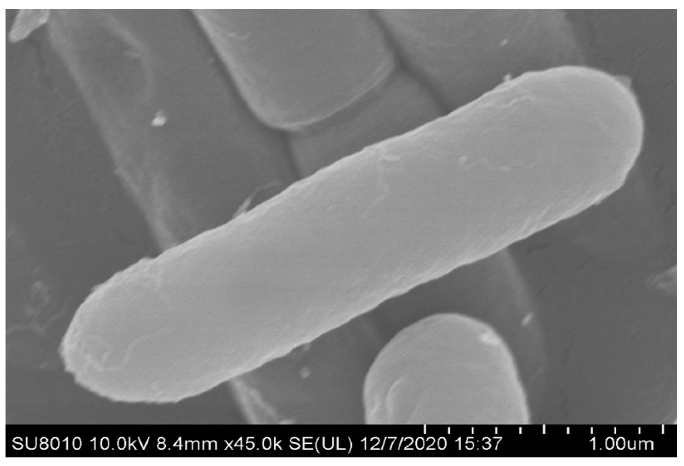
2.2. Characteristics, Microscopic Analysis and Genomic DNA Extraction

The growth characterization and physiological and biochemical characteristics were determined on the basis of reported approaches [27,28]. The strain morphologies were observed by transmission electron microscopy (TEM) and scanning electron microscopy (SEM). Genomic DNA was extracted from the cultured ZF390 cells using the TIANamp Bacteria DNA Kit (TIANGEN BIOTECH, Beijing, China).

2.3. Genome Sequencing and Annotation

According to the manufacturer’s protocol, the whole genome of strain ZF390 was subjected to single-molecule real-time (SMRT) sequencing on the Pacific Biosciences Sequel (Pacific Biosciences, Menlo Park, CA, USA) platform using a library prepared with the SMRTbell Template Prep Kit 1.0 (Pacific Biosciences, Menlo Park, CA, USA). The reads were assembled de novo using SMRT LINK v5.0 (Pacific Biosciences, Menlo Park, CA, USA) after qualitative filtration. The circular genome visualization was generated using CGView [29]. Coding genes of the sequenced genome were predicted by GeneMarkS software. tRNAscan-SE, rRNAmmer, and cmsearch were used to predict tRNA (transfer RNA), rRNA (ribosomal RNA), and snRNAs (small nuclear RNAs), respectively [30,31,32]. Functional gene annotation was carried out through multiple general databases, including nonredundant protein databases (NR) [33], the Gene Ontology database (GO) [34], the Kyoto Encyclopedia of Genes and Genomes (KEGG) [35], the Cluster of Orthologous Groups of proteins (COG) [36], the Transporter Classification Database (TCDB) [37], Pfam3 Swiss-Prot4 [38], and the Carbohydrate-Active enZTmes Database (CAZy) [39]. Furthermore, phiSpy was used to predict prophages, and clustered, regularly interspaced, short palindromic repeat sequences (CRISPR) were identified via CRISPRdigger 1.0 [40,41].

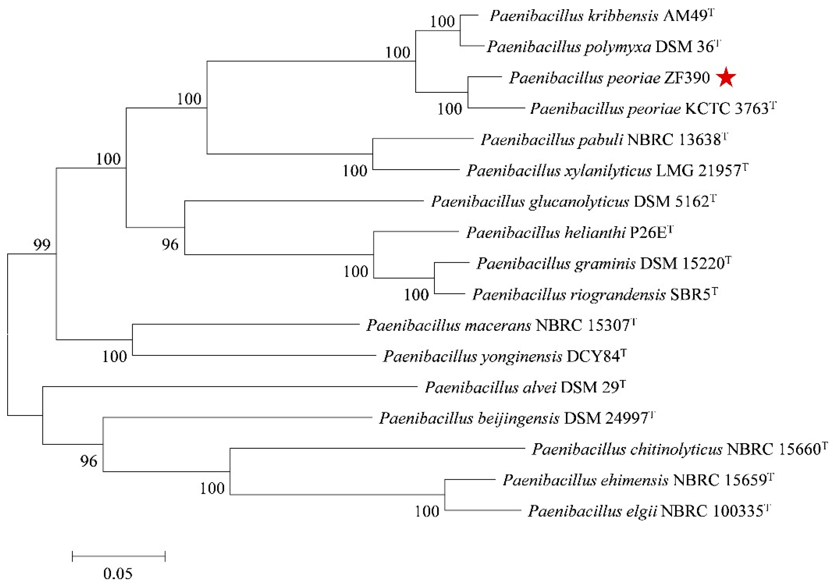
2.4. Phylogenetic Analysis and Genome Comparisons

The taxonomic position of strain ZF390 was determined using multilocus gene sequence analysis (MLSA) based on eight housekeeping genes (gyrB, rpoD, rplA, atpD, holB, rho, dnaA and rpsA). The gene sequences were aligned using MUSCLE and trimmed to remove ambiguously aligned regions (the final sequence lengthes were 9910 bp). Subsequently, the phylogenetic tree was constructed using the maximum likelihood method in MEGA 7.0 [42]. Other available gene sequences of the Paenibacillus strains to be used for phylogenetic tree construction were downloaded from the NCBI database. According to the phylogenetic analysis, four closely related Paenibacillus strains with released complete genome sequences, including P. peoriae HS311 (GenBank Accession No. CP011512.1), P. polymyxa HY96-2 (GenBank Accession No. CP025957.1), P. polymyxa SQR-21 (GenBank Accession No. CP006872.1), and P. kribbensis PS04 (GenBank Accession No. 041731.1), were selected for genome comparison with ZF390. Average nucleotide identities (ANI) and in silico DNA–DNA hybridization (isDDH) analyses were performed using the OrthoANIu algorithm and the Genome-to-Genome Distance Calculator (GGDC), respectively [43]. Moreover, the pairwise alignment of genomes was generated using the Mauve v2.3.1 (SourceForge, San Diego, CA, USA) and Venn diagrams were constructed using the R package [44].

2.5. Analysis of Gene Clusters for Biosynthesis of Secondary Metabolites

The secondary metabolite gene cluster prediction was performed using antiSMASH 5.0 on the authors’ Web servers using the default parameters [45]. Comparative analyses of secondary metabolite gene clusters identified that, among strain ZF390, P. peoriae HS311, P. polymyxa HY96-2, P. polymyxa SQR-21, and P. kribbensis PS04 were performed based on the Kyoto Encyclopedia of Genes and Genomes database (KEGG) [35] and the GenBank database.

2.6. Genome Mining for Genes Encoding Plant Beneficial Traits

The functional genes involved in plant-growth promotion or plant–bacterial interactions, such as the genes responsible for plant-growth promotion, biofilm formation, and induced systemic resistance, were searched in the NCBI and KEGG databases. The identities of different functional genes at the amino acid level were compared between strain ZF390, P. peoriae HS311, P. polymyxa HY96-2, P. polymyxa SQR-21, and P. kribbensis PS04 using BLAST (Basic Local Alignment Search Tool).

2.7. Statistical Analysis

All data were analyzed using the analysis of variance in SPSS v24.0 (IBM, Armonk, NY, USA). The significant differences between means were compared by using the LSD test (Fisher’s protected least significant differences test) at p = 0.05. A p value < 0.05 was considered significant.

3. Results

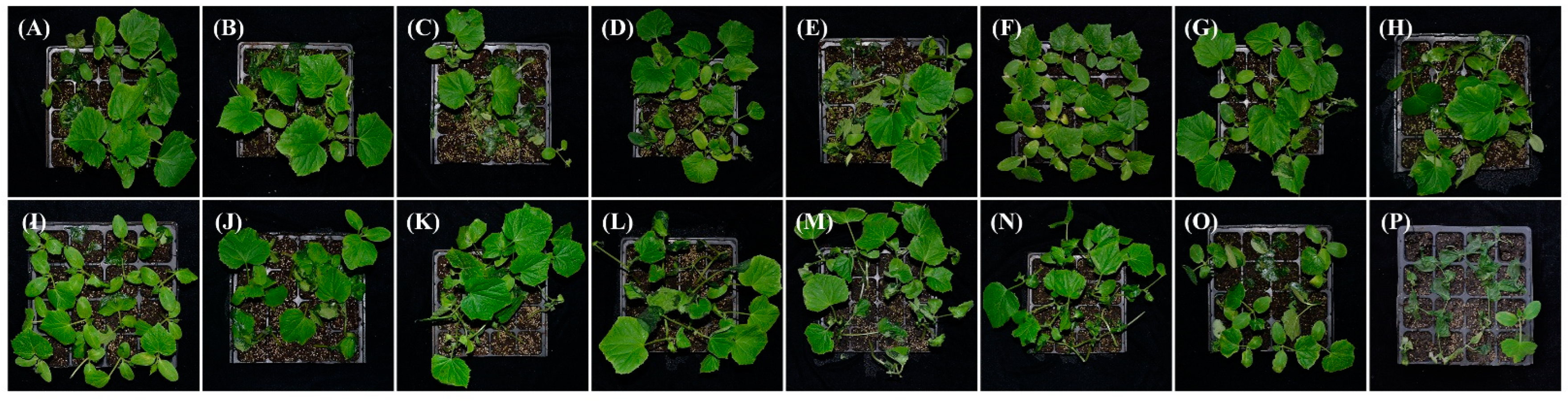
3.1. Biocontrol Effect of Strain ZF390 against Pbr

To identify good biocontrol agents against cucumber soft rot disease caused by Pbr, 161 bacterial strains were segregated from the rhizosphere of cucumber plants. There were 14 strains that showed antagonistic activity against Pbr through plate bioassays (Supplementary Figure S1). The largest transparent halo was generated around the colony of strain ZF428, while only a small ring was produced around the colony of strain ZF390 (Supplementary Figure S1). The control effects of these 14 strains against Pbr were performed by greenhouse experiments on cucumber seedlings. Among these bacteria, the strain ZF390 indicated superb antagonistic activity in greenhouse tests, exhibiting the highest control efficiency of 77.47%, which was much higher than the efficacy of zhongshengmycin (47.89%) [46], which is a ubiquitous aminoglycoside antibiotic against bacterial phytopathogens (Table 1 and Figure 1).
The strain ZF390 also demonstrated prominent antagonistic activities against various plant bacterial and fungal pathogens, including Pbr, X. campestris pv. Campestris, Pseudomonas syringae pv. Tomato, Clavibacter michiganensis subsp. Michiganensis, P. syringae pv. Lachrymans, C. michiganensis subsp. Sepedonicum, Agrobacterium vitis, Acidovorax citrulli, Corynespora cassiicola, F. oxysporum, Colletotrichum capsici, P. capsici, Stemphylium solani, Ascochyta citrulline, Botrytis cinerea, and Rhizoctonia solani in the plate bioassays (Figure 2). In addition, yellow and transparent halos were observed in extracellular enzyme assays, indicating that strain ZF390 produced cellulase, protease, and phosphatase (Supplementary Figure S2A–C), which might be one of its biocontrol mechanisms. Moreover, the strain ZF390 revealed varying degrees of tolerance to Cd2+, Co2+, Mn2+, Pb2+, and Zn2+ (Supplementary Figure S2D). Strain ZF390 exhibited a superior colonization ability in the rhizospheres of cucumbers cultivated in fermentable substrate after 15 days of inoculation (1.36 × 10⁸ CFU/g) (Supplementary Figure S3). In summary, due to the remarkable biocontrol traits of strain ZF390, it was selected for further research.

3.2. Organism Information

Strain ZF390 was demonstrated to be a motile, Gram-positive, and facultative anaerobic bacterium belonging to the Paenibacillaceae family. Strain ZF390 grew readily on LB plates at 28 °C and produced light yellow sticky colonies with irregular margins after 36 h of incubation (Supplementary Figure S4A), and the strain produced rod-shaped cells with a length of 2–4 μm and a diameter of 0.5–1 μm (Figure 3 and Supplementary Figure S4B). As indicated in the physiological and biochemical assays, casein was hydrolyzed by strain ZF390, but arginine dihydrolase and indole were not produced, and the nitrate reduction reaction was negative. Interestingly, the capability of strain ZF390 to liquefy gelatin was negative, which was the complete opposite of that of P. peoriae KCTC 3763T [28]. Acid was produced by ZF390 from the following compounds: glycerol, D-arabinose, L-arabinose, ribose, D-xylose, adonitol, methylxyloside, D-fructose, mannitol, methyl-D-mannoside, salicin, maltose, D-trehalose, gentiobiose, 2-keto-D-gluconate, 5-keto-D-gluconate, rhamnose, D-turanose, and N-acetylglucosamine, and its capacity of utilizing D-turanose was different from that of KCTC 3763T (Supplementary Table S1).

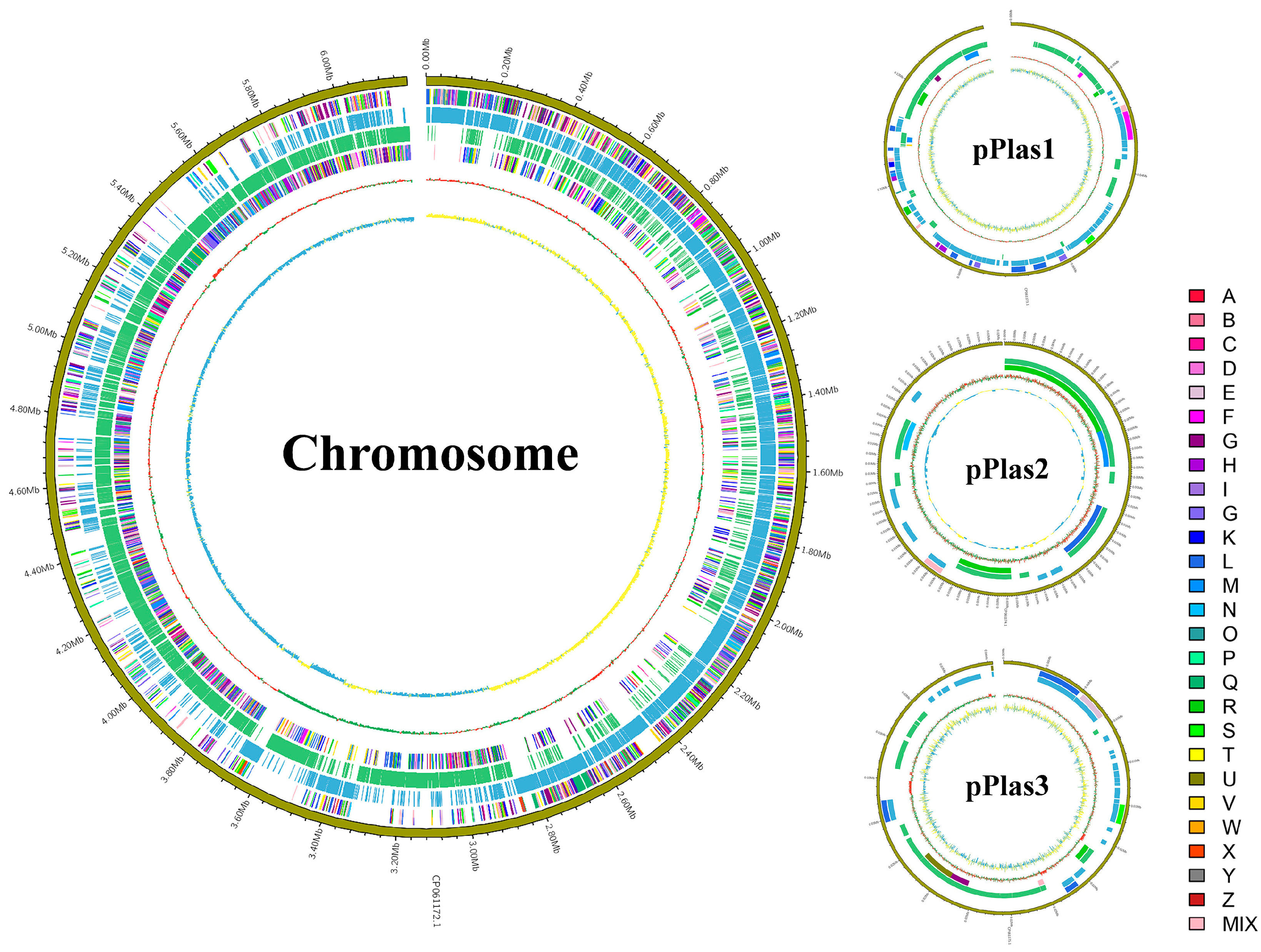
3.3. General Genomic Features of P. peoriae ZF390

The complete genome of P. peoriae ZF390 was composed of a circular 6,193,667 bp chromosome with three additional plasmids, pPlas1 (137,220 bp), pPlas2 (16,869 bp), and pPlas3 (36,233 bp), and the average GC contents were 44.96%, 45.58%, 45.12%, and 41.66%, respectively (Supplementary Table S2). In total, 5890 open reading frames (ORFs) were forecasted in the genome of ZF390. In addition to 5574 protein-coding genes (CDSs), the genome constituted 145 RNA genes, containing 40 rRNA operons, 101 tRNA genes, 4 other RNAs, and 171 pseudogenes (Supplementary Table S3). In addition, the ZF390 genome included 17 genomic islands, 5 prophage regions, and 11 CRISPR-associated genes (Supplementary Table S2). A total of 4001 functional genes of the ZF390 genome were categorized using the Clusters of Orthologous Groups of proteins (COG) database. Most of the genes were related to carbohydrate transport and metabolism (505 genes), followed by transcription (482 genes), general function prediction only (451 genes), amino acid transport and metabolism (355 genes), and signal transduction mechanisms (304 genes). However, 439 genes were not predicted by the COG database, and their specific functions need to be further verified (Supplementary Table S4). The graphical circular genomic maps exhibiting the genome structure and functions of P. peoriae ZF390 are presented in Figure 4. Moreover, 5402 genes were annotated to 204 different KEGG pathway maps, the majority of which were associated with metabolic pathways, biosynthesis of secondary metabolites, ABC transporters, the biosynthesis of antibiotics, microbial metabolism in diverse environments, two-component systems, the biosynthesis of amino acids, and quorum sensing. In addition, 3754, 5645, 481, 3754, 2296, and 342 ORFs were compared to diverse pathways or domains using the GO, NR, TCDB, Pfam, Swiss-Prot, and CAZy databases (Supplementary Table S2).

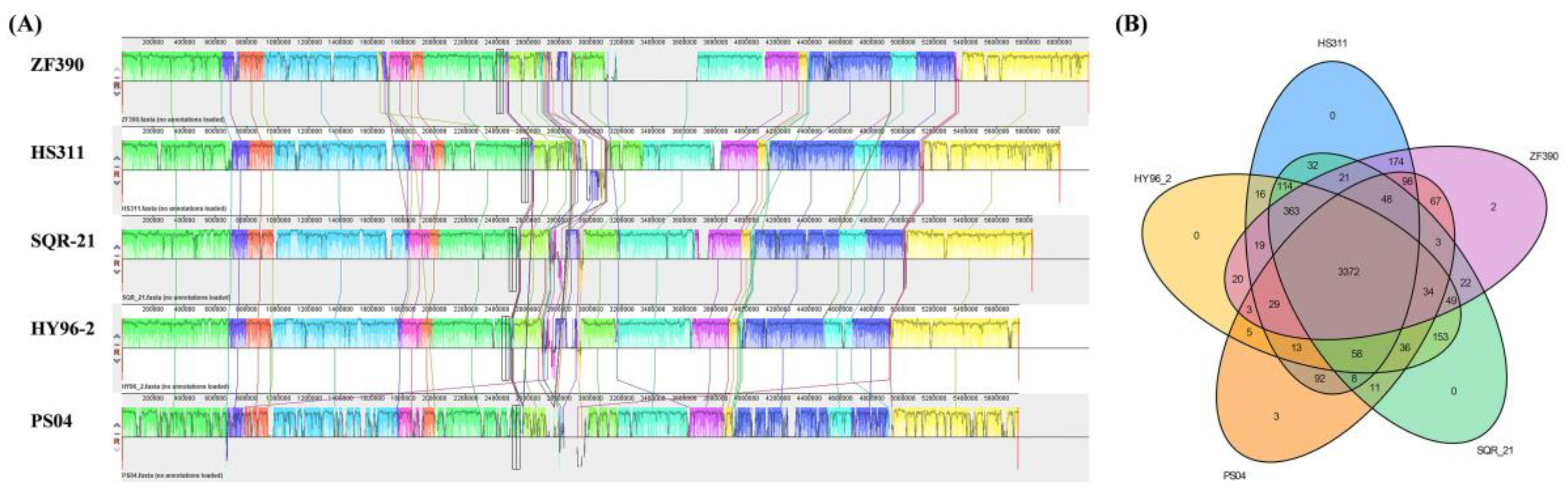
3.4. Comparative Genomics Analysis of P. peoriae ZF390 with Other Paenibacillus Strains

To comprehend the genetic relationship of P. peoriae ZF390 with other Paenibacillus species, sixteen publicly available complete genome sequences of Paenibacillus-type strains were selected to establish a phylogenetic tree based on eight housekeeping genes (gyrB, rpoD, rplA, atpD, holB, rho, dnaA, and rpsA) (Supplementary Table S5). As expected, all of the strains were prominently corroborated by bootstrap values. According to the genetic distance, strain ZF390 was closest to P. peoriae KCTC 3763T, followed by P. polymyxa DSM 36T and P. kribbensis AM49T (Figure 5). The phylogenetic analysis determined an accurate systematic position of ZF390 as P. peoriae.
ANI and isDDH are widely applied analyses that appraise the similarities between bacterial strains by comparing whole genomic sequence data. Pairwise compared strains with ANI values ≥ 96% and isDDH values ≥ 70% are typically considered the same species [47,48]. In this study, the ANI and isDDH values between strains ZF390 and HS311 were 96.61% and 71.80%, respectively (Supplementary Figure S5). Simultaneously, the ANI values were over 96%, and the isDDH values were over 70%, when comparing strain ZF390 with P. polymyxa strains ATCC 15970, CR1, E681, J and YC0573. Nevertheless, lower ANI and isDDH values were obtained when other P. polymyxa strains were used as reference genomes. These findings may suggest inaccurate identities of partial P. polymyxa strains. In addition, the ANI values that contrasted strain ZF390 with P. kribbensis strains AM49T and PS04 were 87.46% and 87.41%, respectively, and the isDDH values were 33.70% and 33.80%, respectively. Altogether, these results revealed that P. polymyxa and P. kribbensis were in more appressed relationships with P. peoriae than other Paenibacillus species. Strain ZF390 was closely related to P. peoriae HS311 and could be assigned to the same taxonomic position (Supplementary Figure S5).
Genomic information was compared between P. peoriae ZF390 and four other accessible complete genomes of Paenibacillus strains (P. peoriae HS311, P. polymyxa SQR-21, P. polymyxa HY96-2 and P. kribbensis PS04) (Supplementary Table S3). The genome size of strain ZF390 (6.38 Mb) was conspicuously large compared to the other four strains, HS311 (6.22 Mb), SQR-21 (5.83 Mb), HY96-2 (5.75 Mb), and PS04 (5.74 Mb). These results also illustrate a maximum number of ORFs and CDSs in the ZF390 genome. Nevertheless, the order of average GC content of these five selected strains was antipodal with the order of genome size results; strain ZF390 had the lowest GC content (44.99%), under 45%. Furthermore, P. peoriae ZF390 and HS311 comprised three plasmids and one plasmid, respectively, and the other three Paenibacillus strains merely contained a single chromosome without a plasmid (Supplementary Table S3).
To estimate the evolutionary distance among these five Paenibacillus strains, their whole genome sequences were compared using Mauve (Figure 6A). The alignments between ZF390 and HS311 showed that several gene inversions and a large region of deletion were detectable in P. peoriae ZF390. Compared to SQR-21 and HY96-2, a number of gene insertions or deletions and local collinear block (LCB) inversions were also detected in strain ZF390. These results show that large LCB inversions occurred among strain PS04 and the other four strains, and the ZF390 genome is much more similar to that of HS311, supporting the relationship described above (Figure 6A).
Whole genome sequences of these five Paenibacillus strains were also compared to identify specific genes in P. peoriae ZF390 (Figure 6B). As shown in Figure 6B, there were 3372 core genes present in ZF390 that were shared with strains HS311, SQR-21, HY96-2, and PS04. It was speculated that these orthologous protein-coding genes were relatively conserved in Paenibacillus. Additionally, 4122 orthologous genes were shared between ZF390 and HS311, while ZF390 shared 3912 and 3889 orthologous genes with SQR-21 and HY96-2, respectively. Moreover, strain ZF390 shared merely 3652 orthologous genes with PS04, whereas it indicated a higher level of similarity with strain HS311. Furthermore, only two and three unique genes were identified in the genomes of strains ZF390 and PS04, which revealed close relationships among the five selected strains.

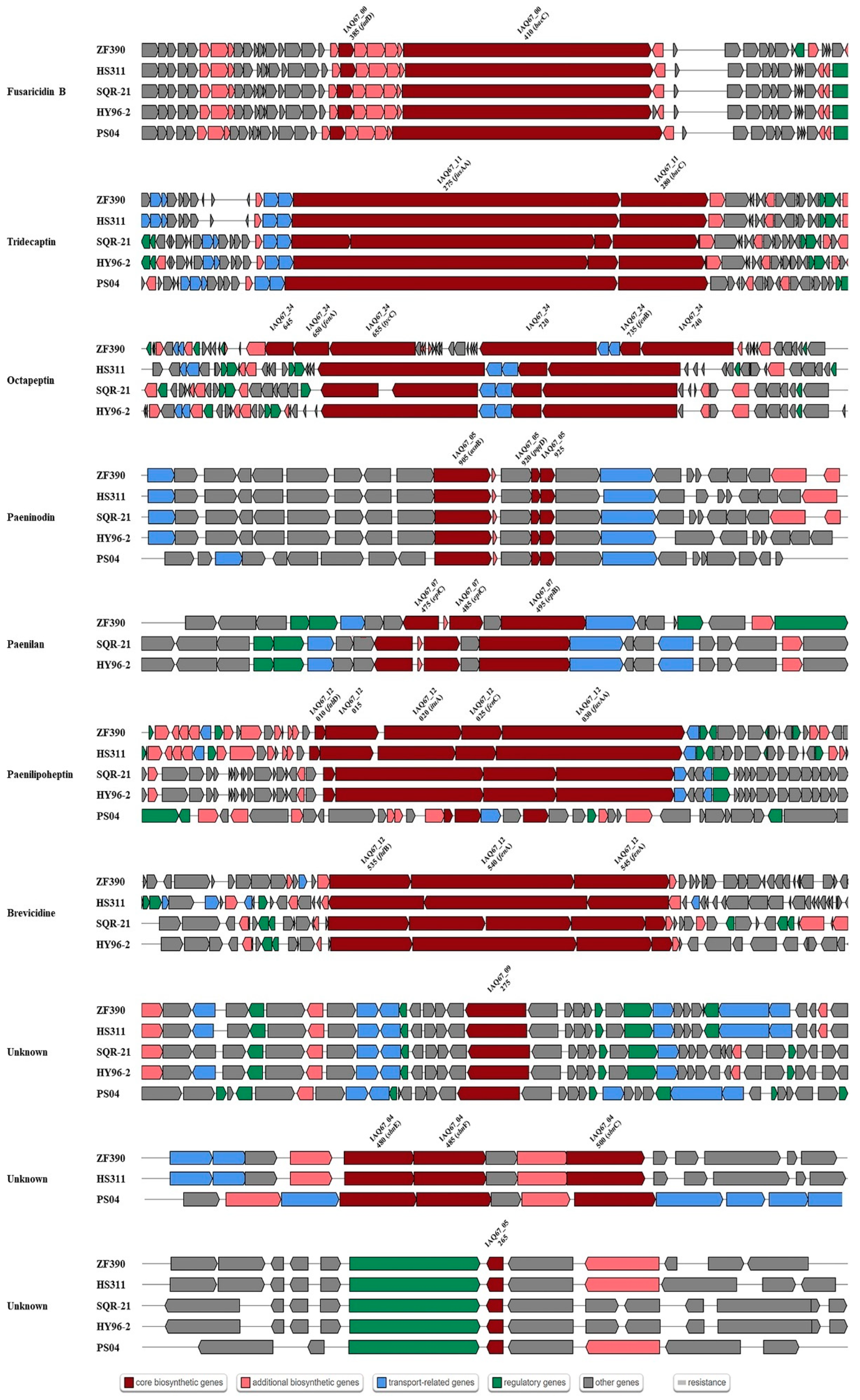
3.5. Gene Clusters Involved in the Synthesis of Secondary Metabolites

With the aim of identifying the antibiotics that may be produced by ZF390, with its broad antagonistic spectrum, the gene clusters for antibiotic biosynthesis were detected in the ZF390 genomic sequence using the antiSMASH database. Ten gene clusters with different biosynthesis pathways involved in secondary metabolite production were retrieved, including three clusters encoding NRPSs (nonribosomal peptide synthetases), one T1PKS (type Ι polyketide synthases) or NRPS, one TransAT-PKS (trans-acyl transferase polyketide synthetases) or NRPS, one lassopeptide, one siderophere, one lanthipeptide-class-i, one RiPP-like (ribosomally synthesized and post-translationally modified peptides) protein, and an NRPS-like protein (Table 2). Gram-negative bacteria can be suppressed by brevicidine, tridecaptin, paeninodin, and octapeptin while paenilan, fusaricidin B, paeninodin, and octapeptin control Gram-positive bacteria [49,50,51,52,53,54,55]. Meanwhile, fusaricidin B, octapeptin, and paeninodin show an outstanding inhibitory effect against fungal pathogens [49,54,55]. The bioactive spectrum of paenilipoheptin and three unknown antibiotics found in the genome is still unclear (Table 2). The comparison analysis of the above five strains revealed a deficiency of the paenilan synthesis gene cluster in strain HS311. An unknown antibiotic synthesized via the siderophore pathway was not retrieved in SQR-21 and HY96-2. The genome of PS04 did not contain the relevant clusters of brevicidine, octapeptin, and paenilan. Interestingly, however, the gene cluster of paenilipoheptin identified in strain PS04 showed poor similarity with the clusters retrieved in strains ZF390, HS311, SQR-21, and HY96-2 (Figure 7). Furthermore, the gene cluster for the biosynthesis of octapeptin detected in the ZF390 genome had a larger core gene cluster (Figure 7).

3.6. Mining for Functional Genes Potentially Associated with Plant–Bacteria Interactions

The key genes related to plant-growth promotion, biofilm formation, and induced systemic resistance were retrieved from P. peoriae ZF390 based on the KEGG database, and the identities of these genes among HS311, SQR-21, HY96-2, and PS04 were compared according to the NCBI database. The identities of all homologous sequences between strain ZF390 and the other four strains were higher than 77%. The sequence identities of strain ZF390 exhibited very high similarities with those in strain HS311; concurrently, lower similarities were observed when comparing ZF390 and PS04 (Supplementary Tables S6–S8).
Five key genes related to phosphate solubilization (phnE, phnC, pstB, pstA, and pstC) and 18 other genes responsible for organic acid biosynthesis were all retrieved from the five Paenibacillus strains with high similarity (87.07–99.21%) (Supplementary Table S6). The results demonstrated a latent capacity of ZF390 to promote plant growth. As data shown in Supplementary Table S7 indicate, genes involved in biofilm formation, such as flagellar assembly, non-ribosomal peptide structures, bacterial chemotaxis, and quorum sensing were mined in the genome sequences of five Paenibacillus strains. Flagellar structure coding genes, including fli gene cluster (fliACEFGHIJKLMNOPQRY), flg gene cluster (flgBCDEFGKLM), gene flhA and gene flhB, and flagellar motivation coding genes motA and motB, were located in the genome of strain ZF390. However, gene fliD and gene fliS were not found in ZF390 genomic sequence. Genes (fenA, fenB, and fenC) related to Fengycin production were also retrieved. The che gene cluster (cheABCDRWXY) and multiple copies of gene mcp were found in five genomes, and the sequence identity exceeded 84.03%. The key gene in AI-2 QS system, luxS, and the four genes (spo0A, spo0B, spo0F, and degU) involved in biofilm formation pathway were found in genome of strain ZF390, and the sequence identity between ZF390 and HS311 (97.64%) exhibited higher values than those between strain ZF390 with SQR-21 (92.42%), HY96-2 (92.42%), or PS04 (91.06%). Key genes related to the plant-resistance inducers in ZF390 were retrieved in light of previous studies, and the identities of these genes among HS311, SQR-21, HY96-2, and PS04 were compared [7,56]. The results demonstrate that the key genes associated with volatile organic compounds belong to systemic resistance inducers, such as 2,3-butanediol, methanethiol, and isoprene synthesis, were all detected in the genomes of the five strains, with sequence identities exceeding 85% (Supplementary Table S8).

4. Discussion

Pbr has caused a devastating soft rot disease of cucumbers in China, and chemical agents did not effectively control it; so, biocontrol has been given higher expectations and attention [4]. Chemical methods are commonly used to control soft rot but are not desirable, due to increased resistance and environmental contamination [57,58]. The biocontrol of soft rot through biocontrol strains is one of the safe, effective, and sustainable methods and research hotspots used to control soft rot [59,60,61]. Abundant biocontrol agents were screened through in vitro inhibition tests, for instance, estimating the inhibition of a target pathogen on agar tablets; however, the bottom line for biocontrol is whether it works under production conditions, and numerous researchers have reported no correlation between in vitro inhibition tests and field performance [62].
In this study, P. peoriae ZF390 showed a conspicuous alleviation effect on cucumber plants infected with soft rot disease in a greenhouse, representing the good potential of biopesticides or microbial inoculants, even though its effect on the plate assays was not particularly noteworthy (Figure 1 and Supplementary Figure S1). In addition, the antimicrobial properties of strain ZF390 were investigated, demonstrating antagonistic activity against a variety of bacteria and fungi (Figure 2), and the results found were similar to those of previous studies [63,64]. Extracellular enzymes, such as protease (Prt) and cellulase (Cel), produced by strain ZF390 might result in antagonistic action against a wide range of microorganisms (Supplementary Figure S2A,B). Due to its superior antimicrobial activity, the genome of P. peoriae ZF390 was completely sequenced to provide insight into its biocontrol mechanisms. Metal-tolerant bacteria could survive in the tissue of plants grown on the metal-polluted soil and perform good adaptability to the environment [65]. Strain ZF390 might be used in a wide range of conditions due to its tolerance to Cd2+, Co2+, Mn2+, Pb2+, and Zn2+ (Supplementary Figure S2D).
P. peoriae was originally recognized as Bacillus peoriae and reclassified as a member of the genus Paenibacillus after performing an amplified ribosomal DNA restriction analysis, a fatty acid methyl ester analysis, sodium dodecyl sulfate–polyacrylamide gel electrophoresis of whole-cell proteins, API, and other routine phenotypic tests [28,66]. Due to its extreme uniformity, 16S rDNA is rarely recommended for examining the relationships between genera or even deeper taxonomic levels, while multilocus sequence typing (MLST) with a sample of between four- and eight-core gene loci is sufficient for the subdivision of most bacterial species [67]. However, there is a general consensus among taxonomists that all taxonomic information about a bacterium is incorporated into the complete nucleotide sequence of its genome [68]. The average nucleotide identity (ANI) and DNA–DNA hybridization (DDH) are considered significant criteria for species identification. The commonly accepted ANI criteria to set bounds on species is generally 95% [69,70], and the critical value of DDH is typically 70% [71]. Nevertheless, an ANI threshold between 93% and 96% can be considered a fuzzy zone where the intra-genus and interspecies boundary of may fall; the definition of the optimal ANI range for prokaryotic species is still controversial [72]. Previous studies corroborated that the ≥95% ANI cutoff alone is unable to resolve closely related species, and the stringent ≥96% ANI cutoff is more accurate with a better correlation with the 70% DDH cutoff [47,73]. It is highlighted that species assignment based on a single algorithm may not be robust enough.
In silico DNA–DNA hybridization (isDDH), determined using the Genome-to-Genome Distance Calculator (Formula 2), is particularly attractive, as it has been shown to be better correlated to the traditional DDH, which is a gold standard for species assignment, than to ANI [43]. Therefore, it is proposed that any genome-based species assignments should be determined by both ANI and isDDH, and the pairwise ANI with a ≥96% cutoff and isDDH with a ≥70% cutoff have been widely used for precise species identification [47,48]. Based on phenotypic, phylogenetic, and genotypic data, P. peoriae ZF390 was subjected to a taxonomic study by using a polyphasic approach. The phylogenetic tree based on eight housekeeping genes revealed an intimate but independent relationship among P. peoriae, P. polymyxa, and P. kribbensis (Figure 5). The ANI and isDDH values for strains ZF390 and HS311 were 96.61% and 71.80%, respectively, which were considerably higher than the values for ZF390 with the selected P. polymyxa and P. kribbensis strains (Supplementary Figure S5).
In contrast to the other four strains, the genome of ZF390 was larger, comprising more plasmids, but with a slightly smaller GC content, and abundant CDSs were predicted (Supplementary Table S3). The analysis of the whole ZF390 genome revealed an impressive capability to produce a diverse spectrum of different secondary metabolites aimed at suppressing harmful phytopathogens. In total, 10 gene clusters (Table 2) representing more than 8.7% of the genome were devoted to synthesizing antimicrobial metabolites, approximately the same proportion as the distinguished biocontrol agent B. amyloliquefaciens FZB42 [74].
On the basis of antiSMASH database, comparisons of the genes and gene clusters related to antibiotic synthesis demonstrated differences among the five Paenibacillus strains (Table 2 and Figure 7). Regarding the control of bacteria, five antibacterial metabolites (brevicidine, tridecaptin, paenilan, fusaricidin B, paeninodin, and octapeptin) were predicted to be produced by P. peoriae ZF390. Brevicidine, a bacterial non-ribosomally-produced cyclic lipododecapeptide, which has low hemolytic activity and cytotoxicity, displays potent antimicrobial activity against Gram-negative pathogens [75]. Brevicidine B was discovered from the producer strain of brevicidine, Brevibacillus laterosporus DSM 25. Recently, researchers found that brevicidine B, compared with an earlier reported member of the brevicidine family, has a broadened antimicrobial spectrum caused by its increased membrane-disruptive capacity against Gram-positive pathogens [52]. The strong antibacterial activity of strain ZF390 might be associated with the production of brevicidine; however, its capability of producing brevicidine B should be further investigated. Tridecaptin is another type of non-ribosomal antibacterial peptide (NRAP) with effective activity against highly problematic strains of Gram-negative bacteria and low levels of resistance development with the potential for safe administration [76]. All five Paenibacillus strains were found to contain tridecaptin-related gene clusters, but more core biosynthetic genes were predicted in SQR-21 and HY96-2 than in the other three strains (Figure 7). With purification and structure analysis, paenilan was identified as a novel class-Ⅰ antibiotic, a ribosomally synthesized peptide that exhibits antimicrobial activity against Gram-positive bacteria and is well conserved in different Paenibacillus sp. isolated from globally distant places [53]. The core genes epiB and epiC and biosynthesis cluster were observed in the genomes of ZF390, SQR-21, and HY96-2, while no genes or gene clusters involved in the synthesis of paenilan were found on the chromosomes of HS311 and PS04 (Table 2). Fusaricidin B, paeninodin, and octapeptin not only possess the antibacterial activity, but also impede the growth of phytopathogenic fungi. According to former reports on its particularly antagonistic activity against Gram-positive bacteria and plant pathogenic fungi, fusaricidin, generated by a non-ribosomal peptide synthetase (NRPS), seems to have immense potential for industrial use [77,78]. Fusaricidin B that was possibly produced by strain ZF390 reported excellent germicidal activity against Candida albicans and Saccharomyces cerevisiae [54]. Octapeptins, which are structurally similar to polymyxins, are lipopeptide antibiotics possessing much broader antimicrobial activity against Gram-negative and Gram-positive bacteria, yeast, fungi, and even protozoa, whereas polymyxins are only active against Gram-negative bacteria [55]. The core gene region belonging to the synthesis gene cluster of octapeptin predicted in strain ZF390 was much larger than that in HS311, SQR-21, and HY96-2; the octapeptin cluster was not found in strain PS04 (Figure 7). Paeninodin, a new lasso peptide, belongs to the ribosomally synthesized and post-translationally modified peptides (RiPPs) that exhibit a wide range of antimicrobial or antiviral activities, and the synthesis cluster was found in all five strains [79]. Paenilipoheptin was recently identified via the genome mining of P. polymyxa E681, and its antimicrobial activity spectrum remains to be elucidated [23,80].
Bacteria belonging to the genus Paenibacillus have been isolated from a variety of environments, and the majority of them are found in soil, demonstrating benefits for plants with mutual interactions. They are considered predominant plant-growth-promoting rhizobacteria (PGPR) and are widely used for agricultural applications [81,82]. Phosphorus is a significant macronutrient for plant nutrition, but its unique characteristics of high fixation and poor solubilization in soil lead to the overuse of chemical phosphorus fertilizers applied to agricultural land, causing severe environmental damage [83,84]. By inoculating fields with phosphorus-solubilizing microorganisms, such as Paenibacillus, the application of chemical phosphorus fertilizer could be reduced, and the principal mechanism of microorganisms to make phosphorus available to plants could be performed by producing organic acids [85]. A differential gene expression study revealed an extremely intricate transcriptional response of P. sonchi SBR5 to soluble and insoluble phosphorus sources, suggesting that the production of organic acids might be the most vital strategy utilized by SBR5 to perform phosphorus solubilization [86]. An evident and clear dissolution halo can be observed around the P. polymyxa inoculum grown on a solid NBRIP medium, in which Ca3(PO4)2 is the sole phosphate source [87]. According to the comparative genomic analysis, 23 genes related to phosphorus solubilization were retrieved in the ZF390 genome, some of which encode organic acid production (Supplementary Table S5). However, the main genes (gcd and gad) responsible for the production of gluconic acid and its conversion were not detected in the genome of ZF390. Similarly, ZF390 does not carry the complete phosphonate gene cluster (phn), except for phnC and phnE, which were deemed a gene gain and loss event during evolution [88]. In addition, the ZF390 genome possesses the pst operon (pstC, pstA, and pstB) that integrates the phosphate-specific transport system. Taken together, P. peoriae ZF390 is a potential PGPR that may become an additive in biofertilizer.
Endospore-forming bacteria such as Paenibacillus have the capacity to survive for long periods in the soil under adverse environmental conditions, which is appropriate for field inoculation [85]. PGPR plays a crucial role in plant growth promotion by effectively colonizing plant roots. Bacteria in natural environments persist by forming biofilms, which are highly structured, surface-attached communities of cells encased in a self-produced extracellular matrix [89]. A series of genes related to flagellar assembly, non-ribosomal peptide structures, bacterial chemotaxis, and quorum sensing play increasingly important roles in biofilm formation. Paenibacillus performs two forms of flagellar-driven motility: swarming on a surface and swimming in liquid culture; swarming motility is crucial during root colonization or for rapid colonization of a specific tissue [20,90]. A threshold amount of power is needed for an organism to swarm, and hyper-flagellation, a dramatic augmentation in flagellum density, may contribute to a cumulative increase in power [90]. According to the KEGG database and comparative genomics results, a complete flagellar assembly gene cluster was revealed in the ZF390 genome, including, but not limited to, the fliC gene encoding the filament, the flgE, flgK, and flgL genes encoding the hook, and multitudinous genes encoding the basal structure [91,92]. Recent research reported that P. polymyxa generates energy through the mechanism of degrading phospholipids to create sn-glycerol 3-phosphate and feed it into the glycolysis/pentose phosphate pathway, which appears likely to be necessary for swarming motility, including flagellar rotation [90]. However, the applicability of this mechanism in ZF390 needs to be further explored. In addition to multicellular rafts of highly flagellated cells, the lipopeptide surfactants or non-ribosomal peptides secreted by bacteria act as wetting agents, providing a mechanism to reduce surface tension between the substrate and the bacterial cells, helping them spread rapidly over surfaces [90,93,94,95]. Except for the non-ribosomal peptide biosynthesis gene clusters identified in the antiSMASH database, several genes belonging to fengycin synthesis were also detected in the genome of ZF390. The presence of these substances distinctly provides the essential requirement for biofilm swarms on plant roots.
Bacterial chemotaxis, a biased movement in chemical gradients, is a motility-based response that generally enhances the efficiency of environmental colonization by motile bacteria [96]. Chemoreceptors called methyl-accepting chemotaxis proteins (MCPs) detect various environmental signals to stimulate the chemotactic response [97]. The basic molecular mechanism of chemotaxis is generally conserved in bacteria, and it was systematically studied in Escherichia coli, revealing only a single chemotaxis operon in its genome [96,98,99]. Nevertheless, multiple parallel chemotaxis systems and a large number of chemoreceptors have been found in motile bacterial species, which is likely to provide a competitive advantage for surviving in the soil, as well as competition for root-surface colonization [100]. We found multiple copies of genes, especially the mcp gene, involved in chemotaxis in the genome of ZF390. Thus, we propose that chemotaxis may play a crucial role in the colonization of ZF390.
Moreover, bacterial biofilm formation is often regulated by quorum sensing (QS), which is a cell-communication mechanism that depends on population density [101]. To date, two typical QS systems using an autoinducing peptide (AIP) or an autoinducer-2 (AI-2) as a signal molecule have been reported in Gram-positive bacteria [102]. The luxS gene, a key regulatory gene of the AI-2-mediated QS system, has been confirmed to impact the biomass and morphology of biofilms formed by Paenibacillus species. In P. polymyxa HY96-2, luxS improved biocontrol efficacy by promoting its biofilm formation ability [103]. On the basis of recent studies, we speculate that luxS can positively regulate biofilm formation, but this assumption needs more experimental arguments [103,104]. The biofilm formation pathway genes spo0A, spo0B, apo0F, and degU were found in the ZF390 genome when searching for QS genes, suggesting that two pathways may be involved in ZF390 [56]. From the above results, it is possible to deduce that ZF390 could colonize well in the rhizosphere by forming biofilms, but the colonization abilities of the five strains might differ because the variety, amount, and expression levels of these genes related to biofilm formation are different.
Many beneficial rhizobacteria including members of Paenibacillus can trigger ISR when present in sufficiently high population densities [85]. Systemic resistance inducers, such as volatile organic compounds (VOCs), including methanethiol, isoprene, 2,3-butanediol and butyl acetate, are generated and released into the surrounding environment by biocontrol agents, offering protection against phytopathogens and insect herbivores [85]. P. polymyxa E681 was interpreted to use VOC elicitors to protect Arabidopsis thaliana plants against the bacterium P. syringae pv. maculicola via the primed transcription of salicylic acid, jasmonic acid, and ethylene-signaling genes [105]. In previous studies, key genes associated with VOC production were investigated in P. polymyxa strains HY96-2, ZF129, and ZF197 [56,88]. The genes responsible for inducers (2,3-butanediol, methanethiol and isoprene) in strains ZF390 and HS311 exhibited 93% identity, which is higher than the identity observed among strains SQR-21, HY96-2, and PS04. These results deduced a similar capability of systemic resistance in these five strains with varying effectiveness owing to the variations in the related genes.

5. Conclusions

P. peoriae ZF390, which was obtained from cucumber rhizosphere soil, exhibited an extraordinary effect on controlling cucumber soft rot caused by Pbr and exhibited broad-spectrum antagonistic activities against 16 distinct bacterial and fungal phytopathogens. Based on the polyphasic analyses combined with phenotypic, phylogenetic, and genotypic data, strain ZF390 was identified as P. peoriae, which is closely related to P. polymyxa and P. kribbensis. According to the antiSMASH database, strain ZF390 possesses miscellaneous secondary metabolite biosynthesis clusters with biocontrol functions, including brevicidine, fusaricidin B, octapeptin, paenilan, paenilipoheptin, paeninodin, tridecaptin, and three other unknown antibiotics, demonstrating preeminent antimicrobial activity. Based on the comparative genomic analysis and phosphate solubilization assay, strain ZF390 may perform as a potential PGPR. In addition, many genes associated with colonization, such as flagellar assembly, bacterial chemotaxis, quorum sensing, and non-ribosomal peptide structures, were detected in the ZF390 genome and shared high homology with P. peoriae strain HS311. Overall, these properties indicate that ZF390 is a promising biocontrol agent for further application in agricultural fields.

Supplementary Materials

The following supporting materials can be downloaded at: https://0-www-mdpi-com.brum.beds.ac.uk/article/10.3390/biology11081172/s1, Figure S1: Inhibition zone of antagonistic strains against Pbr in plate assays. (A): ZF115; (B): ZF119; (C): ZF185; (D): ZF194; (E): ZF278; (F): ZF390; (G): ZF402; (H): ZF405; (I): ZF428; (J): ZF429; (K): ZF436; (L): ZF448; (M): ZF450; (N): ZF453. Figure S2: Plate assays for production of cellulase (A), protease (B) and phosphatase (C) by Paenibacillus peoriae ZF390 and heavy metal resistance of strain ZF390 (D). Figure S3: Colonization of the strain ZF390 in different types of soils. Figure S4: General characteristics of Paenibacillus peoriae ZF390. (A): Image of ZF390 colony morphology; (B): Image of ZF390 cells using transmission electron microscopy. Figure S5: Percentage of average nucleotide indentities (ANI) and in silico DNA-DNA hybridization (isDDH) among the selected Paenibacillus genomes. Table S1: Physiological and biochemical characteristics of strain ZF390. Table S2: Genome statistics. Table S3: Genomic features of P. peoriae ZF390 and other Paenibacillus strains. Table S4: Number of genes associated with general COG functional categories. Table S5: Strains and housekeeping gene locus tag information used for the phylogenetic tree construction in this study. Table S6: Comparison of genes involved in plant growth promotion between strains ZF390, HS311, SQR-21, HY96-2 and PS04. Table S7: Comparison of genes mostly involved in biofilm formation between strains ZF390, HS311, SQR-21, HY96-2, and PS04. Table S8: Comparison of genes involved in synthesis of resistance inducers between strains ZF390, HS311, SQR-21, HY96-2, and PS04.

Author Contributions

Y.Z. and L.L. designed the research. Y.Z., X.X., Y.S., A.C., T.F., B.L. and L.L. performed the experiments. Y.Z. analyzed the data. Y.Z and J.L. wrote the manuscript. All authors have read and agreed to the published version of the manuscript.

Funding

This research was funded by the China Agriculture Research System of MOF and MARA; the Science and Technology Innovation Program, Chinese Academy of Agricultural Sciences (CAAS-ASTIP-IVFCAAS); and the Key Laboratory of Horticultural Crops Genetic Improvement, Ministry of Agriculture in China (IVF2021).

Institutional Review Board Statement

Not applicable.

Informed Consent Statement

Not applicable.

Data Availability Statement

The complete genome sequences of P. peoriae ZF390 were deposited in NCBI GenBank under the accession numbers CP061172.1, CP061173.1, CP061174.1, and CP061175.1, respectively. The Sequence Read Archive (SRA) submission was completed under the accession number SRR19570818. The strain ZF390 was also deposited in the China General Microbiological Culture Collection Center (CGMCC) for Type Culture Collection under the accession number 20322.

Acknowledgments

We would like to acknowledge Yang Cao (Allwegene Technologies Corporation) for the genome data analysis. We thank the reviewers for checking our manuscript and the editors for editing the paper.

Conflicts of Interest

The authors declare no conflict of interest.

References

  1. Portier, P.; Pédron, J.; Taghouti, G.; Fischer-Le Saux, M.; Caullireau, E.; Bertrand, C.; Laurent, A.; Chawki, K.; Oulgazi, S.; Moumni, M.; et al. Elevation of Pectobacterium carotovorum subsp. odoriferum to species level as Pectobacterium odoriferum sp. nov., proposal of Pectobacterium brasiliense sp. nov. and Pectobacterium actinidiae sp. nov., emended description of Pectobacterium carotovorum and description of Pectobacterium versatile sp. nov., isolated from streams and symptoms on diverse plants. Int. J. Syst. Evol. Microbiol. 2019, 69, 3207–3216. [Google Scholar] [PubMed]
  2. Oulghazi, S.; Sarfraz, S.; Zaczek-Moczydłowska, M.A.; Khayi, S.; Ed-Dra, A.; Lekbach, Y.; Campbell, K.; Moleleki, L.N.; O’Hanlon, R.; Faure, D. Pectobacterium brasiliense: Genomics, Host Range and Disease Management. Microorganisms 2021, 9, 106. [Google Scholar] [CrossRef]
  3. Mansfield, J.; Genin, S.; Magori, S.; Citovsky, V.; Sriariyanum, M.; Ronald, P.; Dow, M.; Verdier, V.; Beer, S.V.; Machado, M.A.; et al. Top 10 plant pathogenic bacteria in molecular plant pathology. Mol. Plant Pathol. 2012, 13, 614–629. [Google Scholar] [CrossRef] [PubMed] [Green Version]
  4. Meng, X.; Chai, A.; Shi, Y.; Xie, X.; Ma, Z.; Li, B. Emergence of Bacterial Soft Rot in Cucumber Caused by Pectobacterium carotovorum subsp. brasiliense in China. Plant Dis. 2017, 101, 279–287. [Google Scholar] [CrossRef] [Green Version]
  5. van der Wolf, J.M.; de Boer, S.H.; Czajkowski, R.L.; Cahill, G.; van Gijsegem, F.; Davey, T.; Dupuis, B.; Ellicott, J.; Jafra, S.; Kooman, M.; et al. Management of Diseases Caused by Pectobacterium and Dickeya Species. In Plant Diseases Caused by Dickeya and Pectobacterium Species; van Gijsegem, F., van der Wolf, J.M., Toth, I.K., Eds.; Springer: Berlin/Heidelberg, Germany, 2021; pp. 175–214. [Google Scholar]
  6. Hashem, A.; Tabassum, B.; Fathi Abd_Allah, E. Bacillus subtilis: A plant-growth promoting rhizobacterium that also impacts biotic stress. Saudi J. Biol. Sci. 2019, 26, 1291–1297. [Google Scholar] [CrossRef] [PubMed]
  7. Jiang, A.; Zou, C.; Xu, X.; Ke, Z.; Hou, J.; Jiang, G.; Fan, C.; Gong, J.; Wei, J. Complete genome sequence of biocontrol strain Paenibacillus peoriae HJ-2 and further analysis of its biocontrol mechanism. BMC Genom. 2022, 23, 161. [Google Scholar] [CrossRef] [PubMed]
  8. Ren, X.; Zhang, N.; Cao, M.; Wu, K.; Shen, Q.; Huang, Q. Biological control of tobacco black shank and colonization of tobacco roots by a Paenibacillus polymyxa strain C5. Biol. Fertil. Soils 2012, 48, 613–620. [Google Scholar] [CrossRef]
  9. Abdallah, Y.; Yang, M.; Zhang, M.; Masum, M.M.I.; Ogunyemi, S.O.; Hossain, A.; An, Q.; Yan, C.; Li, B. Plant growth promotion and suppression of bacterial leaf blight in rice by Paenibacillus polymyxa Sx3. Lett. Appl. Microbiol. 2019, 68, 423–429. [Google Scholar] [CrossRef]
  10. Chávez-Ramírez, B.; Rodríguez-Velázquez, N.D.; Mondragón-Talonia, C.M.; Avendaño-Arrazate, C.H.; Martínez-Bolaños, M.; Vásquez-Murrieta, M.S.; Estrada de Los Santos, P. Paenibacillus polymyxa NMA1017 as a potential biocontrol agent of Phytophthora tropicalis, causal agent of cacao black pod rot in Chiapas, Mexico. Antonie Van Leeuwenhoek 2021, 114, 55–68. [Google Scholar] [CrossRef]
  11. Zhai, Y.; Zhu, J.X.; Tan, T.M.; Xu, J.P.; Shen, A.R.; Yang, X.B.; Li, J.L.; Zeng, L.B.; Wei, L. Isolation and characterization of antagonistic Paenibacillus polymyxa HX-140 and its biocontrol potential against Fusarium wilt of cucumber seedlings. BMC Microbiol. 2021, 21, 75. [Google Scholar] [CrossRef]
  12. Soni, R.; Keharia, H. Phytostimulation and biocontrol potential of Gram-positive endospore-forming Bacilli. Planta 2021, 254, 49. [Google Scholar] [CrossRef] [PubMed]
  13. Langendries, S.; Goormachtig, S. Paenibacillus polymyxa, a Jack of all trades. Environ. Microbiol. 2021, 23, 5659–5669. [Google Scholar] [CrossRef]
  14. Cheng, W.; Yang, J.; Nie, Q.; Huang, D.; Yu, C.; Zheng, L.; Cai, M.; Thomashow, L.S.; Weller, D.M.; Yu, Z.; et al. Volatile organic compounds from Paenibacillus polymyxa KM2501-1 control Meloidogyne incognita by multiple strategies. Sci. Rep. 2017, 7, 16213. [Google Scholar] [CrossRef] [PubMed]
  15. Chávez-Ramírez, B.; Kerber-Díaz, J.C.; Acoltzi-Conde, M.C.; Ibarra, J.A.; Vásquez-Murrieta, M.S.; Estrada-de Los Santos, P. Inhibition of Rhizoctonia solani RhCh-14 and Pythium ultimum PyFr-14 by Paenibacillus polymyxa NMA1017 and Burkholderia cenocepacia CACua-24: A proposal for biocontrol of phytopathogenic fungi. Microbiol. Res. 2020, 230, 126347. [Google Scholar] [CrossRef] [PubMed]
  16. Gao, S.; Wu, H.; Wang, W.; Yang, Y.; Xie, S.; Xie, Y.; Gao, X. Efficient colonization and harpins mediated enhancement in growth and biocontrol of wilt disease in tomato by Bacillus subtilis. Lett. Appl. Microbiol. 2013, 57, 526–533. [Google Scholar] [CrossRef]
  17. Allard-Massicotte, R.; Tessier, L.; Lécuyer, F.; Lakshmanan, V.; Lucier, J.F.; Garneau, D.; Caudwell, L.; Vlamakis, H.; Bais, H.P.; Beauregard, P.B. Bacillus subtilis Early Colonization of Arabidopsis thaliana Roots Involves Multiple Chemotaxis Receptors. mBio 2016, 7, e01664-16. [Google Scholar] [CrossRef] [Green Version]
  18. Al-Ali, A.; Deravel, J.; Krier, F.; Béchet, M.; Ongena, M.; Jacques, P. Biofilm formation is determinant in tomato rhizosphere colonization by Bacillus velezensis FZB42. Environ. Sci. Pollut. Res. Int. 2018, 25, 29910–29920. [Google Scholar] [CrossRef]
  19. Zeriouh, H.; de Vicente, A.; Pérez-García, A.; Romero, D. Surfactin triggers biofilm formation of Bacillus subtilis in melon phylloplane and contributes to the biocontrol activity. Environ. Microbiol. 2014, 16, 2196–2211. [Google Scholar] [CrossRef]
  20. Gao, S.; Wu, H.; Yu, X.; Qian, L.; Gao, X. Swarming motility plays the major role in migration during tomato root colonization by Bacillus subtilis SWR01. Biol. Control 2016, 98, 11–17. [Google Scholar] [CrossRef]
  21. Stein, T. Bacillus subtilis antibiotics: Structures, syntheses and specific functions. Mol. Microbiol. 2005, 56, 845–857. [Google Scholar] [CrossRef]
  22. Franco-Sierra, N.D.; Posada, L.F.; Santa-María, G.; Romero-Tabarez, M.; Villegas-Escobar, V.; Álvarez, J.C. Bacillus subtilis EA-CB0575 genome reveals clues for plant growth promotion and potential for sustainable agriculture. Funct. Integr. Genomics 2020, 20, 575–589. [Google Scholar] [CrossRef] [PubMed]
  23. Sulthana, A.; Lakshmi, S.G.; Madempudi, R.S. Genome Sequencing and Annotation of Bacillus subtilis UBBS-14 to Ensure Probiotic Safety. J. Genomics 2019, 7, 14–17. [Google Scholar] [CrossRef] [PubMed]
  24. Wang, B.; Yuan, J.; Zhang, J.; Shen, Z.; Zhang, M.; Li, R.; Ruan, Y.; Shen, Q. Effects of novel bioorganic fertilizer produced by Bacillus amyloliquefaciens W19 on antagonism of Fusarium wilt of banana. Biol. Fertil. Soils 2012, 49, 435–446. [Google Scholar] [CrossRef]
  25. Xu, S.; Xie, X.; Zhao, Y.; Shi, Y.; Chai, A.; Li, L.; Li, B. Whole-genome analysis of bacillus velezensis ZF2, a biocontrol agent that protects cucumis sativus against corynespora leaf spot diseases. 3 Biotech 2020, 10, 186. [Google Scholar] [CrossRef]
  26. Yuan, L.; Li, L.; Zheng, F.; Shi, Y.; Xie, X.; Chai, A.; Li, B. The complete genome sequence of Rahnella aquatilis ZF7 reveals potential beneficial properties and stress tolerance capabilities. Arch. Microbiol. 2020, 202, 483–499. [Google Scholar] [CrossRef] [PubMed]
  27. Sant’Anna, F.H.; Ambrosini, A.; de Souza, R.; de Carvalho Fernandes, G.; Bach, E.; Balsanelli, E.; Baura, V.; Brito, L.F.; Wendisch, V.F.; de Oliveira Pedrosa, F.; et al. Reclassification of Paenibacillus riograndensis as a Genomovar of Paenibacillus sonchi: Genome-Based Metrics Improve Bacterial Taxonomic Classification. Front. Microbiol. 2017, 8, 1849. [Google Scholar] [CrossRef]
  28. Heyndrickx, M.; Vandemeulebroecke, K.; Scheldeman, P.; Kersters, K.; de Vos, P.; Logan, N.A.; Aziz, A.M.; Ali, N.; Berkeley, R.C. A Polyphasic Reassessment of the Genus Paenibacillus, Reclassification of Bacillus lautus (Nakamura 1984) as Paenibacillus lautus comb. nov. and of Bacillus peoriae (Montefusco et al. 1993) as Paenibacillus peoriae comb. nov., and Emended Descriptions of P. lautus and of P. peoriae. Int. J. Syst. Bacteriol. 1996, 46, 988–1003. [Google Scholar] [PubMed]
  29. Stothard, P.; Wishart, D.S. Circular genome visualization and exploration using CGView. Bioinformatics 2005, 21, 537–539. [Google Scholar] [CrossRef] [Green Version]
  30. Lowe, T.M.; Eddy, S.R. tRNAscan-SE: A program for improved detection of transfer RNA genes in genomic sequence. Nucleic Acids Res. 1997, 25, 955–964. [Google Scholar] [CrossRef] [PubMed]
  31. Lagesen, K.; Hallin, P.; Rødland, E.A.; Staerfeldt, H.H.; Rognes, T.; Ussery, D.W. RNAmmer: Consistent and rapid annotation of ribosomal RNA genes. Nucleic Acids Res. 2007, 35, 3100–3108. [Google Scholar] [CrossRef]
  32. Nawrocki, E.P.; Kolbe, D.L.; Eddy, S.R. Infernal 1.0: Inference of RNA alignments. Bioinformatics 2009, 25, 1335–1337. [Google Scholar] [CrossRef] [Green Version]
  33. Li, W.; Jaroszewski, L.; Godzik, A. Tolerating some redundancy significantly speeds up clustering of large protein databases. Bioinformatics 2002, 18, 77–82. [Google Scholar] [CrossRef]
  34. Ashburner, M.; Ball, C.A.; Blake, J.A.; Botstein, D.; Butler, H.; Cherry, J.M.; Davis, A.P.; Dolinski, K.; Dwight, S.S.; Eppig, J.T.; et al. Gene ontology: Tool for the unification of biology. The Gene Ontology Consortium. Nat. Genet. 2000, 25, 25–29. [Google Scholar] [CrossRef] [PubMed] [Green Version]
  35. Kanehisa, M.; Goto, S.; Hattori, M.; Aoki-Kinoshita, K.F.; Itoh, M.; Kawashima, S.; Katayama, T.; Araki, M.; Hirakawa, M. From genomics to chemical genomics: New developments in KEGG. Nucleic Acids Res. 2006, 34, D354–D357. [Google Scholar] [CrossRef]
  36. Galperin, M.Y.; Makarova, K.S.; Wolf, Y.I.; Koonin, E.V. Expanded microbial genome coverage and improved protein family annotation in the COG database. Nucleic Acids Res. 2015, 43, D261–D269. [Google Scholar] [CrossRef]
  37. Saier, M.H., Jr.; Reddy, V.S.; Tsu, B.V.; Ahmed, M.S.; Li, C.; Moreno-Hagelsieb, G. The Transporter Classification Database (TCDB): Recent advances. Nucleic Acids Res. 2016, 44, D372–D379. [Google Scholar] [CrossRef] [Green Version]
  38. Bairoch, A.; Apweiler, R. The SWISS-PROT protein sequence database and its supplement TrEMBL in 2000. Nucleic Acids Res. 2000, 28, 45–48. [Google Scholar] [CrossRef]
  39. Cantarel, B.L.; Coutinho, P.M.; Rancurel, C.; Bernard, T.; Lombard, V.; Henrissat, B. The Carbohydrate-Active EnZymes database (CAZy): An expert resource for Glycogenomics. Nucleic Acids Res. 2009, 37, D233–D238. [Google Scholar] [CrossRef]
  40. You, Z.; Liang, Y.; Lynch, K.H.; Dennis, J.J.; Wishart, D.S. PHAST: A Fast Phage Search Tool. Nucleic Acids. Res. 2011, 39, W347–W352. [Google Scholar]
  41. Grissa, I.; Vergnaud, G.; Pourcel, C. CRISPRFinder: A web tool to identify clustered regularly interspaced short palindromic repeats. Nucleic Acids Res. 2007, 35, W52–W57. [Google Scholar] [CrossRef] [Green Version]
  42. Kumar, S.; Stecher, G.; Tamura, K. MEGA7: Molecular Evolutionary Genetics Analysis Version 7.0 for Bigger Datasets. Mol. Biol. Evol. 2016, 33, 1870–1874. [Google Scholar] [CrossRef] [Green Version]
  43. Meier-Kolthoff, J.P.; Auch, A.F.; Klenk, H.P.; Göker, M. Genome sequence-based species delimitation with confidence intervals and improved distance functions. BMC Bioinform. 2013, 14, 60. [Google Scholar] [CrossRef] [Green Version]
  44. Richter, M.; Rosselló-Móra, R. Shifting the genomic gold standard for the prokaryotic species definition. Proc. Natl. Acad. Sci. USA 2009, 106, 19126–19131. [Google Scholar] [CrossRef] [PubMed] [Green Version]
  45. Blin, K.; Shaw, S.; Steinke, K.; Villebro, R.; Ziemert, N.; Lee, S.Y.; Medema, M.H.; Weber, T. antiSMASH 5.0: Updates to the secondary metabolite genome mining pipeline. Nucleic Acids. Res. 2019, 47, W81–W87. [Google Scholar] [CrossRef] [PubMed] [Green Version]
  46. Guan, Y.; Shen, P.; Lin, M.; Ye, X. Exogenous Alanine Reverses the Bacterial Resistance to Zhongshengmycin with the Promotion of the P Cycle in Xanthomonas oryzae. Antibiotics 2022, 11, 245. [Google Scholar] [CrossRef] [PubMed]
  47. Wu, W.; Feng, Y.; Zong, Z. Precise Species Identification for Enterobacter: A Genome Sequence-Based Study with Reporting of Two Novel Species, Enterobacter quasiroggenkampii sp. nov. and Enterobacter quasimori sp. nov. mSystems 2020, 5, e00527-20. [Google Scholar] [CrossRef] [PubMed]
  48. Zong, Z. Genome-based Taxonomy for Bacteria: A Recent Advance. Trends Microbiol. 2020, 28, 871–874. [Google Scholar] [CrossRef] [PubMed]
  49. Zhu, S.; Hegemann, J.D.; Fage, C.D.; Zimmermann, M.; Xie, X.; Linne, U.; Marahiel, M.A. Insights into the Unique Phosphorylation of the Lasso Peptide Paeninodin. J. Biol. Chem. 2016, 291, 13662–13678. [Google Scholar] [CrossRef] [PubMed] [Green Version]
  50. Ballantine, R.D.; McCallion, C.E.; Nassour, E.; Tokajian, S.; Cochrane, S.A. Tridecaptin-inspired antimicrobial peptides with activity against multidrug-resistant Gram-negative bacteria. Med. Chem. Commun. 2019, 10, 484–487. [Google Scholar] [CrossRef]
  51. Jangra, M.; Kaur, M.; Tambat, R.; Rana, R.; Maurya, S.K.; Khatri, N.; Ghafur, A.; Nandanwar, H. Tridecaptin M, a New Variant Discovered in Mud Bacterium, Shows Activity against Colistin- and Extremely Drug-Resistant Enterobacteriaceae. Antimicrob. Agents Chemother. 2019, 63, e00338-19. [Google Scholar] [CrossRef] [PubMed] [Green Version]
  52. Zhao, X.; Kuipers, O.P. BrevicidineB, a New Member of the Brevicidine Family, Displays an Extended Target Specificity. Front. Microbiol. 2021, 12, 693117. [Google Scholar]
  53. Park, J.E.; Kim, H.R.; Park, S.Y.; Choi, S.K.; Park, S.H. Identification of the biosynthesis gene cluster for the novel lantibiotic paenilan from Paenibacillus polymyxa E681 and characterization of its product. J. Appl. Microbiol. 2017, 123, 1133–1147. [Google Scholar] [CrossRef] [PubMed]
  54. Kajimura, Y.; Kaneda, M. Fusaricidins B, C and D, new depsipeptide antibiotics produced by Bacillus polymyxa KT-8: Isolation, structure elucidation and biological activity. J. Antibiot. 1997, 50, 220–228. [Google Scholar] [CrossRef] [Green Version]
  55. Jiang, X.; Yang, K.; Yuan, B.; Gong, B.; Wan, L.; Patil, N.A.; Swarbrick, J.D.; Roberts, K.D.; Schreiber, F.; Wang, L.; et al. Simulations of octapeptin–outer membrane interactions reveal conformational flexibility is linked to antimicrobial potency. J. Biol. Chem. 2020, 295, 15902–15912. [Google Scholar] [CrossRef] [PubMed]
  56. Luo, Y.; Cheng, Y.; Yi, J.; Zhang, Z.; Luo, Q.; Zhang, D.; Li, Y. Complete Genome Sequence of Industrial Biocontrol Strain Paenibacillus polymyxa HY96-2 and Further Analysis of Its Biocontrol Mechanism. Front. Microbiol. 2018, 9, 1520. [Google Scholar] [CrossRef]
  57. Baz, M.; Lahbabi, D.; Samri, S.; Val, F.; Hamelin, G.; Madore, I.; Bouarab, K.; Beaulieu, C.; Ennaji, M.M.; Barakate, M. Control of potato soft rot caused by Pectobacterium carotovorum and Pectobacterium atrosepticum by Moroccan actinobacteria isolates. World J. Microbiol. Biotechnol. 2012, 28, 303–311. [Google Scholar] [CrossRef]
  58. Charkowski, A.O. Biology and control of Pectobacterium in potato. Am. J. Potato Res. 2015, 92, 223–229. [Google Scholar] [CrossRef]
  59. Raoul des Essarts, Y.; Cigna, J.; Quêtu-Laurent, A.; Caron, A.; Munier, E.; Beury-Cirou, A.; Hélias, V.; Faure, D. Biocontrol of the Potato Blackleg and Soft Rot Diseases Caused by Dickeya dianthicola. Appl. Environ. Microbiol. 2015, 82, 268–278. [Google Scholar] [CrossRef] [Green Version]
  60. Cui, W.; He, P.; Munir, S.; He, P.; He, Y.; Li, X.; Yang, L.; Wang, B.; Wu, Y.; He, P. Biocontrol of Soft Rot of Chinese Cabbage Using an Endophytic Bacterial Strain. Front. Microbiol. 2019, 10, 1471. [Google Scholar]
  61. Kraemer, S.A.; Ramachandran, A.; Perron, G.G. Antibiotic Pollution in the Environment: From Microbial Ecology to Public Policy. Microorganisms 2019, 7, 180. [Google Scholar] [CrossRef] [Green Version]
  62. Fravel, D.R. Commercialization and implementation of biocontrol. Annu. Rev. Phytopathol. 2005, 43, 337–359. [Google Scholar] [CrossRef] [PubMed]
  63. Von der Weid, I.; Alviano, D.S.; Santos, A.L.S.; Soares, R.M.A.; Alviano, C.S.; Seldin, L. Antimicrobial activity of Paenibacillus peoriae strain NRRL BD-62 against a broad spectrum of phytopathogenic bacteria and fungi. J. Appl. Microbiol. 2003, 95, 1143–1151. [Google Scholar] [CrossRef]
  64. Lorentz, R.H.; Ártico, S.; da Silveira, A.B.; Einsfeld, A.; Corção, G. Evaluation of antimicrobial activity in Paenibacillus spp. strains isolated from natural environment. Lett. Appl. Microbiol. 2006, 43, 541–547. [Google Scholar] [CrossRef]
  65. He, H.; Ye, Z.; Yang, D.; Yan, J.; Xiao, L.; Zhong, T.; Yuan, M.; Cai, X.; Fang, Z.; Jiang, Y. Characterization of endophytic Rahnella sp. JN6 from Polygonum pubescens and its potential in promoting growth and Cd, Pb, Zn uptake by Brassica napus. Chemosphere 2013, 90, 1960–1965. [Google Scholar] [CrossRef] [PubMed]
  66. Montefusco, A.; Nakamura, L.K.; Labeda, D.P. Bacillus peoriae sp. nov. Int. J. Syst. Evol. Microbiol. 1993, 43, 388–390. [Google Scholar] [CrossRef] [Green Version]
  67. Cooper, J.E.; Feil, E.J. Multilocus sequence typing—what is resolved? Trends Microbiol. 2005, 12, 373–377. [Google Scholar] [CrossRef] [PubMed]
  68. Goris, J.; Konstantinidis, K.T.; Klappenbach, J.A.; Coenye, T.; Vandamme, P.; Tiedje, J.M. DNA-DNA hybridization values and their relationship to whole-genome sequence similarities. Int. J. Syst. Evol. Microbiol. 2007, 57, 81–91. [Google Scholar] [CrossRef] [PubMed] [Green Version]
  69. Chaumeil, P.A.; Mussig, A.J.; Hugenholtz, P.; Parks, D.H. GTDB-Tk: A toolkit to classify genomes with the Genome Taxonomy Database. Bioinformatics 2019, 36, 1925–1927. [Google Scholar] [CrossRef]
  70. Parks, D.H.; Chuvochina, M.; Chaumeil, P.A.; Rinke, C.; Mussig, A.J.; Hugenholtz, P. A complete domain-to-species taxonomy for Bacteria and Archaea. Nat. Biotechnol. 2020, 38, 1079–1086. [Google Scholar] [CrossRef]
  71. Wayne, L.G.; Brenner, D.J.; Colwell, R.R.; Grimont, P.A.D.; Kandler, O.; Krichevsky, M.I.; Moore, L.H.; Moore, W.E.C.; Murray, R.G.E.; Stackebrandt, E.; et al. Report of the Ad Hoc Committee on Reconciliation of Approaches to Bacterial Systematics. Int. J. Syst. Evol. Microbiol. 1987, 37, 463–464. [Google Scholar] [CrossRef] [Green Version]
  72. Rosselló-Móra, R.; Amann, R. Past and future species definitions for Bacteria and Archaea. Syst. Appl. Microbiol. 2015, 38, 209–216. [Google Scholar] [CrossRef]
  73. Ciufo, S.; Kannan, S.; Sharma, S.; Badretdin, A.; Clark, K.; Turner, S.; Brover, S.; Schoch, C.L.; Kimchi, A.; DiCuccio, M. Using average nucleotide identity to improve taxonomic assignments in prokaryotic genomes at the NCBI. Int. J. Syst. Evol. Microbiol. 2018, 68, 2386–2392. [Google Scholar] [CrossRef] [PubMed]
  74. Chowdhury, S.P.; Hartmann, A.; Gao, X.W.; Borriss, R. Biocontrol mechanism by root-associated Bacillus amyloliquefaciens FZB42—A review. Front. Microbiol. 2015, 6, 780. [Google Scholar] [CrossRef] [PubMed] [Green Version]
  75. Li, Y.X.; Zhong, Z.; Zhang, W.P.; Qian, P.Y. Discovery of cationic nonribosomal peptides as Gram-negative antibiotics through global genome mining. Nat. Commun. 2018, 9, 3273. [Google Scholar] [CrossRef] [PubMed]
  76. Bann, S.J.; Ballantine, R.D.; Cochrane, S.A. The tridecaptins: Non-ribosomal peptides that selectively target Gram-negative bacteria. RSC Med. Chem. 2021, 12, 538–551. [Google Scholar] [CrossRef]
  77. Choi, S.K.; Park, S.Y.; Kim, R.; Lee, C.H.; Kim, J.F.; Park, S.H. Identification and functional analysis of the fusaricidin biosynthetic gene of Paenibacillus polymyxa E681. Biochem. Biophys. Res. Commun. 2008, 365, 89–95. [Google Scholar] [CrossRef] [PubMed]
  78. Yu, W.B.; Yin, C.Y.; Zhou, Y.; Ye, B.C. Prediction of the Mechanism of Action of Fusaricidin on Bacillus subtilis. PLoS ONE 2012, 7, e50003. [Google Scholar] [CrossRef]
  79. Hollensteiner, J.; Poehlein, A.; Granzow, S.; Liesegang, H.; Daniel, R.; Vidal, S.; Wemheuer, F. Draft Genome Sequence of the Endophyte Bacillus mycoides Strain GM5LP Isolated from Lolium perenne. Genome Announc. 2018, 6, e01517-17. [Google Scholar] [CrossRef] [Green Version]
  80. Jeong, H.; Choi, S.K.; Ryu, C.M.; Park, S.H. Chronicle of a Soil Bacterium: Paenibacillus polymyxa E681 as a Tiny Guardian of Plant and Human Health. Front. Microbiol. 2019, 10, 467. [Google Scholar] [CrossRef] [Green Version]
  81. Oleńska, E.; Małek, W.; Wójcik, M.; Swiecicka, I.; Thijs, S.; Vangronsveld, J. Beneficial features of plant growth-promoting rhizobacteria for improving plant growth and health in challenging conditions: A methodical review. Sci. Total Environ. 2020, 743, 140682. [Google Scholar] [CrossRef] [PubMed]
  82. Elnahal, A.S.M.; El-Saadony, M.T.; Saad, A.M.; Desoky, E.S.M.; El-Tahan, A.M.; Rady, M.M.; AbuQamar, S.F.; El-Tarabily, K.A. The use of microbial inoculants for biological control, plant growth promotion, and sustainable agriculture: A review. Eur. J. Plant Pathol. 2022, 162, 759–792. [Google Scholar] [CrossRef]
  83. Attallah, M.F.; Metwally, S.S.; Moussa, S.I.; Soliman, M.A. Environmental impact assessment of phosphate fertilizers and phosphogypsum waste: Elemental and radiological effects. Microchem. J. 2019, 146, 789–797. [Google Scholar] [CrossRef]
  84. Lee, E.K.; Zhang, X.; Adler, P.R.; Kleppel, G.S.; Romeiko, X.X. Spatially and temporally explicit life cycle global warming, eutrophication, and acidification impacts from corn production in the U.S. Midwest. J. Clean. Prod. 2020, 242, 118465. [Google Scholar] [CrossRef]
  85. Grady, E.N.; MacDonald, J.; Liu, L.; Richman, A.; Yuan, Z.C. Current knowledge and perspectives of Paenibacillus: A review. Microb. Cell Fact. 2016, 15, 203. [Google Scholar] [CrossRef] [PubMed] [Green Version]
  86. Brito, L.F.; López, M.G.; Straube, L.; Passaglia, L.M.P.; Wendisch, V.F. Inorganic Phosphate Solubilization by Rhizosphere Bacterium Paenibacillus sonchi: Gene Expression and Physiological Functions. Front. Microbiol. 2020, 11, 588605. [Google Scholar] [CrossRef] [PubMed]
  87. Weselowski, B.; Nathoo, N.; Eastman, A.W.; MacDonald, J.; Yuan, Z.C. Isolation, identification and characterization of Paenibacillus polymyxa CR1 with potentials for biopesticide, biofertilization, biomass degradation and biofuel production. BMC Microbiol. 2016, 16, 244. [Google Scholar] [CrossRef] [PubMed] [Green Version]
  88. Li, J.; Gao, T.; Wang, Q. Comparative and Functional Analyses of Two Sequenced Paenibacillus polymyxa Genomes Provides Insights Into Their Potential Genes Related to Plant Growth-Promoting Features and Biocontrol Mechanisms. Front. Genet. 2020, 11, 564939. [Google Scholar] [CrossRef] [PubMed]
  89. Timmusk, S.; Grantcharova, N.; Wagner, E.G.H. Paenibacillus polymyxa Invades Plant Roots and Forms Biofilms. Appl. Environ. Microbiol. 2005, 71, 7292–7300. [Google Scholar] [CrossRef] [Green Version]
  90. Poudel, S.; Giannone, R.J.; Farmer, A.T.; Campagna, S.R.; Bible, A.N.; Morrell-Falvey, J.L.; Elkins, J.G.; Hettich, R.L. Integrated Proteomics and Lipidomics Reveal That the Swarming Motility of Paenibacillus polymyxa Is Characterized by Phospholipid Modification, Surfactant Deployment, and Flagellar Specialization Relative to Swimming Motility. Front. Microbiol. 2019, 10, 2594. [Google Scholar] [CrossRef] [PubMed]
  91. Terashima, H.; Kojima, S.; Homma, M. Chapter 2 Flagellar Motility in Bacteria: Structure and Function of Flagellar Motor. Int. Rev. Cell Mol. Biol. 2008, 270, 39–85. [Google Scholar]
  92. Guttenplan, S.B.; Kearns, D.B. Regulation of flagellar motility during biofilm formation. FEMS Microbiol. Rev. 2013, 37, 849–871. [Google Scholar] [CrossRef] [PubMed] [Green Version]
  93. Julkowska, D.; Obuchowski, M.; Holland, I.B.; Séror, S.J. Comparative Analysis of the Development of Swarming Communities of Bacillus subtilis 168 and a Natural Wild Type: Critical Effects of Surfactin and the Composition of the Medium. J. Bacteriol. 2005, 187, 65–76. [Google Scholar] [CrossRef] [Green Version]
  94. Mukherjee, S.; Bree, A.C.; Liu, J.; Patrick, J.E.; Chien, P.; Kearns, D.B. Adaptor-mediated Lon proteolysis restricts Bacillus subtilis hyperflagellation. Proc. Natl. Acad. Sci. USA 2015, 112, 250–255. [Google Scholar] [CrossRef] [PubMed] [Green Version]
  95. Kobayashi, K.; Kanesaki, Y.; Yoshikawa, H. Genetic Analysis of Collective Motility of Paenibacillus sp. NAIST15-1. PLoS Genet. 2016, 12, e1006387. [Google Scholar] [CrossRef] [PubMed]
  96. Colin, R.; Ni, B.; Laganenka, L.; Sourjik, V. Multiple functions of flagellar motility and chemotaxis in bacterial physiology. FEMS Microbiol. Rev. 2021, 45, fuab038. [Google Scholar] [CrossRef] [PubMed]
  97. Huang, Z.; Wang, Y.H.; Zhu, H.Z.; Andrianova, E.P.; Jiang, C.Y.; Li, D.; Ma, L.; Feng, J.; Liu, Z.P.; Xiang, H.; et al. Cross Talk between Chemosensory Pathways That Modulate Chemotaxis and Biofilm Formation. mBio 2019, 10, e02876-18. [Google Scholar] [CrossRef] [PubMed] [Green Version]
  98. Wadhams, G.H.; Armitage, J.P. Making sense of it all: Bacterial chemotaxis. Nat. Rev. Mol. Cell Biol. 2004, 5, 1024–1037. [Google Scholar] [CrossRef]
  99. Liu, W.; Sun, Y.; Shen, R.; Dang, X.; Liu, X.; Sui, F.; Li, Y.; Zhang, Z.; Alexandre, G.; Elmerich, C.; et al. A Chemotaxis-Like Pathway of Azorhizobium caulinodans Controls Flagella-Driven Motility, Which Regulates Biofilm Formation, Exopolysaccharide Biosynthesis, and Competitive Nodulation. Mol. Plant Microbe Interact. 2018, 31, 737–749. [Google Scholar] [CrossRef] [PubMed] [Green Version]
  100. Scharf, B.E.; Hynes, M.F.; Alexandre, G.M. Chemotaxis signaling systems in model beneficial plant–bacteria associations. Plant Mol. Biol. 2016, 90, 549–559. [Google Scholar] [CrossRef]
  101. Mangwani, N.; Kumari, S.; Das, S. Taxonomy and Characterization of Biofilm Forming Polycyclic Aromatic Hydrocarbon Degrading Bacteria from Marine Environments. Polycycl. Aromat. Compd. 2019, 41, 1249–1262. [Google Scholar] [CrossRef]
  102. Branda, S.S.; Vik, Å.; Friedman, L.; Kolter, R. Biofilms: The matrix revisited. Trends Microbiol. 2005, 13, 20–26. [Google Scholar] [CrossRef] [PubMed]
  103. Yi, J.; Zhang, D.; Cheng, Y.; Tan, J.; Luo, Y. The impact of Paenibacillus polymyxa HY96-2 luxS on biofilm formation and control of tomato bacterial wilt. Appl. Microbiol. Biotechnol. 2019, 103, 9643–9657. [Google Scholar] [CrossRef] [PubMed] [Green Version]
  104. Voichek, M.; Maaß, S.; Kroniger, T.; Becher, D.; Sorek, R. Peptide-based quorum sensing systems in Paenibacillus polymyxa. Life Sci. Alliance. 2020, 3, e202000847. [Google Scholar] [CrossRef] [PubMed]
  105. Lee, B.; Farag, M.A.; Park, H.B.; Kloepper, J.W.; Lee, S.H.; Ryu, C. Induced Resistance by a Long-Chain Bacterial Volatile: Elicitation of Plant Systemic Defense by a C13 Volatile Produced by Paenibacillus polymyxa. PLoS ONE 2012, 7, e48744. [Google Scholar] [CrossRef] [PubMed]
Figure 1. Control efficiency of antagonistic strains against bacterial soft rot disease on potted cucumbers. (A): ZF115; (B): ZF119; (C): ZF185; (D): ZF194; (E): ZF278; (F): ZF390; (G): ZF402; (H): ZF405; (I): ZF428; (J): ZF429; (K): ZF436; (L): ZF448; (M): ZF450; (N): ZF453; (O): Zhongshengmycin; and (P): Control of sterilized water.
Biology 11 01172 g001
Figure 2. Antagonistic assay of Paenibacillus peoriae ZF390 against eight pathogenic bacteria and eight pathogenic fungi. (A) and (a): Pectobacterium brasiliense; (B) and (b): Xanthomonas campestris pv. campestris; (C) and (c): Pseudomonas syringae pv. tomato; (D) and (d): Clavibacter michiganensis subsp. michiganensis; (E) and (e): Pseudomonas syringae pv. lachrymans; (F) and (f): Clavibacter michiganensis subsp. sepedonicum; (G) and (g): Agrobacterium vitis; (H) and (h): Acidovorax citrulli; (I) and (i): Corynespora cassiicola; (J) and (j): Fusarium oxysporum; (K) and (k): Colletotrichum capsici; (L) and (l): Phytophthora capsici; (M) and (m): Stemphylium solani; (N) and (n): Ascochyta citrulline; (O) and (o): Botrytis cinerea; and (P) and (p): Rhizoctonia solani. Uppercase letters indicated the treatment; lowercase letters indicated the negative control.
Biology 11 01172 g002
Figure 3. Image of ZF390 cells using scanning electron microscope.
Biology 11 01172 g003
Figure 4. Graphical circular maps of the Paenibacillus peoriae ZF390 chromosome and plasmids pPlas1, pPlas2, and pPlas3 generated with CGview Server. From outside to center, rings 2 and 5 show protein-coding genes oriented in the forward (colored by COG categories) and reverse (colored by COG categories) directions, respectively. Rings 3 and 4 show genes on the forward and reverse strands. Ring 6 shows the GC content plot, and the innermost ring shows the GC skews, where yellow indicates positive values, and blue indicates negative values.
Biology 11 01172 g004
Figure 5. Phylogenetic tree highlighting the relative position of P. peoriae ZF390 among other Paenibacillus species. The phylogenetic tree was constructed based on eight housekeeping genes (gyrB, rpoD, rplA, atpD, holB, rho, dnaA and rpsA) according to the aligned gene sequences using the maximum likelihoods method in MEGA 7.0 software. Bootstrap values (1000 replicates) are shown at the branch points. The scale bar indicates 0.05 nucleotide substitution per nucleotide position. GenBank accession numbers associated with the housekeeping loci for all strains are presented in Supplementary Table S5. The red star marks the location of strain ZF390.
Biology 11 01172 g005
Figure 6. Comparison of P. peoriae ZF390 genome sequence against other four Paenibacillus genome sequences. (A) Synteny analysis of the strains P. peoriae ZF390, P. peoriae HS311, P. polymyxa HY96-2, P. polymyxa SQR-21, and P. kribbensis PS04 genomes, pairwise alignments of genomes were generated using Mauve. Strain ZF390 genome as the reference genome. Boxes with same color indicate syntenic regions. Boxes below the horizontal strain line indicate inverted regions. Rearrangements are shown with colored lines. The scale is in nucleotides. (B) Venn diagram showing the number of shared and unique clusters of orthologous genes.
Biology 11 01172 g006
Figure 7. Comparison of gene clusters and core genes involved in antibiotic biosynthesis in strains ZF390 as well as strains HS311, SQR-21, HY96-2, and PS04. Dark red indicates the core biosynthetic genes in different gene clusters among five Paenibacillus strains. The core biosynthetic genes are labeled in each gene cluster.
Biology 11 01172 g007
Table 1. Control efficiency of antagonistic strains against cucumber bacterial soft rot disease caused by Pbr. Data with different lowercase letters indicated significant difference at p < 0.05 level.
TreatmentDisease IndexControl Effect (%)
ZF11576.00 ± 6.93 bcd19.72 ± 7.32 defg
ZF11977.33 ± 4.62 abc18.31 ± 4.88 efg
ZF18588.00 ± 6.93 ab7.05 ± 7.32 g
ZF19434.67 ± 4.62 fg63.38 ± 4.88 ab
ZF27881.33 ± 12.86 ab14.09 ± 13.58 g
ZF39021.33 ± 11.55 g77.47 ± 12.20 a
ZF40258.67 ± 14.05 e38.03 ± 14.84 cd
ZF40578.67 ± 6.11 abc16.90 ± 6.45 fg
ZF42840.00 ± 8.00 f57.75 ± 8.45 b
ZF42985.33 ± 6.11 ab9.86 ± 6.45 g
ZF43660.00 ± 12.00 de36.62 ± 12.68 cde
ZF44878.67 ± 4.62 abc16.90 ± 4.88 fg
ZF45076.00 ± 12.00 bcd19.72 ± 12.68 defg
ZF45362.67 ± 16.17 cde33.81 ± 17.08 cdef
Zhongshengmycin49.33 ± 2.31 ef47.89 ± 2.44 bc
Control94.67 ± 6.11 a-
Table 2. Comparative analysis of secondary metabolites clusters in strains ZF390, HS311, SQR-21, HY96-2, and PS04.
Antibiotic NameTypeCore Gene ClustersSizePositionBioactive SpectrumZF390HS311SQR-21HY96-2PS04
BrevicidineT1PKS, NRPSfenA, ftdB761,14 bp2792116–2868229G-bacteriaIAQ67_12465-
IAQ67_12650
ABE82_12870-ABE82_13050PPSQR21_024840-PPSQR21_025180C1A50_2551-C1A50_2581NA
Fusaricidin BNRPSfadD, bacC68,481 bp62571–
131051
Fungi,
G+
bacteria
IAQ67_00290-
IAQ67_00510
ABE82_00305-ABE82_00500PPSQR21_000590-PPSQR21_001000C1A50_0064-C1A50_0108FOA15_RS06155-
FOA15_RS06360
OctapeptinNRPSfenAB, tycC115,828 bp5324980–5440807Fungi,
bacteria
IAQ67_24570-
IAQ67_24795
ABE82_22040-ABE82_22245PPSQR21_044420-PPSQR21_044780C1A50_4551-C1A50_4603NA
PaenilanLanthipeptide-
class-i
epiBC27,007 bp1620879–1647885G+
bacteria
IAQ67_07435-
IAQ67_07530
NAPPSQR21_015740-PPSQR21_015950C1A50_1599-C1A50_1620NA
PaenilipoheptinTransAT-PKS, NRPSfabD, ituA, fenC, fusAA82,343 bp2634231–2716573NAIAQ67_11935-
IAQ67_12110
ABE82_12325-ABE82_12500PPSQR21_009520-PPSQR21_009940C1A50_0993-C1A50_1036FOA15_RS17665-
FOA15_RS17835
PaeninodinLassopeptideasnB, pqqD24,119 bp1301862–1325980Fungi,
bacteria,
virus
IAQ67_05860-
IAQ67_05975
ABE82_06065-ABE82_06175PPSQR21_012140-PPSQR21_012370C1A50_1276-C1A50_1299FOA15_RS11780-
FOA15_RS11890
TridecaptinNRPSfusAA, bacC92,515 bp2419321–2511835G-bacteriaIAQ67_11205-
IAQ67_11360
ABE82_11520-ABE82_11675PPSQR21_022460-PPSQR21_022870C1A50_2315-C1A50_2359FOA15_RS17145-
FOA15_RS17325
UnknownSiderophoresbnCEF17,402 bp999368–
1016769
NAIAQ67_04460-
IAQ67_04525
ABE82_04615-ABE82_04685NANAFOA15_RS16945-
FOA15_RS17000
UnknownRiPP-likeNA10,237 bp1167935–1178171NAIAQ67_05235-
IAQ67_05290
ABE82_05390-ABE82_05445PPSQR21_010820-PPSQR21_010940C1A50_1133-C1A50_1145FOA15_RS11190-
FOA15_RS11225
UnknownNRPS-likeNA43,119 bp1991107–2034225NAIAQ67_09200-
IAQ67_09365
ABE82_09545-ABE82_09710PPSQR21_018700-PPSQR21_019060C1A50_1915-C1A50_1953FOA15_RS15165-
FOA15_RS15330
NA = not available.
Publisher’s Note: MDPI stays neutral with regard to jurisdictional claims in published maps and institutional affiliations.

