Article

Effect of an Oral Formulation on Skin Lightening: Results from In Vitro Tyrosinase Inhibition to a Double-Blind Randomized Placebo-Controlled Clinical Study in Healthy Asian Participants

1
Activ’Inside, 33750 Beychac et Caillau, France
2
Dermaproof Asia Co., Ltd., Bangkok 10310, Thailand
*
Author to whom correspondence should be addressed.
Submission received: 17 July 2023 / Revised: 12 October 2023 / Accepted: 13 October 2023 / Published: 17 October 2023

Abstract

:
Oral formulations with natural plant-based extracts represent a safe and promising strategy for skin lightening and anti-dark-spot effects, especially in Asia. This study evaluated the effect of an oral formulation including polyphenol-rich extracts and vitamin C (Belight3TM) on in vitro tyrosinase inhibitory activity and investigated its skin lightening and anti-dark-spot effects in vivo. Tyrosinase inhibitory activity of the formulation was measured with spectrophotometry. A randomized, double-blind, placebo-controlled clinical study was carried out on 58 healthy Asian males and females, aged 45–65. Skin color was measured at baseline, 6 weeks and 12 weeks with digital photographs. Color of dark spots was assessed with spectrophotometry. In vitro, the formulation showed a significant synergistic tyrosinase inhibitory activity of 85% compared to the control. In vivo, 12-week oral administration of the formulation significantly lightened the skin and was significantly better than the placebo. In addition, this formulation induced a slight and significant lightening effect of the dark spots after 6 and 12 weeks. Our findings suggest that the daily oral administration of Belight3TM during 12 weeks appears as an efficient and safe nutricosmetic to lighten the color of the facial skin and dark spots in Asian subjects.

1. Introduction

Facial hyperpigmentation with the appearance of dark spots due to ageing, prolonged sun exposure, pregnancy or genetic factors is not only an aesthetic problem but may result in decreased self-esteem and altered quality of life, particularly in Asian women. Since the end of the 19th century, the desire for paler skin in southern Asians was thought to be based in hopes of improving social acceptance and economic opportunities [1]. Hyperpigmentation is characterized by darkening of the skin due to excessive melanin deposition that is secondary to multiple factors such as sunlight exposure, hormonal imbalance or use of some medications. It mainly affects Fitzpatrick skin phototypes III and IV, and nearly 90% of those affected are women [2]. Melasma, one of the most common hyperpigmentary disorders, exceeds 20% in South Asians [3]. Clinical signs of pigmentation may be improved by suppressing melanocyte activation or melanin synthesis or transfer. To protect themselves from hyperpigmentation, Asian populations commonly use preventive strategies such as topical creams and more recently food supplements. In Southeast Asia, the use of skin lightening products is increasing, with recent studies estimating that 50% of Filipino women are using skin lightening products. However, some skin lightening agents mainly used via cutaneous application such as hydroquinone, corticosteroids and mercury, although banned because of their toxicity, are still widely used in some countries [4]. Therefore, developing safe and natural alternatives to lighten the skin is needed.
Natural botanical extracts such as grape, licorice and antioxidants such as vitamin C that can be found in a normal diet may be good candidates as skin lighteners and anti-dark-spot agents. Specifically, grape seed extracts may reduce hyperpigmented spots and prevent UV-induced skin pigmentation. In an open clinical study including Japanese women with melasma, 6-month supplementation with a grape seed extract (GSE) had a significant lightening effect assessed with mexametry and spectrophotometry [5]. These results were supported with an in vivo study demonstrating that oral intake of GSE may lighten the UV-induced skin pigmentation in an animal model [5]. This finding may be explained with the inhibition of melanin synthesis by tyrosinase in melanocytes and the reactive oxygen species (ROS)-related proliferation of melanocytes. In addition, it has been observed that ε-viniferin, a stilbenoid mainly found in grape pomace, demonstrated a powerful tyrosinase inhibitory effect in an in vitro model: it has been suggested that ε-viniferin was four times more effective than kojic acid, a reference among the tyrosinase inhibitor components [6].
Licorice, also known as Glycyrrhiza glabra, has been shown to scavenge ROS, inhibit UVB-induced pigmentation and tyrosinase without affecting DNA synthesis and have anti-inflammatory activity [7]. In an in vitro model that screened Nepalese traditional botanical drugs for their tyrosinase activity, it was found that licorice showed the highest inhibition activity after kojic acid [8]. This result was confirmed in a clinical study that demonstrated the benefits of an oral intake of glycyrrhizic acid from licorice combined with tranexamic acid for recalcitrant Riehl’s melanosis, a pigmented disorder characterized by brown or bluish pigmentation on the forehead, temporal and zygomatic regions [9]. After 2 months of supplementation, the melanin index of the participants was significantly decreased compared with baseline, reflecting a skin brightening effect.
Finally, vitamin C is a well-known antioxidant that inhibits tyrosinase by interacting with copper ions at tyrosinase active sites, thus decreasing melanogenesis [10]. According to a recent review, vitamin C has been cited in the top 10 cosmetics for the prevention of facial hyperpigmentation and is commonly used in nutraceutical formulations with polyphenol-rich extracts for skin health [11,12,13,14]. Thus, combining a grape seed extract, a grape pomace extract, a licorice extract and vitamin C in an oral formulation may be a safe and effective nutritional skin lightening strategy.
The aim of this original article was firstly to explore the tyrosinase inhibitory activity of a new oral formulation, named Belight3TM, that combined a grape seed extract, a grape pomace extract, a licorice root extract and vitamin C using in vitro assays. Secondly, a randomized, double-blind, placebo-controlled clinical trial aimed to investigate the lightening efficacy of Belight3TM on the facial skin color and existing dark spots using cross-polarized light digital photography and spectrophotometry, respectively, and to assess its tolerance.

2. Materials and Methods

2.1. Assay for In Vitro Tyrosinase Activity

Tyrosinase inhibitory activity of Belight3TM in comparison with each individual ingredient that makes up the blend was measured spectrophotometrically according to the modified method described by Chatatikun et al. [15]. Briefly, microplate wells were filled with 40 µL of a sample, 40 µL of mushroom tyrosinase (ref. T3824-25KU, 125 U/mL, Sigma-Aldrich, St. Louis, MO, USA) and 80 µL of a phosphate buffer (50 mM, pH 6.5). Immediately after addition of the substrate (40 µL of L-DOPA, 2.5 mM), the change of absorbance at 475 nm was monitored for 25 min at 25 °C. Samples were tested at the following concentrations: 28 µg/mL for the grape seed extract, 25 µg/mL for the grape pomace extract, 37 µg/mL for the licorice extract and 90 µg/mL for Belight3TM. Concentrations of individual extracts were tested as similar concentrations provided by Belight3TM. As coated vitamin C was used in the formulation, the sample with vitamin C was not tested to avoid the problem of solubilization and bias in the experiment. For all enzyme inhibition assays, a blank solution was prepared using the same respective procedures using a phosphate buffer as a positive control. The percentage of tyrosinase inhibition with each sample was calculated as
Tyrosinase Inhibition (%) = [(A(control) − A(sample))/A(control)] × 100
where A is the mean absorbance measured at 7 min of the reaction. At least 10 replicates using samples were measured.

2.2. Clinical Study—Study Design and Participants

This study adopted a randomized, monocentric, double-blind, placebo-controlled clinical trial design. The total duration of the study was 12 weeks and included 3 testing visits: at baseline, 6 weeks and 12 weeks after product intake. The trial took place from August to November 2022 at Spincontrol Asia (recently changed to DermaProof Asia Co. Ltd.), a clinical trial center located in Bangkok, Thailand. A total of 58 healthy Asian-skin-type female and male subjects were enrolled in this study. The main inclusion criteria were described as well: having between 45 and 65 years of age, with skin phototypes III or IV according to the Fitzpatrick’s scale and with one or several visible dark spots on the face (stable for at least 6 months with a minimum diameter of 3 mm) surrounded by spotless normal skin. Pregnant or breastfeeding women and subjects with erythematous areas on the face, diabetes, non-stabilized thyroid problems, asthma, epilepsy, a diagnosed or highly probable allergy to one or several compounds of cosmetic products or food products, eating disorders such as anorexia and bulimia or an unstable dietary pattern were not eligible. The trial also excluded subjects who had taken medical, hormonal, physical or cosmetic treatments that could interfere with the cutaneous pigmentation, who had a recent history of tobacco, drug and/or alcohol consumption and who changed their dietary habits during the 4 weeks prior to the beginning of the study. At the baseline visit, subjects were instructed to avoid all treatments (topical and systemic) likely to modify cutaneous pigmentation and to risk interfering with the study results, to limit sun exposure and to use sun-protection procedures if sun exposure cannot be avoided for the 12-week trial duration.

2.3. Clinical Study—Products under Investigation

Participants were randomly allocated to receive 300 mg of a proprietary oral formulation including a grape seed extract, a licorice extract, a grape pomace extract and coated vitamin C (Belight3TM, Activ’Inside, France, patent pending WO2022/069416) or a placebo (maltodextrin). Belight3TM was standardized in flavanol monomers ≥ 14.0%, glycyrrhizic acid ≥ 1.0% and vitamin C ≥ 4.0% according to the HPLC method. The products under investigation were hard-shell capsules packaged in bottles and labelled in accordance with a computer-generated randomization list generated by a statistician using a study sponsor’s proprietary software and stratified with a 1:1 allocation. The randomization list was stored in a location not accessible neither to the staff in charge of the evaluations nor to the subjects of the study. Active and placebo capsules were undistinguishable, matched for color, shape, size, smell and taste. Participants, the investigator and all the study staff remained blinded until the completion of the data analysis. Each participant received two bottles according to their randomization number, these bottles being dispensed by an independent technician. Subjects were instructed to take one capsule per day, during breakfast with a glass of water, over a period of 12 weeks. In the event of a missed dose, they were advised to continue a normal dosing. Capsule compliance was evaluated by the study staff by counting the numbers of remaining capsules in the bottles at the testing visits.

2.4. Clinical Study—Study Procedures

All measurements and evaluations were performed after a 20 min period of rest in a controlled environment (temperature: 20–24 °C; 40% < hygrometry < 60%). The participants were in supine position for spectrophotometry measurements and seated for photographic acquisitions. For a given subject, the experimental procedures were always performed at the same time for all the visits (to avoid bias due to the natural circadian variations). All measurements and evaluations were performed by the same blinded technicians and dermatologist over the course of the study (to avoid operator-dependent biases).

2.5. Clinical Study—Measurement of the Skin Color with Image Analysis

High-resolution cross-polarized digital photographs of the face (3/4 profile) of the subjects were captured at each testing visit using the standard in facial imaging, Visia®-CR (Canfield Scientific Inc., Fairfield, CA, USA), which allowed to accurately reposition the subjects and to reproduce similar lighting and photographic conditions [16]. The use of cross-polarized filters allowed the acquisition of pictures where the light reflected by the skin surface has been eliminated, thus enabling a colorimetric assessment. The L*, a* and b* variables were determined from the RGB values of the digital images, on regions of interest reproducibly defined over spotless areas on the face [17]. The luminance L* represents the relative brightness from total darkness (L* = 0) to absolute white (L* = 100) whereas a* and b* are red-green and yellow-blue color coordinates, respectively. As stated by Petit et al. [18], the best description of a lightening effect is given with the so-called Individual Typology Angle, ITA°, where
ITA° = Arctg [(L* − 50)/b*] × (180/π).
The lighter the skin is, the higher the L* and ITA° values are.

2.6. Clinical Study—Measurement of the Color of Dark Spots with Spectrophotometry

At each visit, for each subject, the color of one selected facial dark spot was measured using a spectrophotometer, CM700d (Konica Minolta, Tokyo, Japan), that was calibrated before each measurement according to the recommendations of the manufacturer, using a white ceramic plate, CM-A177. L*, a* and b* values from the CIELAB color space were measured as triplicates and then averaged. The headline carried out the measures over a 3 mm diameter area and used a diffuse light with a pulsed xenon lamp, D65, as well as a sample reading of 2° (specular component included). The L* and ITA° were also used as an indicator of the lightening effect.

2.7. Clinical Study—Safety

Adverse events reported by the subjects or observed by the investigator were collected during the study period. Vital signs including blood pressure, heart rate and body weight were assessed by a dermatologist at each testing visit.

2.8. Statistical Analyses

Statistical analyses were performed using SAS version 9.4 (SAS Institute, Cary, NC, USA), Past 3 (Øyvind Hammer, Natural History Museum, University of Oslo, Norway) and Sigma Stat 3.5 (Systat Software Inc, San José, CA, USA) software. Results were considered significant when p < 0.05. Regarding in vitro data, absorbance and inhibition (%) were expressed as the mean and standard error of the mean (SE). Mean absorbances were compared between conditions using a three-way analysis of variance with interaction. Regarding the clinical study, no formal sample size calculation was deemed necessary since previous clinical studies with similar sizes were successful at evaluating the skin lightening and anti-dark-spot effects of a food supplement. All data were analyzed according to the intention-to-treat principle. Two-way ANOVA for repeated measures was used to analyze the effect of the product (BelightTM (Activ’Inside, Beychac et Caillau, France) vs. placebo), time (Baseline, T + 6 week, T + 12 week) and the interaction (time × product) on L* and ITA° parameters of the normal skin and dark spots. Multiple pairwise comparisons with Tukey–Kramer tests were applied when a significant time × product interaction was found. As additional analyses [1,19,20], comparisons of variations between product groups were also performed using the two-tailed unpaired Student t-test (if the data were normally distributed and the variances were homogeneous) or using the Mann–Whitney Rank Sum Test. A tolerance analysis was performed on the subjects who received at least one dose of the product without statistical comparison between groups.

3. Results

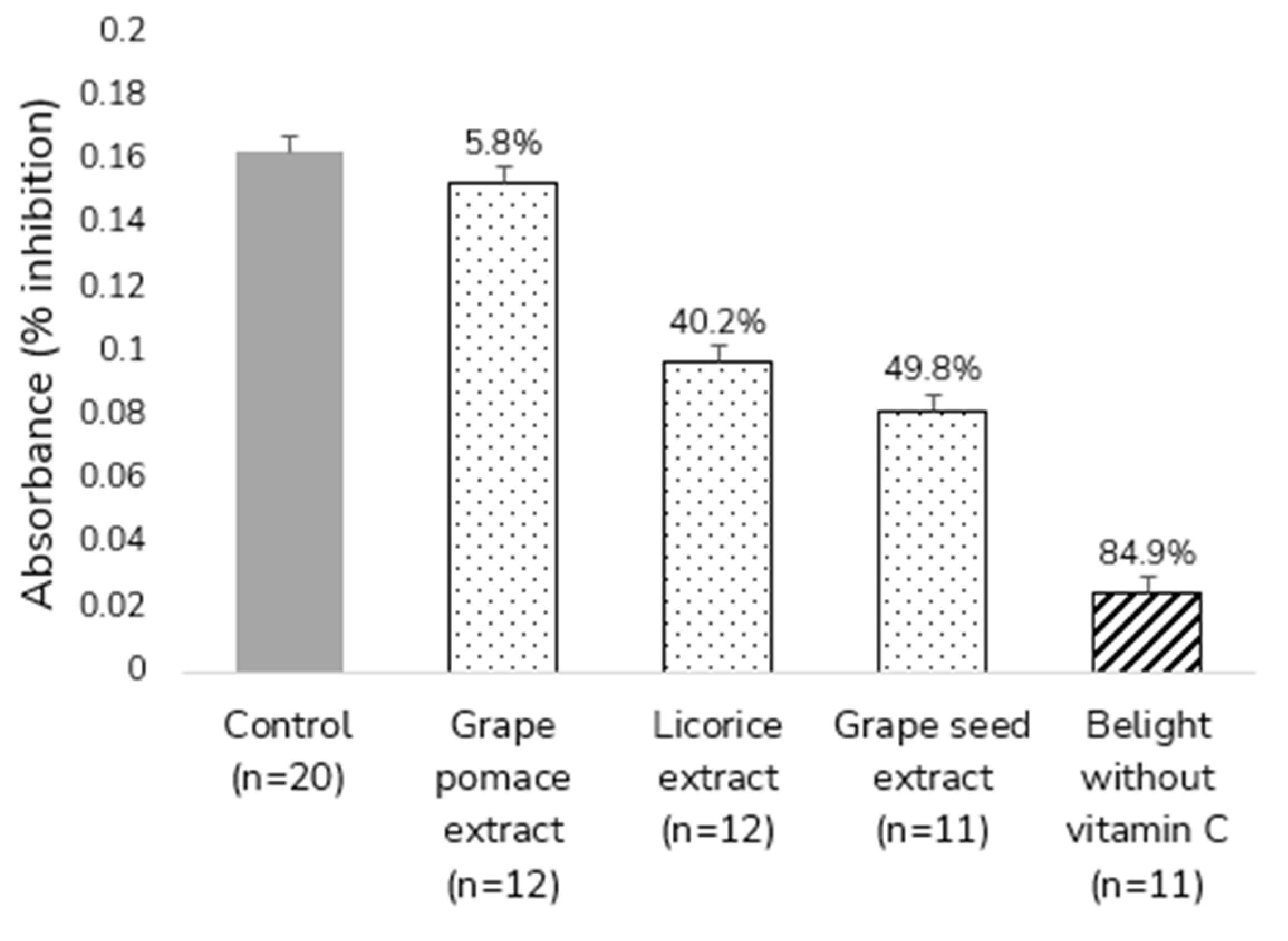
3.1. Tyrosinase Inhibitory Activity

Compared to the control (Figure 1), we observed significant inhibitions of tyrosinase activity for the licorice extract and grape seed extract (40.2% and 49.8% with p < 0.0001, respectively) and a weak inhibitory activity for the grape pomace extract (5.8%). However, a significant interaction between these three ingredients was found (p < 0.0001): the blend composed of the grape seed extract, grape pomace extract and licorice extract showed 84.9% tyrosinase inhibitory activity, which was greater than that observed for individual ingredients compared to the control.

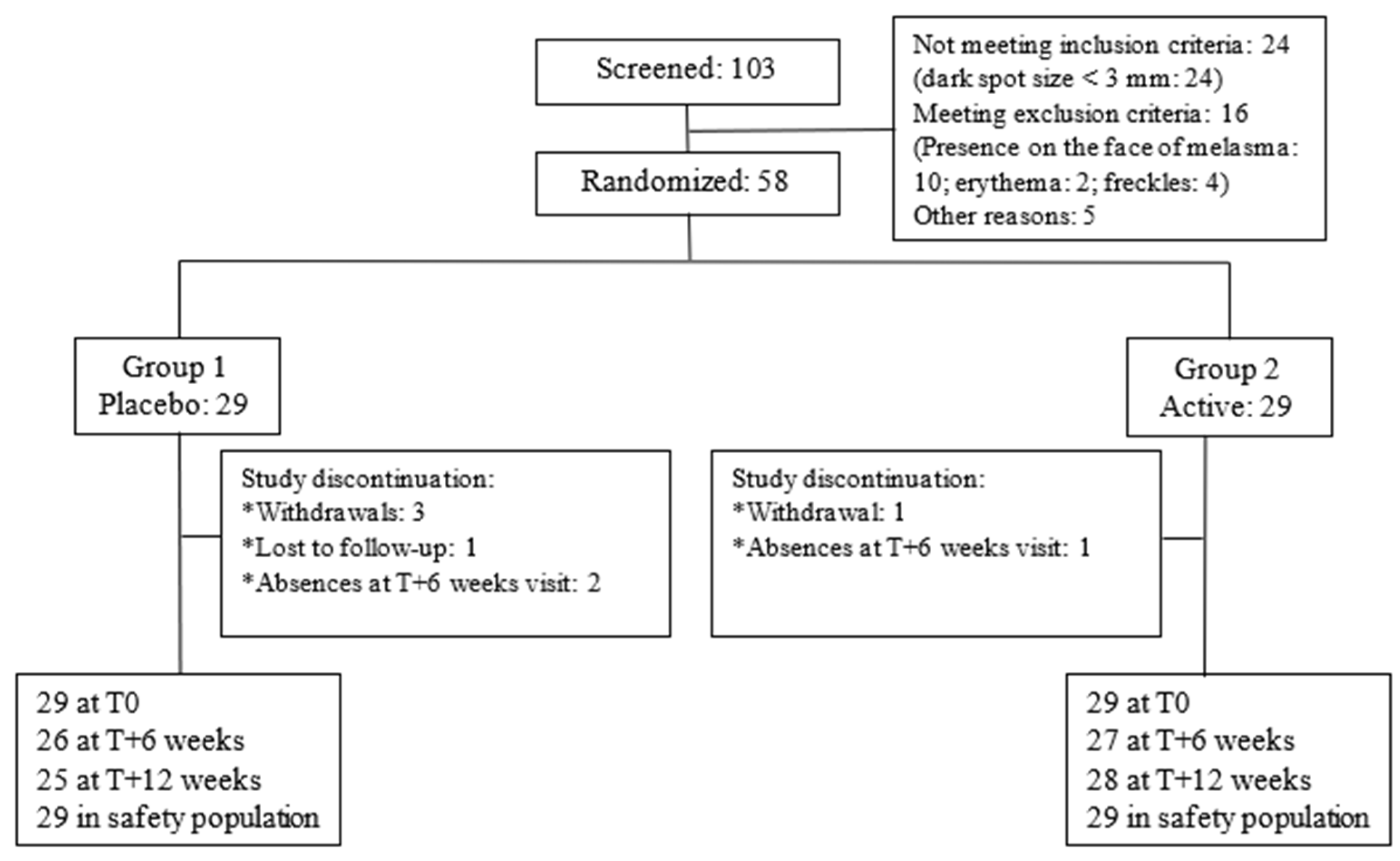
3.2. Clinical Study—Baseline Characteristics

Among the 103 screened participants, 24 participants did not meet the inclusion criteria (they had a dark spot with a diameter < 3 mm), 16 participants met exclusion criteria (10 participants presented melasma on the face, 2 participants presented facial erythema and 2 others presented freckles) and 5 were not eligible for other reasons. Thus, 58 healthy Asian subjects were randomized into the study, including 44 females and 14 males (Figure 2). A total of 53 subjects completed the 12-week study. The demographic, clinical and skin characteristics of the subjects collected at baseline are described in Table 1. The mean age of the product groups was close to 55 years. Heart rate, systolic blood pressure, diastolic blood pressure and body mass index were not significantly different between groups at baseline and were in the normal ranges.

3.3. Clinical Study—Compliance of the Subjects

During 12 weeks, subjects were instructed to take one capsule per day, during breakfast with a glass of water. Bottles with remaining capsules were returned at the testing visits. The number of remaining capsules at 6 and 12 weeks was similar between groups. At 6 weeks, the average numbers of ingested capsules by the participants were 41.5 ± 3.1 capsules for the placebo group and 41.9 ± 1.2 capsules for the active group. At 12 weeks, the average number of consumed capsules was also very similar between groups (82.8 ± 3.9 capsules vs. 84.1 ± 1.8 for the placebo and the active groups, respectively). As the actual consumptions of the tested products were very close to the expected ones (i.e., nearly 84 capsules at 12 weeks), product compliance was well respected by the participants.

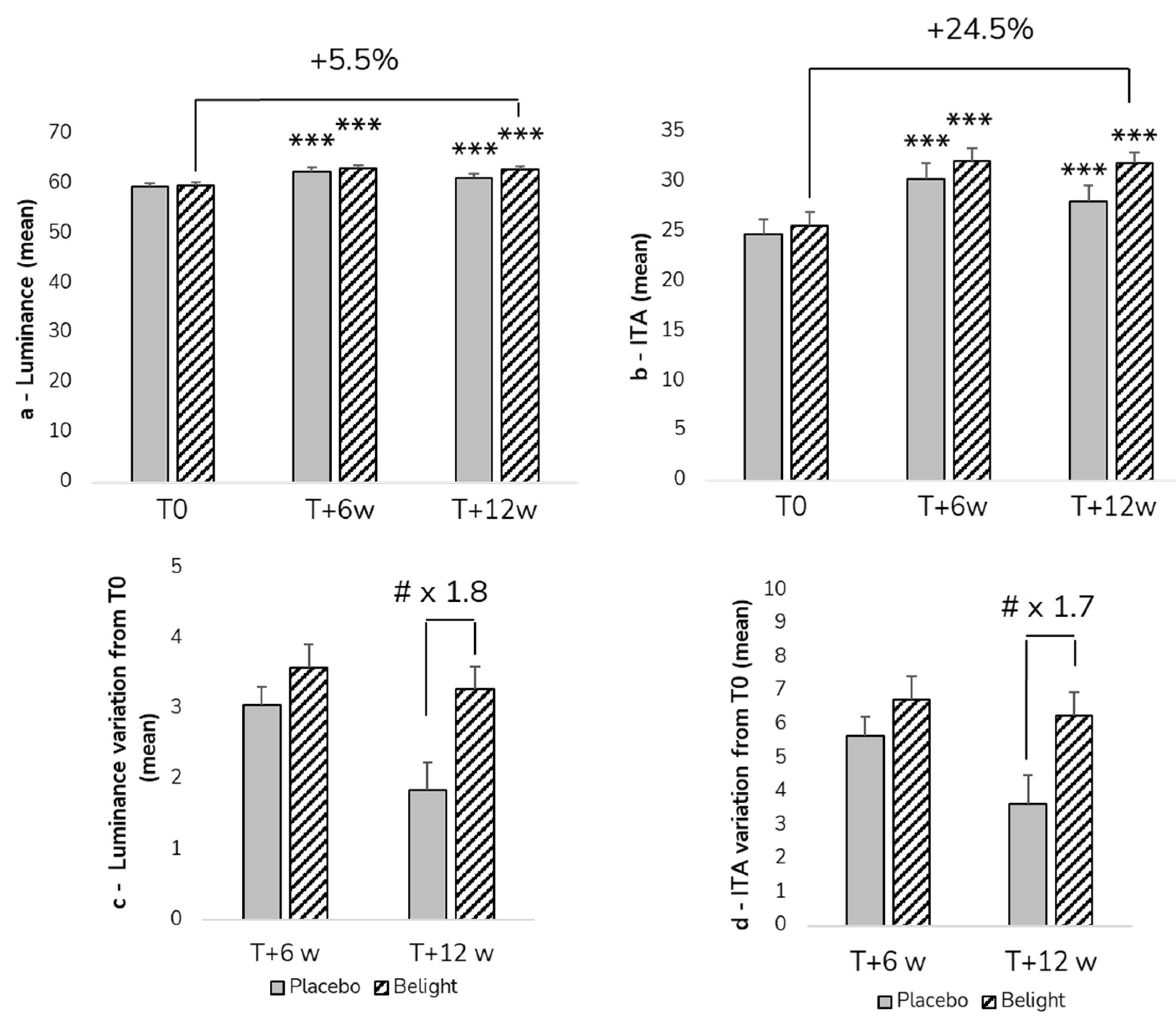
3.4. Clinical Study—Skin Lightening Effect

Color measurements of the facial skin using digital photographs are represented in Figure 3 and Table 2 (data are provided in Supplementary Material Table S1). When the mean L* and ITA° values of the two product groups were compared at baseline, no significant differences were found.
Based on the two-way ANOVA repeated measures, there was a statistically significant time × product interaction on L* indicating that L* was modified using the tested product according to time (p = 0.02). In the BelightTM group, there was a significant +5.5% increase in L* from baseline to week 12 compared to a significant +3.1% increase in the placebo group (Figure 3a).
Regarding ITA°, there was a statistically significant time x product interaction (p = 0.03) indicating that ITA° was modified using the tested product according to time. Indeed, there was a significant + 24.5% increase in ITA° from baseline to week 12 in the BelightTM group compared to a +14.8% increase in the placebo group (Figure 3b).

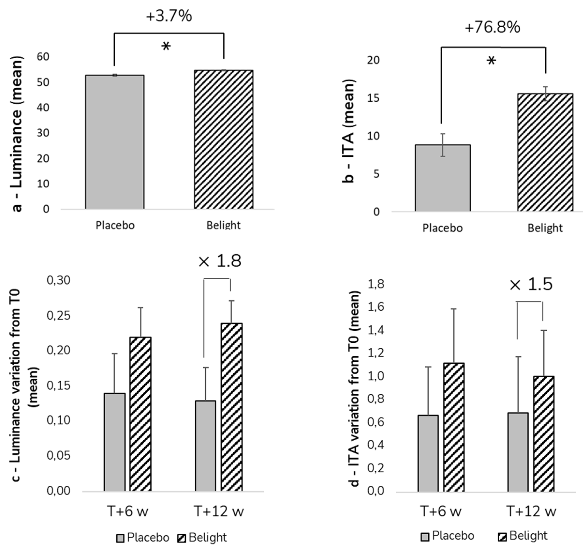
3.5. Clinical Study—Anti-Dark-Spot Effect

Color measurements of the facial dark spots performed with spectrophotometry are summarized in Table 2 and illustrated in Figure 4 (data are provided in Supplementary Material Table S2).
Based on the two-way ANOVA repeated measures, no statistically significant time × product interaction was observed on L* (p = 0.3). In the BelightTM group, there was a +0.4% increase in L* from baseline to week 12 compared to a +0.2% increase in the placebo group. However, independent significant effects of product and time were observed (p = 0.04 and p < 0.0001, respectively). Regarding the group effect, L* was significantly increased by +3.7% in the BelightTM group compared to the placebo group (mean ± SD: 54.70 ± 2.64 vs. 52.74 ± 3.99; Figure 4a).
Regarding ITA°, no statistically significant time × product interaction was observed (p = 0.7). Interestingly, independent significant effects of product and time were also observed on ITA° (p = 0.03 and p < 0.01, respectively). Indeed, ITA° was significantly increased by +76.8% in the BelightTM group compared to the placebo (mean ± SD: 15.57 ± 8.34 vs. 8.81 ± 13.29; Figure 4b). Digital photographs of three participants before and after 12 weeks of BelightTM intake were illustrated in Figure 5.

3.6. Clinical Study—Tolerance

No functional or clinical sign imputable to one of the tested products was reported or observed during the 12-week study period. Blood pressure, heart rate and body weight remained stable at each testing visit. Therefore, the tolerance of the Belight3TM nutricosmetic formulation was considered to be excellent under the test conditions, i.e., a 300 mg capsule per day over a period of 12 weeks.

4. Discussion

In the present study, a new proprietary oral formulation (Belight3TM) combining polyphenol-rich extracts from grape seed, grape pomace, licorice root and coated vitamin C showed a synergistic inhibitory effect on tyrosinase, the key enzyme involved in the process of melanogenesis, responsible for skin pigmentation. Indeed, nearly 85% of tyrosinase inhibition was reached with Belight3TM compared with less than 50% for each extract of the blend individually tested (except coated vitamin C that was not tested). In addition, this finding was supported with complementary research methods. In a randomized double-blind placebo-controlled clinical trial conducted in healthy Asian-skin-type males and females, the administration of 300 mg of Belight3TM for 12 weeks was associated with a significant greater improvement in facial skin lightening variables assessed with image processing of cross-polarized digital photographs, compared with the placebo. In addition, we observed a higher lightening effect of the dietary supplement on existing facial dark spots measured with spectrophotometry. Finally, Belight3TM was safe and as well tolerated as the placebo as no side effect was reported during the study.
Melanin pigments, a natural sunscreen in the skin, are the main determinants of skin color thanks to its ability to absorb, reflect and scatter the incident light, thus contributing to the perceived color [21]. Briefly, the enzyme tyrosinase is involved in the first two steps of melanogenesis, i.e., the hydroxidation of L-tyrosine and L-dihydroxyphenylalanine (L-DOPA) and the subsequent oxidation of L-DOPA to L-Dopaquinone. Thus, most strategies aiming to lighten the skin were based on the modulation of tyrosinase including the inhibition of tyrosinase mRNA transcription, inhibition of tyrosinase catalytic activity or acceleration of tyrosinase degradation [1]. The in vitro assay conducted in our study revealed that the grape seed extract, licorice root extract and, to a lesser extent, grape pomace extract significantly inhibit tyrosinase activity. Interestingly, the blend of botanical extracts that made up Belight3TM showed a synergistic effect on the tyrosinase inhibition that was greater than extracts tested alone. This synergy may be driven by specific flavonoids such as flavanol monomers from grape seed and glabridin from licorice as previously suggested [22,23,24].
In the existing literature, no oral formulation including such a blend of botanical ingredients rich in polyphenols associated with vitamin C has demonstrated both in vitro and in vivo efficacy on skin and dark spot lightening. Although there is a lot of available nutricosmetics claiming a skin lightening, whitening or bleaching effect on the market, very few clinical studies have been designed or published to show their efficacy, probably due to negative results. Comparing our findings with previous clinical research investigating the skin lightening effects of oral formulations including at least one ingredient of Belight3TM seems inadequate due to significant variability in the subjects involved, oral formulations, methods of skin color assessment and study duration [25].
Regarding the skin lightening effect of Belight3TM, we observed an increase in L* and ITA* values (by 5.5% and 24.5%, respectively) of the facial skin from baseline that was significantly higher than the placebo product. These findings are in line with previous clinical studies investigating oral formulation including grape extracts [20,26]. However, contrary to our results, these previous studies showed a lower effect and failed to show a significant difference with the placebo. Indeed, in Japanese healthy women aged 30–59 years, Tsuchiya et al. concluded that 12 weeks of ingestion of a beverage containing red wine oligomeric procyanidins (200 mg daily) significantly increased L values (by +1.1%) of the facial skin (without sunspots) that was measured with spectrophotometry [20]. Similarly, the administration of a multi-plant extract containing a grape seed extract during 12 weeks significantly improved ITA° from baseline of a pigmented lesion in healthy Chinese subjects aged 45–60 years with Fitzpatrick skin types III–IV [26]. Besides the tyrosinase activity inhibition demonstrated in the present study, another potential mechanism that may lighten the skin was the antioxidant properties of flavonoids and vitamin C that had ROS-scavenging properties and the ability to chelate metals at the active site of metalloenzymes [27].
Although the difference between groups was not significant at 6 weeks, we observed that skin lightening peaked at 6 weeks in participants that received Belight3TM but also in the placebo group. Indeed, the trial took place from August to November 2022 in Bangkok, Thailand. As shown with official weather historical records, weather in September was rainier and cloudier than in October [28]. Considering that skin pigmentation in response to UV exposure varies in a delayed way, commonly taking 2–3 weeks to develop noticeable tanning [29], it is more likely that a seasonal effect explains this finding.
As expected, the tolerance of the daily consumption of Belight3TM as 300 mg per capsule over a period of 12 weeks was excellent, as no side effect imputable to the product was reported during the study. Indeed, all the ingredients of the tested product may be widely ingested through a healthy diet including the consumption of fruits and vegetables. A licorice extract and in particular glycyrrhizic acid have been allowed for use in foods by the United States Food and Drug Administration, the Council of Europe and the Joint FAO/WHO Expert Committee on Food Additives and have been recognized as generally safe [30]. Although hair graying has never been associated with the administration of skin lightening food supplements including polyphenols or vitamin C [14,24,31], one limitation of the study design is the absence of hair graying or whitening assessment before and after product intake. As this oral formulation may lead to systemic inhibition of tyrosinase activity, we can hypothesize that melanogenesis may also be inhibited in the hair, leading to hair whitening. However, neither participants nor investigators observed hair whitening nor graying associated with the product administration during 12 weeks.

5. Conclusions

In the growing market of skin lightening/whitening agents, available products or treatments have generally little or no evidence on their safety or efficacy. For the first time, this original study suggests that the daily oral administration of Belight3TM during 12 weeks appears as a safe, natural and efficient lightener nutricosmetic in Asian subjects, with a potential tyrosinase inhibitory effect. This oral formulation may also be useful for the Western nutricosmetic market where having a more uniform and radiant facial complexion is a growing expectation.

Supplementary Materials

The following supporting information can be downloaded at: https://0-www-mdpi-com.brum.beds.ac.uk/article/10.3390/cosmetics10050143/s1, Table S1: Skin color—Digital photographs (raw data and statistics calculation), Table S2: Color of dark spots—Spectrophotometry (raw data and statistics calculation).

Author Contributions

Conceptualization, L.P. and D.G.; methodology, F.P., D.G., L.P. and C.P.; validation, L.P. and K.C.; formal analysis, C.P. and K.C.; investigation, L.P. and K.C.; resources, L.P. and K.C.; writing—original draft preparation, C.P.; writing—review and editing, all authors; visualization, C.P. and K.C.; supervision, F.P.; project administration, K.C.; funding acquisition, D.G. All authors have read and agreed to the published version of the manuscript.

Funding

This research was funded by Activ’Inside.

Institutional Review Board Statement

The study was conducted in accordance with the Declaration of Helsinki and approved by the Human Research Ethics Committee of Thammasat University (Faculty of Medicine) on 16 August 2022 (certificate of approval 160/2022; project no. MTU-EC-IM-5-115/65). The trial was performed according to the most recent recommendations given by the World Medical Association (Helsinki Statement 1964, amended in Fortaleza, Brazil, 2013), and in accordance with Standard Operating Procedures of the contract-testing laboratory. The Good Clinical Practices and Personal Data Protection Act B.E. 2562 (2019) of Thailand was respected.

Informed Consent Statement

Informed consent was obtained from all subjects involved in the study, including the use of their photographs for publication.

Data Availability Statement

The data presented in this study are available on request from the corresponding author.

Acknowledgments

We would like to thank Spincontrol Asia’s technical team for their assistance, as well as all the participants of the clinical trial.

Conflicts of Interest

F.P. and K.C. are employed at Spincontrol Asia. C.P., L.P., I.G. and D.G. are employed at Activ’Inside and provided the Belight3TM and placebo capsules.

References

  1. Duperray, J.; Sergheraert, R.; Chalothorn, K.; Tachalerdmanee, P.; Perin, F. The Effects of the Oral Supplementation of L-Cystine Associated with Reduced L-Glutathione-GSH on Human Skin Pigmentation: A Randomized, Double-Blinded, Benchmark- and Placebo-Controlled Clinical Trial. J. Cosmet. Dermatol. 2022, 21, 802–813. [Google Scholar] [CrossRef] [PubMed]
  2. Vashi, N.A.; Kundu, R.V. Facial Hyperpigmentation: Causes and Treatment. Br. J. Dermatol. 2013, 169 (Suppl. 3), 41–56. [Google Scholar] [CrossRef] [PubMed]
  3. Sarkar, R.; Arora, P.; Garg, K. Cosmeceuticals for Hyperpigmentation: What Is Available? J. Cutan. Aesthet. Surg. 2013, 6, 4. [Google Scholar] [CrossRef] [PubMed]
  4. Juliano, C.C.A. Spreading of Dangerous Skin-Lightening Products as a Result of Colourism: A Review. Appl. Sci. 2022, 12, 3177. [Google Scholar] [CrossRef]
  5. Yamakoshi, J.; Otsuka, F.; Sano, A.; Tokutake, S.; Saito, M.; Kikuchi, M.; Kubota, Y. Lightening Effect on Ultraviolet-Induced Pigmentation of Guinea Pig Skin by Oral Administration of a Proanthocyanidin-Rich Extract from Grape Seeds. Pigment. Cell Res. 2003, 16, 629–638. [Google Scholar] [CrossRef]
  6. Morel-Salmi, C.; Julia, A.; Vigor, C.; Vercauteren, J. A Huge PVDF Adsorption Difference between Resveratrol and ε-Viniferin Allows to Quantitatively Purify Them and to Assess Their Anti-Tyrosinase Property. Chromatographia 2014, 77, 957–961. [Google Scholar] [CrossRef]
  7. Hollinger, J.C.; Angra, K.; Halder, R.M. Are Natural Ingredients Effective in the Management of Hyperpigmentation? A Systematic Review. J. Clin. Aesthet. Dermatol. 2018, 11, 28. [Google Scholar]
  8. Adhikari, A.; Devkota, H.P.; Takano, A.; Masuda, K.; Nakane, T.; Basnet, P.; Skalko-Basnet, N. Screening of Nepalese Crude Drugs Traditionally Used to Treat Hyperpigmentation: In Vitro Tyrosinase Inhibition. Int. J. Cosmet. Sci. 2008, 30, 353–360. [Google Scholar] [CrossRef]
  9. Xu, Z.; Xing, X.; Zhang, C.; Chen, L.; Flora Xiang, L. A Pilot Study of Oral Tranexamic Acid and Glycyrrhizin Compound in the Treatment of Recalcitrant Riehl’s Melanosis. J. Cosmet. Dermatol. 2019, 18, 286–292. [Google Scholar] [CrossRef]
  10. Senol, F.; Khan, M.; Orhan, G.; Gurkas, E.; Orhan, I.; Oztekin, N.; Ak, F. In Silico Approach to Inhibition of Tyrosinase by Ascorbic Acid Using Molecular Docking Simulations. Curr. Top. Med. Chem. 2014, 14, 1469–1472. [Google Scholar] [CrossRef]
  11. Searle, T.; Al-Niaimi, F.; Ali, F.R. The Top 10 Cosmeceuticals for Facial Hyperpigmentation. Dermatol. Ther. 2020, 33, e14095. [Google Scholar] [CrossRef] [PubMed]
  12. Dumoulin, M.; Gaudout, D.; Lemaire, B. Clinical Effects of an Oral Supplement Rich in Antioxidants on Skin Radiance in Women. Clin. Cosmet. Investig. Dermatol. 2016, 9, 315–324. [Google Scholar] [CrossRef] [PubMed]
  13. Sitohang, I.B.S.; Anwar, A.I.; Jusuf, N.K.; Arimuko, A.; Norawati, L.; Veronica, S. Evaluating Oral Glutathione plus Ascorbic Acid, Alpha-Lipoic Acid, and Zinc Aspartate as a Skin-Lightening Agent: An Indonesian Multicenter, Randomized, Controlled Trial. J. Clin. Aesthet. Dermatol. 2021, 14, E53–E58. [Google Scholar] [PubMed]
  14. Handog, E.B.; Galang, D.A.V.F.; De Leon-Godinez, M.A.; Chan, G.P. A Randomized, Double-Blind, Place-bo-Controlled Trial of Oral Procyanidin with Vitamins A, C, E for Melasma among Filipino Women. Int. J. Dermatol. 2009, 48, 896–901. [Google Scholar] [CrossRef] [PubMed]
  15. Chatatikun, M.; Chiabchalard, A. Thai Plants with High Antioxidant Levels, Free Radical Scavenging Activity, Anti-Tyrosinase and Anti-Collagenase Activity. BMC Complement. Altern. Med. 2017, 17, 487. [Google Scholar] [CrossRef] [PubMed]
  16. Morgan, S.P.; Stockford, I.M. Surface-Reflection Elimination in Polarization Imaging of Superficial Tissue. Opt. Lett. 2003, 28, 114–116. [Google Scholar] [CrossRef]
  17. Kim, C.S.; Kim, M.K.; Jung, B.; Choi, B.; Verkruysse, W.; Jeong, M.Y.; Nelson, J.S. Determination of an Optimized Conversion Matrix for Device Independent Skin Color Image Analysis. Lasers Surg. Med. 2005, 37, 138–143. [Google Scholar] [CrossRef]
  18. Petit, L.; Piérard, G.E. Skin-Lightening Products Revisited. Int. J. Cosmet. Sci. 2003, 25, 169–181. [Google Scholar] [CrossRef]
  19. Juturu, V.; Bowman, J.P.; Deshpande, J. Overall Skin Tone and Skin-Lightening-Improving Effects with Oral Supplementation of Lutein and Zeaxanthin Isomers: A Double-Blind, Placebo-Controlled Clinical Trial. Clin. Cosmet. Investig. Dermatol. 2016, 9, 325–332. [Google Scholar] [CrossRef]
  20. Tsuchiya, T.; Fukui, Y.; Izumi, R.; Numano, K.; Zeida, M. Effects of Oligomeric Proanthocyanidins (OPCs) of Red Wine to Improve Skin Whitening and Moisturizing in Healthy Women—A Placebo-Controlled Randomized Double-Blind Parallel Group Comparative Study. Eur. Rev. Med. Pharmacol. Sci. 2020, 24, 1571–1584. [Google Scholar] [CrossRef]
  21. Kollias, N.; Seo, I.S.; Bargo, P.R. Interpreting Diffuse Reflectance for in Vivo Skin Reactions in Terms of Chromophores. J. Biophotonics 2010, 3, 15–24. [Google Scholar] [CrossRef] [PubMed]
  22. Malinowska, M.A.; Billet, K.; Drouet, S.; Munsch, T.; Unlubayir, M.; Tungmunnithum, D.; Giglioli-Guivarc’H, N.; Hano, C.; Lanoue, A. Grape Cane Extracts as Multifunctional Rejuvenating Cosmetic Ingredient: Evaluation of Sirtuin Activity, Tyrosinase Inhibition and Bioavailability Potential. Molecules 2020, 25, 2203. [Google Scholar] [CrossRef] [PubMed]
  23. Tuong, W.; Walker, L.; Sivamani, R.K. Polyphenols as Novel Treatment Options for Dermatological Diseases: A Systematic Review of Clinical Trials. J. Dermatol. Treat. 2015, 26, 381–388. [Google Scholar] [CrossRef] [PubMed]
  24. Yamakoshi, J.; Sano, A.; Tokutake, S.; Saito, M.; Kikuchi, M.; Kubota, Y.; Kawachi, Y.; Otsuka, F. Oral Intake of Proanthocyanidin-Rich Extract from Grape Seeds Improves Chloasma. Phytother. Res. 2004, 18, 895–899. [Google Scholar] [CrossRef] [PubMed]
  25. Lopresti, A.L.; Smith, S.J.; Pouchieu, C.; Pourtau, L.; Gaudout, D.; Pallet, V.; Drummond, P.D. Effects of a Polyphenol-Rich Grape and Blueberry Extract (MemophenolTM) on Cognitive Function in Older Adults with Mild Cognitive Impairment: A Randomized, Double-Blind, Placebo-Controlled Study. Front. Psychol. 2023, 14, 1328. [Google Scholar] [CrossRef] [PubMed]
  26. Xie, Y.; Zhu, G.; Yi, J.; Ji, Y.; Xia, Y.; Zheng, Y.; Ye, C. A New Product of Multi-Plant Extracts Improved Skin Photoaging: An Oral Intake in Vivo Study. J. Cosmet. Dermatol. 2022, 21, 3406–3415. [Google Scholar] [CrossRef]
  27. Gillbro, J.M.; Olsson, M.J. The Melanogenesis and Mechanisms of Skin-Lightening Agents—Existing and New Approaches. Int. J. Cosmet. Sci. 2011, 33, 210–221. [Google Scholar] [CrossRef]
  28. Bangkok Fall 2022 Historical Weather Data (Thailand)—Weather Spark. Available online: https://weatherspark.com/h/s/113416/2022/2/Historical-Weather-Fall-2022-in-Bangkok-Thailand#Figures-Temperature (accessed on 13 April 2023).
  29. Miyamura, Y.; Coelho, S.G.; Wolber, R.; Miller, S.A.; Wakamatsu, K.; Zmudzka, B.Z.; Ito, S.; Smuda, C.; Passeron, T.; Choi, W.; et al. Regulation of Human Skin Pigmentation and Responses to Ultraviolet Radiation. Pigment. Cell Res. 2007, 20, 2–13. [Google Scholar] [CrossRef]
  30. Pastorino, G.; Cornara, L.; Soares, S.; Rodrigues, F.; Oliveira, M.B.P.P. Liquorice (Glycyrrhiza glabra): A Phytochemical and Pharmacological Review. Phytother. Res. 2018, 32, 2323–2339. [Google Scholar] [CrossRef]
  31. Nobile, V.; Schiano, I.; Peral, A.; Giardina, S.; Spartà, E.; Caturla, N. Antioxidant and Reduced Skin-Ageing Effects of a Polyphenol-Enriched Dietary Supplement in Response to Air Pollution: A Randomized, Double-Blind, Placebo-Controlled Study. Food Nutr. Res. 2021, 65. [Google Scholar] [CrossRef]
Figure 1. Tyrosinase inhibitory activity. Absorbance (mean and SE) and inhibition (%) of grape pomace extract, licorice root extract, grape seed extract and Belight3TM (without vitamin C) compared to the control. n: number of replicates. p = 0.7 for grape pomace extract, p < 0.0001 for licorice extract, p < 0.0001 for grape seed extract and p < 0.0001 for the interaction of the 3 extracts (Belight3TM).
Cosmetics 10 00143 g001
Figure 2. Consolidated Standards of Reporting Trials (CONSORT) flow-chart diagram of the participants.
Cosmetics 10 00143 g002
Figure 3. Mean of luminance (a) and ITA° (b) of the normal skin using digital photographs. Mean of the individual variations from T0 for luminance (c) and ITA (d) of the normal skin using digital photographs. T0: baseline timepoint; T + 6 w: visit at 6 weeks; T + 12 w: visit at 12 weeks; p < 0.05 for the interaction time × product using two-way ANOVA for repeated measures. *** p < 0.0001 means significant difference within the group (vs. T0) using two-way ANOVA for repeated measures followed by Tukey’s tests. # p < 0.05 means significant difference between product groups using Student t-tests.
Cosmetics 10 00143 g003
Figure 4. Mean of luminance (a) and ITA (b) of facial dark spot using spectrophotometry. Mean of the individual variations from T0 for luminance (c) and ITA (d) of facial dark spot using spectrophotometry. T0: baseline timepoint; T + 6 w: visit at 6 weeks; T + 12 w: visit at 12 weeks; * p < 0.05 means significant difference between product groups using two-way ANOVA for repeated measures.
Cosmetics 10 00143 g004
Figure 5. Daily oral intake of Belight3TM during 12 weeks visibly lightened the facial skin and dark spots. Before product intake (on the (left) photographs) and after 12 weeks of product intake (on the (right) photographs). Dark spots are delimited with white boxes.
Cosmetics 10 00143 g005
Table 1. The demographic, clinical and skin characteristics of the study population at baseline.
Placebo
(N = 29)
Belight3TM
(N = 29)
Age (years), mean (SD), range55 (6.2), 45–6555 (6.8), 45–65
Gender
Female, n (%)20 (69%)24 (83%)
Male, n (%)9 (31%)5 (17%)
Nature of the skin, n (%)
Greasy9 (31%)3 (10%)
Combination9 (31%)11 (38%)
Normal7 (24%)8 (28%)
Dry4 (14%)7 (24%)
Skin phototype, n (%)
Type III13 (45%)18 (62%)
Type IV16 (55%)11 (38%)
Body mass index (kg·m−2), mean (SD)24 (3.9)24 (4.7)
Heart rate at rest (bpm), mean (SD)73 (11)78 (14)
Systolic blood pressure (mm Hg), mean (SD)128 (21)133 (22)
Diastolic blood pressure (mm Hg), mean (SD)82 (13)82 (13)
Table 2. Colorimetric variables of the facial skin and dark spots (Means ± SD).
BelightTMPlacebop-Value *
T0T + 6 wT + 12 wT0T + 6 wT + 12 wTimeProductTime
× Product
Skin colorL*59.61 ± 3.5863.07 ± 3.5262.89 ± 2.9959.32 ± 3.8162.34 ± 4.2361.09 ± 3.84<0.00010.30.02
ITA°25.60 ± 7.4832.10 ± 6.0631.88 ± 5.3524.69 ± 8.2830.26 ± 7.8828.05 ± 7.83<0.00010.30.03
Dark spotsL*54.60 ± 2.6854.67 ± 2.6554.84 ± 2.6852.72 ± 4.0152.78 ± 4.0352.72 ± 4.10<0.00010.040.3
ITA°14.98 ± 7.9715.77 ± 8.7715.99 ± 8.568.67 ± 13.108.97 ± 13.708.80 ± 13.59<0.010.030.7
T0: baseline timepoint; T + 6 w: visit at 6 weeks; T + 12 w: visit at 12 weeks; * p-values are derived from the two-way ANOVA for repeated measures for the effects of time, product and the interaction time × product.
Disclaimer/Publisher’s Note: The statements, opinions and data contained in all publications are solely those of the individual author(s) and contributor(s) and not of MDPI and/or the editor(s). MDPI and/or the editor(s) disclaim responsibility for any injury to people or property resulting from any ideas, methods, instructions or products referred to in the content.

