Review

Cell-Based Model Systems for Validation of Various Efficacy-Based Claims for Cosmetic Ingredients

Research and Development, Himalaya Wellness Company, Makali, Bangalore 562162, India
*
Author to whom correspondence should be addressed.
These authors contributed equally to this work.
Submission received: 31 July 2022 / Revised: 5 October 2022 / Accepted: 11 October 2022 / Published: 18 October 2022
(This article belongs to the Special Issue Advances in Regulatory and Technological Aspects of Cosmetics)

Abstract

:
Cosmetic formulations have evolved significantly over the years. These are no longer viewed merely as beauty-enhancing products. Rather, they are expected to deliver additional benefits to the skin that positively affect the skin health. There is a renewed interest in using herbal extracts and herbal ingredients in cosmetic products since they offer several advantages over synthetic ingredients. Evaluating the cosmetic ingredients for their efficacy and safety is critical during product development. Several regulatory bodies impose restrictions on using animals for testing these ingredients in cosmetic products. This has increased the need for developing novel cell-based or cell-free biological assays. The current article systematically presents in-vitro/cell-based and/or cell-free strategies for validating the efficacies of cosmetic ingredients for skin health and hair growth. The article focuses on details about various assays for the anti-acne effects, hair-growth-promoting activities, anti-aging activities, skin-rejuvenating properties, wound-healing effects, and skin-depigmentation activities of natural ingredients in cosmetic formulations.

1. Introduction

The usage of cosmetic products has a long history across all cultures and civilizations. In prehistoric times, man applied colors to attract animals to hunt or camouflage the body for protection and provoke fear in the enemy. Over the ages, as humans evolved, the cosmetic market has also evolved globally in terms of quality, safety, and efficacy. Cosmetic products are no longer simply beauty-enhancing agents; rather, consumers now expect them to deliver some additional benefits that will have a positive impact on skin health. In India, a cosmetic is defined under Section 3 (aaa) of the Drugs and Cosmetics Act of 1940 as “Any article intended to be rubbed, poured, sprinkled, or sprayed on, or introduced into, or otherwise applied to, the human body or any part thereof for cleansing, beautifying, promoting attractiveness or altering the appearance, and includes any article intended for use as a component of cosmetic” [1]. The Food and Drug Administration (FDA) defines a cosmetic as “A product (excluding pure soap) intended to be applied to the human body for cleansing, beautifying, promoting attractiveness, or altering the appearance” [2]. According to Article 2.1 (a) of Regulation European Commission (EC) No 1223/2009, “A cosmetic product means any substance or mixture intended to be placed in contact with the external parts of the human body (epidermis, hair system, nails, lips and external genital organs) or with the teeth and the mucous membranes of the oral cavity with a view exclusively or mainly to cleaning them, perfuming them, changing their appearance, protecting them, keeping them in good condition or correcting body odours” [3].
Cosmetic products based on herbal ingredients have gained increased consumer acceptance. Moreover, increased awareness among consumers has enhanced the demand for herbal-based cosmetic products. Several herbal ingredients possess desirable profiles such as mildness, efficacy, biodegradability, and low toxicity [4]. The herbal ingredients serve many purposes for consumers, viz. delay in skin aging, protection against acne and ultraviolet (UV) rays, reduction in hyperpigmentation, wrinkle reduction, hydration improvement, skin elasticity and firmness improvement, scar reduction, hair-loss reduction, dandruff treatment, etc. Safety and efficacy are also two important aspects for cosmetic products. Cosmetic products are not intended to cure dermatological disorders, and according to many regulatory bodies, ‘cosmetics’ and ‘drugs’ are treated separately [5]. However, some cosmetic ingredients can have restorative benefits on the skin. To describe the category of cosmetic products that offer additional skin health benefits, the term ‘cosmeceutical’ is widely used. The term ‘cosmeceutical’ was used in the early 1980s to describe products that exert a pharmaceutical therapeutic benefit but not a biological therapeutic benefit [6,7]. Cosmeceutical ingredients offer health benefits, and they have been evaluated for various conditions such as atopic dermatitis, contact dermatitis, eczema, and other skin disorders [5]. Along with the latest developments in technology, consumer expectations, and competition, there is an increasing need to evaluate cosmetic products for their effect on skin health. Animal model systems provide a translatable validation for efficacy-based claims. However, either due to internal policy or restrictions on the use of animals by legislators, several organizations refrain from animal models for their safety and efficacy validations of personal care products. Hence, cell-based systems have become valuable tools for such validations. Cell-based testing systems have a few advantages over other methods for efficacy validation because they are cost-effective, adaptable for high-throughput, and yield results considerably faster than conventional in vivo methods.
Many personal care products are meant for topical applications and do not involve ingestion through the oral route. Therefore, the testing systems for evaluating personal care ingredients may not consider factors such as metabolic conversion of the ingredients or absorption of the ingredients through tight junctions such as internal epithelia. Hence, cell-based model systems are more employable for validating topical ingredients rather than for those requiring systemic absorption and metabolism.
Providing exhaustive details on the pathophysiology and dermatological aspects is out of the scope of this review. In this review, we focus more on the type of cell lines and the nature of assays that can be used for screening the activity of herbal ingredients in cosmetic formulations.

2. Cell-Based Strategies for Cosmetic Ingredients

2.1. Critical Parameters for In Vitro and Cell-Based Assays

An important parameter that should be carefully considered during evaluation of any cosmetic ingredient’s efficacy is the test substance’s solubility. Aqueous soluble ingredients do not pose much of a problem, since they are compatible with a variety of cell culture media and buffer systems. However, substances soluble in organic solvents pose some challenges since the organic solvent itself can have some background effects in in vitro systems. For example, DMSO, which is very commonly used as a vehicle for dissolving test substances, has been shown to affect cell-based systems at high concentrations [8]. The cytotoxicity of the ingredients is another key parameter to be considered before including them for efficacy-based validations. In general, for all cell-based assays, the ingredient concentration should be non-toxic. This is very important, since cytotoxicity can lead to many effects in the cells, which can lead to artifacts in the interpretation of the assay results. Another important consideration is the assay interference by the test substances, wherein some of the test substances may interfere with the assay in a non-specific manner, which can lead to false positive results [9]. The test substances interfere with the assays because of the following reasons:
  • The color and turbidity of test substances can interfere with assay signals (e.g., absorbance and fluorescence).
  • In many cases, the test compounds can exhibit metal ion chelation and redox cycling properties.
  • The test substance can form protein aggregates or exhibit non-specific protein reactivity, which can be misinterpreted as mechanism-specific inhibition [10].
Hence, appropriate assay controls are essential for the interpretation of the assay results. In the subsequent sections, we provide details on the cell-based methods to evaluate cosmetic ingredients for the following activities:
  • Anti-acne activity
  • Hair-growth-promoting activity
  • Anti-aging and skin-rejuvenating activity
  • Anti-psoriatic activity
  • Wound-healing activity
  • Anti-skin-hyperpigmentation activity
Cosmetic products based on natural ingredients are gaining increased consumer acceptance, and several herbal ingredients are being evaluated in this direction. Keeping this in view, in each section, we have cited examples of herbal ingredients possessing particular biological activity required for the cosmetic product efficacy.

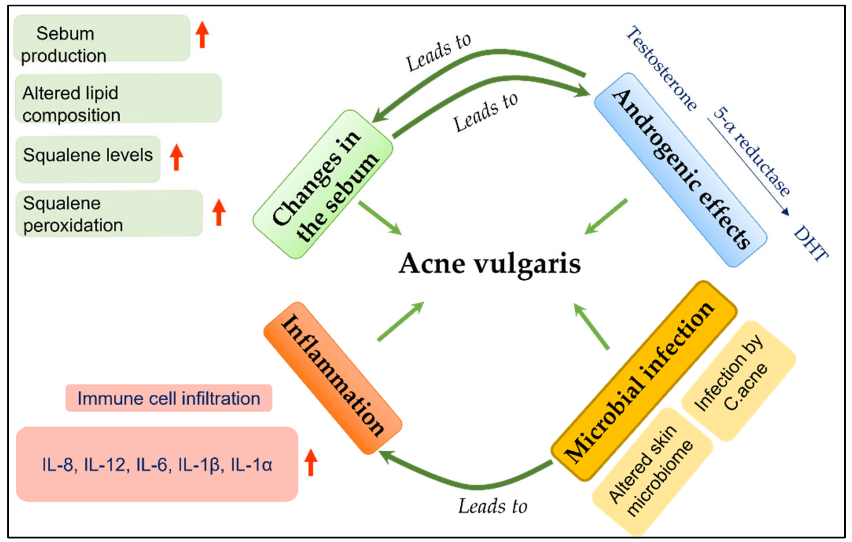
2.2. Evaluation of Effect of Cosmeticingredients on Acne Vulgaris

Acne vulgaris is a common skin condition involving the pilosebaceous follicles [11,12]. The pathological components of acne, as summarized in Figure 1, involve various cellular processes such as microbial infection, inflammation, and changes in sebum quality and quantity [13].
Cutibacterium acnes is one of the major contributing factors for the pathology of acne, and hence, anti-microbial activity against this microbe is generally employed as one of the screening assays for anti-acne products/ingredients. The cells, when challenged with C. acnes, induce several inflammatory processes that lead to the secretion of various cytokines, which play a major role in the pathogenesis of acne. In addition, it has been shown that lipid composition of sebum can greatly influence the progression of acne pathogenesis and may directly or indirectly contribute to aggravation of inflammation [14,15]. Hence, the cosmetic ingredients intended for anti-acne should be evaluated for these processes to capture the most efficacious ingredient. The following sections describe the cellular model systems for capturing these processes.

2.2.1. Evaluating the Anti-Inflammatory Activity of Cosmetic Ingredients against C. acnes-Induced Inflammation

This model system uses human monocytes such as THP-1 cells and heat-inactivated C. acnes or peptidoglycans [16]. THP-1 cells are exposed to heat-inactivated bacteria for 24 h, and the cell culture supernatant is used for measuring inflammatory cytokines such as interleukins (IL) IL-8, IL-10, IL-12, and IL-1a and tumor necrosis factor (TNF-alpha) by using enzyme-linked immunoassay (ELISA). The gene expression of cytokines mentioned above can also be captured at specific time points by real-time PCR. Apart from the immune cells, the keratinocytes also play an important role in inflammation. Hence, human keratinocyte cells can also be used in studying inflammation in monocyte cells. The amount of cytokine produced by keratinoctyes (such as human epidermal keratinocytes (HaCaT)) may be less compared to monocytes (like THP-1 cells). In such cases, induction with high non-toxic concentration of C. acnes can be employed to look for expression levels of cytokine genes in the cells. The herbal ingredients, from Glycyrrhiza glabra [17,18], Azadirachta indica [19], Curcuma longa [20], Myristica fragrans [21], and Camellia sinensis [22] are shown to possess anti-inflammatory properties, and some of these ingredients are incorporated into anti-acne based products such as the purifying neem face wash [23] and charcoal face wash, face gels, and creams manufactured by Himalaya Wellness Company, Bengaluru, India.

2.2.2. Evaluating the Effect of Cosmetic Ingredients on Sebum Secretion

Sebum quality and quantity play important roles in the pathogenesis of acne vulgaris [24]. Sebum is a complex mixture of different types of lipids such as triglycerides, free fatty acids, wax esters, cholesterol esters, and squalene [14]. Changes in the levels of these lipids are known to influence the pathogenesis of acne. Therefore, the human sebocyte cell line would be an ideal model to capture cosmetic ingredients’ effects on sebum quality, quantity, inflammation, and sebostatic activity of the test substances. The sebocytes of the internationally patented SZ95 cell line have been reported to retain several characteristic features of normal human sebocytes [25]. Investigators have also used other sebocyte cell lines, such as SEB-1, Seb-E6E7, and primary sebocytes made in their own laboratories [26,27]. Also, the immortalized human sebocyte cell line (SEBO662) is used by investigators for anti-acne studies [28,29]. Squalene levels and peroxidation of squalene have been shown to play important roles in development of acne [24,30]. Hence, measuring the expression levels of peroxidated squalene or levels of squalene peroxidase activity can be a useful end point for evaluating the effect of cosmetic ingredients on acne [31]. The herbal phytoactive ingredients from Azadirachta indica [19], Curcuma longa [32], Aloe vera [33], and Glycyrrhiza glabra [34] are reported to reduce sebum production, thereby inhibiting the growth of C. acnes.
Table 1 summarizes the in vitro approaches that can be employed to evaluate the ingredients for their effect on the pathology of acne vulgaris.

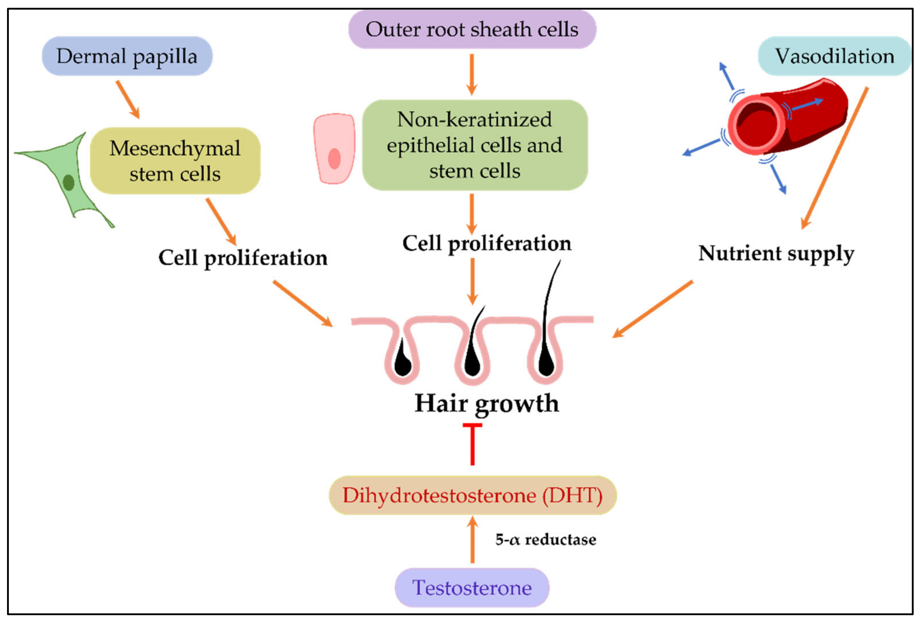
2.3. Evaluation of the Effect of Cosmetic Ingredients on Hair Growth

Hair is made up of two structures: the hair shaft, which is the visible part above the skin, and the hair follicle, which is an invagination of the epidermis into the dermis layer. The hair follicle itself is made up of outer root sheath and inner root sheath cells. At the base of the hair follicles is the dermal papilla, which contains stem cells. Both dermal papilla cells and outer root sheath cells play important roles in hair growth [39,40]. In humans, hair follicle formation occurs during embryogenesis, and no new hair follicles form after birth. Hair fall can be of several etiologies, and not all types of hair fall are reversible. However, some mechanisms are central to the hair growth dynamics of humans. These include the proliferation of stem cells in the dermal papilla, nutrient supply to the dermal papilla, and hormones such as androgens (Figure 2). In general, the cell-based assays for screening anti-hair-loss and hair-growth-promotion activities are designed considering these central mechanisms.

2.3.1. Assay Systems Based on Proliferation of the Hair Follicle Cells

Although the growth of hair follicles is a very complex and dynamic process, when it comes to cell-based systems, two cell types, human follicle dermal papilla cells (HFDPC) and outer root sheath cells (ORScs), are used for assessing the effectiveness of cosmetic ingredients on hair growth. HFDPC cells are derived from hair papilla of normal hair follicles and have been shown to express genes involved in hair growth dynamics [41]. ORScs are derived from hair follicles. A typical assay system for evaluating the effect of cosmetic ingredients involves evaluating the effect of these ingredients on the proliferation of HFDPC or ORSc cells using standard methods such as 3-(4,5-dimethylthiazol-2-yl)-2,5-diphenyltetrazolium bromide (MTT/XTT) dye-based assays, BrDu incorporation, or Ki67 staining-based assays. Fibroblast growth factors (FGFs), mainly FGF-1, FGF-2, and FGF-10, stimulate hair growth [42] and are generally used as a positive control for inducing the cell proliferation in this assay system. In addition, alkaline phosphatase (ALP) and versican are generally investigated as important markers of dermal papilla cells. During cell proliferation, the ALP activity is required for important metabolic pathways that regulate the phosphate and phosphoryl metabolite levels during cell proliferation [43]. ALP activity is a fundamental marker for hair-growth promotion [43]. Versican, on the other hand, is a chondroitin sulfate proteoglycan and is involved in induction and maintenance of the anagenic phase of the hair cycle [44]. The effect of cosmetic ingredients on the expression levels of these two markers makes it worth investing in HFDPC-based model systems. Apart from the expression of key growth factor genes such as insulin growth factor (IGF-1), hepatocyte growth factor (HGF), keratinocyte growth factor (KGF), and vascular endothelial growth factor (VEGF) can also be tested using qPCR-based approaches [45]. The herbal phytoactives present in Phyllanthus emblica [46,47], Eclipta alba [48], Butea frondose [49], Hibiscus rosa [50], and Camellia sinensis [51] have been used in many anti-hair-loss products; some of these have been shown to act directly by influencing the growth of follicular stem cells [52,53].

2.3.2. 5-α Reductase Inhibition

The enzyme 5-α reductase is involved in the local conversion of testosterone into a more potent form, that is, dihydrotestosterone (DHT). DHT is implicated in androgenic alopecia, and hence, inhibiting 5-α reductase is an attractive strategy for anti-hair-loss effects. There are three reported isoforms of this enzyme (5 α-R1, 5 α-R2, and 5 α-R3). In humans, 5 α-R1 is expressed predominantly in sebocytes and keratinocytes [38,54]. The assay system for evaluating the enzyme activity can be cell-based or cell-free. For a cell-based assay, human sebocytes (SZ95) or keratinocytes can be employed [55], since they are known to express measurable levels of 5-α reductase. Some investigators have also employed cells overexpressing specific isoforms of this enzyme [56]. For cell-free systems, purified 5-α reductase or rat liver microsomes can be employed [38]. In both cases (cell-based or cell-free systems), the assay procedure generally involves incubating the system with testosterone in the presence or absence of the inhibitors and detecting the levels of testosterone and/or the DHT. Quantification methods include radiodetection [55], high-performance liquid chromatography (HPLC) [38], thin-layer chromatography (TLC) [38], or liquid chromatography–mass spectrometry (LC-MS) [57]. Finasteride and dutasteride are known inhibitors of this enzyme, and they can be employed as a positive control in these assays.
Table 2 summarizes the assays that can be employed for evaluating the hair-growth-promoting activity of the cosmetic ingredients.

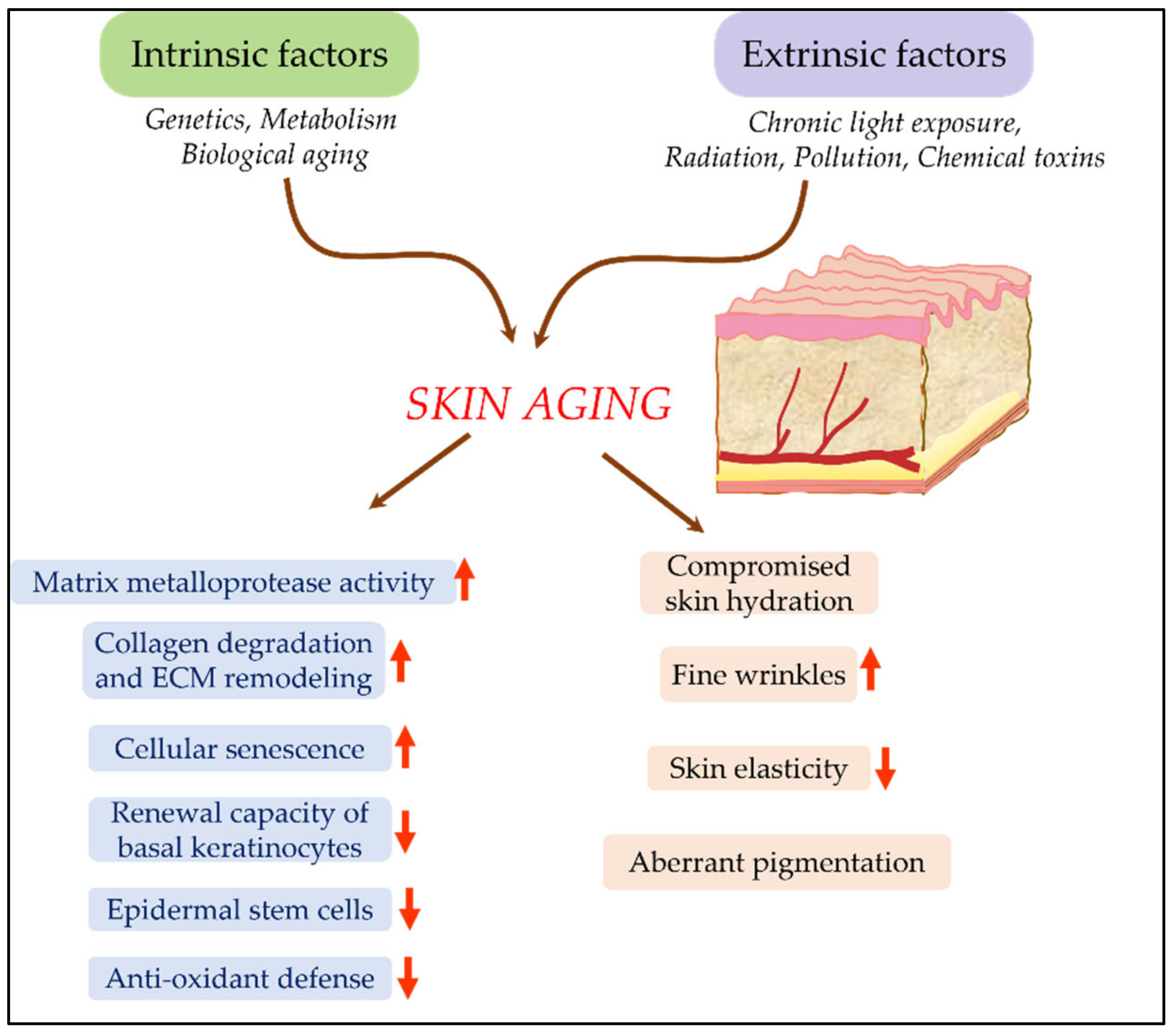
2.4. Evaluation of Effect of Cosmetic Ingredients on Skin-Aging and Rejuvenation

Skin aging can occur due to two factors, intrinsic and extrinsic. Intrinsic aging is chronological aging that occurs naturally due to genomic and hormonal factors (Figure 3). Extrinsic aging, on the other hand, is caused by challenges to the skin from external factors such as UV radiation, environmental pollutants, dietary factors, etc. [58]. These two types of aging show slightly different phenotypic characteristics [59]. The significance of the interplay between extrinsic factors and subsequent internal biological response by organisms is well-recognized by the scientific fraternity. The term ‘skin aging exposome’ has been introduced to explain this phenomenon. This term describes the totality of exposures to which an individual is subjected from conception to death. It considers the interaction between internal and external factors leading to the biological and clinical signs of aging [60]. The literature shows that environmental and nutritional factors modulate the matrix metalloproteases (MMPs), reactive oxygen species (ROS), and inflammatory cytokines expression, thereby causing early signs of skin aging. In addition, various pollutants, including particulate matter and dietary supplements, activate the aryl-hydrocarbon receptor (AhR) in the keratinocytes, which in turn contributes to premature aging and affects skin integrity [61,62].
The cell-based assays to evaluate skin aging and skin rejuvenation potential of the cosmetic ingredients are based on anti-oxidant and anti-inflammatory activities, ability to influence the expression levels of genes involved in aging, effect on collagenase, and elastase and hyaluronidase activities. Subsequent sections describe these platforms in further detail. Herbs such as Aloe barbadensis miller (aloe vera) [63], Leontopodium alpinum (Edelweiss) [64], Withania somnifera [65], triphala [66], Ginkgo biloba (gingko) [67], Curcuma longa [68,69], and Centella asiatica [70] are rich in flavonoids and possess anti-oxidant and anti-inflammatory properties.

2.4.1. Skin Aging Models Based on Extra-Cellular Matrix (ECM) Dynamics

Collagen, elastin, and glycosaminoglycans are essential for the structural integrity of the skin, and they form a major part of the ECM. Enzymes such as collagenases, elastases, and other matrix metalloproteases (MMPs) act on the ECM proteins and contribute to their breakdown. The levels of collagenase and other matrix metalloproteases have been shown to increase with aging [71] and upon exposure to UV [72]. This is thought to be one of the reasons for skin wrinkles, as observed in aged or environmentally exposed skin [73,74]. Hence, assay systems based on the measurement of collagen and elastin dynamics can be employed for studying the anti-aging effects of cosmetic ingredients in cell-based systems [75]. Human dermal fibroblasts (HDF or HS68), upon exposure to UV light, are shown to express elevated levels of matrix-degrading MMPs [76]; this serves as the basis for cell-based photoaging models. This assay system for capturing the features of photoaging involves irradiating the HDFs with UVA for 3–4 days. Following this treatment, the cells exhibit changes in the levels of collagen, MMP1, and MMP3 when compared to control cells, which are cultured under normal conditions. Cosmetic ingredients are tested in this model for their effectiveness in reversing some of these changes. The end point measurements can include collagenase activity, elastase activity, MMP activity, collagen levels (as measured by ELISA), or gene expression levels of several of the genes involved in this process, such as COL1A1, COL1A2, COL3A1, COL4A1, COL7A1, MMP1, MMP2, MMP3, MMP7, MMP8, MMP9, MMP10, MMP12, MMP13, MMP14, TIMP1, TIMP2, TIMP3, TIMP4 [77]. Phytoactives present in herbs such as Aloe barbadensis miller (aloe vera) [63], Centella asiatica [70,73], Calendula officinalis [78], and Garcinia mangostana [79] offer anti-aging properties by effecting collagen and elastin synthesis.

2.4.2. Measurement of Hyaluronic Acid (HA) Levels

Hyaluronic acid is a glycosaminoglycan and serves as a key component of ECM. Of the total HA in the body, 50% is present in the skin [80]. HA is involved in skin hydration and integrity, and its level is shown to decrease with aging [81]. HA levels in the skin are determined by two enzymes: HA synthase, which synthesizes HA, and hyaluronidase, which degrades it. Hence, in vitro systems intended for screening the anti-aging effects of cosmetic ingredients are based on measuring the amount of HA levels in the cells by ELISA or analyzing the enzyme activities or gene expression levels of HA synthase and hyaluronidases. The cosmetic ingredients that can increase the HA and HA synthase levels can be scored as leads for anti-aging effects. Several studies have focused on measuring the levels of HA, hyaluronidase, and HA synthase as cellular markers for aging. Several ingredients from herbal sources, such as almond [82], Curcuma longa [68,69], and Coriandrum sativum [79,83] have been shown to increase the HA levels in skin fibroblasts. Ingredients from Vitis rotundifolia have been shown to inhibit the hyaluronidase [84].

2.4.3. Cellular Senescence Model for Skin Aging

Senescence results in irreversible cessation of cell proliferation, and recent studies support the hypothesis that senescence of the skin fibroblasts is one of the drivers of skin aging [85]. Human dermal fibroblasts isolated from older adults show increased levels of markers of cellular senescence compared to younger counterparts. However, adapting this model for evaluating the anti-aging effects of cosmetic ingredients can be challenging, since it involves the isolation of primary cells from volunteers. Nonetheless, human dermal fibroblasts such as HS68 or HDFa can be employed to induce cellular senescence and aging in vitro [66,86]. HS68 cells exposed to hydrogen peroxide or UVB are demonstrated to show hallmarks of senescence and aging such as increased expression of p53, p21, and p16. UVB or hydrogen peroxide challenge has been shown to increase the number of senescence-associated beta-galactosidase (SA-β-gal)-positive cells [86]. Various investigators employ this model system to test the anti-aging effect of cosmetic ingredients. Since cellular senescence is one of the key contributing factors to skin aging, senolytics are gaining increasing importance as a means to interfere with skin aging [87]. Senolytics are compounds or drugs that can selectively eliminate senescent cells [87]. Cell-based systems for screening the senolytic activity of cosmetic ingredients can be based on scoring senescent cells using SA-β-galactosidase activity. In a recent study, a detailed methodology for screening senolytics is explained [88]. This model system is based on mouse primary embryonic fibroblasts (MEF), and it employs quantitative high-content fluorescent image analysis for scoring senescent cells (based on SA-β-galactosidase activity). This assay is employable in a high-throughput setting, and other adherent cell lines apart from MEFs can also be used. Phytoactive ingredients such as quercetin, hydroxytyrosol, and certain flower extracts have been shown to display senolytic properties, which can be scored by analyzing reductions in SA-β-galactosidase activity using fluorescent microscopy [89,90]. Table 3 summarizes the assays that can be employed for evaluating the anti-skin aging and skin rejuvenation activity of the cosmetic ingredients.

2.5. Gene Expression Studies to Evaluate Skin Hydration, Skin Barrier Function and Skin Rejuvenation

It is quite challenging to develop perfect cell-based models for complex processes such as skin aging, rejuvenation, skin barrier function, and skin hydration. However, several studies over the years have found that many genes regulate the key reactions that are critical to these functions. The expression levels of these genes can be indirect markers and the quantification of expression levels of these genes is commonly employed to evaluate the effect of cosmetic ingredients on skin rejuvenation, skin hydration, and skin barrier functions. To this end, q-PCR-based quantification of several marker genes, such as aquaporin-3 (AQP3), involucrin (INV), transglutaminase 1, 3, and 5 (TGMs), filaggrin (FLG), elastin (ELN), and collagen (COL1A1 or COL1A2), is commonly used to evaluate the efficacy of cosmetic ingredients. These genes are involved in various aspects of skin integrity, skin hydration, and skin barrier functions.
Involucrin is one of the skin barrier proteins involved in the initial step of cornified envelop formation [97]. Similarly, transglutaminases play a major role during the formation of stratified layers by cross-linking various proteins such as filaggrin, involucrin, and small proline-rich proteins [98]. The human FLG gene, encoding profilaggrin and filaggrin, plays an important role in retaining moisture and providing a skin barrier function [99]. Filaggrin (filament-aggregating protein), being an important marker for skin hydration, is routinely used to measure the hydration level. There are 13 mammalian AQPs, and the most abundant is AQP3 [100]. The expression profile of AQP3 is an important parameter to analyze the hydration level of the skin. Cell-based models for these gene expression studies can involve human keratinocytes or dermal fibroblasts, which are treated with the cosmetic ingredients for given time periods (24–48 h). Following the treatment, the expression levels of these genes are quantified using qPCR-based strategies. The expression of the above-mentioned biomarkers ca be quantified with ELISA, western blot, and immunofluorescence methods. Herbs like Aloe vera [63], Centella asiatica [70,73], Calendula officinalis [78], and Garcinia mangostana [79] present cosmetic phytoactive ingredients with skin hydration and rejuvenation properties.

2.6. Evaluation of the Effect of Cosmetic Ingredients on Psoriasis

Psoriasis pathology involves abnormal differentiation and proliferation of keratinocytes, resulting in epidermal hyperplasia and infiltration of immune cells to the epidermis, causing chronic inflammation [101]. The early stages of disease development involve several inflammatory cytokines, including interferons (IFN-γ), IL-1, IL-22, and, mainly, IL-23/IL-17 and TNF-α [102], which are secreted by immune cells. The sustained immune assault in this chronic inflammatory condition leads to keratinocyte hyperproliferation and impaired differentiation of epidermis, causing the clinical manifestation of the disease. The formation and maintenance of psoriatic lesions is affected by this interplay between the keratinocytes and immune cells [103].
Cell-based model systems for capturing the psoriasis phenotype and screening the anti-psoriatic ingredients can include keratinocyte cell lines (e.g., HaCaT) or immune cells (e.g., THP-1cells). Earlier, our laboratory reported an HaCaT-based model system in which the cells exposed to imiquimod (100 µM) exhibited hyperproliferation and increased inflammation (as measured in terms of the levels of pro-inflammatory cytokines–IL-17, TNF-α, IFN-γ, and IL-6). This model system was used for the evaluation of anti-psoriatic effects of curcumin [104]. The S100 protein psoriasin (S100A7) is a characteristic anti-microbial peptide (AMP) found in psoriatic lesions and is thought to have a chemotactic role in psoriasis [105]. Our recent studies in the lab also indicate that this model system exhibits increase expression of psoriasin. Hence, downregulation of this peptide can be scored for anti-psoriatic activity [106]. Since inflammation is an integral part of the development of psoriasis, screening for anti-inflammatory activity in cosmetic ingredients can also add value in terms of their efficacy.
Many USFDA-approved biologics for psoriasis treatment work by inhibiting the inflammatory cytokines. For example, adalimumab, infliximab treatment for tumor necrosis factor alpha (TNF-alpha), secukinumab, brodalumab for interleukin (IL)-17, ustekinumab for IL-12, and tildrakizumab for IL-23. These have shown effectiveness in slowing down epidermal turnover and plaque formation [107,108]. The effect of cosmetic ingredients on these cytokines can be tested in cell-based systems involving human monocytes such as THP-1. The herbal ingredients from Brassica nigra, Linum ussitatissimum [109], Pongamia pinnata [110,111], and Vitis vinifera [112] have been reported to have anti-inflammatory, emollient, anti-microbial, and skin-protectant properties, and therefore can be employed as anti-psoriatic agents.

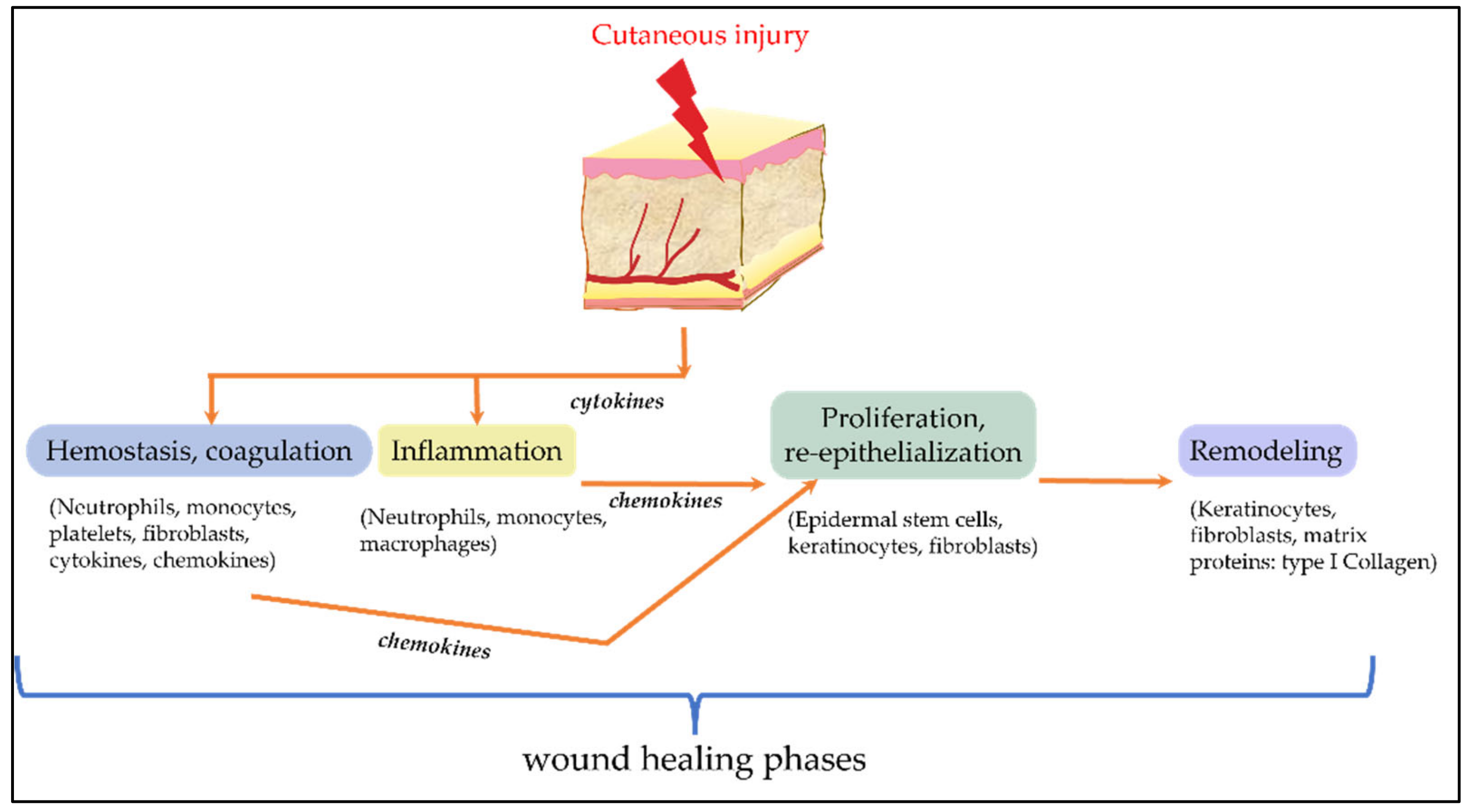
2.7. Cell-Based Systems for Evaluation of Cosmetic Ingredients on Wound-Healing

Skin is subjected to several physical and environmental challenges, and this may lead to injuries such as cuts and rashes on skin. Therefore, many cosmetic products (for example, lip balms, moisturizers, and crack- or burn-healing creams or gels) target wound-healing. Wound-healing is a multinetwork process broadly divided into these phases: hemostasis, inflammation, cell proliferation and re-epithelialization, and finally, remodeling. It involves several cell types such as keratinocytes, fibroblasts, epidermal stem cells, and immune cells [113,114]. It is a complex process which involves a delicate balance between inflammation, resolution, ECM remodeling, cell proliferation, migration, and collagen synthesis, as summarized in Figure 4.
The in vitro assays for evaluating wound-healing are mostly based on cell-migration-based assays. In these assays, to induce wounds, various cell-wounding methods are employed on cells grown on a confluent monolayer. Then, the progress of cell proliferation and migration into the wound site to reduce the wound gap is measured over time, microscopically.
Wounds introduced into the in vitro cultured cell monolayer can be by various methods such as mechanical wounding, thermo-mechanical, electrical wounding, or optical wounding, as shown in Table 4.
Wound-healing is measured visually by calculating the difference in the wound area created on the cell monolayer before and after product treatment. Also, 3D skin models can be employed for this wound-healing assay, where a mechanical wound is introduced in 3D skin models (EpiDermFTTM, StrataTest®, Phenion® FT). Fluorescent labeling and microscopy techniques have been used to study cell migration during wound-healing [120]. Herbal ingredients used in personal care products such as Aleo vera extracts have been shown to improve migration of cells to close the wound caused by scratch assay in in-vitro cell-culture models [121,122]. Software such as the Texture Segmentation algorithm of MATLAB® (uses a texture filter to detect pixel variation), the White Wave Model of ImageJ (cell migration visualization during healing), and TScratch of MATLAB® (graphical and statistical output using curvelet transform) are employed for data analysis [119,123]. Wound-healing is measured visually by calculating the difference in the wound area created on the cell monolayer before and after product treatment.
Monolayer monocultures, co-cultures, or 3D organotypic cultures are used for studying wound-healing. Keratinocytes (epidermal cells), dermal fibroblasts, isolated epidermal stem cells (EPSCs), and endothelial cells are the skin cells studied in wound-healing. Other than studying the migration and closure of wounds in these models, as discussed above, effects on the regulation of expression of key genes involved in wound-healing can also be studied using keratinocyte and dermal fibroblast cells [124,125,126,127,128].
Herbal components such as Aleo polysaccharide, Aleosin from Aloe barbadensis Miller (Aleo vera), and Rubia cordifolia L. (Manjishtha) are known in traditional Indian medicine for their anti-blemish, anti-inflammatory, and anti-oxidant properties and have been shown to regulate IL1A, IL8, IL6, and TNF-α to prevent chronic inflammation [129,130,131]. Aloe Vera extract has been shown to upregulate molecules such as transforming growth factor beta (TGF-β1), TGF-β3 (in scar-free healing), microfibril-associated glycoprotein 4 (MFAP4), vascular endothelial growth factor (VEGF-C), AKT, extracellular signal-regulated kinase (ERK), COL1A, and elastin [122]. Aleosin from Aloe barbadensis Miller (Aleo vera) has been indicated to upregulate growth factors such as TGF-β1 and platelet-derived growth factor C (PDGF-C), which induce fibroblasts to synthesize the type-III collagen that is required in wound-healing [130].

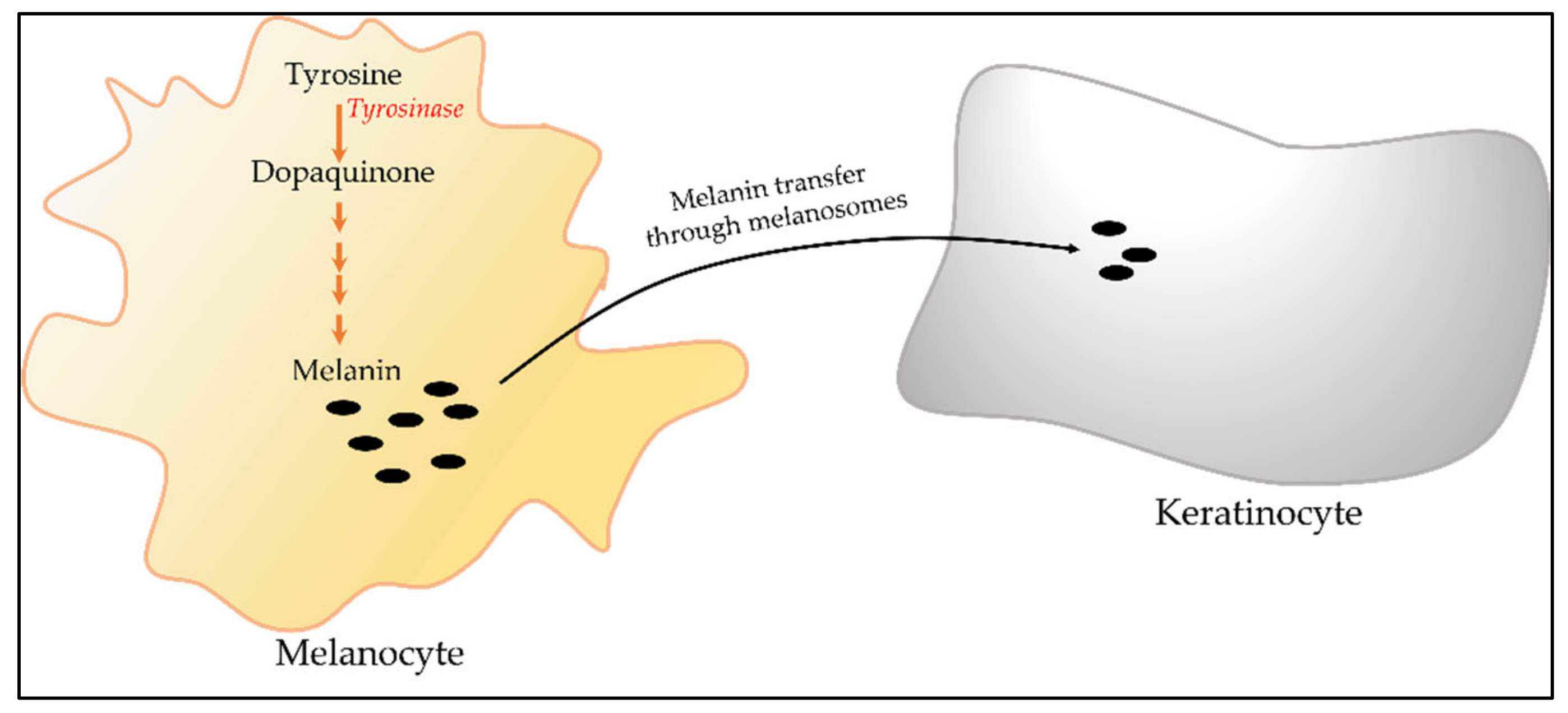
2.8. Cell-Based Systems for Skin Hyperpigmentation

Melanin is a skin pigment responsible for skin coloration. Overproduction of this pigment leads to conditions such as melasma, lentigines, freckles, nevus, and age spots [132]. Such dermatological conditions linked to skin hyperpigmentation may require medical intervention to reduce the pigmentation. Skin lightening, a personal choice that is preferred by many ethnic groups as a cosmetic practice [133], also implicates melanin. In either case, reducing the melanin production is a strategy of choice for reducing skin hyperpigmentation. Two in vitro model systems are routinely used for screening the inhibitors of melanogenesis. One is a cell-based assay, wherein the amount of melanin pigment is measured, and the other assay is based on tyrosinase inhibition.

2.8.1. Cell-Based Assay for Melanogenesis

Biosynthesis of melanin pigment utilizes the specialized cellular organelle called the melanosome, which is present in melanocytes. Melanin synthesized by melanocytes is further distributed to keratinocytes [134] (Figure 5). Mouse melanoma cell line B16F10 and human primary melanocyte NHEM are commonly used for in vitro melanogenesis assay [135,136]. When these cells are treated with cAMP elevating agents such as forskolin or melanocyte-stimulating hormones (α-MSH), they are stimulated to produce cellular melanin [136], which can be measured by absorbance at 405 nm. Potential inhibitors of melanin synthesis or cosmetic ingredients can be included in this system to evaluate their effect on melanogenesis. Kojic acid and phenyl thiourea (PTU) can be used as a positive control inhibitor in these assays [137,138].

2.8.2. Tyrosinase Inhibition Assay

The biosynthetic pathway for melanin production is a cascade of reactions that involves several enzymes and intermediates. The starting point for the biochemical synthesis of melanin is the amino acid tyrosine. This is acted upon by the enzyme tyrosinase and a series of enzymes, finally resulting in the production of melanin. Of all the enzymes of the pathway, tyrosinase is the rate-limiting enzyme. Tyrosinase levels are thought to be the determinants of the color of mammalian skin and hair [139]. Accumulation of this enzyme is shown to result in dermatological disorders such as melisma, age spots, and actinic damage. Several factors influence skin coloration in humans. Reports suggest that more than 150 genes are involved in the determination of skin coloration [140], and the tyrosinase gene is identified as one of them (http://www.ifpcs.org/colorgenes/; accessed on 8 September 2022). Accumulation of tyrosinase is known to result in dermatological disorders. Hence, tyrosinase is a highly targeted enzyme for the inhibition of melanin synthesis. Screening assays for tyrosinase enzyme inhibition involve using purified enzyme tyrosinase from mushroom sources. For the substrate, l-3,4-dihydroxyphenylalanine (L-DOPA) is used, which is converted to dopaquinone during the enzyme reaction and can be monitored by its absorbance at 475 nm [141]. Activity/potency of the inhibitor is expressed as IC50 values, which correspond to the concentration of the inhibitor at which 50% of the enzyme activity is inhibited. Kojic acid is used as a positive control inhibitor in this assay. The phytoactives in Glycyrrhiza glabra [18], Azadiracta indica [19], and Curcuma longa [69] offer anti-blemish properties and can be effectively used as ingredients in cosmetics related to skin hyperpigmentation.

2.9. Melanosome Transfer Assay

As mentioned in previous paragraphs and in Figure 5, melanin pigment is produced in the melanocytes and is transferred to keratinocytes through melanosomes. The transport of melanin pigment from melanocytes to keratinocytes is one of the contributing factors for skin pigmentation [142]. Hence, evaluating the effect of cosmetic ingredients on this process can add value to the anti-hyperpigmentation potential of the ingredients. Several methods are available for studying melanosome transfer. Most of these assays are based on co-culture systems involving melanocytes as donors and keratinocytes as the recipient cells in melanin transfer. A simple cell-based assay system can involve keratinocytes and melanocytes that are prelabeled with carboxyfluorescein diacetate (CFDA). Following co-culturing, the keratinocytes are stained using monoclonal anti-cytokeratin primary antibody and labeled secondary antibodies. In this system, melanin transfer is quantified by counting the keratinocyte cells that are positive both for CFDA and secondary antibody staining [143]. This technique has advantages but also drawbacks, as CFDA is not specific for melanosomes. In another study, investigators identified gp100 as a reliable tracker for melanin transfer in co-culture based assays, and they proposed the silver locus product (Silv/gp100/Pmel17) as a reliable tool for melanosome transfer assays [144]. Few other methods based on co-culturing of melanocytes and keratinocytes have been used by investigators. In these methods, the quantification of melanin transfer is based on scoring the number of cells which are double-positive for melanocyte and keratinocyte specific markers as per the design of the experiment [145].

3. Challenges and Perspectives

In recent years, the cosmetic industry has greatly evolved and is shaping into more of a science- and innovation-driven sector. As a policy, several companies are moving away from using animals in their cosmetic product development. Cell-based models are the best alternatives to animals. Several animal-free safety and toxicity assays prescribed by the Organisation for Economic Co-operation and Development (OECD) have been routinely used in many laboratories. Efficacy validation of cosmetic products is equally important for product development. Since cosmetic products are mostly topical in their application, cell-based model systems are well-suited for efficacy validation. Cell-based assays are more easily adaptable for screening cosmetic products in a high-throughput manner and provide answers in a relatively short time. However, they do have a few limitations that should be carefully considered before initiating screening projects. For example, the single-cell-line-based systems lack cell-to-cell communication with other cell types, and cells in isolation may behave differently when compared to those present in a physiological context. Complex processes such as psoriasis, skin aging, and hair loss involve various cell types and environmental factors. Representing all such components in a cell-based system is not practically possible. Although co-culture-based models can partly consider this, they may not fully account for physiological signal cross-talk between various cell types. Also, 3D skin models may be better alternatives to overcome some of these limitations. However, establishing the 3D models as screening systems is not cost-effective, and validated 3D skin models are unavailable for many indications. Additional parameters such as skin permeability, absorption, and metabolism of topical ingredients (by the enzyme systems in the skin) play an important role in the efficacy of cosmetic ingredients. These parameters need to be carefully considered before extrapolating the in vitro results into possible claims.

Author Contributions

Conceptualization, R.P.R., S.S.B. and P.S.; methodology, P.S., N.S. and S.S.B.; validation, N.S., resources, B.U.V.; writing—R.P.R., S.S.B., P.S. and N.S.; writing—review and editing, R.P.R., B.U.V. and R.M.; supervision, R.P.R. All authors have read and agreed to the published version of the manuscript.

Funding

This research received no external funding.

Institutional Review Board Statement

Not applicable.

Informed Consent Statement

Not applicable.

Conflicts of Interest

The authors declare no conflict of interest.

Abbreviations

ALPalkaline phosphatase
AMPanti-microbial peptide
AQP3aquaporin-3
C. acneCutibacterium acnes
CFDAcarboxyfluorescein diacetate
COL1Acollagen
DHTdihydrotestosterone
ECEuropean Commission
ECMextra-cellular matrix
ELISAenzyme-linked immunoassay
ELNelastin
EPSCsepidermal stem cells
ERKextracellular signal-regulated kinases
FDAFood and Drug Administration
FGFfibroblast growth factor
FLGfilaggrin
HAhylauronic acid
Hacathuman epidermal keratinocytes
HDFhuman dermal fibroblast
HFDPChuman hair follicle dermal papilla cells
HGFhepatocyte growth factor
HPLChigh-performance liquid chromatography
IC50inhibitory concentration 50
IGF-1insulin growth factor 1
ILinterleukins
INVinvolucrin
KGFkeratinocyte growth factor
LC-MSliquid chromatography–mass spectrometry
L-DOPAl-3,4-dihydroxyphenylalanine
MEFmouse primary embryonic fibroblasts
MFAP4microfibril-associated glycoprotein 4
MMPsmatrix metalloprotein
MTT/XTT3-(4,5-dimethylthiazol-2-yl)-2,5-diphenyltetrazolium bromide
OECDOrganisation for Economic Co-operation and Development
ORScouter root sheath cells
PCRpolymerase chain reaction
PDGF-Cplatelet-derived growth factor C
PTUphenyl thiourea
ROSreactive oxygen species
SA-β-galsenescence-associated beta-galactosidase
TGF-β1transforming growth factor beta
TGMstransglutaminase 1, 3, and 5
TIMP1tissue inhibitors of metalloproteinases
TLCthin-layer chromatography
TNF-alphatumor necrosis factor alpha
UVultraviolet
VEGFvascular endothelial growth factor
α-MSHalpha melanocyte-stimulating hormones

References

  1. Cosmetics. Available online: https://cdsco.gov.in/opencms/opencms/en/Cosmetics/cosmetics/ (accessed on 8 September 2022).
  2. Importing Cosmetics|FDA. Available online: https://www.fda.gov/industry/importing-fda-regulated-products/importing-cosmetics#cosmetic (accessed on 8 September 2022).
  3. Regulation (EC) No 1223/2009 of the European Parliament and of the Council of 30 November 2009 On cosmetic products (recast) (Text with EEA relevance). Off. J. Eur. Union. 2009. Available online: https://ec.europa.eu/health/sites/health/files/endocrine_disruptors/docs/cosmetic_1223_2009_regulation_en.pdf (accessed on 8 September 2022).
  4. Chanchal, D.; Swarnlata, S. Novel approaches in herbal cosmetics. J. Cosmet. Derm. 2008, 7, 89–95. [Google Scholar] [CrossRef] [PubMed]
  5. Pandey, A.; Jatana, G.K.; Sonthalia, S. Cosmeceuticals. Adv. Integr. Dermatol. 2019, 393–411. [Google Scholar] [CrossRef]
  6. Brandt, F.S.; Cazzaniga, A.; Hann, M. Cosmeceuticals: Current trends and market analysis. Semin. Cutan. Med. Surg. 2011, 30, 141–143. [Google Scholar] [CrossRef] [PubMed]
  7. Kligman, A. The future of cosmeceuticals: An interview with Albert Kligman, MD, PhD. Interview by Zoe Diana Draelos. Dermatol. Surg. 2005, 31, 890–891. [Google Scholar] [PubMed]
  8. Verheijen, M.; Lienhard, M.; Schrooders, Y.; Clayton, O.; Nudischer, R.; Boerno, S.; Timmermann, B.; Selevsek, N.; Schlapbach, R.; Gmuender, H.; et al. DMSO induces drastic changes in human cellular processes and epigenetic landscape in vitro. Sci. Rep. 2019, 9, 4641. [Google Scholar] [CrossRef] [Green Version]
  9. Baell, J.B.; Holloway, G.A. New substructure filters for removal of pan assay interference compounds (PAINS) from screening libraries and for their exclusion in bioassays. J. Med. Chem. 2010, 53, 2719–2740. [Google Scholar] [CrossRef] [Green Version]
  10. Baell, J.B. Feeling Nature’s PAINS: Natural Products, Natural Product Drugs, and Pan Assay Interference Compounds (PAINS). J. Nat. Prod. 2016, 79, 616–628. [Google Scholar] [CrossRef]
  11. The Role of Inflammation in the Pathology of Acne. Available online: https://pubmed.ncbi.nlm.nih.gov/24062871/ (accessed on 28 July 2022).
  12. Dodou, K. Special Issue “Current and Evolving Practices in the Quality Control of Cosmetics”. Cosmetics 2021, 8, 100. [Google Scholar] [CrossRef]
  13. Dréno, B. What is new in the pathophysiology of acne, an overview. J. Eur. Acad. Dermatol. Venereol. 2017, 31, 8–12. [Google Scholar] [CrossRef] [Green Version]
  14. Picardo, M.; Ottaviani, M.; Camera, E. Mastrofrancesco A. Sebaceous gland lipids. Dermatoendocrinology 2009, 1, 68. [Google Scholar] [CrossRef]
  15. Ottaviani, M.; Camera, E.; Picardo, M. Lipid Mediators in Acne. Mediat. Inflamm. 2010, 2010, 858176. [Google Scholar] [CrossRef] [PubMed] [Green Version]
  16. Fernandéz, J.R.; Rouzard, K.; Voronkov, M.; Feng, X.; Stock, J.B.; Stock, M.; Gordon, J.S.; Shroot, B.; Christensen, M.S.; Pérez, E. SIG1273: A new cosmetic functional ingredient to reduce blemishes and Propionibacterium acnes in acne prone skin. J. Cosmet. Dermatol. 2012, 11, 272–278. [Google Scholar] [CrossRef] [PubMed]
  17. Zadeh, J.B. Licorice (Glycyrrhiza glabra Linn) as a Valuable Medicinal Plant. Available online: https://www.researchgate.net/publication/268502890 (accessed on 29 July 2022).
  18. Joshi, M.D.; Damle, M. Glycyrrhiza glabra (Liquorice)-a potent medicinal herb. Int. J. Herb. Med. 2014, 2, 132–136. Available online: https://www.researchgate.net/publication/305465442 (accessed on 29 July 2022).
  19. Garcia-Jares, C.; Rubio, L.; Baby, A.R.; Freire, T.B.; De Argollo Marques, G.; Rijo, P.; Lima, F.V.; Carlos, J.; De Carvalho, M.; Rojas, J.; et al. Azadirachta indica (Neem) as a Potential Natural Active for Dermocosmetic and Topical Products: A Narrative Review. Cosmetics 2022, 9, 58. [Google Scholar] [CrossRef]
  20. Chainani-Wu, N. Safety and anti-inflammatory activity of curcumin: A component of tumeric (Curcuma longa). J. Altern. Complement. Med. 2003, 9, 161–168. [Google Scholar] [CrossRef] [PubMed] [Green Version]
  21. Lee, J.Y.; Park, W. Anti-Inflammatory Effect of Myristicin on RAW 264.7 Macrophages Stimulated with Polyinosinic-Polycytidylic Acid. Molecules 2011, 16, 7132–7142. [Google Scholar] [CrossRef] [Green Version]
  22. Chattopadhyay, P.; Besra, S.E.; Gomes, A.; Das, M.; Sur, P.; Mitra, S.; Vedasiromoni, J.R. Anti-inflammatory activity of tea (Camellia sinensis) root extract. Life Sci. 2004, 74, 1839–1849. [Google Scholar] [CrossRef]
  23. Yogesh, H.R.; Gajjar, T.; Patel, N.; Kumawat, R. Clinical study to assess efficacy and safety of Purifying Neem Face Wash in prevention and reduction of acne in healthy adults. J. Cosmet. Dermatol. 2021, 21, 2849–2858. [Google Scholar] [CrossRef]
  24. Okoro, O.E.; Adenle, A.; Ludovici, M.; Truglio, M.; Marini, F.; Camera, E. Lipidomics of facial sebum in the comparison between acne and non-acne adolescents with dark skin. Sci. Rep. 2021, 11, 16591. [Google Scholar] [CrossRef]
  25. Zouboulis, C.C.; Seltmann, H.; Neitzel, H.; Orfanos, C.E. Establishment and characterization of an immortalized human sebaceous gland cell line (SZ95). J. Investig. Dermatol. 1999, 113, 1011–1020. [Google Scholar] [CrossRef] [Green Version]
  26. Lo Celso, C.; Berta, M.A.; Braun, K.M.; Frye, M.; Lyle, S.; Zouboulis, C.C.; Watt, F.M. Characterization of bipotential epidermal progenitors derived from human sebaceous gland: Contrasting roles of c-Myc and beta-catenin. Stem Cells 2008, 26, 1241–1252. [Google Scholar] [CrossRef] [PubMed]
  27. Thiboutot, D.; Jabara, S.; McAllister, J.M.; Sivarajah, A.; Gilliland, K.; Cong, Z.; Clawson, G. Human skin is a steroidogenic tissue: Steroidogenic enzymes and cofactors are expressed in epidermis, normal sebocytes, and an immortalized sebocyte cell line (SEB-1). J. Investig. Dermatol. 2003, 120, 905–914. [Google Scholar] [CrossRef] [PubMed] [Green Version]
  28. Barrault, C.; Dichamp, I.; Garnier, J.; Pedretti, N.; Juchaux, F.; Deguercy, A.; Agius, G.; Bernard, F.X. Immortalized sebocytes can spontaneously differentiate into a sebaceous-like phenotype when cultured as a 3D epithelium. Exp. Dermatol. 2012, 21, 314–316. [Google Scholar] [CrossRef]
  29. Barrault, C.; Garnier, J.; Pedretti, N.; Cordier-Dirikoc, S.; Ratineau, E.; Deguercy, A.; Bernard, F.X. Androgens induce sebaceous differentiation in sebocyte cells expressing a stable functional androgen receptor. J. Steroid Biochem. Mol. Biol. 2015, 152, 34–44. [Google Scholar] [CrossRef] [PubMed]
  30. Motoyoshi, K. Enhanced comedo formation in rabbit ear skin by squalene and oleic acid peroxides. Br. J. Dermatol. 1983, 109, 191–198. [Google Scholar] [CrossRef] [PubMed]
  31. Chiba, K.; Yoshizawa, K.; Makino, I.; Kawakami, K.; Onoue, M. Comedogenicity of squalene monohydroperoxide in the skin after topical application. J. Toxicol. Sci. 2000, 25, 77–83. [Google Scholar] [CrossRef] [PubMed] [Green Version]
  32. Zaman, S.U.; Akhtar, N. Effect of Turmeric (Curcuma longa Zingiberaceae) Extract Cream on Human Skin Sebum Secretion. Trop. J. Pharm. Res. 2013, 12, 665–669. [Google Scholar] [CrossRef] [Green Version]
  33. Zhong, H.; Li, X.; Zhang, W.; Shen, X.; Lu, Y.; Li, H. Efficacy of a New Non-drug Acne Therapy: Aloe Vera Gel Combined With Ultrasound and Soft Mask for the Treatment of Mild to Severe Facial Acne. Front. Med. 2021, 8, 662640. [Google Scholar] [CrossRef]
  34. Nam, C.; Kim, S.; Sim, Y.; Chang, I. Anti-Acne Effects of Oriental Herb Extracts: A Novel Screening Method to Select Anti-Acne Agents. Ski. Pharmacol. Physiol. 2003, 16, 84–90. [Google Scholar] [CrossRef]
  35. Balouiri, M.; Sadiki, M.; Ibnsouda, S.K. Methods for in vitro evaluating antimicrobial activity: A review. J. Pharm. Anal. 2015, 6, 71–79. [Google Scholar] [CrossRef] [Green Version]
  36. Nguyen, A.T.; Kim, K.Y. Inhibition of Proinflammatory Cytokines in Cutibacterium acnes-Induced Inflammation in HaCaT Cells by Using Buddleja davidii Aqueous Extract. Int. J. Inflamm. 2020, 2020, 8063289. [Google Scholar] [CrossRef] [PubMed]
  37. Tsai, T.H.; Chuang LTe Lien, T.J.; Liing, Y.R.; Chen, W.Y.; Tsai, P.J. Rosmarinus officinalis Extract Suppresses Propionibacterium acnes–Induced Inflammatory Responses. J. Med. Food 2013, 16, 324–333. [Google Scholar] [CrossRef] [PubMed] [Green Version]
  38. Koseki, J.; Matsumoto, T.; Matsubara, Y.; Tsuchiya, K.; Mizuhara, Y.; Sekiguchi, K.; Nishimura, H.; Watanabe, J.; Kaneko, A.; Hattori, T.; et al. Inhibition of Rat 5α-Reductase Activity and Testosterone-Induced Sebum Synthesis in Hamster Sebocytes by an Extract of Quercus acutissima Cortex. Evid Based Complement. Alternat. Med. 2015, 2015, 853846. [Google Scholar] [CrossRef] [PubMed] [Green Version]
  39. Nilforoushzadeh, M.A.; Aghdami, N.; Taghiabadi, E. Human Hair Outer Root Sheath Cells and Platelet-Lysis Exosomes Promote Hair Inductivity of Dermal Papilla Cell. Tissue Eng. Regen. Med. 2020, 17, 525–536. [Google Scholar] [CrossRef]
  40. Won, C.H.; Jeong, Y.M.; Kang, S.; Koo, T.S.; Park, S.H.; Park, K.Y.; Sung, Y.K.; Sung, J.H. Hair-growth-promoting effect of conditioned medium of high integrin α6 and low CD 71 (α6bri/CD71dim) positive keratinocyte cells. Int. J. Mol. Sci. 2015, 16, 4379–4391. [Google Scholar] [CrossRef] [Green Version]
  41. Watabe, Y.; Tomioka, M.; Watabe, A.; Aihara, M.; Shimba, S.; Inoue, H. The clock gene brain and muscle Arnt-like protein-1 (BMAL1) is involved in hair growth. Arch. Dermatol. Res. 2013, 305, 755–761. [Google Scholar] [CrossRef]
  42. Lin, W.H.; Xiang, L.J.; Shi, H.X.; Zhang, J.; Jiang, L.P.; Cai, P.T.; Lin, Z.L.; Lin, B.B.; Huang, Y.; Zhang, H.L.; et al. Fibroblast growth factors stimulate hair growth through β-catenin and Shh expression in C57BL/6 mice. BioMed Res. Int. 2015, 2015, 730139. [Google Scholar] [CrossRef] [Green Version]
  43. Iida, M.; Ihara, S.; Matsuzaki, T. Hair cycle-dependent changes of alkaline phosphatase activity in the mesenchyme and epithelium in mouse vibrissal follicles. Dev. Growth Differ. 2007, 49, 185–195. [Google Scholar] [CrossRef]
  44. Shin, J.Y.; Kim, J.; Choi, Y.H.; Kang, N.G.; Lee, S. Dexpanthenol Promotes Cell Growth by Preventing Cell Senescence and Apoptosis in Cultured Human Hair Follicle Cells. Curr. Issues Mol. Biol. 2021, 43, 1361–1373. [Google Scholar] [CrossRef]
  45. Saewan, N. Effect of Coffee Berry Extract on Anti-Aging for Skin and Hair—In Vitro Approach. Cosmetics 2022, 9, 66. [Google Scholar] [CrossRef]
  46. Emblica (Phyllanthus emblica Linn.) Fruit Extract Promotes Proliferation in Dermal Papilla Cells of Human Hair Follicle. Available online: https://scialert.net/fulltext/?doi=rjmp.2011.95.100 (accessed on 30 July 2022).
  47. Tiampasook, P.; Chaiyasut, C.; Sivamaruthi, B.S.; Timudom, T.; Nacapunchai, D. Effect of Phyllanthus emblica Linn. on Tensile Strength of Virgin and Bleached Hairs. Appl. Sci. 2020, 10, 6305. [Google Scholar] [CrossRef]
  48. Datta, K.; Singh, A.T.; Mukherjee, A.; Bhat, B.; Ramesh, B.; Burman, A.C. Eclipta alba extract with potential for hair growth promoting activity. J. Ethnopharmacol. 2009, 124, 450–456. [Google Scholar] [CrossRef] [PubMed]
  49. Effect of Trigonella Foenum-Graecum Linn (seeds) and Butea Monosperma Lam (flowers) on Chemotherapy-Induced Alopecia. Available online: https://www.researchgate.net/publication/312163056_Effect_of_Trigonella_foenum-graecum_Linn_seeds_and_Butea_monosperma_Lam_flowers_on_chemotherapy-induced_alopecia (accessed on 30 July 2022).
  50. Adhirajan, N.; Ravi Kumar, T.; Shanmugasundaram, N.; Babu, M. In Vivo and in vitro evaluation of hair growth potential of Hibiscus rosa-sinensis Linn. J. Ethnopharmacol. 2003, 88, 235–239. [Google Scholar] [CrossRef]
  51. Koch, W.; Zagórska, J.; Marzec, Z.; Kukula-Koch, W. Applications of Tea (Camellia sinensis) and Its Active Constituents in Cosmetics. Molecules 2019, 24, 4277. [Google Scholar] [CrossRef] [Green Version]
  52. Evaluation of Clinical Efficacy and Safety of “Anti Dandruff Hair Cream” for the Treatment of Dandruff|Request PDF. Available online: https://www.researchgate.net/publication/284654042_Evaluation_of_clinical_efficacy_and_safety_of_anti_dandruff_hair_cream_for_the_treatment_of_dandruff (accessed on 30 July 2022).
  53. Clinical Evaluation of Herbal Hair Loss Cream in Management of Alopecia Aerata: An Open Study|Semantic Scholar. Available online: https://www.semanticscholar.org/paper/Clinical-evaluation-of-herbal-Hair-Loss-Cream-in-of-Ravichandran-Consultant/b923b7ec4c9eb41f09d37d671d926863de6fb3d5 (accessed on 30 July 2022).
  54. Thiboutot, D.; Harris, G.; Iles, V.; Cimis, G.; Gilliland, K.; Hagari, S. Activity of the type 1 5 alpha-reductase exhibits regional differences in isolated sebaceous glands and whole skin. J. Investig. Dermatol. 1995, 105, 209–214. [Google Scholar] [CrossRef] [Green Version]
  55. Seiffert, K.; Seltmann, H.; Fritsch, M.; Zouboulis, C.C. Inhibition of 5alpha-reductase activity in SZ95 sebocytes and HaCaT keratinocytes in vitro. Horm. Metab. Res. 2007, 39, 141–148. [Google Scholar] [CrossRef]
  56. Jang, S.; Lee, Y.; Hwang, S.L.; Lee, M.H.; Park, S.J.; Lee, I.H.; Kang, S.; Roh, S.S.; Seo, Y.J.; Park, J.K.; et al. Establishment of type II 5alpha-reductase over-expressing cell line as an inhibitor screening model. J. Steroid Biochem. Mol. Biol. 2007, 107, 245–252. [Google Scholar] [CrossRef]
  57. Srivilai, J.; Rabgay, K.; Khorana, N.; Waranuch, N.; Nuengchamnong, N.; Ingkaninan, K. A new label-free screen for steroid 5α-reductase inhibitors using LC-MS. Steroids 2016, 116, 67–75. [Google Scholar] [CrossRef]
  58. Zhang, S.; Duan, E. Fighting against Skin Aging: The Way from Bench to Bedside. Cell Transplant. 2018, 27, 729–738. [Google Scholar] [CrossRef]
  59. Farage, M.A.; Miller, K.W.; Elsner, P.; Maibach, H.I. Intrinsic and extrinsic factors in skin ageing: A review. Int. J. Cosmet. Sci. 2008, 30, 87–95. [Google Scholar] [CrossRef]
  60. Krutmann, J.; Bouloc, A.; Sore, G.; Bernard, B.A.; Passeron, T. The skin aging exposome. J. Dermatol. Sci. 2017, 85, 152–161. [Google Scholar] [CrossRef] [PubMed]
  61. Vogeley, C.; Esser, C.; Tüting, T.; Krutmann, J.; Haarmann-Stemmann, T. Role of the Aryl Hydrocarbon Receptor in Environmentally Induced Skin Aging and Skin Carcinogenesis. Int. J. Mol. Sci. 2019, 20, 6005. [Google Scholar] [CrossRef] [Green Version]
  62. Brinkmann, V.; Ale-Agha, N.; Haendeler, J.; Ventura, N.; Vogeley, C.; Esser, C.; Tüting, T.; Krutmann, J.; Haarmann-Stemmann, T. The Aryl Hydrocarbon Receptor (AhR) in the Aging Process: Another Puzzling Role for This Highly Conserved Transcription Factor. Front. Physiol. 2020, 10, 1561. [Google Scholar] [CrossRef] [PubMed] [Green Version]
  63. Cho, S.; Lee, S.; Lee, M.J.; Lee, D.H.; Won, C.H.; Kim, S.M.; Chung, J.H. Dietary Aloe Vera Supplementation Improves Facial Wrinkles and Elasticity and It Increases the Type I Procollagen Gene Expression in Human Skin in vivo. Ann. Dermatol. 2009, 21, 6–11. [Google Scholar] [CrossRef] [PubMed] [Green Version]
  64. Cho, W.K.; Kim, H.I.; Kim, S.Y.; Seo, H.H.; Song, J.; Kim, J.; Shin, D.S.; Jo, Y.; Choi, H.; Lee, J.H.; et al. Anti-Aging Effects of Leontopodium alpinum (Edelweiss) Callus Culture Extract through Transcriptome Profiling. Genes 2020, 11, 230. [Google Scholar] [CrossRef] [Green Version]
  65. Gupta, G.; Rana, A. Withania somnifera (Ashwagandha): A Review. Pharmacogn. Rev. 2007, 1, 129–136. [Google Scholar]
  66. Varma, S.R.; Sivaprakasam, T.O.; Mishra, A.; Kumar, L.M.S.; Prakash, N.S.; Prabhu, S.; Ramakrishnan, S. Protective Effects of Triphala on Dermal Fibroblasts and Human Keratinocytes. PLoS ONE 2016, 11, e0145921. [Google Scholar] [CrossRef]
  67. Chuarienthong, P.; Lourith, N.; Leelapornpisid, P. Clinical efficacy comparison of anti-wrinkle cosmetics containing herbal flavonoids. Int. J. Cosmet. Sci. 2010, 32, 99–106. [Google Scholar] [CrossRef]
  68. Benameur, T.; Soleti, R.; Panaro, M.A.; La Torre, M.E.; Monda, V.; Messina, G.; Porro, C. Curcumin as Prospective Anti-Aging Natural Compound: Focus on Brain. Molecules 2021, 26, 4794. [Google Scholar] [CrossRef]
  69. Bielak-Zmijewska, A.; Grabowska, W.; Ciolko, A.; Bojko, A.; Mosieniak, G.; Bijoch, Ł.; Sikora, E. The Role of Curcumin in the Modulation of Ageing. Int. J. Mol. Sci. 2019, 20, 1239. [Google Scholar] [CrossRef] [Green Version]
  70. Bylka, W.; Znajdek-Awizeń, P.; Studzińska-Sroka, E.; Brzezińska, M. Centella asiatica in cosmetology. Adv. Dermatol. Allergol. 2013, 1, 46–49. [Google Scholar] [CrossRef] [PubMed]
  71. Quan, T.; Little, E.; Quan, H.; Qin, Z.; Voorhees, J.J.; Fisher, G.J. Elevated matrix metalloproteinases and collagen fragmentation in photodamaged human skin: Impact of altered extracellular matrix microenvironment on dermal fibroblast function. J. Investig. Dermatol. 2013, 133, 1362–1366. [Google Scholar] [CrossRef] [PubMed]
  72. Quan, T.; Qin, Z.; Xia, W.; Shao, Y.; Voorhees, J.J.; Fisher, G.J. Matrix-degrading metalloproteinases in photoaging. J. Investig. Dermatol. Symp. Proc. 2009, 14, 20–24. [Google Scholar] [CrossRef] [PubMed] [Green Version]
  73. Lee, J.; Jung, E.; Kim, Y.; Park, J.; Park, J.; Hong, S.; Kim, J.; Hyun, C.; Kim, Y.S.; Park, D. Asiaticoside induces human collagen I synthesis through TGFbeta receptor I kinase (TbetaRI kinase)-independent Smad signaling. Planta Med. 2006, 72, 324–328. [Google Scholar] [CrossRef] [PubMed]
  74. Bae, J.Y.; Choi, J.S.; Choi, Y.J.; Shin, S.Y.; Kang, S.W.; Han, S.J.; Kang, Y.H. (-)Epigallocatechin gallate hampers collagen destruction and collagenase activation in ultraviolet-B-irradiated human dermal fibroblasts: Involvement of mitogen-activated protein kinase. Food Chem. Toxicol. 2008, 46, 1298–1307. [Google Scholar] [CrossRef]
  75. Ganceviciene, R.; Liakou, A.I.; Theodoridis, A.; Makrantonaki, E.; Zouboulis, C.C. Skin anti-aging strategies. Dermatoendocrinology 2012, 4, 308–319. [Google Scholar] [CrossRef] [Green Version]
  76. Kang, W.; Choi, D.; Park, T. Decanal Protects against UVB-Induced Photoaging in Human Dermal Fibroblasts via the cAMP Pathway. Nutrients 2020, 12, 1214. [Google Scholar] [CrossRef]
  77. Lago, J.C.; Puzzi, M.B. The effect of aging in primary human dermal fibroblasts. PLoS ONE 2019, 14, e0219165. [Google Scholar] [CrossRef] [Green Version]
  78. Kang, C.H.; Rhie, S.J.; Kim, Y.C. Antioxidant and Skin Anti-Aging Effects of Marigold Methanol Extract. Toxicol. Res. 2018, 34, 31–39. [Google Scholar] [CrossRef] [Green Version]
  79. Tan, P.L.; Rajagopal, M.; Chinnappan, S.; Selvaraja, M.; Leong, M.Y.; Tan, L.F.; Yap, V.L. Formulation and Physicochemical Evaluation of Green Cosmeceutical Herbal Face Cream Containing Standardized Mangosteen Peel Extract. Cosmetics 2022, 9, 46. [Google Scholar] [CrossRef]
  80. Papakonstantinou, E.; Roth, M.; Karakiulakis, G. Hyaluronic acid: A key molecule in skin aging. Dermatoendocrinology 2012, 4, 253–258. [Google Scholar] [CrossRef] [PubMed] [Green Version]
  81. Carlomagno, F.; Roveda, G.; Michelotti, A.; Ruggeri, F.; Tursi, F. Anti-Skin-Aging Effect of a Treatment with a Cosmetic Product and a Food Supplement Based on a New Hyaluronan: A Randomized Clinical Study in Healthy Women. Cosmetics 2022, 9, 54. [Google Scholar] [CrossRef]
  82. Foolad, N.; Vaughn, A.R.; Rybak, I.; Burney, W.A.; Chodur, G.M.; Newman, J.W.; Steinberg, F.M.; Sivamani, R.K. Prospective randomized controlled pilot study on the effects of almond consumption on skin lipids and wrinkles. Phyther. Res. 2019, 33, 3212–3217. [Google Scholar] [CrossRef] [Green Version]
  83. Hwang, E.; Lee, D.G.; Park, S.H.; Oh, M.S.; Kim, S.Y. Coriander Leaf Extract Exerts Antioxidant Activity and Protects Against UVB-Induced Photoaging of Skin by Regulation of Procollagen Type I and MMP-1 Expression. J. Med. Food 2014, 17, 985–995. [Google Scholar] [CrossRef]
  84. Bralley, E.; Greenspan, P.; Hargrove, J.L.; Hartle, D.K. Pharmaceutical Biology Inhibition of Hyaluronidase Activity by Vitis rotundifolia. (Muscadine) Berry Seeds and Skins Inhibition of Hyaluronidase Activity by Vitis rotundifolia (Muscadine) Berry Seeds and Skins. Pharm. Biol. 2007, 45, 667–673. [Google Scholar] [CrossRef]
  85. Wlaschek, M.; Maity, P.; Makrantonaki, E.; Scharffetter-Kochanek, K. Connective Tissue and Fibroblast Senescence in Skin Aging. J. Investig. Dermatol. 2021, 141, 985–992. [Google Scholar] [CrossRef] [PubMed]
  86. Lee, J.J.; Ng, S.C.; Hsu, J.Y.; Liu, H.; Chen, C.J.; Huang, C.Y.; Kuo, W.W. Galangin Reverses H 2 O 2-Induced Dermal Fibroblast Senescence via SIRT1-PGC-1α/Nrf2 Signaling. Int. J. Mol. Sci. 2022, 23, 1387. [Google Scholar] [CrossRef]
  87. Ho, C.Y.; Dreesen, O. Faces of cellular senescence in skin aging. Ageing Dev. 2021, 198, 111525. [Google Scholar] [CrossRef]
  88. Fuhrmann-Stroissnigg, H.; Santiago, F.E.; Grassi, D.; Ling, Y.Y.; Niedernhofer, L.J.; Robbins, P.D. SA-β-Galactosidase-Based Screening Assay for the Identification of Senotherapeutic Drugs. J. Vis. Exp. 2019, 148, e58133. [Google Scholar] [CrossRef] [Green Version]
  89. Woo, J.; Shin, S.; Cho, E.; Ryu, D.; Garandeau, D.; Chajra, H.; Fréchet, M.; Park, D.; Jung, E. Senotherapeutic-like effect of Silybum marianum flower extract revealed on human skin cells. PLoS ONE 2021, 16, e0260545. [Google Scholar] [CrossRef]
  90. Jeon, S.; Choi, M. Anti-inflammatory and anti-aging effects of hydroxytyrosol on human dermal fibroblasts (HDFs). Biomed. Dermatol. 2018, 2, 21. [Google Scholar] [CrossRef]
  91. Kim, H.J.; Song, S.B.; Choi, J.M.; Kim, K.M.; Cho, B.K.; Cho, D.H.; Park, H.J. IL-18 downregulates collagen production in human dermal fibroblasts via the ERK pathway. J. Investig. Dermatol. 2010, 130, 706–715. [Google Scholar] [CrossRef] [PubMed]
  92. Yasmin, H.; Kabashima, T.; Rahman, M.S.; Shibata, T.; Kai, M. Amplified and selective assay of collagens by enzymatic and fluorescent reactions. Sci. Rep. 2014, 4, 4950. [Google Scholar] [CrossRef] [Green Version]
  93. Fosang, A.J.; Hey, N.J.; Carney, S.L.; Hardinghami, T.E. An Elisa Plate Based Assay for Hyaluronan Using Biotinylated Proteoglycan G1 Domain (HA-Binding Region). Matrix 1990, 10, 306–313. [Google Scholar] [CrossRef]
  94. Thring, T.S.A.; Hili, P.; Naughton, D.P. Anti-collagenase, anti-elastase and anti-oxidant activities of extracts from 21 plants. BMC Complement. Altern. Med. 2009, 9, 27. [Google Scholar] [CrossRef] [PubMed] [Green Version]
  95. Genc, Y.; Dereli, F.T.G.; Saracoglu, I.; Akkol, E.K. The inhibitory effects of isolated constituents from Plantago major subsp. major L. on collagenase, elastase and hyaluronidase enzymes: Potential wound healer. Saudi Pharm. J. 2019, 28, 101–106. [Google Scholar] [CrossRef] [PubMed]
  96. Dimri, G.P.; Lee, X.; Basile, G.; Acosta, M.; Scott, G.; Roskelley, C.; Medrano, E.E.; Linskens, M.; Rubelj, I.; Pereira-Smith, O.; et al. A biomarker that identifies senescent human cells in culture and in aging skin in vivo. Proc. Natl. Acad. Sci. USA 1995, 92, 9363–9367. [Google Scholar] [CrossRef] [Green Version]
  97. Furue, M. Regulation of Filaggrin, Loricrin, and Involucrin by IL-4, IL-13, IL-17A, IL-22, AHR, and NRF2: Pathogenic Implications in Atopic Dermatitis. Int. J. Mol. Sci. 2020, 21, 5382. [Google Scholar] [CrossRef]
  98. Eckert, R.L.; Sturniolo, M.T.; Broome, A.M.; Ruse, M.; Rorke, E.A. Transglutaminase Function in Epidermis. J. Investig. Dermatol. 2005, 124, 481–492. [Google Scholar] [CrossRef] [Green Version]
  99. Sandilands, A.; Sutherland, C.; Irvine, A.D.; McLean, W.H.I. Filaggrin in the frontline: Role in skin barrier function and disease. J. Cell Sci. 2009, 122, 1285–1294. [Google Scholar] [CrossRef] [Green Version]
  100. Boury-Jamot, M.; Sougrat, R.; Tailhardat, M.; Le Varlet, B.; Bonté, F.; Dumas, M.; Verbavatz, J.-M. Expression and function of aquaporins in human skin: Is aquaporin-3 just a glycerol transporter? Biochim. Biophys. Acta Biomembr. 2006, 1758, 1034–1042. [Google Scholar] [CrossRef] [PubMed] [Green Version]
  101. Bochénska, K.; Smolińska, E.; Moskot, M.; Jakóbkiewicz-Banecka, J.; Gabig-Cimińska, M. Models in the Research Process of Psoriasis. Int. J. Mol. Sci. 2017, 18, 2514. [Google Scholar] [CrossRef] [PubMed]
  102. Marinoni, B.; Ceribelli, A.; Massarotti, M.S.; Selmi, C. The Th17 axis in psoriatic disease: Pathogenetic and therapeutic implications. AutoImmun. Highlights 2014, 5, 9–19. [Google Scholar] [CrossRef] [PubMed] [Green Version]
  103. Albanesi, C.; Madonna, S.; Gisondi, P.; Girolomoni, G. The Interplay Between Keratinocytes and Immune Cells in the Pathogenesis of Psoriasis. Front. Immunol. 2018, 9, 1549. [Google Scholar] [CrossRef] [PubMed] [Green Version]
  104. Varma, S.R.; Sivaprakasam, T.O.; Mishra, A.; Prabhu, S.; Rafiq, M.; Rangesh, P. Imiquimod-induced psoriasis-like inflammation in differentiated Human keratinocytes: Its evaluation using curcumin. Eur. J. Pharmacol. 2017, 813, 33–41. [Google Scholar] [CrossRef] [PubMed]
  105. Morizane, S.; Gallo, R.L. Antimicrobial peptides in the pathogenesis of psoriasis. J. Dermatol. 2012, 39, 225–230. [Google Scholar] [CrossRef] [Green Version]
  106. Ekman, A.K.; Vegfors, J.; Bivik Eding, C.; Enerbäck, C. Overexpression of psoriasin (S100A7) contributes to dysregulated differentiation in psoriasis. Acta Derm. Venereol. 2017, 97, 441–448. [Google Scholar] [CrossRef] [Green Version]
  107. New Biologics in Psoriasis: An Update on IL-23 and IL-17 Inhibitors. Available online: https://pubmed.ncbi.nlm.nih.gov/28319618/ (accessed on 29 July 2022).
  108. Rønholt, K.; Iversen, L. Old and New Biological Therapies for Psoriasis. Int. J. Mol. Sci. 2017, 18, 2297. [Google Scholar] [CrossRef] [Green Version]
  109. Ríos, J.L.; Schinella, G.R.; Andújar, I. Antipsoriatic Medicinal Plants: From Traditional Use to Clinic. In Ethnobotany, 1st ed.; CRC Press: Boca Raton, FL, USA, 2019; pp. 158–186. [Google Scholar] [CrossRef]
  110. Wadher, K.; Dabre, S.; Gaidhane, A.; Trivedi, S.; Umekar, M. Evaluation of antipsoriatic activity of gel containing Pongamia pinnata extract on Imiquimod-induced psoriasis. Clin. Phytosci. 2021, 7, 1–6. [Google Scholar] [CrossRef]
  111. Divakara, P.; Nagaraju, B.; Buden, R.P.; Sekhar, H.S.; Ravi, C.M. Antipsoriatic activity of ayurvedic ointment containing aqueous extract of the bark of Pongamia Pinnata using the rat ultraviolet ray photodermatitis model. Adv. Med. Plant Res. 2013, 1, 8–16. [Google Scholar]
  112. Sangiovanni, E.; Di Lorenzo, C.; Piazza, S.; Manzoni, Y.; Brunelli, C.; Fumagalli, M.; Magnavacca, A.; Martinelli, G.; Colombo, F.; Casiraghi, A.; et al. Vitis vinifera L. Leaf Extract Inhibits In Vitro Mediators of Inflammation and Oxidative Stress Involved in Inflammatory-Based Skin Diseases. Antioxidants 2019, 8, 134. [Google Scholar] [CrossRef] [PubMed] [Green Version]
  113. Reinke, J.M.; Sorg, H. Wound repair and regeneration. Eur. Surg. Res. 2012, 49, 35–43. [Google Scholar] [CrossRef] [PubMed]
  114. Pastar, I.; Stojadinovic, O.; Yin, N.C.; Ramirez, H.; Nusbaum, A.G.; Sawaya, A.; Patel, S.B.; Khalid, L.; Isseroff, R.R.; Tomic-Canic, M. Epithelialization in Wound Healing: A Comprehensive Review. Adv. Wound Care 2014, 3, 445–464. [Google Scholar] [CrossRef] [Green Version]
  115. Yue, P.Y.K.; Leung, E.P.Y.; Mak, N.K.; Wong, R.N.S. A simplified method for quantifying cell migration/wound healing in 96-well plates. J. Biomol. Screen 2010, 15, 427–433. [Google Scholar] [CrossRef] [PubMed] [Green Version]
  116. Lee, J.; Wang, Y.L.; Ren, F.; Lele, T.P. Stamp wound assay for studying coupled cell migration and cell debris clearance. Langmuir 2010, 26, 16672–16676. [Google Scholar] [CrossRef]
  117. Hettler, A.; Werner, S.; Eick, S.; Laufer, S.; Weise, F. A New In Vitro Model to Study Cellular Responses after Thermomechanical Damage in Monolayer Cultures. PLoS ONE 2013, 8, e82635. [Google Scholar] [CrossRef]
  118. Keese, C.R.; Wegener, J.; Walker, S.R.; Giaever, I. Electrical wound-healing assay for cells in vitro. Proc. Natl. Acad. Sci. USA 2004, 101, 1554–1559. [Google Scholar] [CrossRef] [Green Version]
  119. Zordan, M.D.; Mill, C.P.; Riese, D.J.; Leary, J.F. A high throughput, interactive imaging, bright-field wound healing assay. Cytom. Part A 2011, 79, 227–232. [Google Scholar] [CrossRef] [Green Version]
  120. Fronza, M.; Heinzmann, B.; Hamburger, M.; Laufer, S.; Merfort, I. Determination of the wound healing effect of Calendula extracts using the scratch assay with 3T3 fibroblasts. J. Ethnopharmacol. 2009, 126, 463–467. [Google Scholar] [CrossRef]
  121. Fox, L.T.; Mazumder, A.; Dwivedi, A.; Gerber, M.; du Plessis, J.; Hamman, J.H. In Vitro wound healing and cytotoxic activity of the gel and whole-leaf materials from selected aloe species. J. Ethnopharmacol. 2017, 200, 1–7. [Google Scholar] [CrossRef]
  122. Razia, S.; Park, H.; Shin, E.; Shim, K.S.; Cho, E.; Kang, M.C.; Kim, S.Y. Synergistic effect of Aloe vera flower and Aloe gel on cutaneous wound healing targeting MFAP4 and its associated signaling pathway: In-vitro study. J. Ethnopharmacol. 2022, 290, 115096. [Google Scholar] [CrossRef] [PubMed]
  123. Matsubayashi, Y.; Razzell, W.; Martin, P. ‘White wave’ analysis of epithelial scratch wound healing reveals how cells mobilise back from the leading edge in a myosin-II-dependent fashion. J. Cell Sci. 2011, 124, 1017–1021. [Google Scholar] [CrossRef] [PubMed]
  124. Zaja-Milatovic, S.; Richmond, A. CXC chemokines and their receptors: A case for a significant biological role in cutaneous wound healing. Histol. Histopathol. 2008, 23, 1399–1407. [Google Scholar] [CrossRef] [PubMed]
  125. Yamamoto, T.; Eckes, B.; Mauch, C.; Hartmann, K.; Krieg, T. Monocyte Chemoattractant Protein-1 Enhances Gene Expression and Synthesis of Matrix Metalloproteinase-1 in Human Fibroblasts by an Autocrine IL-1α Loop. J. Immunol. 2000, 164, 6174–6179. [Google Scholar] [CrossRef] [Green Version]
  126. Ridiandries, A.; Tan, J.T.M.; Bursill, C.A. The role of chemokines in wound healing. Int. J. Mol. Sci. 2018, 19, 3217. [Google Scholar] [CrossRef] [Green Version]
  127. Cole, J.; Tsou, R.; Wallace, K.; Gibran, N.; Isik, F. Early gene expression profile of human skin to injury using high-density cDNA microarrays. Wound Repair Regen. 2001, 9, 360–370. [Google Scholar] [CrossRef]
  128. Wiegand, C.; Hipler, U.C.; Elsner, P.; Tittelbach, J. Keratinocyte and fibroblast wound healing in vitro is repressed by non-optimal conditions but the reparative potential can be improved by water-filtered infrared A. Biomedicines 2021, 9, 1802. [Google Scholar] [CrossRef]
  129. Nowinski, D.; Lysheden, A.S.; Gardner, H.; Rubin, K.; Gerdin, B.; Ivarsson, M. Analysis of Gene Expression in Fibroblasts in Response to Keratinocyte-Derived Factors In Vitro: Potential Implications for the Wound Healing Process. J. Investig. Dermatol. 2004, 122, 216–221. [Google Scholar] [CrossRef] [Green Version]
  130. Leng, H.; Pu, L.; Xu, L.; Shi, X.; Ji, J.; Chen, K. Effects of aloe polysaccharide, a polysaccharide extracted from Aloe vera, on TNF-α-induced HaCaT cell proliferation and the underlying mechanism in psoriasis. Mol. Med. Rep. 2018, 18, 3537–3543. [Google Scholar] [CrossRef] [Green Version]
  131. Wahedi, H.M.; Jeong, M.; Chae, J.K.; Do, S.G.; Yoon, H.; Kim, S.Y. Aloesin from Aloe vera accelerates skin wound healing by modulating MAPK/Rho and Smad signaling pathways in vitro and in vivo. Phytomedicine 2017, 28, 19–26. [Google Scholar] [CrossRef]
  132. Ju Woo, H.; Jun, D.Y.; Lee, J.Y.; Park, H.S.; Woo, M.H.; Park, S.J.; Kim, S.C.; Yang, C.H.; Kim, Y.H. Anti-inflammatory action of 2-carbomethoxy-2,3-epoxy-3-prenyl-1,4-naphthoquinone (CMEP-NQ) suppresses both the MyD88-dependent and TRIF-dependent pathways of TLR4 signaling in LPS-stimulated RAW264.7 cells. J. Ethnopharmacol. 2017, 205, 103–115. [Google Scholar] [CrossRef]
  133. Evaluation of Antioxidant and Anti-Melanogenic Activities of Different Extracts from Aerial Parts of Nepeta Binaludensis Jamzad in Murine Melanoma B16F10 Cells. Available online: https://pubmed.ncbi.nlm.nih.gov/27482348/ (accessed on 29 July 2022).
  134. Burger, P.; Landreau, A.; Azoulay, S.; Michel, T.; Fernandez, X. Skin Whitening Cosmetics: Feedback and Challenges in the Development of Natural Skin Lighteners. Cosmetics 2016, 3, 36. [Google Scholar] [CrossRef]
  135. Maranduca, M.A.; Branisteanu, D.; Serban, D.N.; Branisteanu, D.C.; Stoleriu, G.; Manolache, N.; Serban, I.L. Synthesis and physiological implications of melanic pigments. Oncol. Lett. 2019, 17, 4183–4187. [Google Scholar] [CrossRef] [PubMed] [Green Version]
  136. Zhou, S.; Yotsumoto, H.; Tian, Y.; Sakamoto, K. α-Mangostin suppressed melanogenesis in B16F10 murine melanoma cells through GSK3β and ERK signaling pathway. Biochem. Biophys. Rep. 2021, 26, 100949. [Google Scholar] [CrossRef] [PubMed]
  137. Kang, M.C.; Lee, J.W.; Lee, T.H.; Subedi, L.; Wahedi, H.M.; Do, S.G.; Shin, E.; Moon, E.Y.; Kim, S.Y. UP256 Inhibits Hyperpigmentation by Tyrosinase Expression/Dendrite Formation via Rho-Dependent Signaling and by Primary Cilium Formation in Melanocytes. Int. J. Mol. Sci. 2020, 21, 5341. [Google Scholar] [CrossRef]
  138. Lajis, A.F.B.; Hamid, M.; Ariff, A.B. Depigmenting effect of Kojic acid esters in hyperpigmented B16F1 melanoma cells. J. Biomed. Biotechnol. 2012, 2012, 952452. [Google Scholar] [CrossRef] [Green Version]
  139. D’Mello, S.A.N.; Finlay, G.J.; Baguley, B.C.; Askarian-Amiri, M.E. Signaling Pathways in Melanogenesis. Int. J. Mol. Sci. 2016, 17, 1144. [Google Scholar] [CrossRef] [Green Version]
  140. Yamaguchi, Y.; Hearing, V.J. Physiological factors that regulate skin pigmentation. BioFactors 2009, 35, 193–199. [Google Scholar] [CrossRef] [Green Version]
  141. Kim, C.S.; Noh, S.G.; Park, Y.; Kang, D.; Chun, P.; Chung, H.Y.; Jung, H.J.; Moon, H.R. A Potent Tyrosinase Inhibitor, ( E)-3-(2,4-Dihydroxyphenyl)-1-(thiophen-2-yl)prop-2-en-1-one, with Anti-Melanogenesis Properties in α-MSH and IBMX-Induced B16F10 Melanoma Cells. Molecules 2018, 23, 2725. [Google Scholar] [CrossRef] [Green Version]
  142. Moreiras, H.; Seabra, M.C.; Barral, D.C. Melanin Transfer in the Epidermis: The Pursuit of Skin Pigmentation Control Mechanisms. Int. J. Mol. Sci. 2021, 22, 4466. [Google Scholar] [CrossRef]
  143. Greatens, A.; Hakozaki, T.; Koshoffer, A.; Epstein, H.; Schwemberger, S.; Babcock, G.; Bissett, D.; Takiwaki, H.; Arase, S.; Wickett, R.R.; et al. Effective inhibition of melanosome transfer to keratinocytes by lectins and niacinamide is reversible. Exp. Dermatol. 2005, 14, 498–508. [Google Scholar] [CrossRef] [PubMed]
  144. Singh, S.K.; Nizard, C.; Kurfurst, R.; Bonte, F.; Schnebert, S.; Tobin, D.J. The silver locus product (Silv/gp100/Pmel17) as a new tool for the analysis of melanosome transfer in human melanocyte-keratinocyte co-culture. Exp. Dermatol. 2008, 17, 418–426. [Google Scholar] [CrossRef] [PubMed]
  145. Lin, H.C.; Shieh, B.H.; Lu, M.H.; Chen, J.Y.; Chang, L.T.; Chao, C.F. A method for quantifying melanosome transfer efficacy from melanocytes to keratinocytes in vitro. Pigment Cell Melanoma Res. 2008, 21, 559–564. [Google Scholar] [CrossRef] [PubMed]
Figure 1. Key components of pathophysiology of acne vulgaris. Changes in the microbiome and infection by C. acne lead to the initiation of acne vulgaris. Although inflammation is beneficial for fighting against the infection, it can lead to aggravation of acne. In addition to this, androgenic factors can influence sebum quality and quantity and dihydrotestosterone (DHT). Changes in the sebum quality and quantity can create a conducive atmosphere for microbes during acne. Cell-based model systems to evaluate anti-acne ingredients take these components of pathology into consideration. The “up arrow” marked in red color indicates increase in expression.
Cosmetics 09 00107 g001
Figure 2. Key factors contributing for hair follicle growth. The proliferation of mesenchymal stem cells from dermal papilla and non-keratinized epithelial cells contributes to the growth of the hair follicles. Agents that can stimulate the growth of these cell types can be potential hair-growth promoters. The enzyme 5-alpha reductase converts testosterone into a more potent form, i.e., dihydrotestosterone (DHT), which has inhibitory effects on hair growth. Inhibiting this enzyme is one of the strategies for promoting hair growth. Finally, vasodilation facilitates nutrient supply to hair follicle cells and promotes hair growth.
Cosmetics 09 00107 g002
Figure 3. Cellular and morphological characteristics of skin aging. Skin aging can be due to either intrinsic or extrinsic factors. Along with age, there is an increase in the enzymes that degrade collagen and extracellular matrix. This results in reduced skin elasticity, leading to the appearance of external features of aging such as fine wrinkles, skin dehydration, and aberrant pigmentation. The up and down arrows in red indicate increase and decrease, respectively.
Cosmetics 09 00107 g003
Figure 4. Cellular events involved in wound-healing. Wound-healing is a multi-phasic process involving cellular hemostasis, inflammation, proliferation, re-epithelialization, and remodeling. Cytokines and chemokines act as messengers to regulate these phases. Platelets, fibroblasts, immune cells, keratinocytes, and epidermal stem cells participate in this process. Remodeling through changes in ECM proteins and connective tissues comprises the final phase of wound-healing.
Cosmetics 09 00107 g004
Figure 5. Schematic of melanin biosynthesis. Melanin biosynthesis takes place in melanocytes using tyrosine. Tyrosinase is a key enzyme for the synthesis of melanin. Once produced, melanin is transported to keratinocytes. Inhibition of tyrosinase, inhibition of melanogenesis, and melanin transfer are the main functions considered when screening cosmetic ingredients for their anti-hyperpigmentation activity.
Cosmetics 09 00107 g005
Table 1. Strategies for validation of anti-acne activity.
Type of AssaySignificance of AssayModel System Used and Evaluation StrategyEnd Point Measured
Anti-microbial activityAnti-microbial activity against C. acnes is important for the anti-acne effects of the ingredientsC. acnes bacterial culture Anti-bacterial activity of the ingredients using assays such as agar diffusion assay [35]
Anti-inflammatory activityInflammation is an integral part of the pathogenesis of acne vulgaris, and the anti-inflammatory activity of the ingredients can offer good protection against acne. Monocyte cell lines (THP-1), keratinocytes (HaCaT cell line), or human sebocyte cell line. (inflammation induced in the cells by exposure to heat-inactive C. acnes or peptidoglycans [16]Effect of cosmetic ingredients on C. acnes-induced inflammation as measured by the levels of inflammatory cytokines in the cell culture supernatant or expression of the respective genes in the cells [36,37]
Sebum quality and sebostatic activitySebum quality and quantity is an important parameter that can contribute to the progression of the pathology of acne vulgarisHuman sebocytic cell lines
  • Effect of cosmetic ingredients on the total amount of lipid accumulation in sebocytes as measured by oil-o-red staining [38]
  • Expression profiling of lipogenic genes in the sebocytes [15]
  • Effect of the ingredients on proliferation of sebocytes using standard cell proliferation based assays [26,27].
  • Qualitative nature of the lipids secreted by the sebocytes to the cell culture supernatant; Gene expression profiling of the sebocytes for key genes involved in squalene synthesis and squalene peroxidation [31]
Table 2. Strategies for validation of hair-growth-promoting activity.
Type of AssaySignificance of AssayModel System Used and Evaluation StrategyEnd Point Measured
Proliferation of the follicle cellsHair follicle cells are primarily responsible for the growth of hair.HFPDPC or ORSc cells are used as model systems. The cells are grown in the presence of different doses of the test substance for different time points: 24, 48, and 72 hEffect of the ingredients on proliferation of HFPDPC or ORSc cells using standard cell-proliferation-based assays [40].
Inhibition of 5-α reductase5-α reductase converts testosterone into a more potent form, i.e., DHT. DHT is known to be involved in male pattern baldness.Keratinocyte and sebocytes that are known to exhibit detectable levels of 5-α reductase activity (should be individually established in the labs since the activity levels may depend on the cell type)Effect of the ingredients on the enzyme activity of 5-α reductase using testosterone as substrate. The detection of the DHT can be based on radioactive detection, TLC, HPLC, or LC-MS [38,57]
Table 3. Strategies for validation of skin-aging.
Type of AssaySignificance of AssayModel System Used and Evaluation StrategyEnd Point Measured
Estimation of collagen levelsCollagen is important for skin integrity. Its levels are known to reduce with aging, which results in visible phenotypes such as skin wrinklesHuman dermal fibroblasts (e.g., HDF or HS68) are treated with test substances, and collagen levels are estimated by using biochemical methods or ELISA [91,92]Collagen levels
Estimation of hyaluronic acid levelsHyaluronic acid is an important component of ECM, and it is known to reduce with agingELISA-based estimation of HA in human fibroblasts such as HDF in presence of test substances [93]Hyaluronic acid levels
Estimation of enzyme activities of collagenase, elastase, hyaluronidase, and HA synthasesThese enzymes affect the levels of key ECM components such as collagen, elastin, and hyaluronic acidEffect of cosmetic ingredients on the activities of these enzymes are tested by respective biochemical methods. Human fibroblasts (HDF) or keratinocytes (HaCaT) can be employed [94,95]Enzyme activities
PhotoagingExposure to UV is one of the triggers for age associated changes in the skin Human dermal fibroblasts (HDF or HS68) cells are exposed to UV light and the effect of the test substance is evaluated [72]
  • Activities of collagenase, elastase, and various other MMPs
  • Biochemical estimation of collagen levels
  • Gene expression levels of MMPs
Cellular senescenceCellular senescence causes irreversible cessation of cell proliferation and increases with agingHuman dermal fibroblasts are exposed to either UV light or hydrogen peroxide to induce cellular senescence; test substances are evaluated for their effectiveness to regulate or reverse the senescence [87,96]
  • Gene or protein expression levels of of p53, p21, and p16
  • Cellular staining for SA-β-gal
Table 4. Strategies for validation of wound-healing activity.
Wound Creation MethodsProsCons
Mechanical wounding (scratch assay)Scratch introduced mechanically on cell monolayer using a pipette tip or cell scraper or stamping with stamp molds [115,116]Simple technique, easily available toolsIrregular wound area created, scraped cells may be retained at the edges, impairing the data
Thermo-mechanical woundingTemperature-controlled stamp molds [117]Thermal and mechanical damage healing can be analyzedHeat may affect surrounding cells of the wound area, impairing data
Electrical woundingCells grown on electrodes, scratch made on the cell layer, change in impedance studiedAccurate, reproducible dataHeating of bystander cells surrounding the wound; specialized tools required to perform electrical cell-substrate impedance sensing (ECIS) [118]
Optical woundingLaser beam pulses used to create precise woundsPrecise, high-throughput, reproducible wounds and data createdSpecialized equipment required: Laser-enabled analysis and processing (LEAP) instrument [119]
Publisher’s Note: MDPI stays neutral with regard to jurisdictional claims in published maps and institutional affiliations.

