Article

Compounds in Indonesian Ginger Rhizome Extracts and Their Potential for Anti-Skin Aging Based on Molecular Docking

by
Shadila F. Asoka
1,
Irmanida Batubara
1,2,*,
Ayu Rahmania Lestari
1,
Wulan Tri Wahyuni
1,2 and
Setyanto Tri Wahyudi
2,3
1
Department of Chemistry, Faculty of Mathematics and Natural Sciences, IPB University, Bogor 16680, Indonesia
2
Tropical Biopharmaca Research Center, IPB University, Bogor 16128, Indonesia
3
Department of Physics, Faculty of Mathematics and Natural Sciences, IPB University, Bogor 16680, Indonesia
*
Author to whom correspondence should be addressed.
Submission received: 16 October 2022 / Revised: 14 November 2022 / Accepted: 24 November 2022 / Published: 1 December 2022
(This article belongs to the Special Issue Advances in Regulatory and Technological Aspects of Cosmetics)

Abstract

:
Skin aging is a condition caused by reactive oxygen species (ROS) and advanced glycation end products (AGEs). Indonesian gingers (Zingiber officinale), which consists of Gajah (GG), Red (MM), and Emprit (EE) ginger, are thought to produce anti-skin aging compounds through enzyme inhibition. The enzymes used in the molecular docking study were collagenase, hyaluronidase, elastase, and tyrosinase. This study aimed to determine the compounds contained in Indonesian ginger rhizome ethanolic extracts using liquid chromatography–mass spectrometry/mass spectrometry to differentiate metabolites contained in the different Indonesian ginger rhizome extracts. A principal component analysis (PCA) and a heat map analysis were used in order to determine which compounds and extracts contained potential anti-skin aging properties based on a molecular docking study. Ascorbic acid was used as a control ligand in the molecular docking study. Ninety-eight compounds were identified in three different ginger rhizomes extracts and were grouped into three separate quadrants. The most potent compound for anti-skin aging in the Indonesian ginger rhizome extracts was octinoxate. Octinoxate showed a high abundance in the EE ginger rhizome extract. Therefore, the EE ginger extract was the Indonesian ginger rhizome extract with the greatest potential for anti-skin aging.

1. Introduction

Aging is a natural process to grow old in an organism. According to Rose et al., aging is a progressive condition throughout life; or after a certain stage, the probability that a given individual will die during the next unit of time from randomly distributed causes [1]. The World Health Organization states that aging results from the accumulated impact of various molecular and cellular damages over time. This leads to a gradual decline in physical, psychological, and mental capacities, and to social changes, whereby increasing the risk of disease and ultimately death [2]. These changes are related to a person’s age in years. Aging can occur in cells [3], tissues [4], and organs [5]. Skin is one of the tissues that experiences aging, and skin aging is a concern because the signs are clearly visible, thereby reducing the aesthetics of the body’s appearance.
Skin is a body tissue consisting of the epidermis, dermis, and hypodermis layers. The skin’s main function is to protect the body from the external environment. Skin tissue is one of the most frequently cared for parts of the body because people want to look healthy. This is evident from the Central Bureau of Statistics, Republic of Indonesia (2021), which stated that skin care sales in an e-commerce store grew 5.59% in the first quarter of 2021. The skincare products in demand included a range of topical products for significant problems such as acne, hyperpigmentation, and skin aging. Skin aging is the process of changes in the skin over time. Skin aging is characterized by the appearance of wrinkles and hyperpigmented spots on the outermost layer of the skin [6].
Intrinsic and extrinsic factors cause skin aging. The intrinsic factor that causes skin aging is that the natural cycle of skin cell production during a person’s life slows down. Skin cells will undergo a regeneration process, namely replacing old skin cells with new ones, every 14–28 days [7]. Genetic, hormonal, gender, and biochemical changes in the body are also intrinsic factors of skin aging, while pollution and ultraviolet (UV) light are extrinsic factors that cause skin aging [8]. Excessive exposure to pollution and UV rays can cause skin aging. The process of skin aging is called photoaging [9]. Photoaging occurs due to the formation of free radicals in the skin. These free radicals accelerate photoaging because free radicals are unstable molecules, so they are reactive and damage surrounding cells [10]. Generally, these reactive molecules are called reactive oxygen species (ROS). In addition to ROS, other compounds, namely advanced glycation end products (AGEs), accelerate the aging process. This compound is the Maillard reaction’s product in the body. AGEs cause stiffness and loss of skin elasticity through the mechanism of connective tissue accumulation in melanocytes [11].
Ginger is a plant often used as a base for traditional medicines. Ginger in Indonesia is divided into three main varieties, namely Gajah ginger (Zingiber officinale var. roscoe, GG), Emprit ginger (Z. officinale var. amarum, EE), and Red ginger (Z. officinale var. rubrum, MM). Treatment using ginger was first carried out in China to treat nausea [12] and increase immunity [13]. The ginger rhizome contains many active compounds that can be found in its extracts. Geranial compounds in ginger rhizome essential oil have antibacterial activity [14]. The compounds α-zingiberene, camphene, sesquiphellandrene, and bisabolene in ginger rhizome essential oil have antioxidant activity [15]. The compound 6-shogaol in ginger rhizome extract reduced the effects of wrinkling and age in experimental rats that had been exposed to UV B radiation for eight weeks [16]. Ginger extract has been used in several skin care products with claims of providing soothing benefits, regenerating skin, and acting as a bleaching agent. In addition, ginger can be applied as a mask to increase collagen production and prevent acne. Exploration of the anti-skin aging potential in ginger rhizome extract still has to be done because there are still many compounds whose potential is not yet known. One of the methods to determine anti-skin aging activity is the molecular docking study.
The anti-skin aging activity determination of ginger rhizome extracts can be predicted using the molecular docking study. Molecular docking is a computational method that can estimate the interactions between compounds and receptors [17,18]. Molecular docking is a direct and rational drug discovery approach, and a low-cost and effective screening [19,20]. The interaction between a small molecule and a protein at the atomic level can be modeled using the molecular docking approach, allowing us to characterize the behavior of small molecules in the binding site of target proteins as well as elucidate fundamental biochemical processes [21]. Molecular docking can predict an optimized orientation of the ligand on its target [22]. It can predict different binding modes of ligands in the groove of the target molecule. This can be used to develop more potent, selective, and efficient drug candidates [23].
In this study, the receptors and their binding compounds are expected to be able to inhibit aging activity on the skin. The receptors used in this study were collagenase (2TCL), hyaluronidase (2PE4), elastase (3F19), and tyrosinase (5M8R). Collagenase is an enzyme that plays a role in the process of cleavage of the extracellular matrix, especially collagen types 1, 2, and 3. If the collagenase enzyme is inhibited, the partition of the extracellular matrix that causes collagen degradation can be prevented [24]. The hyaluronidase enzyme plays a role in the breakdown of hyaluronic acid, which causes dry skin. If the hyaluronidase enzyme’s activity is inhibited, then hyaluronic acid’s breakdown will also be inhibited, so skin moisture is hoped to be maintained [25]. Elastase is an enzyme that has the function of degrading elastin whereby causing the skin to become stiff. Excess elastase activity can be inhibited so that skin elasticity can be maintained [26]. The enzyme that plays a role in producing melanin in the skin is tyrosinase. Excessive melanin production will cause hyperpigmentation, making the skin look aged. Melanin production will be reduced if this enzyme is inhibited so that hyperpigmentation can be prevented [27].
A full exploration of the compounds in the three Indonesian ginger rhizome extracts remains to be done. Therefore, this study aimed to identify only those compounds showing a potential for anti-skin aging based on a molecular docking study and to differentiate the metabolites contained in the extracts of the three types of Indonesian ginger rhizome (GG, EE, MM).

2. Materials and Methods

2.1. Preparation and Sample Extraction

The types of ginger used were GG, EE, and MM, aged >8 months, referring to the guidelines of the Agricultural Technology Study Center, The Ministry of Agriculture of the Republic of Indonesia [28] regarding Ginger Plant Cultivation. These ginger samples were obtained from Nagrak, Sukabumi, West Java, Indonesia (6°52′11.6″ S 106°48′27.1″ E) in a fresh state. A quantity of 25 g of fresh ground ginger rhizome was extracted using ultrasonic waves by Ultrasonic Bath 60 Hz (Ovan, Barcelona, ES) (Barcelona, Spanyol). The solvent used was ethanol absolute for analysis from Merck 1.00983.2500 (Darmstadt, Germany) in a quantity of as much as 125 mL. Extraction time lasted for 30 min at a temperature of ±35 °C. The extract was filtered and stored in the refrigerator prior to the next step of analysis.

2.2. Identification Compounds in Indonesian Ginger Rhizome Extracts Using Liquid Chromatography–Mass Spectrometry/Mass Spectrometry (LC–MS/MS)

Ginger rhizome extracts were analyzed using the UHPLC Vanquish Tandem Q Exactive Plus Orbitrap HRMS Thermo Scientific instrument (Thermo Fisher Scientific Inc., Waltham, MA, USA). The extracts were dissolved in methanol (LC–MS grade) and filtered by a PTFE 0.2 µm membrane. The column used was Accucore C18 Thermo Scientific (2.1 mm × 100 mm, 1.5 µm) (Thermo Fischer Scientific, Waltham, MA, USA) operated at 30 °C. The mobile phase consisted of 0.1% formic acid in water (A) and 0.1% formic acid in acetonitrile (B). The flow rate used was 0.2 mL/minute, and the injection volume was 2.5 µL. The type of elution used was gradient elution. At 0–1.00 min, the eluent composition was 5% B. At 1.01–25.00 min, the eluent composition changed linearly to 95% B. Finally, at 25.01–28.00 min, the eluent composition was maintained at 95% B. Finally, the composition of the eluent became 5% B at 28.01–30.00 min. The analysis time used was 30 min. Electrospray Ionization (ESI) was the ionization source, with a spraying voltage of 5500 V. A quadrupole-orbitrap mass analyzer was used.

2.3. Statistical Analysis

The abundance values for each compound were analyzed using an ANOVA: single factor. The significant differences in compound abundance among the three extracts (GG, EE, and MM) were determined using a t-test. An ANOVA and a t-test analysis were performed using Microsoft Excel, while Orange data mining software was used for a PCA and a heat map analysis to differentiate the three extracts. Imported data were preprocessed using normalized data from the software. The data obtained were grouped using the heat map analysis and the PCA with a percentage of the variance of both PCs of at least 70%.

2.4. Ligands and Receptors Preparation for Molecular Docking

The test ligands used in this study were compounds chosen from the ginger rhizome extract based on an LC–MS/MS analysis using ascorbic acid as a control ligand. The test and control ligands were downloaded from the PubChem (https://pubchem.ncbi.nlm.nih.gov/) (accessed on 26 August 2022) in *sdf. The 3D ligand structure was partially charged using the Gasteiger charge method in AutoDock Tools 1.5.7 software and saved in *pdbqt. The 3D crystal structures of the collagenase (2TCL), hyaluronidase (2PE4), elastase (3F19), and tyrosinase (5M8R) were downloaded from the Protein Data Bank (https://www.rcsb.org/) (accessed on 19 April 2022) in *pdb. The downloaded structure was checked for missing residues. Water and natural ligands were removed using UCSF Chimera. The prepared structure was saved in *pdb format using UCSF Chimera. The receptor structure was optimized using AutoDock Tools 1.5.7 and then saved in *pdbqt format.

2.5. Molecular Docking Simulation, Visualization, and admetSAR Analysis

Molecular docking simulation was carried out by targeted docking because the receptor’s active site was already known. The gridbox was made according to the size of the test ligand and then adjusted to the receptor’s crystal ligand binding site area. The files were collected in a folder containing the ligands to use and the configuration script containing the gridbox description. The molecular docking simulations were run with Autodock Vina. The molecular docking results were analyzed to obtain the test ligand with the highest negative affinity energy. Ten compounds with the highest negative affinity energy from each receptor would be entered into the visualization and admetSAR test stages. Ligands that appear in 2 or 3 receptors were selected as ligands with great potential as anti-skin aging compounds. After being docked using LigPlus, the complex was screen-captured. Next, selected ligands were tested by admetSAR using the site http://lmmd.ecust.edu.cn/admetSAR2/ (accessed on 12 October 2022) and then tested using the advance predict option. The SMILES ligand format was used in this test which can be downloaded from the site https://pubchem.ncbi.nlm.nih.gov (accessed on 12 October 2022).

3. Results

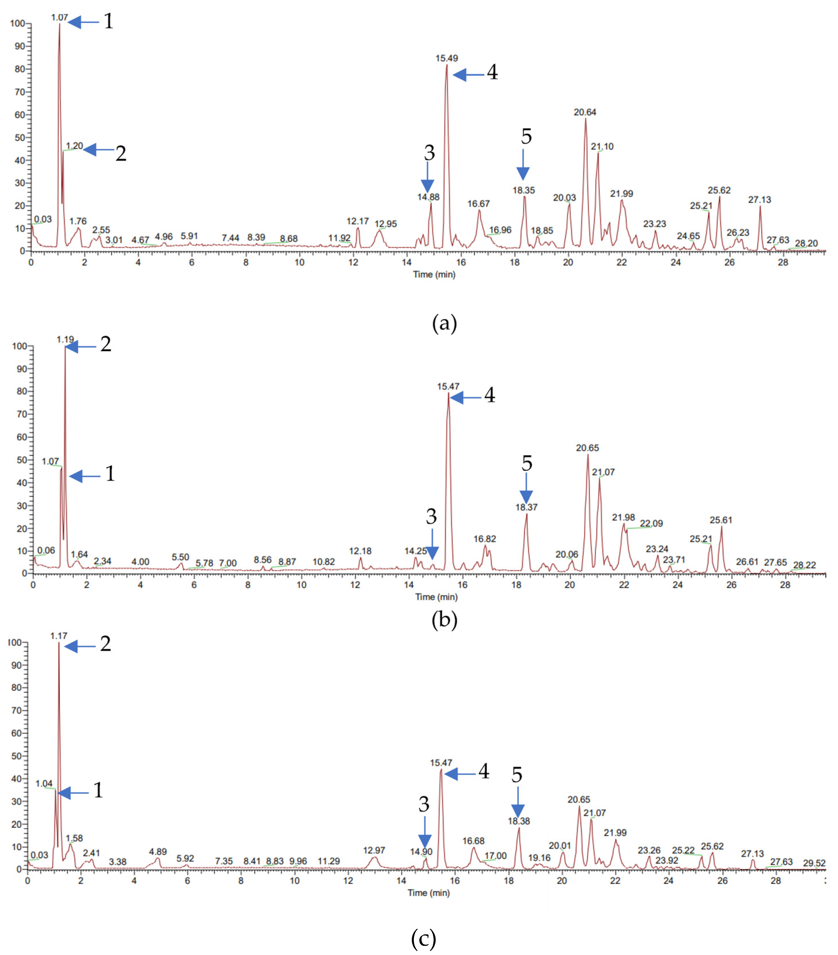
3.1. Compounds in Indonesian Ginger Rhizome Extracts

In general, the separation pattern of the compounds in Figure 1 showed almost the same pattern. The only difference was in the height of the peaks. The five peaks emphasized here were the peaks of the identified compounds, all of which also had a fairly high abundance (Figure 1). There were 98 compounds identified in the Indonesian ginger rhizome extracts based on the LC–MS/MS analysis results. Choline was the compound which had the highest abundance in the GG extract (4.0408 ± 2.0042%), while D–(+)–pipecolinic acid had an abundance in the MM (7.6154 ± 3.1640%), and the EE (7.9358 ± 0.0000%) ginger rhizome extracts. Ginger-characterizing compounds such as gingerol and shogaol were also extracted, but had a low abundance (Table S1). Almost none of the components differed significantly between the groups based on the multivariate analysis, except for 17–α–21-dihydroxypregnenolone, isoamyl–4–methoxycinnamate, bisoprolol, ubiquinone–2, D–glutamine, γ–aminobutyric acid, L–(+)–arginine, and octinoxate (Table S1).

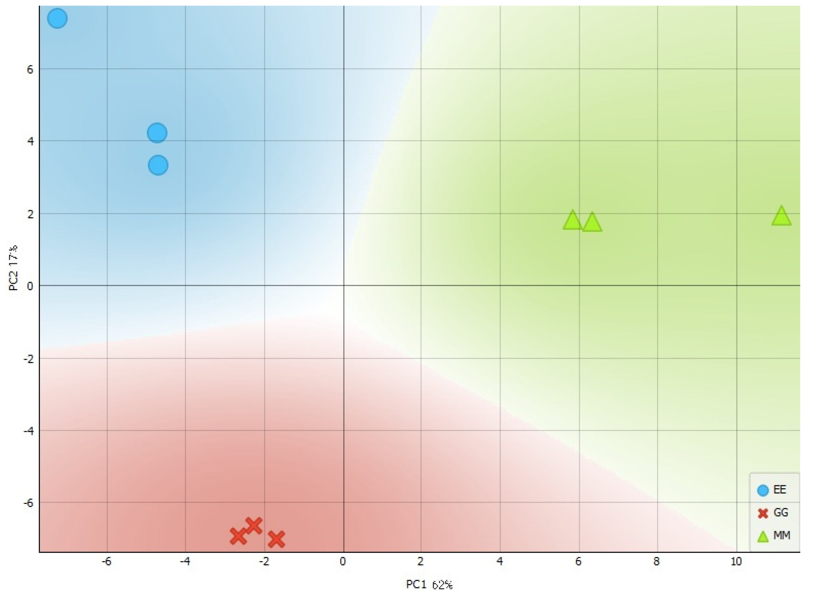
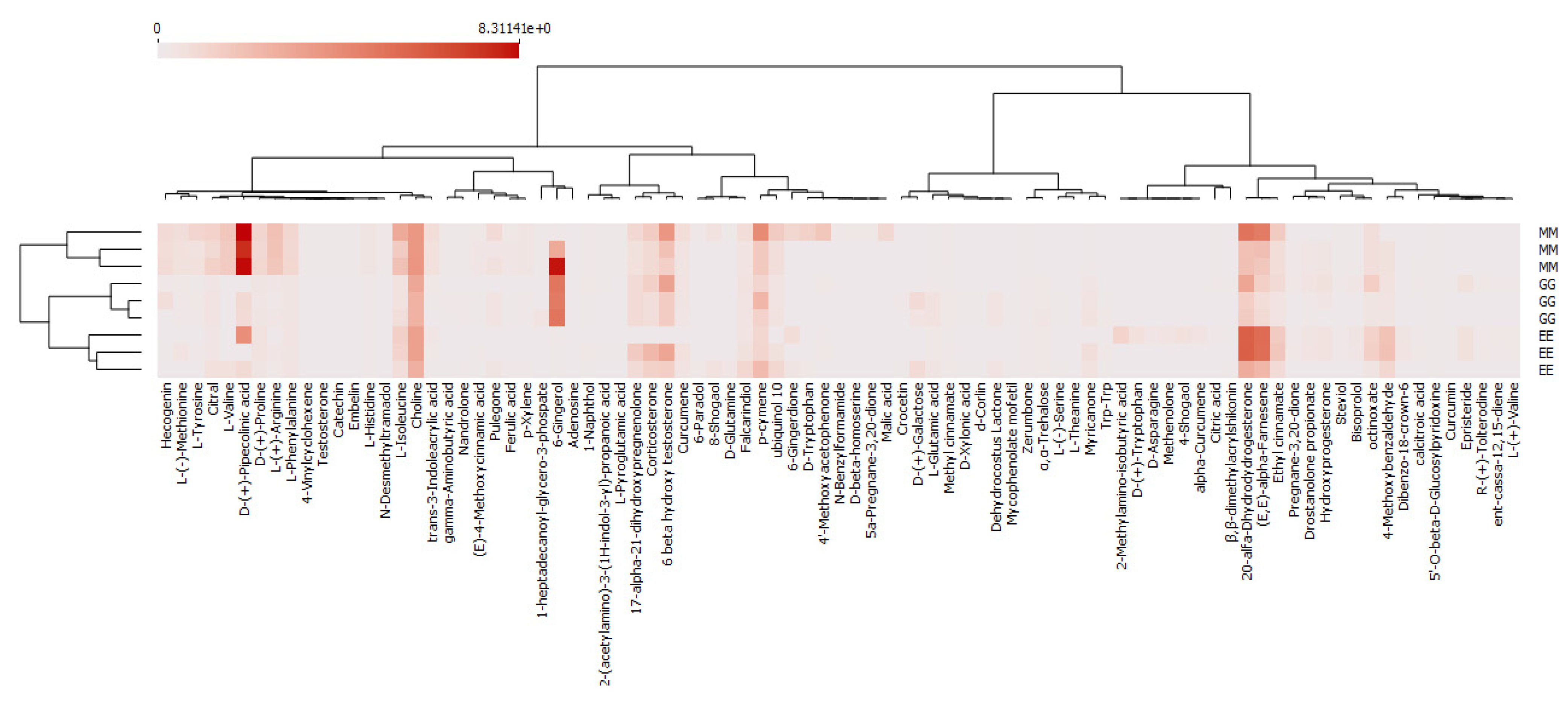
3.2. Principal Component Analysis

A principal component analysis (PCA) is a multivariate analysis with the principle of subtracting variables from several observations. The PCA aims to classify and identify a reduced set of data in order to represent the original data with fewer variables without losing essential information [29]. This study used the PCA to find patterns and differentiate compounds in three Indonesian ginger rhizome extracts (GG, MM, and EE ginger).
The peak area data for each compound were used in the PCA. The output of the PCA is the score plot representing the variety of the analyzed data. The farther a group is from another group, the more different the composition of the metabolites. Based on the plot of scores obtained, the PCA could classify the Indonesian ginger rhizome extract. The total PC score in this plot score was 79%, with PC 1 and PC 2 at 62% and 17%, respectively. The three Indonesian ginger rhizome extracts were grouped very well in different quadrants (Figure 2). The heat map was used to determine differential metabolites among the three Indonesian ginger rhizome extracts because it can visualize and differentiate the distribution of experimental data [30]. The row represented the metabolites’ abundance in each sample, and the column represented the metabolites’ abundance in different samples. The abundance of D–(+)–pipecolinic acid was very high in the Red ginger rhizome extract (MM), while it was low in the other samples (Figure 3). The abundance of 20–α–dhydrodydrogesterone and (E,E)–α–farnesene in the EE extract was higher than in the other samples. Intriguingly, both compounds were significantly lower in the GG ginger rhizome extract than in the MM and EE ginger rhizome extracts (Figure 3).

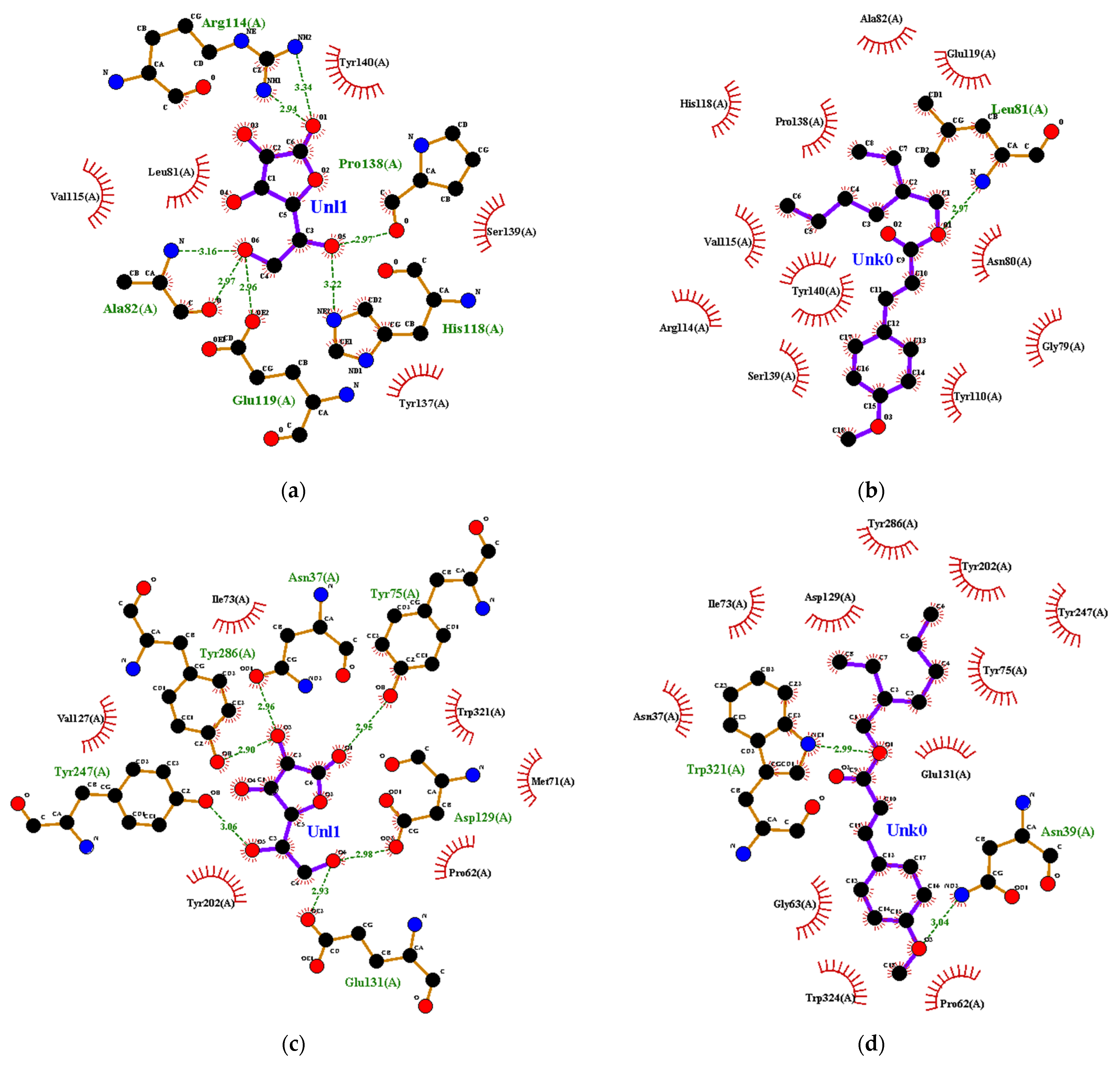
3.3. Molecular Docking for Anti-Skin Aging Activity Evaluation

The test ligands used in this study were compounds found in Indonesian ginger extracts. The control ligand used was ascorbic acid. Ascorbic acid was chosen as a control ligand because it can prevent aging by inhibiting the formation of reactive oxygen species and stratum corneum formation so that dead skin cells can be replaced quickly [31]. In addition, ligand preparation produces ligands whose geometric structure is optimized. These conditions are expected to increase the accuracy and validity of the molecular docking results. The results of the ligand preparation are saved in *pdbqt format. The results of the preparation of the test and control ligands used are listed in Table S2. The receptors used were collagenase (2TCL), hyaluronidase (2PE4), elastase (3F19), and tyrosinase (5M8R). Information about the coordinates of each receptor’s active site was generated in the grid box from Autodock Vina. The coordinates of each active site can be seen in Table 1.
Most of the test ligands’ affinity energies in each complex had higher negative values than the crystal and control ligands (Table S3). This indicated that the test ligands that were used had the potential to be inhibitors of the selected receptor. For example, the d-Corlin ligand appeared in four receptor complexes with the highest negative affinity energies for the collagenase, hyaluronidase, elastase, and tyrosinase complexes, namely −14.6; −13.9; −16.0; and −13.4 kcal/mol (Table 2). The 20-α-dhydrodydrogesterone also appeared in four receptor complexes with an average affinity energy of −14.6 kcal/mol (Table 2), but was most likely not chosen because the compound is listed as a potent drug causing miscarriage and bleeding in pregnant women [32]. The catechin ligand appeared in four complexes, namely the collagenase complex with an affinity energy of −8.8 kcal/mol, the hyaluronidase complex with an affinity energy of −7.9 kcal/mol, the elastase complex with an affinity energy of −9.7 kcal/mol, and the elastase complex with an affinity energy of −8.4 kcal/mol (Table 2). Unfortunately, despite having a low affinity energy, the abundance of d-Corlin and catechin in Indonesian ginger extracts was low. Therefore, this study selected ligands showing a reasonable negative affinity energy and a high enough abundance in Indonesian ginger rhizome extracts.
The 6-gingerol, 8-shogaol, 4-shogaol, 10-shogaol, and 6-paradol had an average affinity energy of −6.9; −6.8; −6.7; −6.7; and −6.5 kcal/mol, respectively (Table 2). The decrease in affinity energy of gingerol, shogaol, and paradol compounds possibly occurred due to the decreasing the -OH group in gingerol and the double-bond change in shogaol. In another study, the different constituents of gingerol, shogaol, and paradol caused differences in their antioxidant activity. According to Dugasani et al., 6-gingerol has higher antioxidant activity than shogaol and paradol compounds [33]. However, this study found that the abundance of 6-gingerol, 8-shogaol, 4-shogaol, 10-shogaol, and 6-paradol was very low. The compounds octinoxate, 6-gingerdione, p-cymene, and ethyl cinnamate had an average affinity energy of −6.9; −6,7; −6.2; and −6.2 kcal/mol (Table 2). Based on the results of the molecular docking analysis, octinoxate, 6-gingerdione, p-cymene, and ethyl cinnamate were most likely to be potential anti-skin aging compounds in the Indonesian ginger rhizome extracts. These four compounds had negative affinity energies and showed a high abundance in the three Indonesian ginger rhizome extracts. However, octinoxate, 6-gingerdione, p-cymene, and ethyl cinnamate still had to pass the physicochemical tests.

3.4. Visualization and Determination of Physicochemical Properties of Ligands Based on admetSAR Parameters

A comparison of the test and control ligand complex’s interaction is one of the appropriate parameters to determine which test ligands have the greatest potential as anti-aging compounds. The comparison resulted in the percentage of binding site similarity (%BSS), which was observed from the similarity of amino acid residues that interacted between the test and control ligands. The similarity of binding sites is helpful in drug development, the analysis of protein–ligand complexes, and drug function prediction in chemistry [34]. Ligand–receptor interactions can occur in hydrophobic or hydrogen bonds. Hydrophobic interactions occur because of the contact between the alkyl chains in both the ligands and the receptors. Hydrogen bonds are formed due to the electrostatic attraction between hydrogen atoms attached to more electronegative atoms or groups and other electronegative atoms with lone pairs of electrons. In this case, a hydrogen bond occurred between the amino acid residue from the receptor and the ligand.
The amino acid residues of collagenase that interacted hydrophobically with ascorbic acid were Leu-81, Val-115, Tyr-137, Ser-139, and Tyr-140, as well as Ala-82, Arg-114, His-118, Glu-119, and Pro-138 which hydrogen-bonded (Figure 4a). The Leu-81, Ala-82, His-118, Pro-138, and Tyr-140 residues are directly involved in the ligand binding process on collagenase. Molecular docking to collagenase yielded various %BSS. The octinoxate ligand had a 90%BSS because it interacted with nine amino acid residues (Leu-81, Ala-82, Arg-114, Val-115, His-118, Glu-119, Pro-138, Ser-139, and Tyr-140) that interacted with ascorbic acid (Figure 4b). Only the N-atom at Leu-81 in collagenase hydrogen-bonded with the O-atom in octinoxate, and the distance between them was 2.97 Å (Figure 4b).
The amino acid residues Pro-62, Met-71, Ile-73, Val-127, Tyr-202, and Trp-321 of hyaluronidase interacted hydrophobically while Asn-37, Tyr-75, Asp-129, Glu-131, Tyr-247, and Tyr-286 hydrogen-bonded with ascorbic acid (Figure 4c). According to Chao et al., Tyr-75, Asp-129, Glu-131, Tyr-202, Tyr-247, Tyr-286, and Trp-321 are residues that play a critical role in hyaluronidase’s active site [35]. The octinoxate ligand had hydrophobic interactions with amino acid residues Asn-37, Pro-62, Ile-73, Tyr-75, Asp-129, Glu-131, Tyr-202, Tyr-247, and Tyr-286; it hydrogen-bonded with Asn-39 at a distance of 3.04 Å and with Trp-321 at a distance of 2.99 Å (Figure 4d). The %BSS for the octinoxane ligand was 83%.
Ascorbic acid interacted hydrophobically and hydrogen-bonded with amino acid residues in the elastase enzyme. The amino acid residues His-218, His-222, His-228, Phe-237, and Tyr-240 were hydrogen-bonded, while the residues Thr-215, Pro-238, and Thr-239 interacted hydrophobically (Figure 4e). The octinoxate ligand had only hydrogen-bonded with Leu-181 at a distance of 2.52 Å (Figure 4f). Octinoxate had a 50%BSS because it only had four of the eight interactions that were the same as the ascorbic acid–elastase interaction.
In tyrosinase, ascorbic acid hydrogen-bonded with His-192, His-215, His-224, Glu-360, His-377, Asn-378, His-381, Gly-389, and Ser-394; it interacted hydrophobically with Phe-362, Glu-390, and Val-391 (Figure 4g). The octinoxate had a 46%BSS because it only interacted with six of the thirteen ascorbic acid–tyrosinase interactions (Figure 4h). This was due to ascorbic acid ligands hydrogen-bonding with amino acid residues of tyrosinase, whereas octinoxate only interacted hydrophobically.
The structure and shape similarity between ligand and substrate [36], as well as the interaction of the ligand and amino acid residues [37], are the things that determine the inhibitory ability of the ligand–receptor activity. Most of the interactions formed between the test ligands and the receptors were hydrophobic interactions. In contrast to the test ligands, most of the interactions between the control ligand and the amino acid residues of the receptors were hydrogen bonds. All hydrogen bonds formed between ligands and amino acid residues are >1.85 Å long.
In addition to the parameters previously described, several parameters are related to the physicochemical properties of compounds in the body. These parameters are contained in Lipinski’s rules and the admetSAR test. Lipinski’s rules and the admetSAR test have several parameters, including relative atomic mass, partition coefficient value (log p), number of hydrogen-bond acceptors and donors, number of rotatable bonds, mutagenesis, carcinogenicity, and eye irritation. All ligands with the potential for anti-skin aging passed Lipinski’s rules (Table 3). However, all ligands, except for octinoxate and 20-α-dhydrodydrogesterone, could cause eye irritation, while catechin, 8-shogaol, 6-gingerdione, and 10-shogaol had the potential to cause mutation. Therefore, the use of these ligands must be considered in order to avoid adverse effects on the body. Based on the values of compound abundance, affinity energy, Lipinski’s rules, and the admetSAR test, octinoxate in the Indonesian ginger rhizome extracts was the only compound with anti-skin aging activity potential and was abundant in the EE ginger rhizome extract. Therefore, the EE ginger rhizome extract was the ginger rhizome extract with the greatest potential for anti-skin aging.

4. Discussion

Ginger rhizome extract was analyzed using liquid chromatography–mass spectrometry/mass spectrometry (LC–MS/MS). This was non-targeted because the aim was to find all the compounds contained in Indonesian ginger rhizome extracts. The output of the LC–MS/MS analysis was a chromatogram that provided information on retention time, separation pattern, and abundance. The retention time and separation pattern were the initial pieces of information needed for identifying compounds. The retention times and separation patterns on the identical chromatograms with available databases indicated the same compounds. Peak intensity was interpreted as the abundance of a compound in a sample. The higher the peak formed, the more abundant the compound in the analyzed sample. Based on this study, choline was the high-abundance compound in the GG ginger rhizome extract, and D-(+)-pipecolinic acid in the MM and the EE ginger extracts. Using a principal component analysis (PCA), Indonesian ginger rhizome extracts could be grouped based on their components. The plot scores proved that the Indonesian ginger rhizome extracts were well separated into three groups in three different quadrants. The compounds D-(+)-pipecolinic acid, 20-α-dhydrodydrogesterone, and (E,E)-α-farnesene were screened out as differential metabolites because these compounds had significantly unique and distinctive patterns in the heat map analysis.
Before the molecular docking process started, several preparatory steps had to be carried out for a specific purpose. First, the removal of water molecules and their natural ligands from the receptor aimed to prevent interference with the interaction of the receptor with other molecules that could cause inaccuracies in the molecular docking result. The tyrosinase structure used was only ring A. This was because the other rings were identical to each other, the binding site was not in the space between the rings, and the ring collection did not bind to each other, so it could be ascertained that it would not affect the molecular docking results. The use of either ring could also shorten the docking time.
Molecular docking was used to determine the components in ginger rhizome extracts that had the potential for anti-skin aging. Affinity energy was based on the output data for the molecular docking. The affinity energy indicates the spontaneity of the interaction between the ligand and the receptor [38]. The test ligand–receptor complex is categorized as a good complex if it has an affinity energy of less than or the same as the affinity energy of the control ligand–receptor complex [17]. Test ligands were derived from Indonesian ginger rhizome extracts, and the control ligand was ascorbic acid. Those compounds were docked to collagenase, hyaluronidase, elastase, and tyrosinase enzymes. The affinity energy in this study varied from one ligand to another and one receptor to another. This was due to differences in the presence of hydrogen bonds and hydrophobic interactions. If traced more deeply, the difference was also influenced by the ligand’s structure that binds to the amino acid residues on the receptor. Unfortunately, there are no reports on the structure-dependent in silico method.
After the molecular docking process, the ligands that had anti-skin aging potential were tested for their physicochemical properties. One of the tests was performed using Lipinski’s rules. Lipinski’s rules describe the solubility and permeability of a ligand in drug development [39]. The parameters of Lipinski’s rules are that the compound has a relative atomic mass of <500 g/mol, a log p value < 5, some hydrogen-bond acceptors <10, some hydrogen-bond donors <5, and some rotatable bonds <10. The compound must have a relative atomic mass of <500 g/mol to pass through the cell membrane by passive diffusion to reach the receptor [40]. The log p value describes the compound’s solubility in nonpolar and polar species. This value can predict the hydrophobicity of compounds associated with the admetSAR process. The greater the log p value, the more hydrophobic a compound will remain in the lipid bilayer [41]. The number of hydrogen-bond acceptors and donors is related to the number of hydrogen bonds formed. According to Cheng et al., the ideal number of hydrogen-bond acceptors and donors was 10 and 5, respectively [42]. A great number of hydrogen bonds causes the compound to be more bound to polar solvents so that the absorption process into cells to reach the receptor is inhibited. The ideal number of rotatable bonds in a compound is <10 because more will cause the affinity energy between the ligand receptors to decrease [43]. All ligands that have anti-skin aging potential passed the Lipinski’s rules test.
The other physicochemical test carried out in this study was the admetSAR test. The admetSAR test describes the absorption, distribution, metabolism, excretion, toxicity, and structure–activity relationships of a compound in the body or test organism. This test is included in the pharmacokinetic and pharmacodynamic analysis in drug development. The admetSAR test enables drug developers to understand the safety and efficacy of drug candidates. This is required to approve regulations for issuing distribution permits for prospective drugs. The admetSAR parameters were considered, namely mutagenesis, carcinogenicity, and eye irritation. The enzyme inhibitor parameter indicates the general strength of the compound against the enzyme.
After considering several aspects, namely affinity energy, compound abundance, Lipinski’s test results, and the admetSAR test results, octinoxate was the compound in the Indonesian ginger rhizome extracts that hadthe greatest potential as an anti-skin aging agent. In addition, the EE ginger rhizome extract can be considered an anti-skin aging agent for future development. However, there are some disadvantages of molecular docking which include the lack of a synergistic computational model [44], the lack of a quality database [45], the lack of method standardization and validation [46], and the lack of n accurate scoring function [47]. Molecular docking simulation progresses in small steps and thus has difficulties in stepping over high energy-conformational barriers, which may lead to inadequate sampling [48]. The challenges include the need for greater emphasis on protein docking to molecules of different types, proper accounting for conformational flexibility upon binding, new promising methodologies based on residue co-evolution and deep learning, affinity prediction, and further development of a fully automated docking server [49].
There are suggestions for the continuation of this research in the future. Optimization of Indonesian ginger rhizome extraction needs to be performed because it is suspected that many compounds still have not been extracted. The condition of the analytical instrument also needs to be considered so that the detected compounds can be more diverse. Compounds that have potential anti-skin aging activity need to be tested in vitro and in vivo to validate the molecular docking results. For further research, it would be better to use different receptors from the receptors used in this study in order to further explore the enzymes that cause aging.

5. Conclusions

Indonesian ginger rhizome extracts contain ninety-eight compounds and can be grouped into three different quadrants. The differential metabolites among the Indonesian ginger rhizome extracts were D-(+)-pipecolinic acid, 20-α-dhydrodydrogesterone, and (E,E)-α-farnesene. The compound that had the greatest potential for anti-skin aging, as found in Indonesian ginger rhizome extracts, was octinoxate. Octinoxate was able to inhibit all receptors, and showed a high abundance in the EE ginger rhizome extract. Therefore, the EE ginger rhizome extract had the greatest potential as an anti-skin aging ingredient compared with the other Indonesian ginger rhizome extracts.

Supplementary Materials

The following supporting information can be downloaded at: https://0-www-mdpi-com.brum.beds.ac.uk/article/10.3390/cosmetics9060128/s1, Table S1: Compounds in ginger rhizome extracts based on the results of a liquid chromatography–mass spectrometry/mass spectrometry analysis, Table S2: Examples of test and control ligand structures, Table S3: The affinity energy of the ligand–receptor complex.

Author Contributions

Conceptualization, I.B., W.T.W. and S.T.W.; methodology, I.B., W.T.W. and S.T.W.; software, S.T.W.; validation, I.B., W.T.W. and S.T.W.; formal analysis, I.B., W.T.W. and S.T.W.; research investigation, S.F.A.; data curation, I.B., W.T.W. and S.T.W.; writing—original draft preparation, S.F.A. and A.R.L.; writing—review and editing, A.R.L., I.B., W.T.W. and S.T.W.; visualization, S.F.A.; supervision, I.B., W.T.W. and S.T.W.; project administration, I.B. All authors have read and agreed to the published version of the manuscript.

Funding

This research was funded by the Directorate General of Higher Education, Research, and Technology; Ministry of Education, Culture, Research, and Technology, Republic of Indonesia, which funded this research through contract number: 001/E5/PG.02.00PT/2022 to the Institute of Research and Community Services, IPB University.

Institutional Review Board Statement

Not applicable.

Informed Consent Statement

Not applicable.

Data Availability Statement

Data are contained within the article and the Supplementary Material.

Acknowledgments

The authors acknowledge the Advanced Research Laboratory, IPB University, for analyzing ginger rhizome extract using LC–MS/MS.

Conflicts of Interest

The authors declare no conflict of interest.

References

  1. Rose, M.R.; Flatt, T.; Graves, J.L.; Greer, L.F.; Martinez, D.E.; Matos, M.; Mueller, L.D.; Shmookler Reis, R.J.; Shahrestani, P. What is aging? Front. Genet. 2012, 3, 2011–2013. [Google Scholar] [CrossRef] [PubMed] [Green Version]
  2. Cohen, A.A. Complex systems dynamics in aging: New evidence, continuing questions. Biogerontology 2016, 17, 205–220. [Google Scholar] [CrossRef] [PubMed] [Green Version]
  3. Chen, F.; Liu, Y.; Wong, N.K.; Xiao, J.; So, K.F. Oxidative Stress in Stem Cell Aging. Cell Transplant. 2017, 26, 1483–1495. [Google Scholar] [CrossRef]
  4. Risques, R.A.; Kennedy, S.R. Aging and the rise of somatic cancer-associated mutations in normal tissues. PLoS Genet. 2018, 14, e1007108. [Google Scholar] [CrossRef] [PubMed] [Green Version]
  5. Sirard, M.A. Why is the human ovary aging faster than other organs? Women Health 2022, 62, 577–579. [Google Scholar] [CrossRef] [PubMed]
  6. Li, M.; Vierkötter, A.; Schikowski, T.; Hüls, A.; Ding, A.; Matsui, M.S.; Deng, B.; Ma, C.; Ren, A.; Zhang, J.; et al. Epidemiological evidence that indoor air pollution from cooking with solid fuels accelerates skin aging in Chinese women. J. Dermatol. Sci. 2014, 79, 148–154. [Google Scholar] [CrossRef]
  7. Tottoli, E.M.; Dorati, R.; Genta, I.; Chiesa, E.; Pisani, S.; Conti, B. Skin wound healing process and new emerging technologies for skin wound care and regeneration. Pharmaceutics 2020, 12, 735. [Google Scholar] [CrossRef]
  8. Keaney, T.C. Aging in the Male Face: Intrinsic and Extrinsic Factors. Dermatol. Surg. 2016, 42, 797–803. [Google Scholar] [CrossRef] [Green Version]
  9. Rabe, J.H.; Mamelak, A.J.; McElgunn, P.J.S.; Morison, W.L.; Sauder, D.N. Photoaging: Mechanisms and repair. J. Am. Acad. Dermatol. 2006, 55, 1–19. [Google Scholar] [CrossRef]
  10. Harman, D. Forum Mini Review. Antioxid. Redox Signal. 2003, 5, 557–561. [Google Scholar] [CrossRef]
  11. Lee, E.J.; Kim, J.Y.; Oh, S.H. Advanced glycation end products (AGEs) promote melanogenesis through receptor for AGEs. Sci. Rep. 2016, 6, 27848. [Google Scholar] [CrossRef] [PubMed] [Green Version]
  12. Lete, I.; Allué, J. The Effectiveness of Ginger in the Prevention of Nausea and Vomiting during Pregnancy and Chemotherapy. Integr. Med. Insights 2016, 11, 11–17. [Google Scholar] [CrossRef] [PubMed] [Green Version]
  13. Mahassni, S.H.; Bukhari, O.A. Journal of Nutrition & Intermediary Metabolism Bene fi cial e ff ects of an aqueous ginger extract on the immune system cells and antibodies, hematology, and thyroid hormones in male smokers and. J. Nutr. Intermed. Metab. 2019, 15, 10–17. [Google Scholar] [CrossRef]
  14. Singh, G.; Kapoor, I.P.S.; Singh, P.; de Heluani, C.S.; de Lampasona, M.P.; Catalan, C.A.N. Chemistry, antioxidant and antimicrobial investigations on essential oil and oleoresins of Zingiber officinale. Food Chem. Toxicol. 2008, 46, 3295–3302. [Google Scholar] [CrossRef]
  15. Höferl, M.; Stoilova, I.; Wanner, J.; Schmidt, E.; Jirovetz, L.; Trifonova, D.; Stanchev, V.; Krastanov, A. Composition and Comprehensive Antioxidant Activity of Ginger (Zingiber officinale) Essential Oil from Ecuador. Nat. Prod. Commun. 2015, 10, 1085–1090. [Google Scholar] [CrossRef] [Green Version]
  16. Feng, J.; Du, Z.; Zhang, L.; Luo, W.; Zheng, Y.; Chen, D.; Pan, W.; Yang, Z.; Lin, L.; Xi, L. Chemical Composition and Skin Protective Effects of Essential Oil Obtained from Ginger (Zingiber officinale Roscoe). J. Essent. Oil-Bear. Plants 2018, 21, 1542–1549. [Google Scholar] [CrossRef]
  17. Lestari, A.R.; Batubara, I.; Wahyudi, S.T.; Ilmiawati, A.; Achmadi, S.S. Bioactive Compounds in Garlic (Allium sativum) and Black Garlic as Antigout Agents, Using Computer Simulation. Life 2022, 12, 1131. [Google Scholar] [CrossRef]
  18. Rampogu, S.; Baek, A.; Gajula, R.G.; Zeb, A.; Bavi, R.S.; Kumar, R.; Kim, Y.; Kwon, Y.J.; Lee, K.W. Ginger (Zingiber officinale) phytochemicals-gingerenone-A and shogaol inhibit SaHPPK: Molecular docking, molecular dynamics simulations and in vitro approaches. Ann. Clin. Microbiol. Antimicrob. 2018, 17, 16. [Google Scholar] [CrossRef] [Green Version]
  19. Moitessier, N.; Englebienne, P.; Lee, D.; Lawandi, J.; Corbeil, C.R. Towards the development of universal, fast and highly accurate docking/scoring methods: A long way to go. Br. J. Pharmacol. 2008, 153, 7–26. [Google Scholar] [CrossRef] [Green Version]
  20. Bailey, D.; Brown, D. High-throughput chemistry and structure-based design: Survival of the smartest. Drug Discov. Today 2001, 6, 57–59. [Google Scholar] [CrossRef]
  21. McConkey, B.J.; Sobolev, V.; Edelman, M. The performance of current methods in ligand-protein docking. Curr. Sci. 2002, 83, 845–856. [Google Scholar]
  22. Agarwal, S.; Mehrotra, R. An Overview of Molecular Simulation. JSM Chem. 2016, 4, 1024–1028. [Google Scholar]
  23. Lee, H.S.; Jo, S.; Lim, H.S.; Im, W. Application of binding free energy calculations to prediction of binding modes and affinities of MDM2 and MDMX inhibitors. J. Chem. Inf. Model. 2012, 52, 1821–1832. [Google Scholar] [CrossRef] [PubMed] [Green Version]
  24. Arakaki, P.A.; Marques, M.R.; Santos, M.C.L.G. MMP-1 polymorphism and its relationship to pathological processes. J. Biosci. 2009, 34, 313–320. [Google Scholar] [CrossRef] [PubMed]
  25. Weber, G.; Buhren, B.; Schrumpf, H.; Wohlrab, J.; Gerber, P. Clinical applications of hyaluronidase. Adv. Exp. Med. Biol. 2019, 1148, 255–277. [Google Scholar] [PubMed]
  26. Wittenauer, J.; MäcKle, S.; Sußmann, D.; Schweiggert-Weisz, U.; Carle, R. Inhibitory effects of polyphenols from grape pomace extract on collagenase and elastase activity. Fitoterapia 2015, 101, 179–187. [Google Scholar] [CrossRef] [PubMed]
  27. Eun Lee, K.; Bharadwaj, S.; Yadava, U.; Gu Kang, S. Evaluation of caffeine as inhibitor against collagenase, elastase and tyrosinase using in silico and in vitro approach. J. Enzyme Inhib. Med. Chem. 2019, 34, 927–936. [Google Scholar] [CrossRef] [Green Version]
  28. Agricultural Technology Study Center. BPTP Sumatera Utara. In Petunjuk Teknis Budidaya Tanaman Jahe.; Ministry of Agricultural Republic of Indonesia: Jakarta, Indonesia, 2012; pp. 25–27. Available online: http://repository.pertanian.go.id/handle/123456789/8585 (accessed on 23 December 2021).
  29. Kherif, F.; Latypova, A. Principal component analysis. Mach. Learn. Methods Appl. Brain Disord. 2019, 1, 209–225. [Google Scholar] [CrossRef]
  30. Marlin, M.; Maharijaya, A.; Purwito, A.; Sobir, S. Secondary metabolites change under vernalization and its relation to flowering competency in shallot (Allium cepa var. aggregatum). Rasayan J. Chem. 2019, 12, 1418–1425. [Google Scholar] [CrossRef]
  31. Ravetti, S.; Clemente, C.; Brignone, S.; Hergert, L.; Allemandi, D.; Palma, S. Ascorbic acid in skin health. Cosmetics 2019, 6, 58. [Google Scholar] [CrossRef] [Green Version]
  32. Carp, H. A systematic review of dydrogesterone for the treatment of threatened miscarriage. Gynecol. Endocrinol. 2012, 28, 983–990. [Google Scholar] [CrossRef] [PubMed] [Green Version]
  33. Dugasani, S.; Pichika, M.R.; Nadarajah, V.D.; Balijepalli, M.K.; Tandra, S.; Korlakunta, J.N. Comparative antioxidant and anti-inflammatory effects of [6]-gingerol, [8]-gingerol, [10]-gingerol and [6]-shogaol. J. Ethnopharmacol. 2010, 127, 515–520. [Google Scholar] [CrossRef] [PubMed]
  34. Ehrt, C.; Brinkjost, T.; Koch, O. A benchmark driven guide to binding site comparison: An exhaustive evaluation using tailor-made data sets (ProSPECCTs). PLoS Comput. Biol. 2018, 14, e1006483. [Google Scholar] [CrossRef] [PubMed]
  35. Chao, K.L.; Muthukumar, L.; Herzberg, O. Structure of human hyaluronidase-1, a hyaluronan hydrolyzing enzyme involved in tumor growth and angiogenesis. Biochemistry 2007, 46, 6911–6920. [Google Scholar] [CrossRef] [PubMed] [Green Version]
  36. Tang, Y.B.; Liu, J.Z.; Zhang, S.E.; Du, X.; Nie, F.; Tian, J.Y.; Ye, F.; Huang, K.; Hu, J.P.; Li, Y.; et al. 3-Phenylpropanoic acid-based phosphotyrosine (pTyr) mimetics: Hit evolution to a novel orally active protein tyrosine phosphatase 1B (PTP1B) inhibitor. ChemMedChem 2014, 9, 918–921. [Google Scholar] [CrossRef]
  37. Chhabra, N.; Aseri, M.; Padmanabhan, D. A review of drug isomerism and its significance. Int. J. Appl. Basic Med. Res. 2013, 3, 16–18. [Google Scholar] [CrossRef] [Green Version]
  38. Rahman, A.J.; Sharma, D.; Kumar, D.; Pathak, M.; Singh, A.; Kumar, V.; Chawla, R.; Ojha, H. Spectroscopic and molecular modelling study of binding mechanism of bovine serum albumin with phosmet. Spectrochim. Acta—Part A Mol. Biomol. Spectrosc. 2021, 244, 118803. [Google Scholar] [CrossRef]
  39. Benet, L.Z.; Hosey, C.M.; Ursu, O.; Oprea, T.I. BDDCS, the Rule of 5 and drugability. Adv. Drug Deliv. Rev. 2016, 101, 89–98. [Google Scholar] [CrossRef] [Green Version]
  40. Doak, B.C.; Over, B.; Giordanetto, F.; Kihlberg, J. Oral druggable space beyond the rule of 5: Insights from drugs and clinical candidates. Chem. Biol. 2014, 21, 1115–1142. [Google Scholar] [CrossRef] [Green Version]
  41. Mishra, A.; Dixit, S.; Ratan, V.; Srivastava, M.; Trivedi, S.; Srivastava, Y.K. Identification and in silico screening of biologically active secondary metabolites isolated from Trichoderma harzianum. Ann. Phytomed. An Int. J. 2018, 7. [Google Scholar] [CrossRef]
  42. Cheng, F.; Li, W.; Zhou, Y.; Shen, J.; Wu, Z.; Liu, G.; Lee, P.W.; Tang, Y. AdmetSAR: A comprehensive source and free tool for assessment of chemical ADMET properties. J. Chem. Inf. Model. 2012, 52, 3099–3105. [Google Scholar] [CrossRef] [PubMed]
  43. Andrews, P.R.; Craik, D.J.; Martin, J.L. Functional Group Contributions to Drug-Receptor Interactions. J. Med. Chem. 1984, 27, 1648–1657. [Google Scholar] [CrossRef] [PubMed]
  44. Gu, Y.; Han, J.; Jiang, C.; Zhang, Y. Biomarkers, oxidative stress and autophagy in skin aging. Ageing Res. Rev. 2020, 59, 101036. [Google Scholar] [CrossRef] [PubMed]
  45. Yuriev, E.; Holien, J.; Ramsland, P.A. Improvements, trends, and new ideas in molecular docking: 2012–2013 in review. J. Mol. Recognit. 2015, 28, 581–604. [Google Scholar] [CrossRef] [PubMed]
  46. Corbeil, C.R.; Williams, C.I.; Labute, P. Variability in docking success rates due to dataset preparation. J. Comput. Aided. Mol. Des. 2012, 26, 775–786. [Google Scholar] [CrossRef] [Green Version]
  47. Chen, F.; Liu, H.; Sun, H.; Pan, P.; Li, Y.; Li, D.; Hou, T. Assessing the performance of the MM/PBSA and MM/GBSA methods. 6. Capability to predict protein-protein binding free energies and re-rank binding poses generated by protein-protein docking. Phys. Chem. Chem. Phys. 2016, 18, 22129–22139. [Google Scholar] [CrossRef]
  48. Meng, X.-Y.; Zhang, H.-X.; Mezei, M.; Cui, M. Molecular Docking: A Powerful Approach for Structure-Based Drug Discovery. Curr. Comput. Aided-Drug Des. 2012, 7, 146–157. [Google Scholar] [CrossRef]
  49. Vakser, I.A. Challenges in protein docking. Curr. Opin. Struct. Biol. 2020, 64, 160–165. [Google Scholar] [CrossRef]
Figure 1. LC–MS/MS chromatogram of (a) GG, (b) MM, (c) EE rhizome extract, peak 1 (choline), peak 2 (D–(+)–pipecolinic acid), peak 3 (6–paradol), peak 4 (6–gingerol), and peak 5 (8–shogaol).
Cosmetics 09 00128 g001
Figure 2. Score plot of PCA. The circle shape represents EE ginger rhizome extract; the cross shape represents GG ginger rhizome extract; and the triangle shape represents MM ginger rhizome extract.
Cosmetics 09 00128 g002
Figure 3. Heat map of LC–MS/MS metabolite profile of Indonesian ginger rhizome extracts.
Cosmetics 09 00128 g003
Figure 4. Visualization of the binding of: (a) ascorbic acid and (b) octinoxate to the collagenase (2TCL); (c) ascorbic acid and (d) octinoxate to the hyaluronidase (2PE4); (e) ascorbic acid and (f) octinoxate to the elastase (3F19); and (g) ascorbic acid and (h) octinoxate to the tyrosinase (5M8R).
Cosmetics 09 00128 g004aCosmetics 09 00128 g004b
Table 1. Active site coordinates of the receptor.
ReceptorCoordinates
XYZ
Collagenase (2TCL)73.9868.6559.279
Hyaluronidase (2PE4)41.877−22.142−16.287
Elastase (3F19)−11.338−4.481−18.305
Tyrosinase (5M8R)−30.322−4.944−24.773
Table 2. The affinity energy of the ligand–receptor complex.
LigandAffinity Energy (kcal/mol)
2TCL 22PE4 33F19 45M8R 5
Ascorbic acid−6.4−5.6−6.1−6.0
d-Corlin−14.6−13.9−16.0−13.4
DG 1−10.6−11.4−13.0−10.6
Catechin−8.8−7.9−9.7−8.4
6-Gingerol−6.1−6.7−8.0−6.8
8-Shogaol−6.0−6.9−8.0−6.4
4-Shogaol−6.1−6.8−7.6−6.3
10-Shogaol−5.5−7.0−7.7−6.6
6-Paradol−5.7−6.8−7.9−5.5
Octinoxate−6.1−7.2−7.8−6.3
6-Gingerdione−5.9−6.4−7.9−6.6
p-Cymene−5.9−6.6−6.5−5.8
Ethyl cinnamate−6.1−6.1−7.0−5.5
1 20-α-Dhydrodydrogesterone, 2 Collagenase, 3 Hyaluronidase, 4 Elastase, and 5 Tyrosinase.
Table 3. Ligand evaluation using Lipinski’s rules and admetSAR parameters.
LigandLipinski’s RulesadmetSAR ParametersEnzyme Inhibitor
a, b, c, d, eMutagenicityCarcinogenicityEye Irritation
CatechinPass+-+0.47
6-ParadolPass--+0.18
6-GingerolPass--+0.38
EC 1Pass--+−0.45
p-CymenePass--+−0.78
OctinoxatePass---0.02
8-ShogaolPass+-+0.27
6G 2Pass+-+0.21
4-ShogaolPass--+0.27
10-ShogaolPass+-+0.25
DG 3Pass---0.56
1 Ethyl cinnamate, 2 6-gingerdione, 3 20-α-Dhydrodydrogesterone, a—molecular weight, b—rotatable bonds, c—hydrogen-bond acceptors, d—hydrogen-bond donors, e—partition coefficient, (-)—non-mutagen/non-carcinogen/non-irritating, and (+)—mutagen/carcinogen/irritant.
Publisher’s Note: MDPI stays neutral with regard to jurisdictional claims in published maps and institutional affiliations.

