Article

A Combination of Structural, Genetic, Phenotypic and Enzymatic Analyses Reveals the Importance of a Predicted Fucosyltransferase to Protein O-Glycosylation in the Bacteroidetes

1
NanoGlycobiology Unit, Institute of Biologically Inspired Materials, Department of NanoBiotechnology, Universität für Bodenkultur Wien, Muthgasse 11, A-1190 Vienna, Austria
2
Institute of Biochemistry, Department of Chemistry, Universität für Bodenkultur Wien, Muthgasse 18, A-1190 Vienna, Austria
3
Department of Microbiology and the Duchossois Family Institute, University of Chicago, KCBD, 900 E. 57th Street, Chicago, IL 60637, USA
4
Institute of Organic Chemistry, Department of Chemistry, Universität für Bodenkultur Wien, Muthgasse 18, A-1190 Vienna, Austria
5
Department of Organic Chemistry, Faculty of Chemistry, University of Vienna, Währinger Strasse 38, A-1090 Vienna, Austria
*
Author to whom correspondence should be addressed.
These authors contributed equally to this work.
Submission received: 18 October 2021 / Revised: 21 November 2021 / Accepted: 23 November 2021 / Published: 30 November 2021
(This article belongs to the Special Issue Glycosylation—The Most Diverse Post-Translational Modification)

Abstract

:
Diverse members of the Bacteroidetes phylum have general protein O-glycosylation systems that are essential for processes such as host colonization and pathogenesis. Here, we analyzed the function of a putative fucosyltransferase (FucT) family that is widely encoded in Bacteroidetes protein O-glycosylation genetic loci. We studied the FucT orthologs of three Bacteroidetes species—Tannerella forsythia, Bacteroides fragilis, and Pedobacter heparinus. To identify the linkage created by the FucT of B. fragilis, we elucidated the full structure of its nine-sugar O-glycan and found that l-fucose is linked β1,4 to glucose. Of the two fucose residues in the T. forsythia O-glycan, the fucose linked to the reducing-end galactose was shown by mutational analysis to be l-fucose. Despite the transfer of l-fucose to distinct hexose sugars in the B. fragilis and T. forsythia O-glycans, the FucT orthologs from B. fragilis, T. forsythia, and P. heparinus each cross-complement the B. fragilis ΔBF4306 and T. forsythia ΔTanf_01305 FucT mutants. In vitro enzymatic analyses showed relaxed acceptor specificity of the three enzymes, transferring l-fucose to various pNP-α-hexoses. Further, glycan structural analysis together with fucosidase assays indicated that the T. forsythia FucT links l-fucose α1,6 to galactose. Given the biological importance of fucosylated carbohydrates, these FucTs are promising candidates for synthetic glycobiology.

Graphical Abstract

1. Introduction

Bacteroidetes is a phylum of Gram-negative bacteria that colonize diverse ecological niches. Within this phylum are members of the order Bacteroidales, which include abundant anaerobic gut symbionts such as Bacteroides species that provide benefits to their host [1,2], as well as pathogenic anaerobic species such as the periodontal pathogens Tannerella forsythia and Porphyromonas gingivalis [3]. Flavobacteriales and Sphingobacteriales are other orders of this phylum that are generally aerobes or facultative anaerobes and typically colonize diverse environmental and non-mammalian host ecosystems. Members of this phylum have a tremendous capability to degrade high molecular-weight polysaccharides using dedicated polysaccharide utilization loci, many of which are shared between distantly related members [4].
One outstanding feature of many Bacteroidales species is their synthesis of numerous capsular polysaccharides (CPS) and glycoproteins. For example, the gut symbiont B. fragilis NCTC 9343 produces eight CPSs that are subject to phase variation, each synthesized by the products of separate operons [5,6]. This strain also has a general protein O-glycosylation system that targets hundreds of proteins of diverse biological functions at a conserved (D)(S/T)(A/I/L/V/M/T/S/C/G/F) motif [7,8]. A genetic locus termed lfg (locus for glycosylation) is necessary for protein glycosylation with the full-length oligosaccharide in B. fragilis, and lfg-like regions are present in the genomes of Bacteroides species. In all Bacteroides species analyzed, the lfg region begins with metG and terminates with an ortholog of the B. fragilis NCTC 9343 gene BF4306 (alternate designation BF9343_4192), predicted to encode a fucosyltransferase [7]. The transcriptional linkage of the lfg region to metG, a gene required for translation, implies the importance of protein glycosylation in these bacteria [7]. T. forsythia is a periodontal pathogen, also of the Bacteroidales order, which synthesizes a prominent glycosylated cell surface (S-) layer comprised of the glycosylated proteins TfsA and TfsB. In addition, several other proteins of T. forsythia are glycosylated using this general protein O-glycosylation system [9,10]. The mature O-glycan is pivotal to the establishment of T. forsythia in the oral biofilm community (dental plaque) [11], its recognition by the immune system in a macrophage cell culture model [12], and the modulation of dendritic cell effector functions upon infection [10]. Cross-glycosylation analyses between T. forsythia and B. fragilis confirmed that the conserved three-amino acid glycosylation motif identified in B. fragilis is also the site of protein glycosylation in T. forsythia [7,8].
The presence of fucose (Fuc) residues in carbohydrate structures is crucial for many biological and pathological processes in both eukaryotic and prokaryotic organisms. Fucosylated bacterial oligosaccharides have been suggested to be involved in molecular mimicry, adhesion, colonization, and modulation of immune responses [13,14,15]. Previous studies have shown that both the CPSs and O-glycans of Bacteroidales contain Fuc [7,16]. A B. fragilis Δgmd-fclΔfkp mutant [16], with deletions of genes involved in both the de novo and salvage pathways for synthesis of GDP-l-Fuc [15], the precursor for incorporation of l-Fuc into bacterial glycans and polysaccharides, is rapidly outcompeted by wild-type bacteria in a mouse model of intestinal colonization [16,17]. Structural elucidation of the O-glycan of T. forsythia revealed that this critical virulence compound also contains Fuc [9,10].
In bacteria, fucosyltransferases (FucTs) transfer Fuc from either GDP-l-Fuc or TDP-d-Fuc into a growing undecaprenol-P linked oligosaccharide at the cytoplasmic face of the inner membrane. Several putative bacterial FucT genes have been identified to date, but only a few are functionally characterized (reviewed previously [14,18]). The best-studied bacterial enzymes are the α1,2-, α1,3-, and α1,3/4-l-FucTs from Helicobacter pylori responsible for the last steps in the synthesis of Lewis blood antigen structures [19]. Structural data for bacterial FucTs are available for the H. pylori α1,3-l-FucT [20] and the Bradyrhizobium sp. WM9 α1,6-l-FucT NodZ involved in chitooligosaccharide Nod-factor biosynthesis [21], highlighting the limited characterization of prokaryotic FucTs.
A predicted FucT, Tanf_01305 (named GtfE [10]) is involved in the biosynthesis of the complex O-linked decasaccharide of T. forsythia that modifies several of the bacterium’s proteins [8,9]. Analysis of the O-glycan of a Tanf_01305 deletion mutant showed that the glycan was truncated and lacked the fucose residue branching from the reducing end galactose [10]. Analysis of the deletion mutant of the FucT ortholog in B. fragilis NCTC9343 (ΔBF4306) showed that only the first two sugars are added to the glycan, despite the fact that mass spectrometry analysis revealed a deoxyhexose (i.e., the predicted l-Fuc) to be the fourth sugar of the glycan [7,22]. The same phenotype results in the gmd-fcl/fkp mutant, which is unable to synthesize GDP-l-fucose, the precursor for addition of l-Fuc into the glycan. Therefore, the data support that the B. fragilis FucT ortholog is an l-Fuc transferase.
In this study, we sought to study the BF4306 gene product of B. fragilis, determine the linkage it creates in the O-glycan structure, and compare it to the predicted FucT of T. forsythia (Tanf_01305) and the bioinformatically predicted FucT of the distantly related Bacteroidetes species Pedobacter heparinus (Phep_4048). We elucidated the B. fragilis NCTC 9343 O-glycan structure by 1D and 2D NMR spectroscopy and identified the Fuc linkage. In addition, we performed cross-complementation experiments using the FucT from T. forsythia ATCC 43037, B. fragilis NCTC 9343 and P. heparinus DSM 2366 accompanied by mass spectrometry together with an in vitro enzyme assay to reveal relaxed acceptor specificity of these novel FucTs.

2. Materials and Methods

2.1. Construction of a Cladogram of Bacteroidetes Fucosyltransferases

The proteomes of each of thirty-five Bacteroidetes genomes were compiled into a custom blast database using makeblastdb from the BLAST+ suite (version 2.10.0) (National Library of Medicine (US), National Center for Biotechnology Information, Bethesda, MD, USA). Two predicted FucT orthologs (BF4306, B. fragilis NCTC 9343 NC_003228: 5112881..5113649; Tanf_01305, T. forsythia ATCC 43037 NZ_JUET01000030: 51799..52566) were used to query this database using blastp, and the best hits from each genome to each query by bitscore were retained. In all cases, the best target protein sequence found was the same for both queries. MEGA X (version 10.2.2) (Molecular Evolutionary Genetics Analysis; Megasoftware at www.megasoftware.net) [23] was used to generate a Clustal W alignment of these 35 FucT proteins and to generate the maximum likelihood phylogenetic tree using 250 bootstrap replicates [24] and JTT model [25] for amino acid substitutions.

2.2. Bacterial Strains and Cultivation Conditions

T. forsythia ATCC 43037 (American Type Culture Collection, Manassas, VA, USA), characterized T. forsythia mutants, and B. fragilis NCTC 9343 (National Collection of Type Cultures, Salisbury, UK) were grown anaerobically in Brain Heart Infusion (BHI) medium as described previously [10]. P. heparinus DSM 2366 (German Collection of Microorganisms and Cell Cultures, Braunschweig, Germany) was grown in peptone (5 g/L)-meat extract (3 g/L) medium at 28 °C under aerobic conditions. The following antibiotics were added when appropriate: 50 µg/mL gentamicin, 5 µg/mL erythromycin or 10 µg/mL chloramphenicol. Escherichia coli strains were grown under standard conditions in Luria Bertani medium (LB; Sigma-Aldrich, Vienna, Austria) containing 100 μg/mL ampicillin and 50 µg/mL kanamycin, when appropriate. All bacterial strains and plasmids used in this study are listed in Table 1.

2.3. General Methods

Genomic DNA was extracted according to a published protocol [32]. Plasmid DNA was isolated with the GeneJET Plasmid Miniprep Kit (Thermo Fisher Scientific, Vienna, Austria). Oligonucleotides (Thermo Fisher Scientific) used in this study are listed in Table S1. PCR amplification was performed with Phusion High-Fidelity DNA polymerase (Thermo Fisher Scientific) according to the manufacturer’s instructions. The GeneJET Gel Extraction Kit (Thermo Fisher Scientific) was used to purify DNA fragments and restriction enzyme (Thermo Fisher Scientific)-digested plasmids. Transformation of chemically competent E. coli DH5α and BL21(DE3) cells (Thermo Fisher Scientific) was performed according to the manufacturer’s protocol. Transformants were screened by PCR using the REDTaq ReadyMix (Sigma-Aldrich, Vienna, Austria) and confirmed by sequencing (Microsynth, Vienna, Austria).
SDS-PAGE was performed according to a standard protocol [33] in a Mini-Protean II electrophoresis apparatus (Bio-Rad, Vienna, Austria). Carbohydrates were visualized with ProQ-Emerald dye [34]. Protein bands were visualized with colloidal Coomassie Brilliant Blue R-250 (Serva, Heidelberg, Germany) or were transferred onto a nitrocellulose or polyvinylidene difluoride (PVDF) membrane (Bio-Rad) for Western-blot analysis. Polyclonal rabbit antisera raised against the recombinant T. forsythia S-layer proteins TfsA (α-TfsA) and TfsB (α-TfsB) [35] were used as primary antibodies in combination with a monoclonal goat α-rabbit IgG secondary antibody labeled with IRDye 800CW (LI-COR Biosciences, Lincoln, NE, USA). B. fragilis cell lysates were probed with antiserum to the unglycosylated His-tagged BF2494 protein [36] followed by a monoclonal goat α-rabbit IgG secondary antibody as above. Bands were visualized at 800 nm using an Odyssey Infrared Imaging System (LI-COR Biosciences). Protein concentrations were determined using the Bradford Assay Kit (Bio-Rad) and carbohydrates in fractions after column purification were determined using the orcinol assay [37].

2.4. Preparation of Bacteroides fragilis Glycopeptides for Glycan Structure Elucidation

To obtain sufficient material for B. fragilis O-glycan structure elucidation by NMR spectroscopy, we took advantage of the bacterium’s general protein O-glycosylation system where the same O-glycan is added to numerous extracytoplasmic proteins [7]. To obtain glycopeptides, 5-g batches of B. fragilis wild-type biomass (wet pellet) were digested with 100 mg of Pronase E (Sigma-Aldrich) in 150 mM Tris-HCl buffer, pH 7.8, containing 1 mM CaCl2 and 0.02% NaN3, at 37 °C for 24 h [38]. These digests were pre-purified using a Dowex 50WX2 cation-exchange resin (H+-form; Sigma-Aldrich) according to the manufacturer’s instruction, followed by size exclusion chromatography on a Sephadex G50 superfine column (120 × 2.5 cm) with 1% acetic acid as eluent. Elution was monitored by measuring the absorbance at 235 nm and fractions of 10 mL were collected. Final isolation and fractionation of glycopeptides from a prominent carbohydrate-positive pool was performed by preparative porous graphitized carbon (PGC) HPLC, employing a Hypercarb column (150 × 3 mm, 5 µm particle size; Thermo Fisher Scientific) with a gradient of 1% to 80% solvent B in solvent A over 60 min at flow rate of 0.5 mL/min (solvent A: 80 mM ammonium formate, pH 3.0; solvent B: 20% solvent A, 80% acetonitrile (ACN)) and a fraction size of 0.25 mL [9].
To facilitate NMR analysis of the protein-linked reducing-end sugar of the O-glycan, the glycopeptide preparation procedure was also performed for the B. fragilis ΔBF4306 mutant, which produces a heavily truncated O-glycan comprising only two sugar residues [36]. PGC-LC-ESI-MS screening of B. fragilis ΔBF4306 glycopeptides was performed as described in Section 2.5 for the analysis of β-eliminated O-glycans.

2.5. β-Elimination of Glycans and Mass Spectrometry Analysis

O-glycans were released from the purified pool of B. fragilis wild-type glycopeptides by reductive β-elimination with 1 M NaBH4 in 0.5 M NaOH at 50 °C overnight [39], followed by purification of the reduced O-glycans by preparative PGC-HPLC as described above. O-glycans from T. forsythia wild-type and cross-complemented T. forsythia ΔTanf_01305 strains were released from glycoproteins after separation by SDS-PAGE by in-gel reductive β-elimination [9,10]. After enrichment and clean-up via PGC SPE cartridges (10 mg HyperSep Hypercarb, Thermo Scientific) using the same solvents and elution strength as in the subsequent chromatography, the glycan mixtures were analyzed on a PCG-Hypercarb column (100 × 0.32 mm, 5 µm particle size; Thermo Fisher Scientific) with a gradient of 1% to 80% solvent B in solvent A over 15 min at flow rate of 6 µL/min (solvent A and B as in Section 2.4 above), using a Dionex Ultimate 3000 system directly linked to an ion trap instrument (amaZon speed ETD, Bruker, Germany) equipped with the standard ESI source in positive-ion, data-dependent acquisition (DDA) mode (performing MS2 on signals based on their intensity and LC elution, at 30% collision energy using CID with helium gas). MS-scans were recorded over an m/z range of 450–1650; the ICC target was set to 100,000 and maximum accumulation time to 200 ms. The top 10 highest peaks were selected for fragmentation with an absolute intensity threshold above 50,000. Instrument calibration was performed using ESI Tuning Mix (Agilent Technologies, Vienna, Austria) as per the manufacturer’s recommendations.
Data were evaluated manually using the DataAnalysis 4.0 software (Bruker) and Glycoworkbench 2.1 build 146 [40].

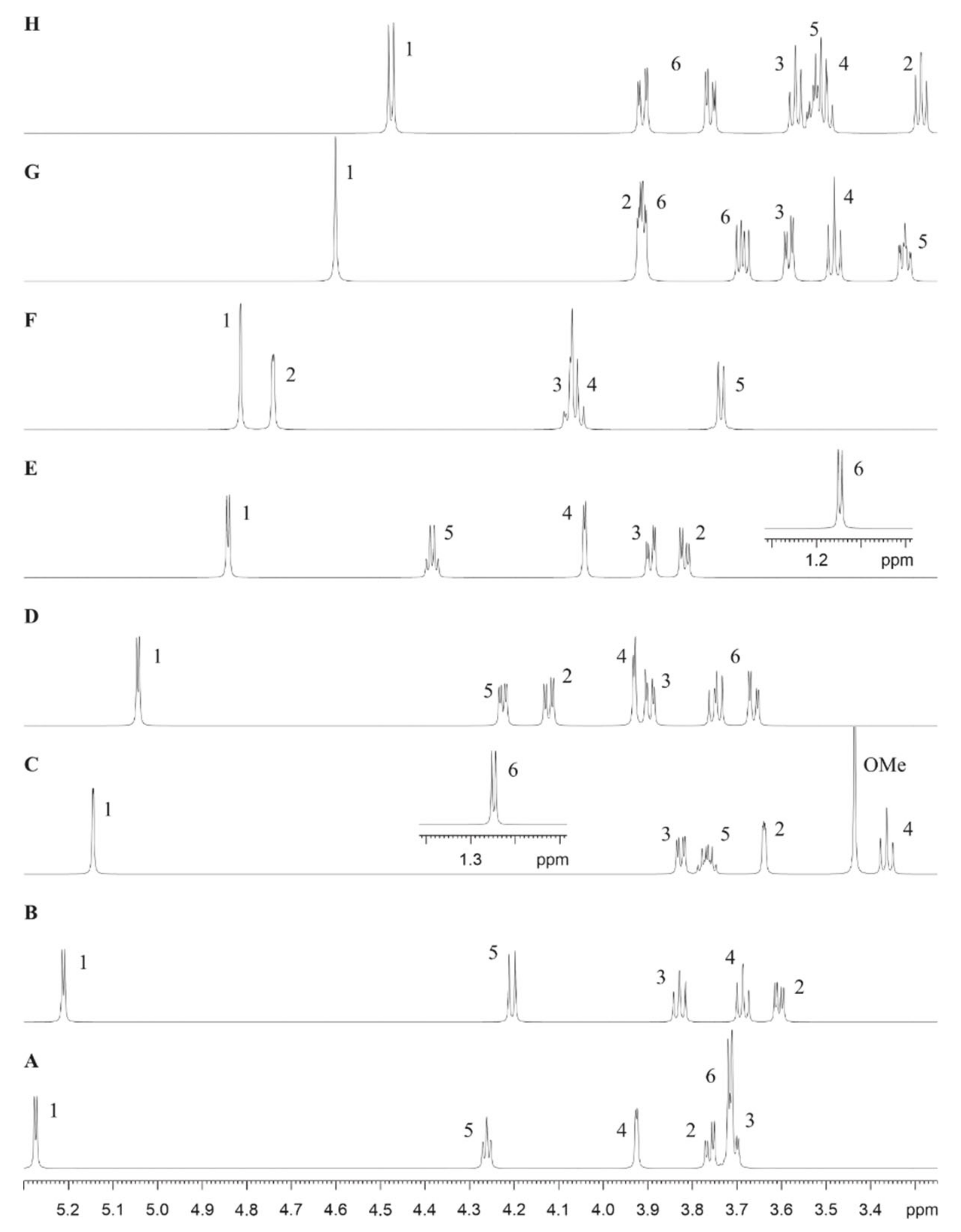
2.6. NMR Spectroscopy for Structure Analysis of the Bacteroides fragilis O-Glycan

NMR spectra were recorded on a Bruker AV III HD 700 MHz NMR spectrometer (Bruker BioSpin, Rheinstetten, Germany), equipped with a quadruple (1H, 13C, 15N, 19F) inverse helium-cooled cryo-probe, operating at 700.40 MHz for 1H and 176.12 MHz for 13C, respectively. A 500-µg sample of purified, β-eliminated B. fragilis wild-type glycan dissolved in 300 µL of D2O and transferred into a Shigemi tube, as well as three individual fractions of B. fragilis ΔBF4306 glycopeptides dissolved in 600 µL of D2O and transferred into standard 5-mm NMR tubes, were measured at a temperature of 25 °C. The spectra were referenced for 1H to the signal of the methyl groups of DSS (δ = 0 ppm), and for 13C on a unified scale relative to 1H using the Ξ value for DSS [41]. The following experiments were performed using pulse sequences as supplied by the manufacturer: 1H NMR with and without suppression of the HDO signal using presaturation or a diffusion filter (diffusion delay of 100 ms), 13C DEPTq-135, 2D DQF-COSY, 2D TOCSY (100 ms MLEV17 spin-lock), 2D NOESY (500 ms mixing time), 2D HSQC with and without 13C decoupling (GARP), and 2D HMBC. In addition, 1D selective TOCSY experiments were performed for high resolution spectra of the sugar units using a selective pulsed-field gradient spin echo sequence (80 ms 180° Gaussian pulse, 100 to 300 ms MLEV17 spin-lock).
Processing and detailed analysis of the spectra were performed within the TopSpin software (Bruker BioSpin). The spin coupling network was elucidated by spin simulations using DAISY within the TopSpin software, by fitting the calculated spectra mainly to the 1D TOCSY traces or partly to the normal 1H NMR spectrum.

2.7. Cross-Complementation in Tannerella forsythia

A cross-complementation gene cassette was constructed to replace the native Tanf_01305 gene with the homologous genes BF4306 from B. fragilis and Phep_4048 from P. heparinus, respectively. A detailed description of the cloning procedure and the transformation of vectors into T. forsythia is published elsewhere [30]. Briefly, the native Tanf_01305 upstream region was amplified with primers 490 and JB_6. The BF4306 gene was amplified using primer pair JB_7/JB_23 containing the restriction sites KpnI and SacI. Next, this fragment was added to the upstream region by overlap-extension (OE) PCR and subcloned into the blunt-end cloning vector pJET1.2. The chloramphenicol (cat) resistance gene was amplified from pJET1.2/ΔTanf_01245+ using primers JB_24 (KpnI) and JB_10 [31] (Table S1), and cloned to the native down-stream homology region, JB_11/JB_19 (SacI), by OE-PCR. Via the introduced restriction sites KpnI and SacI, the combined cat gene and the down-stream homology region were inserted, creating the final cross-complementation cassette pJET1.2/ΔTanf_01305+BF4306.
Analogously, the cross-complementation cassette pJET1.2/ΔTanf_01305+Phep_4048 for the integration of the homologous gene Phep_4048 from P. heparinus was created. The sole exception was the use of primers 490/JB_13 for the amplification of the upstream homology region and JB_14/JB_25 (KpnI, SacI) for the amplification of the Phep_4048 gene. Clones were selected on chloramphenicol-containing BHI plates and tested for correct integration on the genomic level after transformation into electrocompetent T. forsythia ΔTanf_01305 cells [31] (Figure S1).

2.8. Cross-Complementation in Bacteroides fragilis

Genes Tanf_01305 and Phep_4048 were cloned into an expression vector for complementation studies in B. fragilis ΔBF4306. The Phep_4048 gene was PCR-amplified using P. heparinus DSM 2366 genomic DNA as template, with primers Phep_4048_F/Phep_4048_R, which included a BamHI site (Table S1), and this product was inserted into BamHI-linearized pCMF118 [22], creating pMT2. Analogously, the Tanf_01305 gene was PCR-amplified using T. forsythia ATCC 43037 genomic DNA as template with primers Tanf_01305_F/Tanf_01305_R, which included BamHI sites, and was inserted into BamHI-linearized pCMF118 [22], creating pMT21.
These plasmids were transferred from E. coli DH5α into B. fragilis ΔBF4306 by conjugation using helper plasmid RK231. Transconjugants were selected using erythromycin and gentamycin.

2.9. Construction of a T. forsythia GDP-l-Fucose Synthase Deletion Mutant

A knock-out vector was constructed to exchange the GDP-l-fucose synthase gene of T. forsythia (Tanf_07535, fcl) in frame with the erythromycin resistance gene, erm. The plasmid contains regions of approximately 1-kbp upstream and downstream of Tanf_07535, flanking erm. Primer pairs 486/487 and 488/489, respectively, were used to amplify the up- and down-stream homology regions from genomic DNA of T. forsythia. The erm gene (805 bp, without the promotor region) was amplified from pJET/TF0955ko [30] using primers 460 and 461 (Table S1). This gene cassette was blunt-end cloned into the cloning vector pJET1.2, creating the final knock-out vector pJET1.2/ΔTanf_07535. Transconjugants selected on plates containing erythromycin and gentamycin were further confirmed by screening PCR (Figure S2). The deletion mutant carries an ermF marker in place of the fcl gene and, accordingly, was named ΔTanf_07535::ermF.

2.10. Cloning of Fucosyltransferase Genes

The fucosyltransferase genes Tanf_01305 (KKY62509.1), BF4306 (CAH09973.1), and Phep_4048 (ACU06239.1) were PCR amplified from genomic DNA using primers 516/517, JB_174/JB_175, and JB_172/JB_173, respectively. The amplification products were digested with EcoRI/HindIII (Tanf_01305, BF4306) and EcoRI/BamHI (Phep_4048) and inserted into the linearized vector pMAL_c2E digested with the same restriction enzymes (amino acid sequences of the maltose binding protein (MBP)-fusion proteins can be found in Figure S3). These plasmids, pMAL_c2E/Tanf_01305, pMAL_c2E/BF4306, and pMAL_c2E/Phep_4048, were transformed into E. coli BL21(DE3) cells for FucT expression. All constructs were verified by sequencing.

2.11. Expression and Purification of Recombinant Fucosyltransferases

Overnight cultures of E. coli BL21(DE3) harboring each the FucT encoding plasmids were inoculated into 400 mL of 2× LB medium with ampicillin and cells were grown at 37 °C and 200 rpm until an OD600 of 0.4 to 0.6 was reached. The cultures were shifted to 30 °C and expression was induced by addition of 0.5 mM IPTG. After incubation for 4 h, cells were harvested by centrifugation and cell pellets were stored at −20 °C. Cell pellets were thawed and resuspended in 20 mM Tris-HCl, pH 7.5 (15 mL buffer per g of wet cell pellet) in the presence of a protease inhibitor cocktail (cOmplete, Roche, Vienna, Austria). Cells were lysed by sonication and cell debris was removed by centrifugation (20,000× g, 20 min, 4 °C). Supernatants containing the soluble recombinant proteins were purified via an amylose resin (New England Biolabs, Vienna, Austria; running buffer: 20 mM Tris-HCl, pH 5.0, 200 mM NaCl; elution buffer: 20 mM Tris-HCl, pH 7.5, 200 mM NaCl, 10 mM maltose). Fractions were monitored and those containing the enzyme, based on SDS-PAGE analysis, were pooled. rTfFuc1 was expressed and purified as described previously [42].

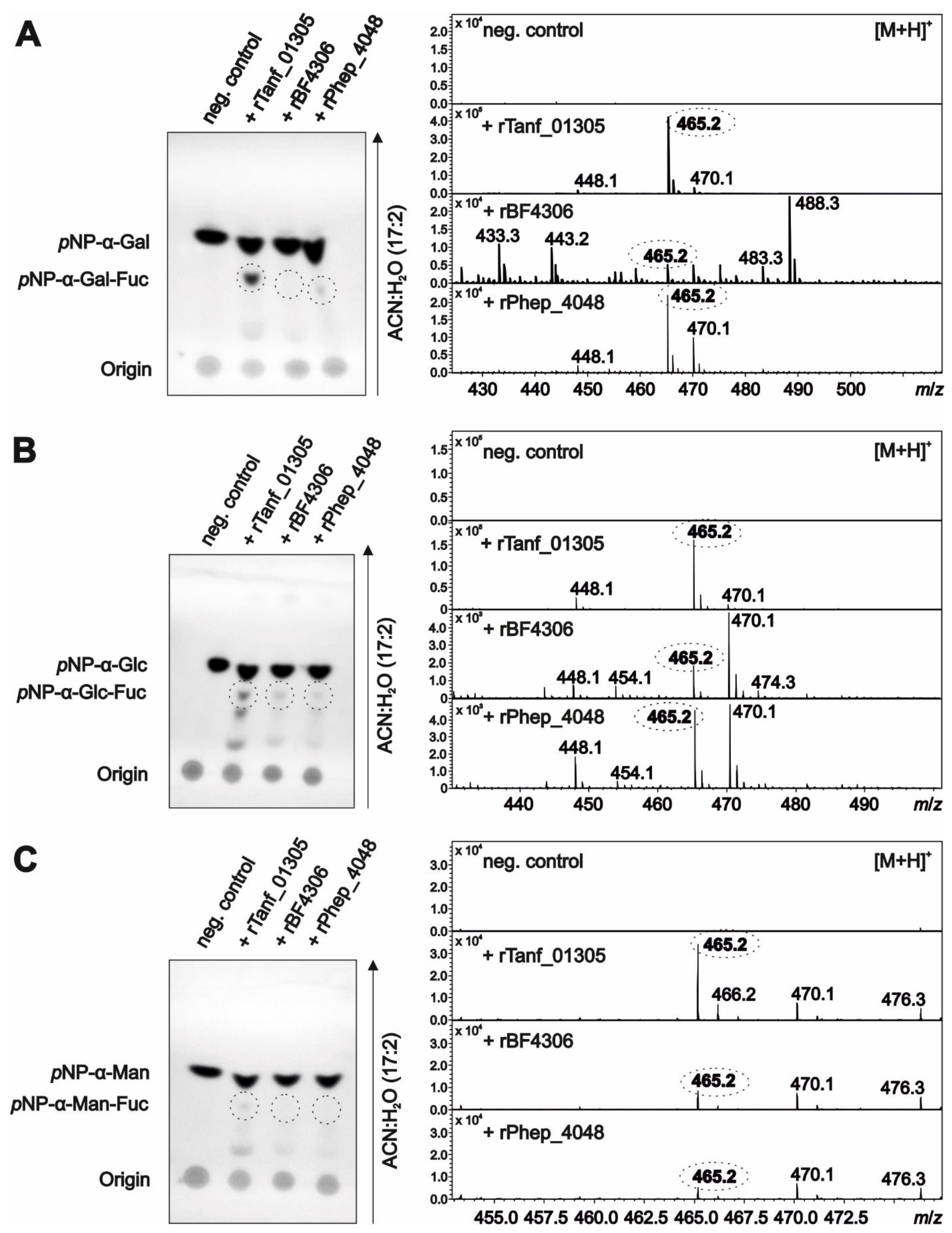
2.12. In Vitro Fucosyltransferase Activity Assays Using pNP-Sugar Substrates

The activity of the recombinant FucTs was determined by using 4-nitrophenyl (pNP)-α-d-Gal, pNP-α-d-Glc, pNP-α-d-Man, pNP-β-d-Xyl, pNP-α-d-GlcA, and pNP-β-d-GlcNAc as substrates (Sigma-Aldrich). The standard assay conditions at 37 °C were: 5 µL (~15–20 µg) of rFucT enzyme, 8 mM pNP-sugar substrate, 20 mM Tris-HCl, pH 7.4, 5 mM MgCl2, 5 mM MnCl2, and 2 mM GDP-L-Fuc (Sigma-Aldrich). After overnight incubation, reactions were terminated by the addition of 1:1 (v/v) stop solution (80% ACN). Samples were analyzed by thin-layer chromatography on silica plates (TLC Silica gel 60 F254, Merck Millipore, Vienna, Austria) with ACN:H2O (17:2) as running solvent. Separated spots were detected on the dried plates under UV-light at 254 nm.
For product analysis by ESI-MS, reactions were terminated by heating at 60 °C for 10 min followed by centrifugation to remove precipitated proteins. The supernatant was injected in the iontrap mass analyzer; instrument parameters were as described in Section 2.5, except for the recorded m/z range, which was 200–1650.

2.13. Preparation of pNP-α-d-Gal-Fuc as a Fucosidase Substrate

The preparation of pNP-α-d-Gal-Fuc is described in Supplementary Materials.

3. Results

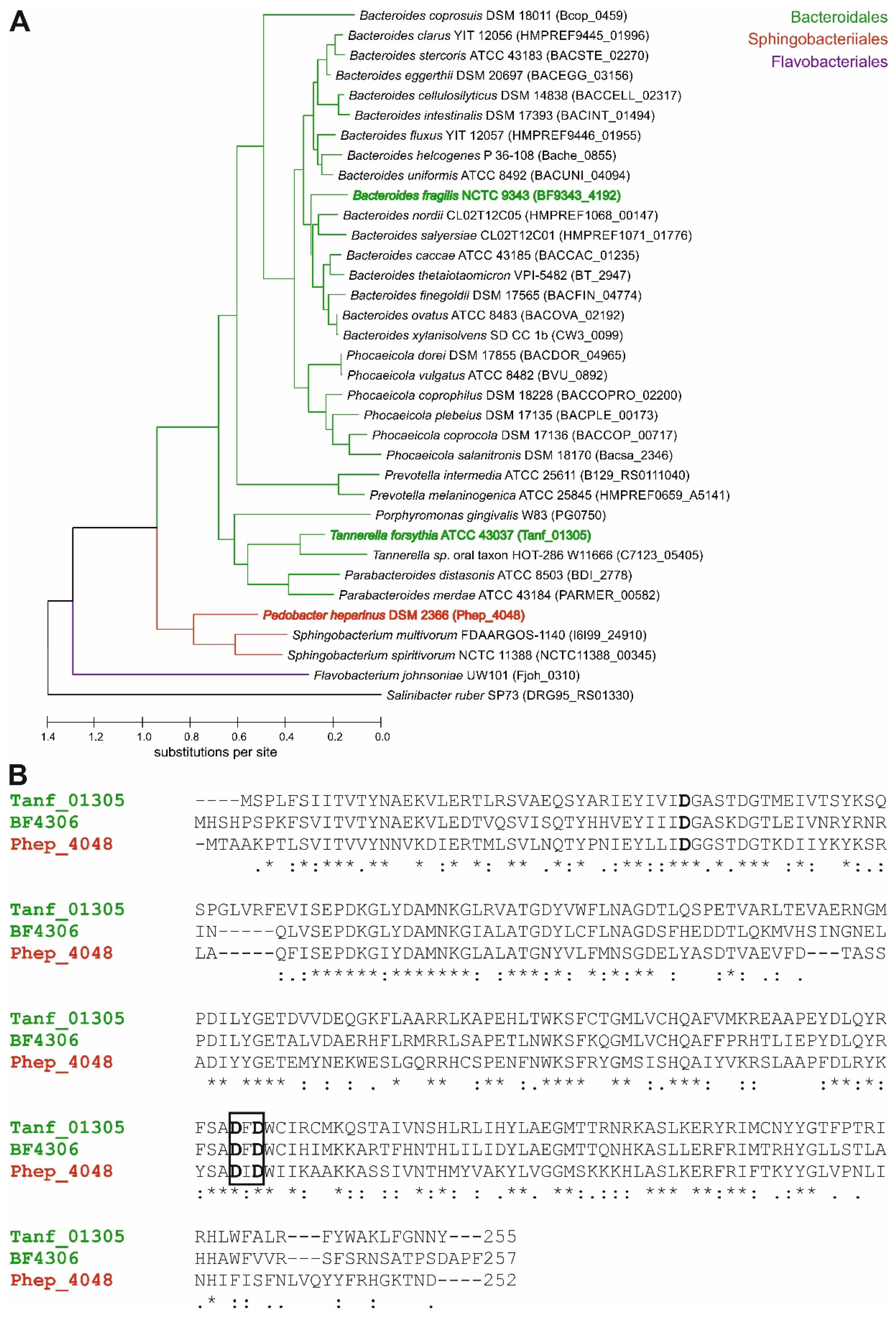
3.1. Presence of FucT Orthologs in Diverse Bacteroidetes Genomes

Mutational analysis of BF4306 and Tanf_01305 suggested that these orthologs are FucTs. In the O-glycan biosynthesis regions of all Bacteroides species analyzed, and in T. forsythia, this gene is conserved and is the terminal gene of these biosynthesis loci. These predicted FucTs are from the broad GT2 family of glycosyltransferases with a predicted GT-A type structural fold [10]. To determine how prevalent these FucT orthologs are in Bacteroidetes species, we searched the genomes of 35 diverse Bacteroidetes strains for orthologs. Despite the fact that the B. fragilis and T. forsythia FucT are only 71% similar to each other, separate Blastp searches using the B. fragilis and T. forsythia FucT proteins retrieved the same ortholog in each genome. The cladogram (Figure 1A) illustrates that the relationships among these FucT orthologs seemed to parallel species phylogeny. Glycosyltransferase-encoding genes of Bacteroidales species are also commonly found in non-conserved segments of the genome, like CPS biosynthesis loci, so this phylogenic distribution was unexpected.
Alignment of the B. fragilis, T. forsythia, and distantly related P. heparinus FucT orthologs show that they all contain a DXD motif typical of glycosyltransferases [43] (Figure 1B).

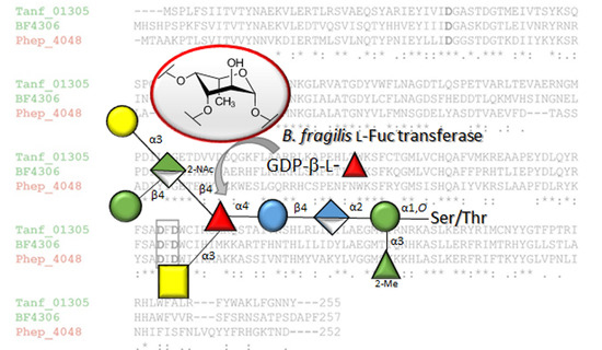
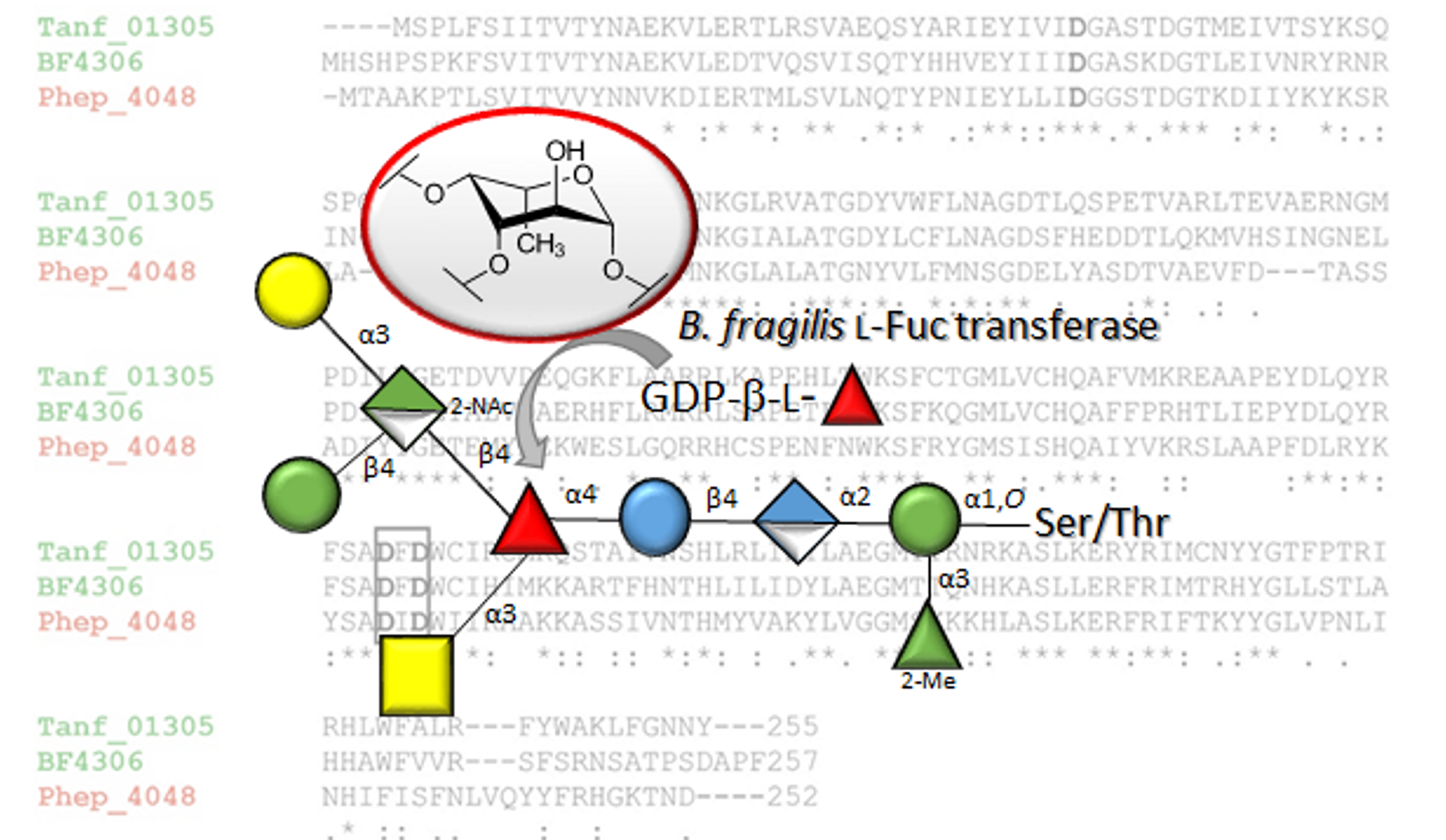
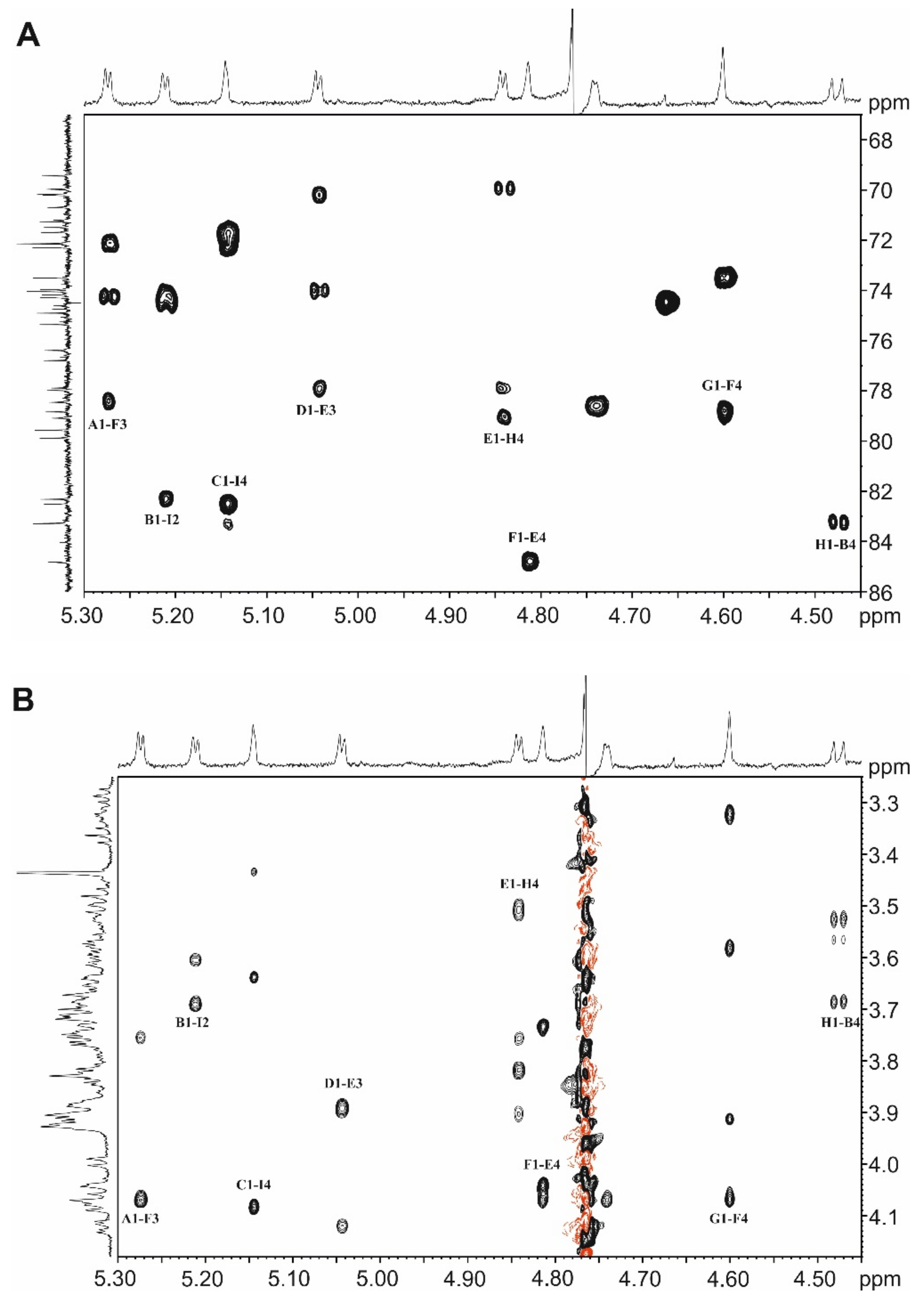
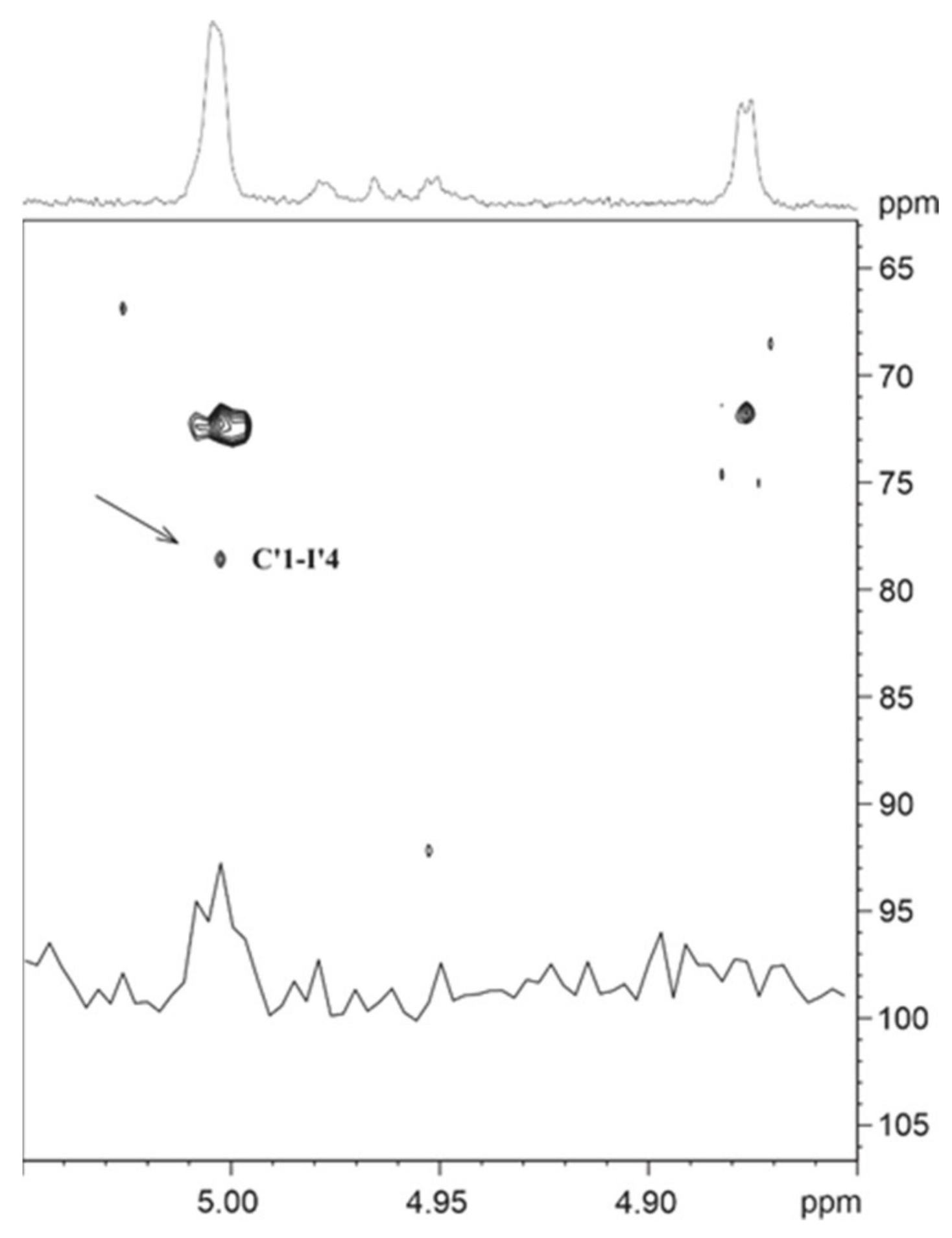
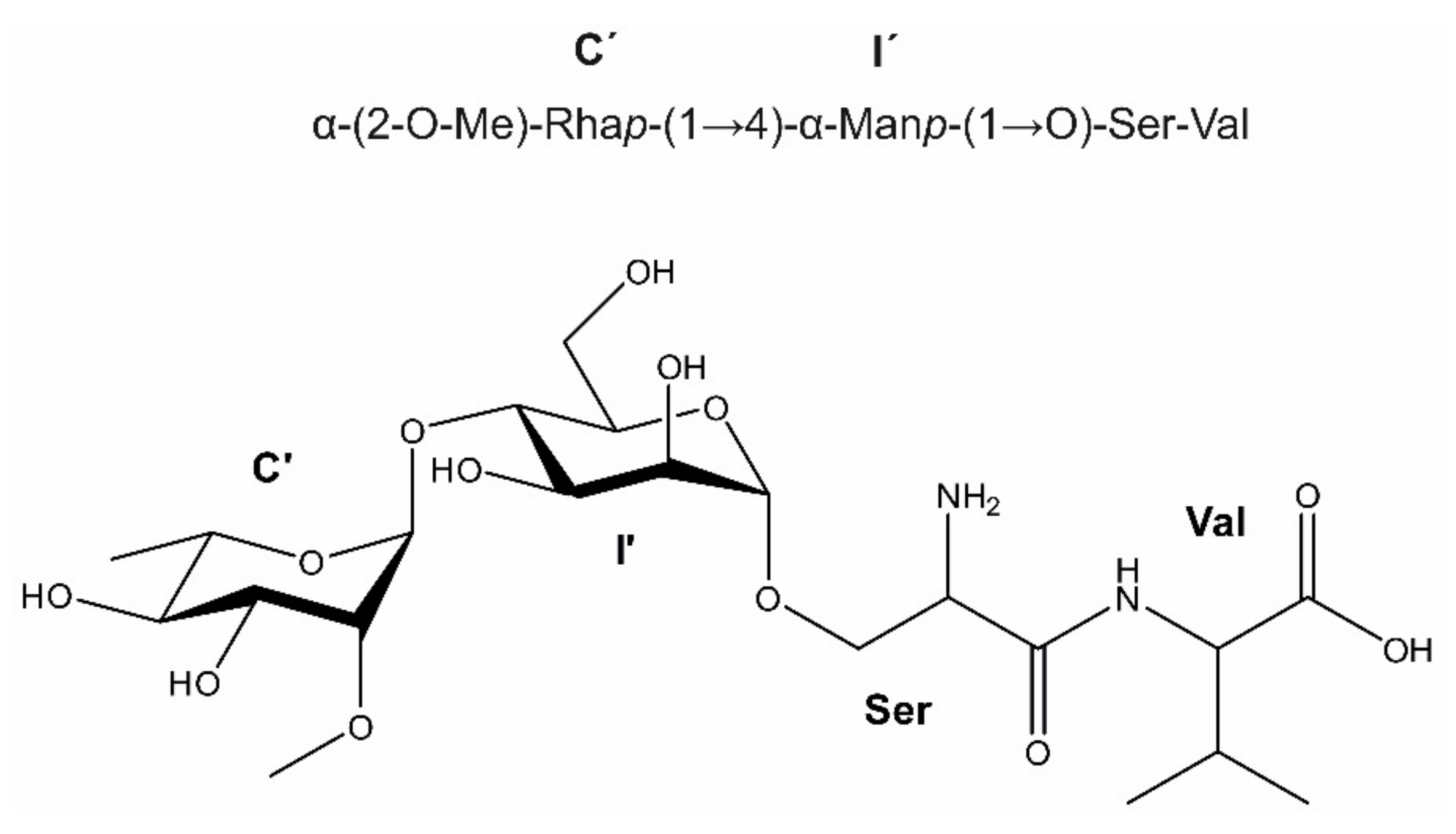
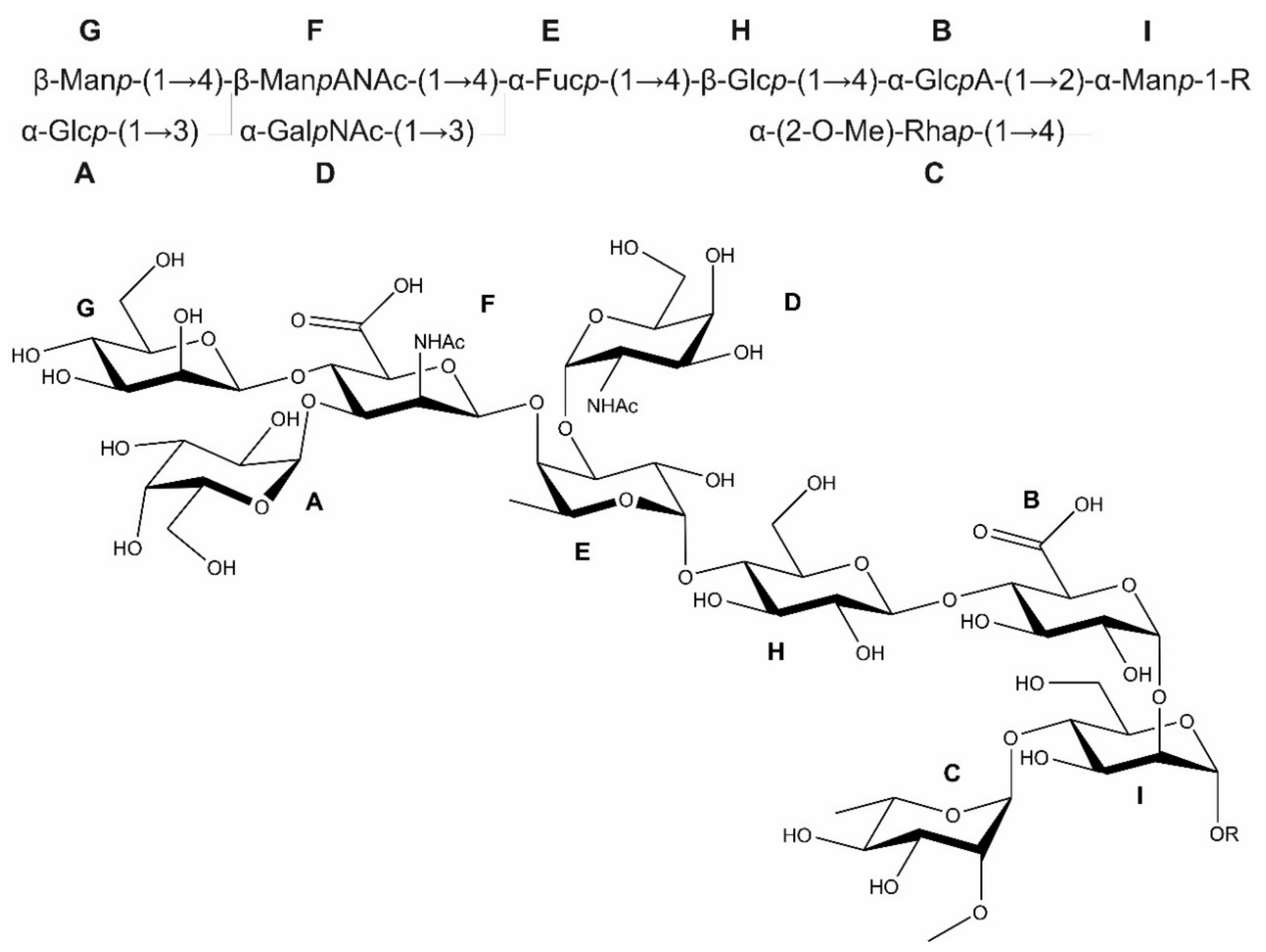
3.2. The Bacteroides fragilis O-Glycan Is a Complex Nonasaccharide Containing Fucose Linked α1,4 to Glucose

The analysis of Figure 1 illustrates the conservation of this FucT in Bacteroidetes species, suggesting its importance in O-glycosylation in this phylum. Our previous analysis of the B. fragilis protein O-glycan showed that it is composed of nine monosaccharides categorized broadly (i.e., hexose, deoxyhexose, etc.) [7,22]. By analysis of mutants unable to synthesize GDP-l-fucose, we showed that l-Fuc is a component of the outer glycan of B. fragilis and is also present in the O-linked glycans of diverse other Bacteroidetes species [7,16]. To unambiguously determine the position and linkage of the fucose moiety in the B. fragilis O-glycan and the various linkages between residues, we sought to elucidate the complete structure of this glycan. Reductive β-elimination was employed to release the O-glycan from purified B. fragilis glycopeptides. Subsequent PGC-ESI-MS analysis of the derived glycan revealed a molecule with a monoisotopic value of m/z = 1571.5 Da, in agreement with previous data from our laboratory and corresponding to a nonasaccharide [22].
The 1H NMR of the B. fragilis O-glycan preparation showed a typical carbohydrate spectrum with anomeric and core signals in the narrow chemical shift range between 3.2 and 5.3 ppm (Figure S4). Within this region, a sharp singlet at 3.44 ppm hinted a methoxy group and signals in the aliphatic region were indicative of an acetyl group (~2.0 ppm) and deoxy sugars (1.2 ppm).
The anomeric region in a 2D HSQC spectrum showed eight signals that gave a cross-peak at a 13C chemical shift between 100 and 105 ppm, characteristic of anomeric carbons (Figure S5). To identify the individual monosaccharides, the anomeric signals were chosen as starting point, providing a unique reporter for each monomer (AH) due to clear separation. The anomeric proton of A had a splitting of 4 Hz; the cross-peak in the DQF-COSY to proton 2 showed a significant larger coupling, therefore, not representing a mannose-type sugar. The 2D TOCSY gave a cross-peak with a very narrow shape for proton 4, assigning sugar A as α-Galp. To verify this sugar, the derived chemical shifts and the estimated coupling constants were used as input for a spin simulation and optimized by fitting to the experimental spectra, essentially to a 1D TOCSY trace. Figure 2 shows the calculated spectra together with the assignment of sugar A as well as of all following residues. For sugar B, besides the small splitting of 4 Hz for the anomeric proton, only large coupling constants can be detected (Figure 2). The proton spin system ends at position 5 with a doublet a 4.205 ppm. The 2D HMBC spectrum showed a cross-peak at this proton frequency to the carbon region related to carboxylic groups, thus identifying B as α-GlcA. The 2D TOCSY trace for the anomeric signal of residue C showed a cross-peak in the aliphatic region indicating a 6-deoxy sugar. Together with only small J couplings for H-2, C is Rhap (Figure 2). The anomeric configuration was deduced from the size of the proton-carbon coupling constant in a 2D HSQC experiment without 13C decoupling during acquisition. With an experimental value of 168.4 Hz, C refers to α-Rhap. In addition, this building block was methylated at position 2, indicated by HMBC cross-peaks from the CH3 protons at 3.44 ppm to the appropriate carbon C-2 at 83.30 ppm, and from the methyl carbon at 61.09 ppm to H-2 at 3.64 ppm (Figure 3A). For building block D, the coupling pattern of the 2D DQF-COSY and 2D TOCSY cross-peaks with small J values between 1 and 2, large J values from 2 to 3, and small J values from 3 to 4, was similar as for residue A (Figure 2). However, the 13C chemical shift for C-2 was found at 52.51 ppm, in the region for amino sugars. Proven by a 2D HMBC cross-peak from H-2 to a carbon in the carboxylic region, the amino function was acetylated and D is therefore α-GalpNAc (Figure 3A). The anomeric proton of E was close to the residual solvent signal (Figures S4 and S5). The spin system showed the same features, like sugars D or A, so E has a galacto-configuration. A 2D HMBC cross-peak at the carbon shift from C-4 identifies H-5, which, in the 2D DQF-COSY, showed a correlation in the aliphatic region to a doublet at 1.15 ppm. The spin simulation manifested all features of the 6-deoxy sugar E as α-Fucp (Figure 2). Going to higher field, the next signal was completely covered by the residual solvent (Figures S4 and S5). A 2D HMBC cross-peak from the anomeric proton to a CH signal at a carbon shift of 55.7 ppm identified F as a 2-amino-2-deoxy sugar, and the corresponding proton at this carbon shift was located at 4.74 ppm, covered by the solvent as well. This proton and also another doublet at 3.74 ppm had long-range correlations to carbonyl carbon signals in the HMBC spectrum. Monosaccharide F, thus, has features of 2-N-acetylamino-2-deoxy-uronic acid (ManpANAc). The analysis of the coupling network in the 1D TOCSY spectrum together with the spin simulation finally resulted in ManpANAc in β-configuration, due to a CH coupling constant of 161.5 Hz (Figure 2). At the right side of the residual solvent, the anomeric proton G was clearly visible, with the J coupling in the range of the line width and, thus, not resolved. As starting point for the selective 1D TOCSY, the complete spin system can be derived by applying a long spin-lock period. The analysis together with the spin simulation identified sugar G as Man, again in the β-configuration derived from a heteronuclear coupling constant of 160.4 Hz (Figure 2). Finally, starting from the anomeric proton of building block H with the large splitting of 8 Hz, the pathway to H-2 and further to H-3 can be easily followed in the 2D DQF-COSY experiment, and the fine structure of these cross-peaks displayed only large J values, implicating a glucose-type sugar (Figure 2). This was confirmed with the selective 1D TOCSY spectrum and, as a result, residue H is β-Glcp. All identified aldohexoses formed pyranoses, evidenced by appropriate 2D HMBC cross-peaks from the anomeric proton to C-5 or from the anomeric carbon to H-5, respectively.
The inter-glycosidic linkage information was derived from appropriate cross-peaks in a 2D HMBC experiment (Figure 3A), assisted by a 2D NOESY spectrum (Figure 3B). Further confirmation was obtained from the 13C chemical shift, as the involved carbons should exhibit a down-field shift up to 10 ppm due to glycosylation (Table 2). Residue A had an HMBC cross-peak from H-1 to C-3 of building block F (Figure 3A). All carbons except C-1 had chemical shifts in the range of unsubstituted carbons.
In total, these data provided the following information. Sugar A was the terminal residue at the non-reducing site. The oligosaccharide started from this end with the disaccharide unit α-Galp-(1→3)-β-ManpANAc. The carbon chemical shift of C-3 from unit F, which was deshielded due to glycosylation with A, was similar as for C-4, suggesting another substitution site (Table 2). Since an HMBC cross-peak could be found from this C-4 to the anomeric proton from sugar G (Figure 3A), β-ManpANAc F was a branching point of the glycan. Like A, this β-Manp G was the end of this side chain, since none of the carbons except C-1 were shifted due to the absence of glycosylation (Table 2). The next residue in the direction of the reducing end was identified by the HMBC cross-peak from H-1 of sugar F to C-4 of the Fucp E. Unit E was disubstituted, shown by the HMBC cross-peak from C-3 to the anomeric proton of the α-GalpNAc D. Next, H-1 from E had a long-range correlation to C-4 of the Glcp H. Finally, an HMBC cross-peak linked H-1 of H with C-4 of the GlcA B (Figure 3A). The next connectivity going onward from B was not to an aldose, although one residue would be still available, namely the α-(2-O-Me)-Rhap C. On the other hand, from both of these anomeric protons from B and C, HMBC cross-peaks to two individual carbons were visible at around 82 ppm (Figure 3A), of which none corresponded to the sugars analyzed so far. As the glycan was obtained by β-elimination under reductive conditions, the last part of the oligosaccharide was an alditol denoted as I, glycosylated with B at position 2 and with C at position 4 (Table 2). The nature of the starting hexose leading to this sugar alcohol could not be deduced from the NMR data at this point. As a cross check for all inter-glycosidic HMBC cross-peaks, a 2D-NOESY spectrum affirmed all connectivities with cross-peaks between the anomeric protons and the appropriate protons at the linkage position (Figure 3B).
The combined data of the NMR analysis (Table 2) and simulated 1H NMR spectra of the sugar units (Figure 2) reveal that the isolated glycan from B. fragilis is a nonasaccharide (Figure S6), which agrees with the experimental mass of 1571.8 Da. While the biochemical data are in full support of the presence of l-fucose, the absolute configuration of the rhamnose unit remains to be firmly established and has only been tentatively assigned to l-configuration in Figure S6.

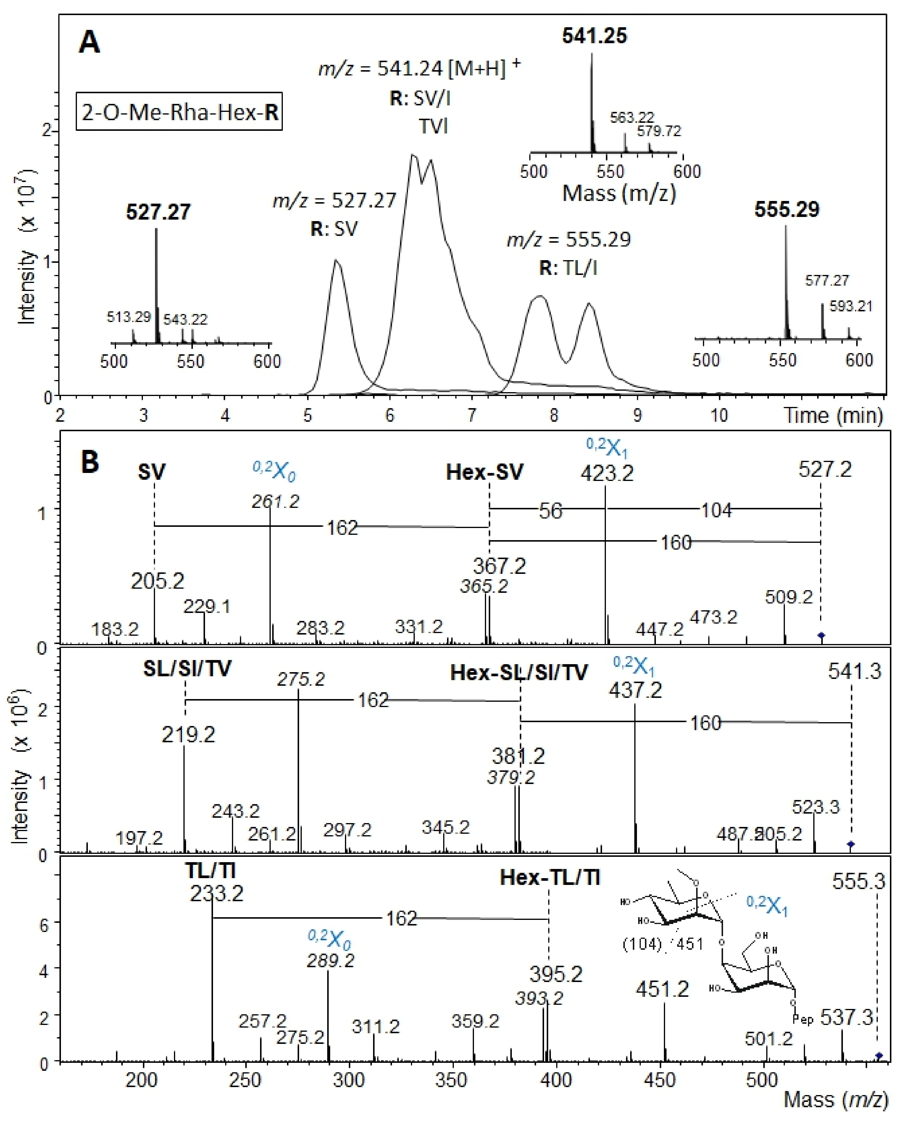
3.3. Determination of the Reducing-End Sugar of the Bacteroides fragilis O-Glycan

To identify the reducing-end sugar of the B. fragilis nonasaccharide, glycopeptides were prepared from the B. fragilis ΔBF4306 mutant, which produces a truncated O-glycan containing only two of the nine protein-linked sugar residues [7]. Glycopeptides, pre-purified by size exclusion chromatography, were further separated via PGC-HPLC and collected fractions screened by PGC-LC-MS. Three dominant fractions identified by MS and MS2 (Figure 4) were chosen for subsequent NMR analysis. In the MS-spectra, peaks with m/z = 527.27 [M + H]+ conforming to 2-O-Me-Rha-Hex-Ser-Val (fraction f28), m/z = 541.24 [M + H]+ conforming to 2-O-Me-Rha-Hex-Ser-Leu/Ile or 2-O-Me-Rha-Hex-Thr-Val (fraction f32), and m/z = 555.29 [M + H]+ conforming to 2-O-Me-Rha-Hex-Thr-Leu/Ile (fraction f36) were observed (Figure 4A). CID MS2 in the ion trap typically left the dipeptide portion intact with additional fragments showing mass increments of a hexose plus a methylated deoxyhexose (Figure 4B). Thus, the three glycopeptide samples (f28, f32, f36) differed in the peptide portion while containing the identical disaccharide, as expected. The MS2 spectra exhibited signs of considerable rearrangements as usual for positive mode CID [44,45,46].
For f28, f32, and f36, 1H NMR revealed a high peptide background with high coverage of the carbohydrate chemical shift region; furthermore, the amount of the samples was very low (Figure S7). In the anomeric region of the 1H NMR spectrum, the signal with the highest intensity was at approximately 5.00 ppm in all three spectra. Additional signals are visible in upfield direction, but they differed in number, intensity, and chemical shift in the three preparations. The main reason for this heterogeneity seems to originate from the different peptides. Omitting the signals near baseline in the spectra, f28 showed only one additional anomeric proton at approximately 4.88 ppm (Figure S7, upper trace). In f32, three anomeric signals were visible for the second sugar, one identical to that in f28, suggesting that the disaccharide-peptide structure from f28 is also present in f32. The two remaining anomeric protons derived either from the sugar linked to the Ser-Leu or the Ser-Ile amino acid sequence (Figure S7, middle trace). Finally, the preparation with the lowest intensity, f36, showed two anomeric protons for the second sugar, which can be attributed to two different peptides (Figure S7, lower trace). As f28 was the most promising candidate with regard to intensity and homogeneity in the peptide portion, it was chosen for detailed NMR analysis.
Starting with the anomeric proton of the first sugar at 5.00 ppm, one dominant TOCSY cross-peak under standard spin-lock conditions of 100 ms occurred at 3.61 ppm. Analysis of the appropriate frequency in the 1D 1H NMR spectrum revealed the corresponding multiplet was formed only by small J couplings, suggesting a mannose-type sugar. The 2D TOCSY trace at this chemical shift showed the rest of the spin system, with one signal located in the aliphatic region and a CH3 group as a doublet at 1.24 ppm. Applying a selective 1D TOCSY experiment by excitation of the methyl protons followed by a spin simulation, this sugar was identified as a Rhap. This Rhap is substituted as the carbon at position 2 was deshielded to 83.13 ppm, and an HMBC cross-peak was present at this 13C frequency connecting a sharp single line proton signal at 3.45 ppm. The anomeric configuration was α as proven by a heteronuclear J coupling of 169.7 Hz. Taken together, these data showed that this building block is an α-(2-O-Me)-Rhap and therefore matches unit C of the intact B. fragilis glycan. For this reason, in glycopeptide fraction f28, this residue was named C’ (Figure S8). The 2D TOCSY trace starting from the anomeric proton of the second sugar at 4.88 ppm was similar as for the first sugar. Again, essentially only one cross-peak was visible at a chemical shift of 3.9 ppm. The coupling pattern of this H-2 originated only from small J values, implicating a mannose-type sugar. Access to the complete spin system for this unit was only possible by a selective 1D TOCSY experiment starting from the anomeric proton using a long 300 ms spin-lock. Based on the interpretation of the 1D TOCSY spectrum, a spin simulation confirmed the Manp (Figure S8). The anomeric configuration was α according to a heteronuclear J coupling of 170.3 Hz. This α-Manp residue represents the reducing-end sugar before the reductive β-elimination of the B. fragilis nonasaccharide and was named I’.
Due to the low sample amount and large peptide background signals in the spectra, the elucidation of the linkage between the two monosaccharides of the disaccharide was challenging. In the 2D NOESY spectrum, cross-peaks could be traced from H-1 of the Rhap to signals at 3.77 and 3.90 ppm. The positions of the first chemical shift H-3 of Rhap and of H-4 of Manp were located more or less on top of each other. As the anomeric configuration of the Rha is α, a cross-peak from the equatorial H-1 to the axial H-3 is rather unlikely. In a 2D ROESY experiment with 200 ms mixing time, this cross-peak was more intense, which enabled the analysis of the cross-peak fine structure. The shape implied a triplet structure, thus resulting from two J couplings of similar size, which only fits H-4 of the Manp. The second cross-peak at 3.90 ppm with a much lower intensity was H-3 of the Manp. These results revealed either a 3 or a 4 linkage from Rhap to the Manp, although the higher cross-peak intensity representing a shorter distance between the two involved protons at the linkage site suggested a higher probability for position 4. In addition, the linkage from the α-(2-O-Me)-Rhap C to the alditol in the previously analyzed oligosaccharide is also position 4 (compared with Figure S6). Final verification was possible with an HMBC spectrum showing a cross-peak from the anomeric proton of the Rhap to C-4 of the Manp (Figure 5). These data prove that the missing hexose, which was reduced to the sugar alcohol during preparation of the B. fragilis glycan, is α-Manp.
As shown by the mass spectrometry analysis, the disaccharide portion of f28 had an O-glycosidic linkage to a dipeptide fragment. Starting from the sugar portion, the corresponding peptide-Man linkage was revealed in the 2D NOESY spectrum, showing a cross-peak from H-1 of the α-Manp I’ to a chemical shift of 3.93 ppm, one of the protons from the CH2 group of serine. From this chemical shift, the rest of this amino acid spin system could be assigned. For the valine, two doublets at 0.95 and 0.91 could be assigned as methyl groups, and the rest of the amino acid spin system is accessible in the 2D TOCSY spectrum. The simulated 1H NMR spectra of the two sugar units from the glycopeptide f28 is shown in Figure S8, and the elucidated structure of the glycopeptide is shown in Figure 6.
The complete structure of the B. fragilis O-glycan including the linkage sugar to the protein portion is shown in Figure 7. All NMR data of the nonasaccharide preparations from B. fragilis wild-type and the disaccharide glycopeptide f28 from a B. fragilis ΔBF4306 mutant are summarized in Table 2.

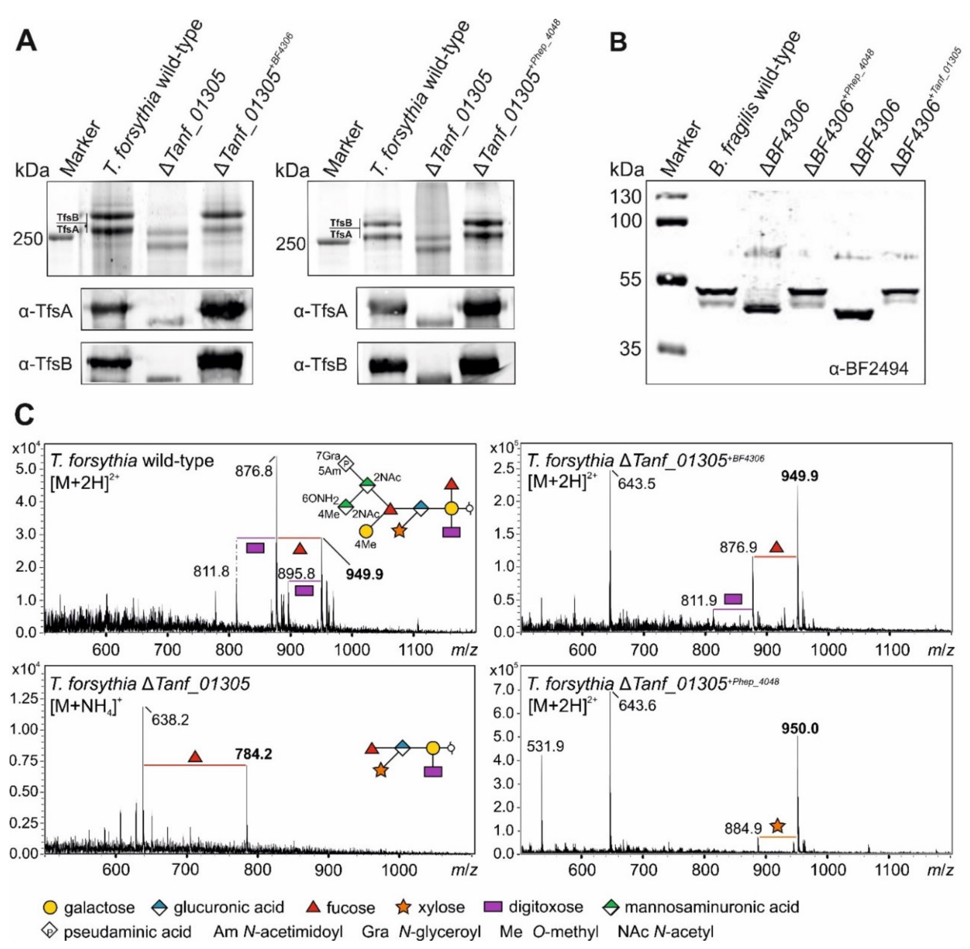
3.4. Bacteroidetes FucTs Can Cross-Complement

The structures of the T. forsythia and B. fragilis O-glycans suggest that their FucTs transfer Fuc to different hexose sugars (Figure 8). Here, we tested the ability of each of these FucTs to complement mutants in the heterologous species. In addition, we tested the ability of the FucT from P. heparinus, Phep_4048, of a phylogenetically distant Bacteroidetes species whose FucT is only 48.1% and 48.6% similar to the FucT of B. fragilis and T. forsythia respectively, to complement these mutants. As read-out for glycosylation, we used specific antisera to the protein portion of the abundant T. forsythia S-layer glycoproteins TfsA and TfsB [8] and the protein portion of the B. fragilis glycoprotein BF2494, an abundant soluble periplasmic protein [37].
Western immunoblots using α-TfsA and α-TfsB antibodies (Figure 9A) and α-BF2494 antiserum (Figure 9B) showed the reduction in the size of the glycoproteins in the ΔfucT mutants—indicative of glycan truncation in these bacteria. The T. forsythia glycan was reduced from a decasaccharide to a pentasaccharide [9] and the B. fragilis nonasaccharide was reduced to a disaccharide [7] (Figure 8). When the heterologous genes were added to these mutants in trans, the glycoproteins migrated identical to those in the wild-type bacteria in SDS-PAGE. For T. forsythia, the detection of the MW-shifts was also possible using CBB-staining due to the cellular abundance of the S-layer glycoproteins and their large size (Figure 9A). In addition, MS analysis of released S-layer O-glycans verified that cross-complementation of T. forsythia ΔTanf_01305 with BF4306 and Phep_4048, respectively, restored the complete T. forsythia wild-type O-glycan (Figure 9C).

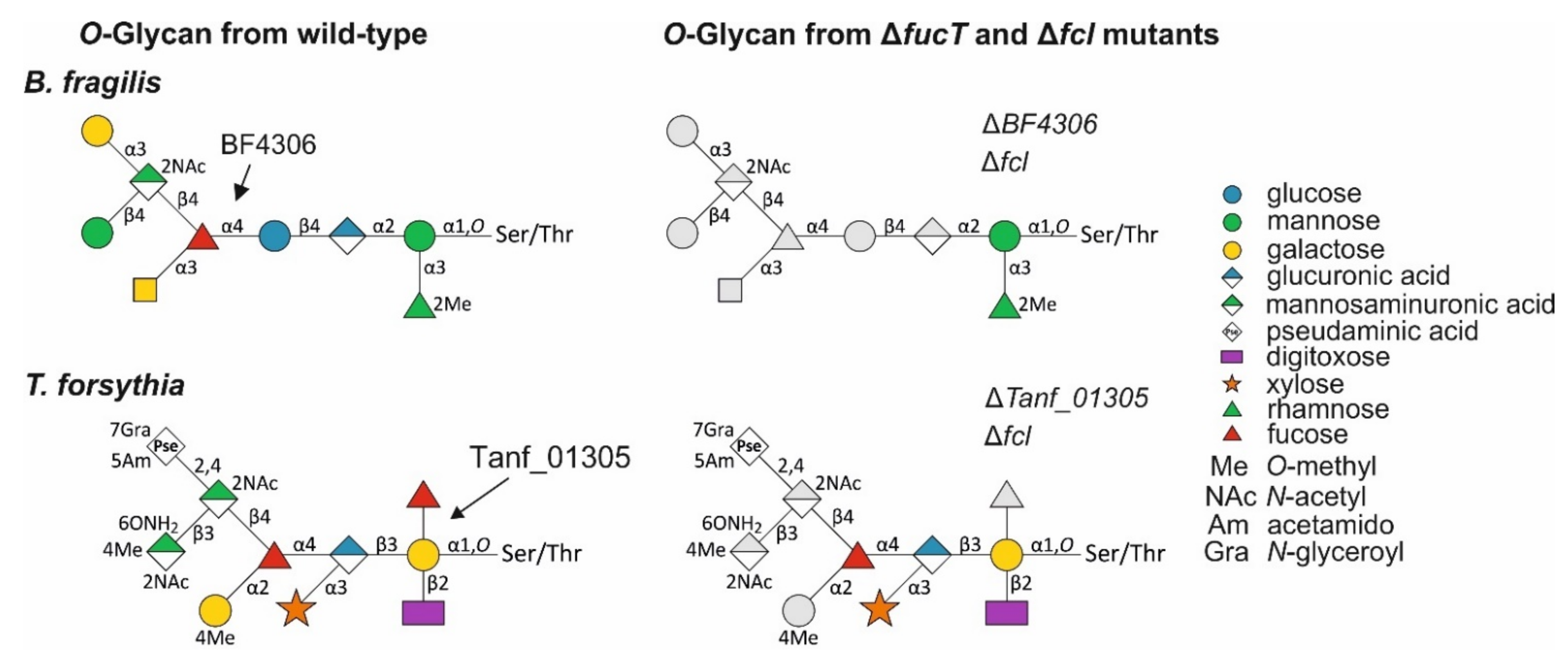
3.5. A T. forsythia GDP-l-Fucose Synthase fcl Knock-Out Strain Produces the Same Truncated O-Glycan Phenotype as the FucT-Deficient Strain

The T. forsythia O-glycan contains two Fuc residues, of which only the galactose-bound, inner Fuc is affected in the ΔTanf_01305 mutant (Figure 8), as previously demonstrated by detailed MS2 analyses of that mutant [9]. Based on the loss of fucosylation in a B. fragilis mutant deficient in GDP-l-Fuc biosynthesis [15] and the demonstrated functional homology of the FucTs from B. fragilis and T. forsythia, we reasoned that the Fuc residue branching from the protein-linked Gal of the T. forsythia glycan is l-Fuc, and the GlcA-linked Fuc at the branching point of the glycan is d-Fuc. To confirm this, we created a T. forsythia ΔTanf_07535 mutant defective in the GDP-l-Fuc synthase Fcl, which catalyzes the conversion of GDP-d-Man to GDP-l-Fuc [18]. Unlike Bacteroides and Parabacteroides species, where there are both de novo and salvage pathways for the synthesis of GDP-l-Fuc [16], T. forsythia does not contain the gene encoding Fkp of the salvage pathway, and therefore, deletion of fcl is sufficient to abrogate GDP-l-Fuc synthesis and prevent incorporation of l-Fuc into oligo- and polysaccharides. As expected, the ESI-MS spectrum of the O-glycan of T. forsythia Δfcl showed the same phenotype as T. forsythia ΔTanf_01305, revealing the significant m/z = 784.3 [M + NH4]+ indicative of a pentasaccharide lacking the Fuc branching from the protein-linked Gal (Figure 10). These data showed that Tanf_01305 transfers an l-Fuc and that the Fuc attached to the GlcA at the branching point of the T. forsythia glycan is likely a d-Fuc.

3.6. A T. forsythia GDP-l-Fucose Synthase fcl Knock-Out Strain Produces the Same O-Glycan Phenotype as the FucT-Deficient Strain

Although all three FucT orthologs can cross-complement, we showed that two of these FucTs create different linkages, one to Gal (T. forsythia) and one to Glc (B. fragilis) (Figure 8). To further demonstrate that Tanf_01305, BF4306, and Phep_4048 are l-FucTs, and to determine if they are specific for Fuc transfer to hexose, an in vitro FucT assay was performed. Each protein was translationally fused to MBP and produced in and purified from E. coli. SDS-PAGE analysis of these purified recombinant proteins showed that rPhep_4048 and rBF4306 have lower MW forms in addition to the full-length form, likely due to degradation (Figure S9).
All enzymes were tested for their ability to fucosylate different pNP-hexose acceptor substrates, including pNP-α-Gal, pNP-α-Glc and pNP-α-Man and, in addition, pNP-α-GlcA (GlcA is the attachment site of the second Fuc in the T. forsythia O-glycan structure; Figure 8), pNP-β-Xyl and pNP-β-GlcNAc (negative controls), with GDP-l-Fuc serving as the Fuc donor. All three enzymes transferred l-Fuc to pNP-α-Gal (Figure 11A), pNP-α-Glc (Figure 11B), and pNP-α-Man (Figure 11C), albeit with different preferences according to semiquantitative detection of the reaction products on TLC plates and by ESI-MS, evidenced by m/z of pNP-Hex-Fuc = 465.2 [M + H]+. Low activities could be due to the recombinant form of the proteins and/or suboptimal reaction conditions. rTanf_01305 transferred an l-Fuc residue to all three tested acceptor substrates with pNP-α-Gal as the best substrate—confirming reactivity on its native acceptor in the T. forsythia O-glycan—and pNP-α-Man as the least favorable substrate (pNP-α-Gal > pNP-α-Glc > pNP-α-Man). rBF4306 demonstrated very low activity with all substrates; pNP-α-Glc was the best substrate, which is in agreement with the finding that l-Fuc is linked to a Glc residue in the B. fragilis O-glycan structure (Figure 8). rBF4306 also demonstrated slight transfer to pNP-α-Man substrate, while only traces of modified pNP-α-Gal were detected. However, cross-complementation showed that in the native organism, BF4306 complements the function of Tanf_01305. rPhep_4048 showed comparable preferences for the pNP-hexose acceptor substrates as rTanf_01305, but was only minimally active. The structure of the P. heparinus glycan has not been determined and therefore its natural preferred substrate is unknown; however, these data combined with the cross-complementation data suggest that rPhep_4048 links l-Fuc to a hexose residue of its O-glycan. Neither pNP-α-GlcA, pNP-β-Xyl, nor pNP-α-GlcNAc was a suitable acceptor substrate for any of the three enzymes (Figure S10).

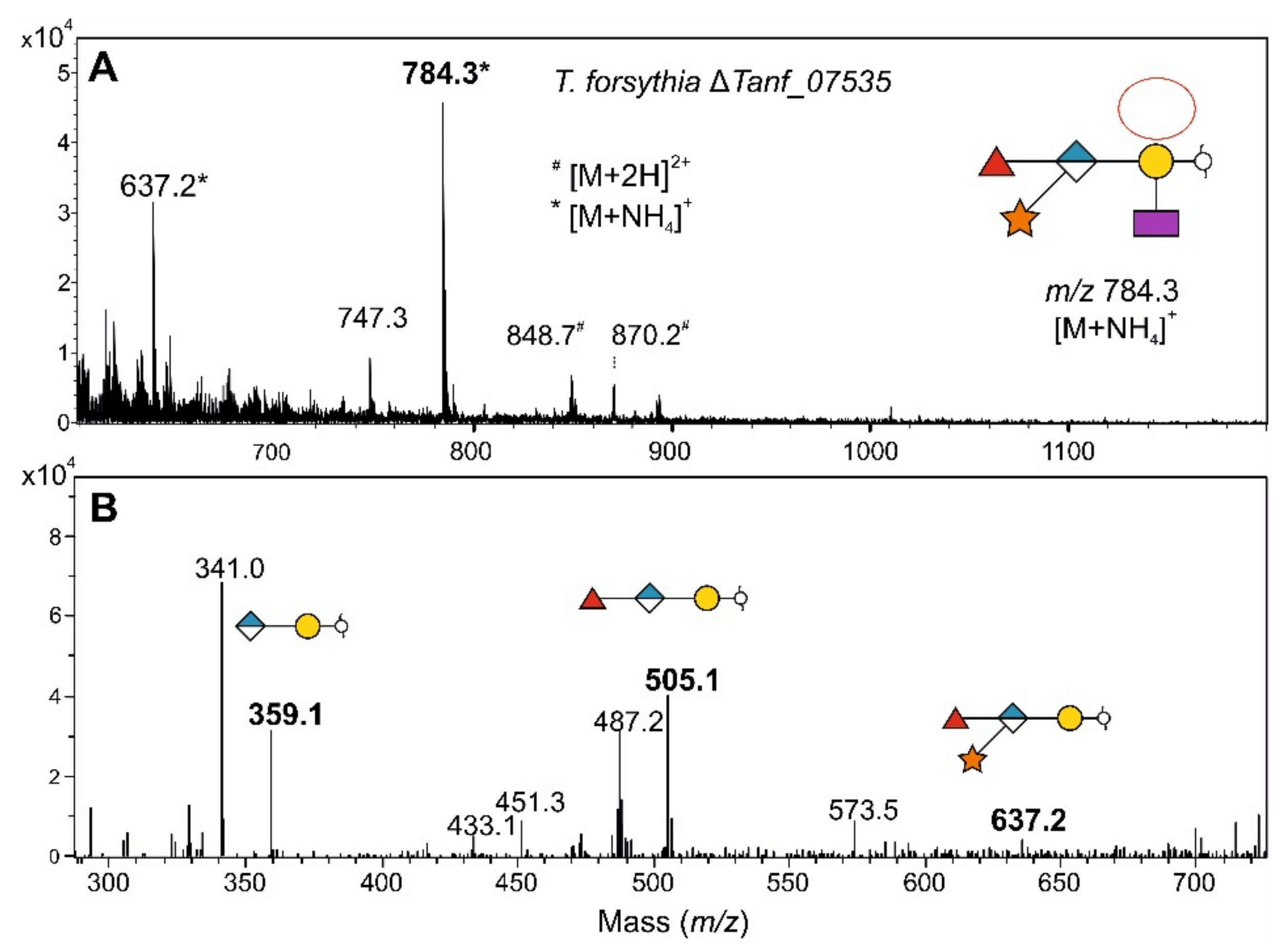
3.7. Fucosidase Treatment of pNP-α-d-Gal-Fuc Suggests an α1,6-Linkage of the l-Fuc in the T. forsythia O-Glycan

Since the linkage between the l-Fuc residue and the reducing-end Gal in the T. forsythia O-glycan structure is unknown, we used the pNP-α-Gal-Fuc reaction product from the rTanf_01305 activity assay to elucidate this linkage (Figure 11A). We purified the compound from the in vitro reaction by preparative TLC and used various fucosidases of known specificities to investigate the linkage. We used commercially available fucosidases (α1,2 fucosidase, α1,3/4 fucosidase, and α1,2/4/6 fucosidase O) as well as T. forsythia fucosidase TfFuc1 that was recently characterized in our laboratory to be an α1,2-fucosidase with additional α1,6 specificity on small unbranched substrates [35]. TLC showed that treatment with α1,2/4/6 fucosidase O and rTfFuc1 resulted in cleavage of the terminal Fuc from pNP-α-d-Gal-Fuc (Figure S11). From these data, together with the knowledge of the T. forsythia O-glycan structure where the protein-linked Gal residue is substituted at the C2 with a Dig residue, we conclude that the linkage between l-Fuc and the Gal residue is most likely α1,6.

4. Discussion

Members of the phylum Bacteroidetes are colonizers of numerous habitats on Earth [4]. They are among the major members of the microbiota of animals, especially in the gastrointestinal tract, can act as pathogens, and are frequently found in soils, oceans, and freshwater. Despite these diverse ecological niches, we revealed commonalities in Bacteroidetes with regards to the biosynthesis of O-linked glycoproteins involving a conserved FucT that is widely distributed in the phylum (Figure 1).
For B. fragilis and T. forsythia, the presence of a general protein O-glycosylation system targeting various cellular proteins was demonstrated previously and showed the biological importance of protein glycosylation [7,22,49]. In these bacteria, the O-glycan is a complex oligosaccharide with both a core (inner glycan) and outer glycan. In Bacteroides species, the outer glycan enzymes are encoded by the lfg region [7,10], whereas the genes involved in core glycan synthesis are located elsewhere on the bacterial genome.
To determine the role of the conserved FucT from T. forsythia (Tanf_01305), B. fragilis (BF4306), and P. heparinus (Phep_4048) as representatives of the phylum, we first elucidated the complete structure of the B. fragilis O-glycan by 1D and 2D NMR spectroscopy and revealed that it is a branched nonasaccharide with a Fuc residue serving as a branching point in is backbone structure (Figure 7). The O-glycan structure analysis of the B. fragilis ΔBF4306 mutant, in contrast, revealed a heavily truncated structure devoid of Fuc (Figure 6 and Figure 10) further supporting that BF4306 is a FucT. These data also support the previous finding that the core glycan is devoid of Fuc and that the BF4306 enzyme is essential for outer glycan biosynthesis. Interestingly, the branched disaccharide α-2-O-Me-Rhap-(1→3)-Manp-(1→O of the B. fragilis core glycan (this study) is identical with those of the Elizabethkingia meningoseptica hydrolase glycoproteins, according to MS-based evidence [50], and P. heparinus heparinase I, however without provision of a detailed structural analysis [51].
The observation that deletion mutants of conserved FucT in B. fragilis ΔBF4306 and T. forsythia ΔTanf_01305 leads to glycans that are lacking not only the Fuc residue transferred by the FucT but also proximal sugar residues in the O-glycans—i.e., to a disaccharide in B. fragilis and a pentasaccharide in T. forsythia [7,10] (compared with Figure 10)—raises questions about the biosynthetic pathway for the protein-linked O-glycans. For instance, it might involve a signalling function of the FucT to upstream-acting biosynthetic enzymes or the FucT might be part of a multienzyme complex. To investigate the latter possibility, pull-down experiments with the different FucTs are currently underway in our laboratory.
The analyses of the Δfcl mutant of T. forsythia revealed that this O-glycan likely contains d-Fuc in addition to l-Fuc. In most cases, FucTs catalyze an inverting reaction in which GDP-β-l-Fuc serves as a donor substrate; this is de novo synthesized from GDP-d-Man involving the enzymes Gmd (GDP-mannose-4,6-dehydratase) and Fcl (GDP-keto-6-deoxymannaose 3,5-epimerase/4-reductase) [18]. Rare examples of bacterial carbohydrate structures contain d-configurated Fuc—e.g., the O-specific polysaccharide of the LPS from Mesorhizobium huakuii strain S-52 [52], the O-antigen of Aggregatibacter actinomycetemcomitans (previously, Actinobacillus actinomycetemcomitans) Y4 (serotype b) [53] or the S-layer glycan from Geobacillus tepidamans GS5-97T [54]. For the biosynthesis of these, TDP-d-Fuc serves as the d-Fuc donor and is produced along the RmlA (glucose-1-phosphate thymidylyltransferase)/RmlB (dTDP-glucose-4,6-dehydratase)/Fcd (dTDP-4-dehydro-6-deoxyglucose reductase) pathway [53,54]. A Δfcl mutant of T. forsythia (Figure 8) and of B. fragilis [16] (Figure 8) clearly revealed that GDP-l-Fuc is the substrate for the conserved FucTs, allowing the assignment of the targeted Fuc residue in the O-glycans as an l-Fuc. As the second Fuc present in the T. forsythia glycan (Figure 10) was not affected by the lack of GDP-l-fucose in the T. forsythia Δfcl mutant, it is likely transferred by another FucT requiring TDP-d-Fuc. This would imply that the T. forsythia O-glycan is among the rare examples of bacterial carbohydrate structures that contain both an l-Fuc and a d-Fuc residue.
The conserved FucTs Tanf_01305, BF4306, and Phep_4048 are functional orthologs based on the successful cross-complementation of the T. forsythia ΔTanf_01305 and the B. fragilis ΔBF4306 mutants with the non-native fucT genes leading to the restoration of the respective wild-type glycans (Figure 9).
We showed in an in vitro enzyme assay that BF4306, Tanf_01305, and Phep_4048 are active on various pNP-hexose acceptor substrates, including pNP-Gal, pNP-Glc, and pNP-Man (Figure 11). Under the experimental conditions used, the enzymes had different preferences for the pNP-hexose substrates, indicative of substrate promiscuity of the conserved Bacteroidetes FucTs. Further, these FucTs might also exhibit some degree of promiscuity with regards to the linkage specificity. BF4306 is an α1,4-FucT as concluded from the linkage data of the Fuc residues in the B. fragilis O-glycan (Figure 7). The linkage information of the l-Fuc in the O-glycan structure of T. forsythia is missing, however, the data of this study suggest that Tanf_01305 is an α1,6 FucT (Figure S11). Thus, the conserved Bacteroidetes FucTs investigated within the frame of this study might represent the first bacterial FucTs with α1,4/6 specificity. The α1,3/α1,4 FucTs, which are mostly classified as GT10 family enzymes and have a DXD motif, are evolutionary distinct form the superfamily of α1,2/α1,6/O-FucTs. The Bacteroidetes FucT family described here has a DXD motif, fitting with α1,4-linked Fuc observed in the B. fragilis O-glycan, but not with α1,6-linked Fuc deduced for the T. forsythia O-glycan. Furthermore, these new FucTs group as GT2 and not with other known FucTs CAZy families.
These remaining questions illustrate the need for further research on FucTs to obtain a more detailed understanding of their functional capabilities. Furthermore, FucTs are promising tools for biosynthetic glycobiology. Significant examples of Fuc-containing glycoconjugates include the biotechnological production of fucosylated human milk oligosaccharides [55], engineering of cancer vaccines presenting Fuc-containing tumor-associated carbohydrate antigens (such as Lewis antigens, Globo H, fucosyl-GM1) [56], and chemoenzymatic synthesis of cholera toxin inhibitors [57]. Therefore, a more detailed analysis of the activity of the FucTs is warranted to understand their true potential in glycoengineering.

5. Conclusions

  • The structure of the protein-linked O-glycan of B. fragilis NCTC 9343 was elucidated by 1D and 2D NMR spectroscopy and shown to be a complex, branched nonasaccharide with an l-Fuc residue in the backbone structure.
  • The described l-FucT is relatively conserved among different members of the bacterial phylum Bacteroidetes. Functional orthologs of the FucT BF4306 were demonstrated in the periodontal pathogen Tannerella forsythia (Tanf_01305) and the soil bacterium Pedobacter heparinus (Phep_4048), using in vivo cross-complementation and an in vitro enzyme assay.
  • Enzymatic analyses revealed that these l-FucTs exhibit relaxed acceptor substrate specificity transferring l-Fuc from GDP-l-Fuc to galactose, glucose, and mannose residues, with α1,4/6 linkage specificity.
  • Given the biological importance of fucosylated carbohydrates, the Bacteroidetes l-FucTs are promising candidates for glycobiology applications.

Supplementary Materials

The following are available online at https://0-www-mdpi-com.brum.beds.ac.uk/article/10.3390/biom11121795/s1, Supplementary Methods: Preparation of pNP-α-d-Gal-Fuc As a Fucosidase Substrate. Table S1: Primers used in this study. Figure S1: Strategy for cross-complementation of a T. forsythia ΔTanf_01305 knock-out mutant with the homologous genes BF4306 from B. fragilis and Phep_4048 from P. heparinus, respectively, and confirmation by screening PCR. (A) The genomic organization of the Tanf_01305 locus is shown for the parent strain T. forsythia ATCC 43037, the ΔTanf_01305 mutant and the cross-complemented mutants ΔTanf_01305+BF4306 and ΔTanf_01305+Phep_4048. Black coloured arrows represent primers used for PCR amplification of genes and homologous regions, red coloured primers represent those used to screen for correct integration of the cross-complementation cassettes; restriction sites used for cloning are indicated (not drawn to scale). (B) Agarose gel electrophoresis (left) confirms the successful integration of BF4306 at the Tanf_01305 locus using the screening primers 494/JB_23 (1888 bp) and 48/495 (1141 bp) when using genomic DNA of the ΔTanf_01305+BF4306 strain. Screening primers 494/495 yield in a 3649-bp PCR product on genomic DNA of the cross-complemented mutant ΔTanf_01305+BF4306, whereas the same primer pair results in a 3023-bp product on genomic DNA of the T. forsythia wild-type. The successful integration of Phep_4048 at the Tanf_01305 locus was likewise confirmed using the screening primers 494/JB_25 (1867 bp) and 48/495 (1137 bp) when using genomic DNA of the ΔTanf_01305+Phep4048 strain (right). Screening primers 494/495 yield in a 3630-bp PCR product from genomic DNA of the cross-complemented mutant ΔTanf_01305+Phep_4048, whereas the same primer pair results in a 3023-bp product from genomic DNA of the T. forsythia wild-type. O’Gene Ruler 1 kb Plus DNA Ladder (Thermo Fisher Scientific) was used as a gene ladder). Figure S2: Strategy for the generation of a T. forsythia ATCC 43037 fcl deficient mutant and confirmation by PCR. (A) The genomic organization of the Tanf_07535 locus is shown for the parent strain T. forsythia ATCC 43037 and the ΔTanf_07535 mutant. Black coloured arrows represent primers used for PCR amplification of genes and homologous regions, red coloured primers represent those used to screen for correct integration of the knock-out (not drawn to scale). (B) Agarose gel electrophoresis confirms the deletion of Tanf_07535 using the up-stream primers 532/524 (1212 bp) and down-stream primers 525/533 (1085 bp) from genomic DNA of T. forsythia ATCC 43037 ΔTanf_07535 mutant with integrated erm. Primers 534/535 yield in a 7898-bp PCR fragment when using T. forsythia wild-type genomic DNA, whereas this fragment is absent from genomic DNA of the ΔTanf_07535 mutant confirming the loss of the gene (log); O´Gene Ruler 1 kb Plus DNA Ladder (Thermo Fisher Scientific) was used as a gene ladder. Figure S3: Amino acid sequences of rFucT-MBP chimera used in this study. Sequences are color-coded as follows: MBP (maltose binding protein); linker sequence; rFucT. Figure S4: 1H NMR spectra of the B. fragilis O-glycan released from glycopeptides by reductive β-elimination. The bottom trace shows the conventional 1H NMR, whereas in the top trace, the residual solvent signal HDO at 4.7 ppm was reduced significantly by applying a 100 ms diffusion filter. Figure S5: 2D-HSQC spectrum of the B. fragilis O-glycan released from glycopeptides by reductive β-elimination correlating the carbon signals (left projection) with the directly attached protons (top projection) over one bond. The insert shows the expanded region for the anomeric cross-peaks. The sugar units AH are denoted on the corresponding 1H NMR signals in the top trace. Figure S6: Structure elucidated by 700 MHz NMR spectroscopy of the B. fragilis wild-type nonasaccharide obtained after reductive β-elimination. While the biochemical data are in full support of the presence of l-fucose, the absolute configuration of the rhamnose unit C remains to be firmly established and has only been tentatively assigned to l-configuration. Figure S7: 1H NMR spectra of three glycopeptides derived from a B. fragilis ΔBF4306 mutant. The bottom trace is the spectrum from preparation f28, the middle trace from f32, and the top trace from f36. The residual solvent signal HDO (at 4.7 ppm) was suppressed by using presaturation for 3 s. The expansions on the right side show the anomeric regions. Preparation f28 was used for further NMR investigations as essentially only two intense anomeric signals for the disaccharide peptide show up. Preparations f32 and f36 show more signals due to heterogeneity of the peptide part. Figure S8: Simulated and experimental 1H NMR spectra from the B. fragilis ΔBF4306 glycopeptide f28. The top trace shows the experimental spectrum with an expansion as insert for the aliphatic methyl region. Below are two traces with the simulated spin systems of the two sugar units C’ and I’, the assignment of the individual protons is denoted at the corresponding signals. Trace C’ shows an expansion as insert showing the methyl group of the Rha. Figure S9: SDS-PAGE analysis of purified FucTs used in this study. Recombinantly expressed MBP chimera (rTanf_01305-MBP; calculated molecular weight: 72.5 kDa; rBF4306-MBP; calculated molecular weight: 72.7 kDa; rPhep_4048-MBP, calculated molecular weight: 72.0 kDa) were purified via an amylose resin (GE Healthcare; running buffer: 20 mM Tris-HCl pH 7.5, 200 mM NaCl; elution buffer: 20 mM Tris-HCl, pH 7.5, 200 mM NaCl, 10 mM maltose). 10 µg of recombinant enzyme was loaded per lane. Proteins were stained with CBB after separation on a 7.5% SDS-PA gel. PageRuler Plus Prestained Protein Ladder (Thermo Fisher Scientific) was used as a protein molecular weight marker. Figure S10: In vitro fucosyltransferase activity assays with other pNP-sugar substrates. Developed TLC plates show no formation of (A) pNP-α-d-Xyl-Fuc, (B) pNP-α-d-GlcA-Fuc or (C) pNP-β-d-GlcNAc-Fuc when using recombinant FucT from T. forsythia (rTanf_01305), B. fragilis (rBF4306) and P. heparinus (rPhep_4048), respectively. Figure S11: Fucosidase treatment of pNP-Gal-Fuc generated through Tanf_01305 activity on pNP-Gal. Commercially available fucosidases α1,2 fucosidase, α1,3/4 fucosidase and α1,2/4/6 fucosidase O as well as T. forsythia fucosidase rTfFuc1 were tested for their ability to cleave Fuc from pNP-α-Gal-Fuc. Reactions were analyzed by TLC on silica gel 60 F254 plates. Fucosidase O and rTfFuc1 were active on pNP-α-Gal-Fuc.

Author Contributions

Conceptualization, L.E.C. and C.S.; formal analysis, M.B.T., B.J., M.L.B., M.T., C.G.-G., R.F., M.J.C., M.B., H.K. and P.K.; funding acquisition, C.S.; investigation, M.B.T., B.J., H.K. and P.K.; methodology, M.B.T., B.J., F.A., H.K., P.K., L.E.C. and C.S.; supervision, L.E.C., C.S.; writing—original draft, M.B.T., B.J., H.K., R.F., M.B. and F.A.; writing—review and editing, M.B.T., L.E.C. and C.S. All authors have read and agreed to the published version of the manuscript.

Funding

Open Access Funding by the Austrian Science Fund (FWF), projects P24317-B22, I2875-B22 and P31521-B22 (to C.S.), and the PhD Program “Biomolecular Technology of Proteins”, FWF project W1224.

Institutional Review Board Statement

Not applicable.

Informed Consent Statement

Not applicable.

Data Availability Statement

The data presented in this study are available in the Supplementary Materials or upon request from C.S.

Acknowledgments

The authors thank Alois K. Köll for assistance with the production of recombinant fucosyltransferases and the in vitro enzyme assay.

Conflicts of Interest

The authors declare no conflict of interest. The funders had no role in the design of the study; in the collection, analyses, or interpretation of data; in the writing of the manuscript, or in the decision to publish the results.

Abbreviations

ACN, acetonitrile; BHI, brain heart infusion; cat, chloramphenicol; CBB, Coomassie Brilliant Blue; CID, collision-induced dissociation; COSY, Correlation Spectroscopy; DQF, double quantum filtered; erm, erythromycin; ESI-MS, electrospray ionization mass spectrometry; Fcl, GDP-l-fucose synthase; Fuc, fucose; FucT, fucosyltransferase; Gal, galactose; GDP, guanosine diphosphate; Glc, glucose; GlcA, glucuronic acid; GlcNAc, N-acetylglucosamine; GT, glycosyltransferase; HMBC, Heteronuclear Multiple Bond Correlation Spectroscopy; HSQC, Heteronuclear Single Quantum Coherence Spectroscopy; IPTG, isopropyl-β-d-1-thiogalactopyranoside; LB medium, Luria Bertani medium; LC, liquid chromatography; lfg, locus for glycosylation; LPS, lipopolysaccharide; Man, mannose; MBP, maltose binding protein; ManpANAc, 2-N-acetylamino-2-deoxy-uronic acid; MS, mass spectrometry; NOESY, Nuclear Overhauser Effect Spectroscopy; PGC, porous graphitized carbon; pNP, para-nitrophenylphosphate; r, recombinant; TDP, thymidine diphosphate; TfsA/B, Tannerella forsythia surface-layer protein A/B; TOCSY, Total Correlation Spectroscopy; TLC, thin-layer chromatography; Xyl, xylose.

References

  1. Wexler, H.M. Bacteroides: The good, the bad, and the nitty-gritty. Clin. Microbiol. Rev. 2007, 4, 593–621. [Google Scholar] [CrossRef] [PubMed] [Green Version]
  2. Wexler, A.G.; Goodman, A.L. An insider’s perspective: Bacteroides as a window into the microbiome. Nat. Microbiol. 2017, 5, 17026. [Google Scholar] [CrossRef] [PubMed] [Green Version]
  3. Lamont, R.J.; Koo, H.; Hajishengallis, G. The oral microbiota: Dynamic communities and host interactions. Nat. Rev. Microbiol. 2018, 12, 745–759. [Google Scholar] [CrossRef] [PubMed]
  4. Thomas, F.; Hehemann, J.H.; Rebuffet, E.; Czjzek, M.; Michel, G. Environmental and gut Bacteroidetes: The food connection. Front. Microbiol. 2011, 2, 93. [Google Scholar] [CrossRef] [Green Version]
  5. Krinos, C.M.; Coyne, M.J.; Weinacht, K.G.; Tzianabos, A.O.; Kasper, D.L.; Comstock, L.E. Extensive surface diversity of a commensal microorganism by multiple DNA inversions. Nature 2001, 6863, 555–558. [Google Scholar] [CrossRef]
  6. Baumann, H.; Tzianabos, A.O.; Brisson, J.R.; Kasper, D.L.; Jennings, H.J. Structural elucidation of two capsular polysaccharides from one strain of Bacteroides fragilis using high-resolution NMR spectroscopy. Biochemistry 1992, 16, 4081408–4081409. [Google Scholar] [CrossRef]
  7. Coyne, M.J.; Fletcher, C.M.; Chatzidaki-Livanis, M.; Posch, G.; Schäffer, C.; Comstock, L.E. Phylum-wide general protein O-glycosylation system of the Bacteroidetes. Mol. Microbiol. 2013, 4, 772–783. [Google Scholar] [CrossRef] [Green Version]
  8. Veith, P.D.; Scott, N.E.; Reynolds, E.C. Characterization of the O-glycoproteome of Tannerella forsythia. mSphere 2021, 6, e00649-21. [Google Scholar] [CrossRef]
  9. Posch, G.; Pabst, M.; Brecker, L.; Altmann, F.; Messner, P.; Schäffer, C. Characterization and scope of S-layer protein O-glycosylation in Tannerella forsythia. J. Biol. Chem. 2011, 44, 38714–38724. [Google Scholar] [CrossRef] [Green Version]
  10. Tomek, M.B.; Maresch, D.; Windwarder, M.; Friedrich, V.; Janesch, B.; Fuchs, K.; Neumann, L.; Nimeth, I.; Zwickl, N.F.; Dohm, J.C.; et al. A general protein O-glycosylation gene cluster encodes the species-specific glycan of the oral pathogen Tannerella forsythia: O-glycan biosynthesis and immunological implications. Front. Microbiol. 2018, 9, 2008. [Google Scholar] [CrossRef]
  11. Bloch, S.; Thurnheer, T.; Murakami, Y.; Belibasakis, G.N.; Schäffer, C. Behavior of two Tannerella forsythia strains and their cell surface mutants in multispecies oral biofilms. Mol. Oral Microbiol. 2017, 5, 404–418. [Google Scholar] [CrossRef] [Green Version]
  12. Sekot, G.; Posch, G.; Messner, P.; Matejka, M.; Rausch-Fan, X.; Andrukhov, O.; Schäffer, C. Potential of the Tannerella forsythia S-layer to delay the immune response. J. Dent. Res. 2011, 1, 109–114. [Google Scholar] [CrossRef] [Green Version]
  13. Staudacher, E.; Altmann, F.; Wilson, I.B.; März, L. Fucose in N-glycans: From plant to man. Biochim. Biophys. Acta 1999, 1, 216–236. [Google Scholar] [CrossRef]
  14. Ma, B.; Simala-Grant, J.L.; Taylor, D.E. Fucosylation in prokaryotes and eukaryotes. Glycobiology 2006, 12, 158r–184r. [Google Scholar] [CrossRef] [Green Version]
  15. Garber, J.M.; Hennet, T.; Szymanski, C.M. Significance of fucose in intestinal health and disease. Mol. Microbiol. 2021, 6, 1086–1093. [Google Scholar] [CrossRef]
  16. Coyne, M.J.; Reinap, B.; Lee, M.M.; Comstock, L.E. Human symbionts use a host-like pathway for surface fucosylation. Science 2005, 5716, 1778–1781. [Google Scholar] [CrossRef]
  17. Comstock, L.E.; Kasper, D.L. Bacterial glycans: Key mediators of diverse host immune responses. Cell 2006, 5, 847–850. [Google Scholar] [CrossRef] [Green Version]
  18. Oriol, R.; Mollicone, R.; Cailleau, A.; Balanzino, L.; Breton, C. Divergent evolution of fucosyltransferase genes from vertebrates, invertebrates, and bacteria. Glycobiology 1999, 4, 323–334. [Google Scholar] [CrossRef] [Green Version]
  19. Wang, G.; Ge, Z.; Rasko, D.A.; Taylor, D.E. Lewis antigens in Helicobacter pylori: Biosynthesis and phase variation. Mol. Microbiol. 2000, 6, 1187–1196. [Google Scholar] [CrossRef]
  20. Sun, H.Y.; Lin, S.W.; Ko, T.P.; Pan, J.F.; Liu, C.L.; Lin, C.N.; Wang, A.H.; Lin, C.H. Structure and mechanism of Helicobacter pylori fucosyltransferase. A basis for lipopolysaccharide variation and inhibitor design. J. Biol. Chem. 2007, 13, 9973–9982. [Google Scholar] [CrossRef] [Green Version]
  21. Brzezinski, K.; Dauter, Z.; Jaskolski, M. Structures of NodZ alpha1,6-fucosyltransferase in complex with GDP and GDP-fucose. Acta Crystallogr. D Biol. Crystallogr. 2012, 68, 160–168. [Google Scholar] [CrossRef] [Green Version]
  22. Posch, G.; Pabst, M.; Neumann, L.; Coyne, M.J.; Altmann, F.; Messner, P.; Comstock, L.E.; Schäffer, C. “Cross-glycosylation” of proteins in Bacteroidales species. Glycobiology 2013, 5, 568–577. [Google Scholar] [CrossRef] [Green Version]
  23. Kumar, S.; Stecher, G.; Li, M.; Knyaz, C.; Tamura, K. MEGA X: Molecular evolutionary genetics analysis across computing platforms. Mol. Biol. Evol. 2018, 6, 1547–1549. [Google Scholar] [CrossRef]
  24. Felsenstein, J. Confidence limits on phylogenies: An approach using the bootstrap. Evolution 1985, 4, 783–791. [Google Scholar] [CrossRef]
  25. Jones, D.T.; Taylor, W.R.; Thornton, J.M. The rapid generation of mutation data matrices from protein sequences. Comput. Appl. Biosci. 1992, 3, 275–282. [Google Scholar] [CrossRef]
  26. Friedrich, V.; Pabinger, S.; Chen, T.; Messner, P.; Dewhirst, F.E.; Schäffer, C. Draft genome sequence of Tannerella forsythia type strain ATCC 43037. Genome Announc. 2015, 3, e00660-15. [Google Scholar] [CrossRef] [Green Version]
  27. Cerdeno-Tarraga, A.M.; Patrick, S.; Crossman, L.C.; Blakely, G.; Abratt, V.; Lennard, N.; Poxton, I.; Duerden, B.; Harris, B.; Quail, M.A.; et al. Extensive DNA inversions in the B. fragilis genome control variable gene expression. Science 2005, 5714, 1463–1465. [Google Scholar] [CrossRef] [Green Version]
  28. Han, C.; Spring, S.; Lapidus, A.; Del Rio, T.G.; Tice, H.; Copeland, A.; Cheng, J.F.; Lucas, S.; Chen, F.; Nolan, M.; et al. Complete genome sequence of Pedobacter heparinus type strain (HIM 762-3(T)). Stand. Gen. Sci. 2009, 1, 54–62. [Google Scholar] [CrossRef] [Green Version]
  29. Guiney, D.G.; Hasegawa, P.; Davis, C.E. Plasmid transfer from Escherichia coli to Bacteroides fragilis—Differential expression of antibiotic-resistance phenotypes. Proc. Natl. Acad. Sci. USA 1984, 22, 7203–7206. [Google Scholar] [CrossRef] [Green Version]
  30. Tomek, M.B.; Neumann, L.; Nimeth, I.; Koerdt, A.; Andesner, P.; Messner, P.; Mach, L.; Potempa, J.S.; Schäffer, C. The S-layer proteins of Tannerella forsythia are secreted via a type IX secretion system that is decoupled from protein O-glycosylation. Mol. Oral Microbiol. 2014, 6, 307–320. [Google Scholar] [CrossRef] [Green Version]
  31. Tomek, M.B.; Janesch, B.; Maresch, D.; Windwarder, M.; Altmann, F.; Messner, P.; Schäffer, C. A pseudaminic acid or a legionaminic acid derivative transferase is strain-specifically implicated in the general protein O-glycosylation system of the periodontal pathogen Tannerella forsythia. Glycobiology 2017, 6, 555–567. [Google Scholar] [CrossRef] [PubMed] [Green Version]
  32. Cheng, H.R.; Jiang, N. Extremely rapid extraction of DNA from bacteria and yeasts. Biotechnol. Lett. 2006, 1, 55–59. [Google Scholar] [CrossRef] [PubMed]
  33. Laemmli, U.K. Cleavage of structural proteins during the assembly of the head of bacteriophage T4. Nature 1970, 5259, 680–685. [Google Scholar] [CrossRef]
  34. Hart, C.; Schulenberg, B.; Steinberg, T.H.; Leung, W.Y.; Patton, W.F. Detection of glycoproteins in polyacrylamide gels and on electroblots using Pro-Q Emerald 488 dye, a fluorescent periodate Schiff-base stain. Electrophoresis 2003, 4, 588–598. [Google Scholar] [CrossRef] [PubMed]
  35. Sekot, G.; Posch, G.; Oh, Y.J.; Zayni, S.; Mayer, H.F.; Pum, D.; Messner, P.; Hinterdorfer, P.; Schäffer, C. Analysis of the cell surface layer ultrastructure of the oral pathogen Tannerella forsythia. Arch. Microbiol. 2012, 6, 525–539. [Google Scholar] [CrossRef] [Green Version]
  36. Fletcher, C.M.; Coyne, M.J.; Villa, O.F.; Chatzidaki-Livanis, M.; Comstock, L.E. A general O-glycosylation system important to the physiology of a major human intestinal symbiont Bacteroides fragilis. Cell 2009, 2, 321–331. [Google Scholar] [CrossRef] [Green Version]
  37. Schonenberger, M.; Kellner, H.; Sudhof, H.; Haupt, H. Method of hexose determination in serum proteins with orcinol. Hoppe Seylers Z. Physiol. Chem. 1957, 309, 145–157. [Google Scholar] [PubMed]
  38. Zarschler, K.; Janesch, B.; Pabst, M.; Altmann, F.; Messner, P.; Schäffer, C. Protein tyrosine O-glycosylation -a rather unexplored prokaryotic glycosylation system. Glycobiology 2010, 6, 787–798. [Google Scholar] [CrossRef] [PubMed] [Green Version]
  39. Pabst, M.; Kolarich, D.; Poltl, G.; Dalik, T.; Lubec, G.; Hofinger, A.; Altmann, F. Comparison of fluorescent labels for oligosaccharides and introduction of a new postlabeling purification method. Anal. Biochem. 2009, 2, 263–273. [Google Scholar] [CrossRef] [PubMed]
  40. Ceroni, A.; Maass, K.; Geyer, H.; Geyer, R.; Dell, A.; Haslam, S.M. GlycoWorkbench: A tool for the computer-assisted annotation of mass spectra of glycans. J. Proteome Res. 2008, 4, 1650–1659. [Google Scholar] [CrossRef] [Green Version]
  41. Harris, R.K.; Becker, E.D.; De Menezes, S.M.C.; Granger, P.; Hoffman, R.E.; Zilm, K.W. Further conventions for NMR shielding and chemical shifts (IUPAC recommendations 2008). Pure Appl. Chem. 2008, 1, 59–84. [Google Scholar] [CrossRef]
  42. Megson, Z.A.; Koerdt, A.; Schuster, H.; Ludwig, R.; Janesch, B.; Frey, A.; Naylor, K.; Wilson, I.; Stafford, G.P.; Messner, P.; et al. Characterization of an α-L-fucosidase from the periodontal pathogen Tannerella forsythia. Virulence 2015, 3, 282–292. [Google Scholar] [CrossRef] [Green Version]
  43. Breton, C.; Šnajdrová, L.; Jeanneau, C.; Koča, J.; Imberty, A. Structures and mechanisms of glycosyltransferases. Glycobiology 2006, 2, 29R–37R. [Google Scholar] [CrossRef]
  44. Harvey, D.J.; Mattu, T.S.; Wormald, M.R.; Royle, L.; Dwek, R.A.; Rudd, P.M. "Internal residue loss": Rearrangements occurring during the fragmentation of carbohydrates derivatized at the reducing terminus. Anal. Chem. 2002, 4, 734–740. [Google Scholar] [CrossRef]
  45. Lettow, M.; Mucha, E.; Manz, C.; Thomas, D.A.; Marianski, M.; Meijer, G.; von Helden, G.; Pagel, K. The role of the mobile proton in fucose migration. Anal. Bioanal. Chem. 2019, 19, 4637–4645. [Google Scholar] [CrossRef] [Green Version]
  46. Wuhrer, M.; Koeleman, C.A.; Hokke, C.H.; Deelder, A.M. Mass spectrometry of proton adducts of fucosylated N-glycans: Fucose transfer between antennae gives rise to misleading fragments. Rapid Commun. Mass Spectrom. 2006, 11, 1747–1754. [Google Scholar] [CrossRef]
  47. Domon, B.; Costello, C.E. A systematic nomenclature for carbohydrate fragmentations in FAB-MS MS spectra of glycoconjugates. Glycoconj. J. 1988, 4, 397–409. [Google Scholar] [CrossRef]
  48. Neelamegham, S.; Aoki-Kinoshita, K.; Bolton, E.; Frank, M.; Lisacek, F.; Lütteke, T.; O’Boyle, N.; Packer, N.H.; Stanley, P.; Toukach, P.; et al. Updates to the Symbol Nomenclature for Glycans guidelines. Glycobiology 2019, 9, 620–624. [Google Scholar] [CrossRef]
  49. Fletcher, C.M.; Coyne, M.J.; Comstock, L.E. Theoretical and experimental characterization of the scope of protein O-glycosylation in Bacteroides fragilis. J. Biol. Chem. 2011, 5, 3219–3226. [Google Scholar] [CrossRef] [Green Version]
  50. Reinhold, B.B.; Hauer, C.R.; Plummer, T.H.; Reinhold, V.N. Detailed structural analysis of a novel, specific O-linked glycan from the prokaryote Flavobacterium meningosepticum. J. Biol. Chem. 1995, 22, 13197–13203. [Google Scholar] [CrossRef] [Green Version]
  51. Huang, L.; Van Halbeek, H.; Eggimann, B.; Zimmcrmann, J. Structural characterization of the novel O-linked carbohydrate structure of Flavobacterium heparinum heparinase I. Glycobiology 1995, 5, 712. [Google Scholar] [CrossRef]
  52. Turska-Szewczuk, A.; Russa, R. Structural studies of the O-specific polysaccharide from the lipopolysaccharide of Mesorhizobium huakuii strain S-52, the symbiotic partner of Astragalus sinicus. Carbohydr. Res. 2011, 8, 1065–1069. [Google Scholar] [CrossRef]
  53. Yoshida, Y.; Nakano, Y.; Nezu, T.; Yamashita, Y.; Koga, T. A novel NDP-6-deoxyhexosyl-4-ulose reductase in the pathway for the synthesis of thymidine diphosphate-D-fucose. J. Biol. Chem. 1999, 24, 16933–16939. [Google Scholar] [CrossRef] [Green Version]
  54. Zayni, S.; Steiner, K.; Pföstl, A.; Hofinger, A.; Kosma, P.; Schäffer, C.; Messner, P. The dTDP-4-dehydro-6-deoxyglucose reductase encoding fcd gene is part of the surface layer glycoprotein glycosylation gene cluster of Geobacillus tepidamans GS5-97T. Glycobiology 2007, 4, 433–443. [Google Scholar] [CrossRef] [Green Version]
  55. Petschacher, B.; Nidetzky, B. Biotechnological production of fucosylated human milk oligosaccharides: Prokaryotic fucosyltransferases and their use in biocatalytic cascades or whole cell conversion systems. J. Biotechnol. 2016, 235, 61–83. [Google Scholar] [CrossRef]
  56. Heimburg-Molinaro, J.; Lum, M.; Vijay, G.; Jain, M.T.; Almogren, A.; Rittenhouse-Olson, K. Cancer vaccines and carbohydrate epitopes. Vaccine 2011, 48, 8802–8826. [Google Scholar] [CrossRef] [Green Version]
  57. Kumar, V.; Turnbull, W.B. Carbohydrate inhibitors of cholera toxin. Beilstein J. Org. Chem. 2018, 14, 484–498. [Google Scholar] [CrossRef]
Figure 1. (A) Cladogram of the FucT orthologs in diverse species of the Bacteroidetes phylum. The maximum likelihood tree with the highest log likelihood (−9554.64) is shown, with initial trees generated using the Neighbor-Join and BioNJ methods applied to a JTT matrix-based model [25] of pairwise distances. The branch lengths of the tree indicate the number of substitutions per site (35 amino acid sequences comprising 359 positions). The tree was produced using MEGA X [23]. (B) Amino acid sequence alignment of T. forsythia Tanf_01305 (KKY62509.1), B. fragilis BF4306 (CAH09973.1), and P. heparinus Phep_4048 (ACU06239.1) illustrates the sequence similarity for compared sequences. The conserved DXD-motif typical of glycosyltransferases is indicated within a black box, and predicted metal binding sites are written in boldface. Amino acids identical in all three proteins are indicated with *. The alignment was calculated with the software Clustal Omega (www.ebi.ac.uk/Tools/msa/clustalo; accessed on 1 July 2021).
Biomolecules 11 01795 g001
Figure 2. Simulated 1H NMR spectra of the sugar units (AH) of the B. fragilis wild-type O-glycan released from glycopeptides. The traces AH show the individual spin systems together with the assignment of the sugar protons. OMe, O-methyl.
Biomolecules 11 01795 g002
Figure 3. (A) Region of the 2D-HMBC spectrum and (B) region of the 2D-NOESY spectrum of the B. fragilis wild-type O-glycan released from glycopeptides by reductive β-elimination, showing the inter-glycosidic cross-peaks. (A) Long-range cross-peaks over three bonds between the anomeric protons of residues AH to the carbon at the linkage position of the next following sugar and (B) NOE cross-peaks between the anomeric protons of residues AH to the proton at the linkage position of the next following sugar.
Biomolecules 11 01795 g003
Figure 4. (A) Overlaid extracted ion chromatograms of PGC-LC-ESI-MS analysis of B. fragilis ΔBF4306 glycopeptides obtained by PCG-HPLC fractionation. A mass of 527.27 Da indicates glycopeptide 2-O-Me-Rha-Hex-Ser-Val (fraction f28), of 541.24 Da glycopeptide 2-O-Me-Rha-Hex-Ser-Leu/Ile or 2-O-Me-Rha-Hex-Thr-Val (fraction f32), and of 555.29 Da glycopeptide 2-O-Me-Rha-Hex-The-Leu/Ile (fraction f36). Small insets show the respective MS1 spectrum. (B) The corresponding MS2 spectra confirm the glycopeptide nature of B. fragilis ΔBF4306 glycopeptides f28, f32, and f36 as obtained by PGC-HPLC separation and fractionation in the previous purification. The spectra exhibit prominent peaks from cross-ring cleavages. Peaks explicable by rearrangements—as frequently observed in positive mode fragment spectra—are labeled with italic numbers. Cross-ring fragments indicated in blue are described as proposed by Domon and Costello [47].
Biomolecules 11 01795 g004
Figure 5. Region of the 2D-HMBC spectrum from the B. fragilis ΔBF4306 glycopeptide f28. The arrow marks the inter-glycosidic cross-peak from the anomeric proton of Rha C’ to carbon 4 of the Man I’. The inserted trace shows the 1H row at the carbon frequency of the inter-glycosidic cross-peak C’1-I’4.
Biomolecules 11 01795 g005
Figure 6. Structure of the B. fragilis ΔBF4306 glycopeptide f28, revealing the glycan-peptide linkage. The absolute configuration of the rhamnose unit C‘ remains to be firmly established and has only been tentatively assigned to l-configuration.
Biomolecules 11 01795 g006
Figure 7. Complete structure of the B. fragilis O-glycan by combining the 700 MHz NMR spectroscopy results of the structure elucidation from B. fragilis wild-type O-glycan released from glycopeptides by reductive β-elimination and from the B. fragilis ΔBF4306 glycopeptide f28. R at the reducing end denotes the peptide portion. While the biochemical data are in full support of the presence of l-fucose, the absolute configuration of the rhamnose unit C remains to be firmly established and has only been tentatively assigned to the l-configuration. AI refers to the individual sugars as described in the text.
Biomolecules 11 01795 g007
Figure 8. Comparison of the O-linked glycan structures from B. fragilis wild-type (this study), B. fragilis ΔBF4306 (this study), B. fragilis Δgmd-fclΔfkp [16], T. forsythia wild-type [10], T. forsythia ΔTanf_01305 [10], and T. forsythia Δfcl (this study). The severe truncation of the glycans upon deletion of the fucT or the fcl gene is indicated in grey. Glycan structures are drawn according to the symbol nomenclature of glycans SNFG [48].
Biomolecules 11 01795 g008
Figure 9. (A) Analysis of crude cell extracts from T. forsythia ATCC 43037 wild-type, the FucT-deficient mutant (ΔTanf_01305) and cross-complemented strains (ΔTanf_01305+BF4306; ΔTanf_01305+Phep_4048) after separation on 7.5% SDS-PAGE gels. Top panels—CBB staining of the S-layer glycoproteins (labeled TfsA and TfsB) showing the downshifts resulting from glycan truncation in the mutant. S-layer glycoprotein bands were further processed for MS analyses. Bottom panels—same samples processed for Western immunoblot analysis probed with a α-TfsA and α-TfsB antiserum, respectively. PageRuler Plus Prestained Protein Ladder (Thermo Fisher Scientific) was used as a protein molecular weight marker. (B) Western immunoblot analyses of crude cell extracts from B. fragilis wild-type, the FucT-deficient mutant (ΔBF4306) and cross-complemented strains (ΔBF4306+Phep_4048; ΔBF4306+Tanf_01305) probed with α-BF2494 antiserum. (C) ESI-MS sum spectra of β-eliminated TfsB O-glycans from T. forsythia wild-type, ΔTanf_01305 and cross-complemented strains. The glycan structures of the signals corresponding to the largest mass (bold m/z values) are drawn according to the symbol nomenclature of glycans SNFG [48]. O-glycan signals detected for the mutant and cross-complemented strains were assigned based on the m/z mass differences corresponding to the loss of individual sugar units and/or modifications. Peak intensities are shown on the y axis.
Biomolecules 11 01795 g009
Figure 10. MS analysis of the β-eliminated TfsA O-glycans from T. forsythia ΔTanf_07535fcl). In the deconvoluted ESI-MS sum spectrum (A) of β-eliminated TfsA O-glycans from T. forsythia ΔTanf_07535fcl), the glycan structure of the signal corresponding to the m/z 784.3 value is drawn according to the symbol nomenclature of glycans SNFG [48]. *, [M + NH4]+; #, [M + 2H]2+. The MS2 spectrum (B) confirms the glycan nature of the m/z 784.3 peak [10]. Peak intensities are provided on the y axis.
Biomolecules 11 01795 g010
Figure 11. In vitro fucosyltransferase activity assays on pNP-α-l-hexoses. Developed TLC plates in combination with MS analysis show the formation of (A) pNP-α-d-Gal-Fuc, (B) pNP-α-d-Glc-Fuc, and (C) pNP-α-d-Man-Fuc when using recombinant FucTs from T. forsythia (rTanf_01305), B. fragilis (rBF4306), and P. heparinus (rPhep_4048) respectively. To determine the product masses corresponding to the detected spots on TLC plates, the reaction product (marked by a dashed circle) was eluted from the TLC plate and analyzed by ESI-MS. The mass corresponding to a pNP-α-d-Hex-Fuc product (465.2 m/z) is written in bold in a dashed circle. Peak intensities are given on the y axis. Negative controls were assay mixtures without enzyme.
Biomolecules 11 01795 g011
Table 1. Bacterial strains and plasmids used in this study.
Strain or PlasmidGenotype and Use or DescriptionSource or Reference
Escherichia coli Strains
DH5αF Φ80lacZ∆M15 ∆(lacZYA-argF) U169 recA1 endA1 hsdR17 (rK–, mK+) phoA supE44 λ- thi-1 gyrA96 relA1; cloning strainThermo Fisher Scientific
BL21(DE3)F ompT hsdSB (rBmB) gal dcm (DE3); expression strainThermo Fisher Scientific
Tannerella forsythia Strains
ATCC 43037Type strain, wild-typeATCC; [26]
ATCC 43037 ΔTanf_01305ΔTanf_01305:(Perm)-ermF; knock-out strain of Tanf_01305[10]
ATCC 43037 ΔTanf_01305+BF4306ΔTanf_01305::BF4306 cat; cross-complemented knock-out strainThis study
ATCC 43037 ΔTanf_01305+Phep_4048ΔTanf_01305::Phep_4048 cat; cross-complemented knock-out strainThis study
ATCC 43037 ΔTanf_07535ΔTanf_07535::ermF; knock-out strain of Tanf_07535This study
Bacteroides fragilis Strains
NCTC 9343Type strain, wild-typeNCTC; [27]
NCTC 9343 ΔBF4306ΔBF4306; knock-out strain of BF4306[7]
Pedobacter heparinus strain
DSM 2366Type strain, wild-typeDSMZ; [28]
Plasmids
pJET1.2/bluntCloning vector; ampRThermo Fisher Scientific
pMAL_c2EExpression vector, ampRNew England Biolabs
RK231Broad-host-range mobilizing IncP plasmid, RK2 derivative; kanR[29]
pCMF118E. coli-Bacteroides shuttle vector, pFD340 derivative; ampR ermR[22]
pJET/TF0955koTemplate for amplifying the erm gene[30]
pJET1.2/ΔTanf_01245+Template for amplifying the cat gene; ampR catR[31]
pJET1.2/ΔTanf_01305+BF4306Cassette for reconstitution of ΔTanf_01305 with BF4306; ampR catRThis study
pJET1.2/ΔTanf_01305+Phep_4048Cassette for reconstitution of ΔTanf_01305 with Phep_4048; ampR catRThis study
pMT2Cassette for reconstitution of ΔBF4306 with Tanf_01305; ampR catRThis study
pMT21Cassette for reconstitution of ΔBF4306 with Phep_4048; ampR catRThis study
pJET1.2/ΔTanf_07535ΔTanf_07535::ermF; Tanf_07535 knock-out cassette; ampR ermRThis study
pMAL_c2E/Tanf_01305MBP-Tanf_01305 fusion; 72.5 kDa; ampRThis study
pMAL_c2E/BF4306MBP-BF4306 fusion; 72.7 kDa; ampRThis study
pMAL_c2E/Phep_4048MBP-Phep_4048 fusion; 72.0 kDa; ampRThis study
Table 2. 1H and 13C chemical shifts (ppm) and in parenthesis J couplings (Hz) of the nonasaccharide preparation from B. fragilis wild-type and the disaccharide glycopeptide from a B. fragilis ΔBF4306 mutant.
Chemical Shift δ 1H (JHH) and 13C (JCH)
Unit123456CH3CO
A5.274 (4.0)3.760 (4.0, 10.4)3.707 (10.4, 3.1)3.925 (3.1, 1.2)4.261 (1.2, 5.5, 7.0)3.719 (5.5, 11.5)
α-Galp 3.712 (7.0, 11.5)
102.87 (173.1)71.2672.1472.1474.3064.24
B5.212 (4.1)3.606 (4.1, 10.0)3.828 (10.0, 8.8)3.687 (8.8, 9.7)4.205 (9.7)
α-GlcpA102.44 (171.6)73.9774.1883.2874.62178.51
C5.145 (1.6)3.639 (1.6, 3.5)3.826 (3.5, 9.7)3.364 (9.7, 9.7)3.767 (9.7, 6.3)1.248 (6.3)3.436
α-(2-O-Me)-Rhap101.56 (168.4)83.3072.3174.9171.6919.2961.09
D5.044 (4.0)4.122 (4.0, 11.2)3.896 (11.2, 3.3)3.930 (3.3, 0.8)4.225 (0.8, 8.7, 3.5)3.747 (8.7, 12.0)2.010
α-GalpNAc 3.664 (3.5, 12.0)
102.32 (172.1)52.5170.2171.5074.0464.5824.80177.24
E4.842 (4.1)3.819 (4.1, 10.3)3.893 (10.3, 3.2)4.042 (3.2, 0.8)4.384 (0.8, 6.2)1.147 (6.2)
α-Fucp102.25 (172.2)70.6977.8984.8369.9818.11
F4.814 (1.2)4.741 (1.2, 3.3)4.077 (3.3, 9.8)4.059 (9.8, 9.3)3.736 (9.3) 2.097
β-ManpANAc103.23 (161.5)55.7078.4678.8479.89177.7224.78177.61
G4.601 (1.0)3.914 (1.0, 3.7)3.583 (3.7, 9.8)3.482 (9.8, 9.7)3.323 (9.7, 2.2, 7.0)3.913 (2.2, 12.1)
β-Manp 3.687 (7.0, 12.1)
102.27 (160.4)73.5175.3669.4479.5763.70
H4.476 (8.0)3.287 (8.0, 9.3)3.568 (9.3, 8.8)3.501 (8.8, 9.8)3.529 (9.8, 3.5, 4.0)3.911 (3.5, 11.4)
β-Glcp 3.760 (4.0, 11.4)
104.77 (161.2)76.3976.8079.0977.9962.42
I3.9143.6913.9984.0853.8843.837
Alditol3.869 3.669
63.7982.3270.1882.5274.0565.11
C’5.004 (1.5)3.608 (1.5, 3.5)3.770 (3.5, 9.4)3.365 (9.4, 9.4)3.956 (9.4, 6.3)1.240 (6.3)3.454
α-(2-O-Me)-Rhap100.49 (169.7)83.1372.6874.8971.6119.1561.30
I’4.877 (1.9)3.941 (1.9, 3.7)3.888 (3.7, 9.4)3.768 (9.4, 9.2)3.708 (9.2, 1.8, 5.2)3.855 (1.8, 12.3)
α-Manp 3.773 (5.2, 12.3)
103.09 (170.3)72.5571.6578.5874.7663.27
AβγCO
Ser4.335 (3.9, 5.7)4.181 (3.9, 11.5)
3.916 (5.7, 11.5)
55.6968.56 n.d.
Val4.081 (6.1)2.099 (6.1, 6.9)0.934 (6.9)0.903 (6.9)
55.7533.1021.5620.07n.d.
n.d. not detected.
Publisher’s Note: MDPI stays neutral with regard to jurisdictional claims in published maps and institutional affiliations.

