Next Issue
Volume 14, May
Previous Issue
Volume 14, March


Biomolecules, Volume 14, Issue 4 (April 2024) – 125 articles

Cover Story (view full-size image): 
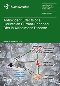
The administration of dietary Corinthian currant in a mouse model of Alzheimer’s disease using 5xFAD increases the activity of the antioxidant enzyme Paraoxonase-1 (PON1) and reduces oxidative stress in circulation and the brain during the early stages of the disease. However, during the later stages of the disease, no beneficial effects are observed. The increased activity of PON1 in plasma can be attributed to the capacity of Corinthian currant phenolics to increase PON1 expression in hepatocytes, the major source of this enzyme in organisms. Given that oxidative stress starts early in the progression of Alzheimer’s disease, the current findings suggest that any intervention with Corinthian currants should commence in the early stages of disease pathogenesis before the onset of significant neurodegeneration. View this paper
  • Issues are regarded as officially published after their release is announced to the table of contents alert mailing list.
  • You may sign up for e-mail alerts to receive table of contents of newly released issues.
  • PDF is the official format for papers published in both, html and pdf forms. To view the papers in pdf format, click on the "PDF Full-text" link, and use the free Adobe Reader to open them.
Order results
Result details
Section
Select all
Export citation of selected articles as:
18 pages, 7169 KiB  
Article
The Role of Tenascin-C on the Structural Plasticity of Perineuronal Nets and Synaptic Expression in the Hippocampus of Male Mice
by Ana Jakovljević, Vera Stamenković, Joko Poleksić, Mohammad I. K. Hamad, Gebhard Reiss, Igor Jakovcevski and Pavle R. Andjus
Biomolecules 2024, 14(4), 508; https://0-doi-org.brum.beds.ac.uk/10.3390/biom14040508 - 22 Apr 2024
Viewed by 383
Abstract
Neuronal plasticity is a crucial mechanism for an adapting nervous system to change. It is shown to be regulated by perineuronal nets (PNNs), the condensed forms of the extracellular matrix (ECM) around neuronal bodies. By assessing the changes in the number, intensity, and [...] Read more.
Neuronal plasticity is a crucial mechanism for an adapting nervous system to change. It is shown to be regulated by perineuronal nets (PNNs), the condensed forms of the extracellular matrix (ECM) around neuronal bodies. By assessing the changes in the number, intensity, and structure of PNNs, the ultrastructure of the PNN mesh, and the expression of inhibitory and excitatory synaptic inputs on these neurons, we aimed to clarify the role of an ECM glycoprotein, tenascin-C (TnC), in the dorsal hippocampus. To enhance neuronal plasticity, TnC-deficient (TnC-/-) and wild-type (TnC+/+) young adult male mice were reared in an enriched environment (EE) for 8 weeks. Deletion of TnC in TnC-/- mice showed an ultrastructural reduction of the PNN mesh and an increased inhibitory input in the dentate gyrus (DG), and an increase in the number of PNNs with a rise in the inhibitory input in the CA2 region. EE induced an increased inhibitory input in the CA2, CA3, and DG regions; in DG, the change was also followed by an increased intensity of PNNs. No changes in PNNs or synaptic expression were found in the CA1 region. We conclude that the DG and CA2 regions emerged as focal points of alterations in PNNs and synaptogenesis with EE as mediated by TnC. Full article
(This article belongs to the Section Biological Factors)
Show Figures

Figure 1

17 pages, 3243 KiB  
Case Report
Phenotypic Variability of Andersen–Tawil Syndrome Due to Allelic Mutation c.652C>T in the KCNJ2 Gene—A New Family Case Report
by Maria Elena Onore, Esther Picillo, Paola D’Ambrosio, Salvatore Morra, Vincenzo Nigro and Luisa Politano
Biomolecules 2024, 14(4), 507; https://0-doi-org.brum.beds.ac.uk/10.3390/biom14040507 - 22 Apr 2024
Viewed by 258
Abstract
Andersen–Tawil syndrome (ATS) is a multisystem channelopathy characterized by periodic paralysis, ventricular arrhythmias, prolonged QT interval, and facial dysmorphisms occurring in the first/second decade of life. High phenotypic variability and incomplete penetrance of the genes causing the disease make its diagnosis still a [...] Read more.
Andersen–Tawil syndrome (ATS) is a multisystem channelopathy characterized by periodic paralysis, ventricular arrhythmias, prolonged QT interval, and facial dysmorphisms occurring in the first/second decade of life. High phenotypic variability and incomplete penetrance of the genes causing the disease make its diagnosis still a challenge. We describe a three-generation family with six living individuals affected by ATS. The proband is a 37-year-old woman presenting since age 16, with episodes of muscle weakness and cramps in the pre-menstrual period. The father, two brothers, one paternal uncle and one cousin also complained of cramps, muscle stiffness, and weakness. Despite normal serum potassium concentration, treatment with potassium, magnesium, and acetazolamide alleviated paralysis attacks suggesting a dyskalemic syndrome. Dysmorphic features were noted in the proband, only later. On the ECG, all but one had normal QT intervals. The affected males developed metabolic syndrome or obesity. The father had two myocardial infarctions and was implanted with an intracardiac cardioverter defibrillator (ICD). A genetic investigation by WES analysis detected the heterozygous pathogenic variant (NM_000891.2: c.652C>T, p. Arg218Trp) in the KCNJ2 gene related to ATS, confirmed by segregation studies in all affected members. Furthermore, we performed a review of cases with the same mutation in the literature, looking for similarities and divergences with our family case. Full article
Show Figures

Figure 1

12 pages, 4939 KiB  
Article
Transduction and Genome Editing of the Heart with Adeno-Associated Viral Vectors Loaded onto Electrospun Polydioxanone Nonwoven Fabrics
by Kotoko Furuno, Keiichiro Suzuki and Shinji Sakai
Biomolecules 2024, 14(4), 506; https://0-doi-org.brum.beds.ac.uk/10.3390/biom14040506 - 22 Apr 2024
Viewed by 378
Abstract
In this study, we introduce electrospun polydioxanone (PDO) nonwoven fabrics as a platform for the delivery of adeno-associated virus (AAV) vectors for transduction and genome editing by adhering them to organ surfaces, including the heart. AAV vectors were loaded onto the PDO fabrics [...] Read more.
In this study, we introduce electrospun polydioxanone (PDO) nonwoven fabrics as a platform for the delivery of adeno-associated virus (AAV) vectors for transduction and genome editing by adhering them to organ surfaces, including the heart. AAV vectors were loaded onto the PDO fabrics by soaking the fabrics in a solution containing AAV vectors. In vitro, the amount of AAV vectors loaded onto the fabrics could be adjusted by changing their concentration in the solution, and the number of cells expressing the green fluorescent protein (GFP) encoded by the AAV vectors increased in correlation with the increasing amount of loaded AAV vectors. In vivo, both transduction and genome editing resulted in the observation of GFP expression around AAV vector-loaded PDO fabrics attached to the surfaces of mouse hearts, indicating effective transduction and expression at the target site. These results demonstrate the great potential of electrospun PDO nonwoven fabrics carrying therapeutic AAV vectors for gene therapy. Full article
(This article belongs to the Special Issue Biomolecules and Biomaterials for Tissue Engineering)
Show Figures

Figure 1

17 pages, 3825 KiB  
Article
Metformin Induces MeCP2 in the Hippocampus of Male Mice with Sex-Specific and Brain-Region-Dependent Molecular Impact
by Khatereh Saei Arezoumand, Chris-Tiann Roberts and Mojgan Rastegar
Biomolecules 2024, 14(4), 505; https://0-doi-org.brum.beds.ac.uk/10.3390/biom14040505 - 21 Apr 2024
Viewed by 431
Abstract
Rett Syndrome (RTT) is a progressive X-linked neurodevelopmental disorder with no cure. RTT patients show disease-associated symptoms within 18 months of age that include developmental regression, progressive loss of useful hand movements, and breathing difficulties, along with neurological impairments, seizures, tremor, and mental [...] Read more.
Rett Syndrome (RTT) is a progressive X-linked neurodevelopmental disorder with no cure. RTT patients show disease-associated symptoms within 18 months of age that include developmental regression, progressive loss of useful hand movements, and breathing difficulties, along with neurological impairments, seizures, tremor, and mental disability. Rett Syndrome is also associated with metabolic abnormalities, and the anti-diabetic drug metformin is suggested to be a potential drug of choice with low or no side-effects. Previously, we showed that in vitro exposure of metformin in a human brain cell line induces MECP2E1 transcripts, the dominant isoform of the MECP2 gene in the brain, mutations in which causes RTT. Here, we report the molecular impact of metformin in mice. Protein analysis of specific brain regions in the male and female mice by immunoblotting indicated that metformin induces MeCP2 in the hippocampus, in a sex-dependent manner. Additional experiments confirm that the regulatory role of metformin on the MeCP2 target “BDNF” is brain region-dependent and sex-specific. Measurement of the ribosomal protein S6 (in both phosphorylated and unphosphorylated forms) confirms the sex-dependent role of metformin in the liver. Our results can help foster a better understanding of the molecular impact of metformin in different brain regions of male and female adult mice, while providing some insight towards its potential in therapeutic strategies for the treatment of Rett Syndrome. Full article
(This article belongs to the Special Issue Brain-Derived Neurotrophic Factor in Health and Diseases)
Show Figures

Figure 1

19 pages, 6585 KiB  
Article
Catalytic Synthesis of (S)-CHBE by Directional Coupling and Immobilization of Carbonyl Reductase and Glucose Dehydrogenase
by Yadong Wang, Ruiqi Sun, Peng Chen and Fenghuan Wang
Biomolecules 2024, 14(4), 504; https://0-doi-org.brum.beds.ac.uk/10.3390/biom14040504 - 21 Apr 2024
Viewed by 279
Abstract
Ethyl (S)-4-chloro-3-hydroxybutyrate ((S)-CHBE) is an important chiral intermediate in the synthesis of the cholesterol-lowering drug atorvastatin. Studying the use of SpyTag/SpyCatcher and SnoopTag/SnoopCatcher systems for the asymmetric reduction reaction and directed coupling coenzyme regeneration is practical for efficiently synthesizing (S)-CHBE. In this study, [...] Read more.
Ethyl (S)-4-chloro-3-hydroxybutyrate ((S)-CHBE) is an important chiral intermediate in the synthesis of the cholesterol-lowering drug atorvastatin. Studying the use of SpyTag/SpyCatcher and SnoopTag/SnoopCatcher systems for the asymmetric reduction reaction and directed coupling coenzyme regeneration is practical for efficiently synthesizing (S)-CHBE. In this study, Spy and Snoop systems were used to construct a double-enzyme directed fixation system of carbonyl reductase (BsCR) and glucose dehydrogenase (BsGDH) for converting 4-chloroacetoacetate (COBE) to (S)-CHBE and achieving coenzyme regeneration. We discussed the enzymatic properties of the immobilized enzyme and the optimal catalytic conditions and reusability of the double-enzyme immobilization system. Compared to the free enzyme, the immobilized enzyme showed an improved optimal pH and temperature, maintaining higher relative activity across a wider range. The double-enzyme immobilization system was applied to catalyze the asymmetric reduction reaction of COBE, and the yield of (S)-CHBE reached 60.1% at 30 °C and pH 8.0. In addition, the double-enzyme immobilization system possessed better operational stability than the free enzyme, and maintained about 50% of the initial yield after six cycles. In summary, we show a simple and effective strategy for self-assembling SpyCatcher/SnoopCatcher and SpyTag/SnoopTag fusion proteins, which inspires building more cascade systems at the interface. It provides a new method for facilitating the rapid construction of in vitro immobilized multi-enzyme complexes from crude cell lysate. Full article
(This article belongs to the Section Biomacromolecules: Proteins)
Show Figures

Figure 1

38 pages, 2786 KiB  
Review
Recent Findings on Therapeutic Cancer Vaccines: An Updated Review
by Sara Sheikhlary, David Humberto Lopez, Sophia Moghimi and Bo Sun
Biomolecules 2024, 14(4), 503; https://0-doi-org.brum.beds.ac.uk/10.3390/biom14040503 - 21 Apr 2024
Viewed by 689
Abstract
Cancer remains one of the global leading causes of death and various vaccines have been developed over the years against it, including cell-based, nucleic acid-based, and viral-based cancer vaccines. Although many vaccines have been effective in in vivo and clinical studies and some [...] Read more.
Cancer remains one of the global leading causes of death and various vaccines have been developed over the years against it, including cell-based, nucleic acid-based, and viral-based cancer vaccines. Although many vaccines have been effective in in vivo and clinical studies and some have been FDA-approved, there are major limitations to overcome: (1) developing one universal vaccine for a specific cancer is difficult, as tumors with different antigens are different for different individuals, (2) the tumor antigens may be similar to the body’s own antigens, and (3) there is the possibility of cancer recurrence. Therefore, developing personalized cancer vaccines with the ability to distinguish between the tumor and the body’s antigens is indispensable. This paper provides a comprehensive review of different types of cancer vaccines and highlights important factors necessary for developing efficient cancer vaccines. Moreover, the application of other technologies in cancer therapy is discussed. Finally, several insights and conclusions are presented, such as the possibility of using cold plasma and cancer stem cells in developing future cancer vaccines, to tackle the major limitations in the cancer vaccine developmental process. Full article
(This article belongs to the Special Issue Advance in Nanoparticles for Tumor Targeting)
Show Figures

Figure 1

19 pages, 1257 KiB  
Review
Glycogen Synthase Kinase-3 Beta (GSK3β) as a Potential Drug Target in Regulating Osteoclastogenesis: An Updated Review on Current Evidence
by Sok Kuan Wong
Biomolecules 2024, 14(4), 502; https://0-doi-org.brum.beds.ac.uk/10.3390/biom14040502 - 21 Apr 2024
Viewed by 257
Abstract
Glycogen synthase kinase 3-beta (GSK3β) is a highly conserved protein kinase originally involved in glucose metabolism, insulin activity, and energy homeostasis. Recent scientific evidence demonstrated the significant role of GSK3β in regulating bone remodelling through involvement in multiple signalling networks. Specifically, the inhibition [...] Read more.
Glycogen synthase kinase 3-beta (GSK3β) is a highly conserved protein kinase originally involved in glucose metabolism, insulin activity, and energy homeostasis. Recent scientific evidence demonstrated the significant role of GSK3β in regulating bone remodelling through involvement in multiple signalling networks. Specifically, the inhibition of GSK3β enhances the conversion of osteoclast progenitors into mature osteoclasts. GSK3β is recognised as a pivotal regulator for the receptor activator of nuclear factor-kappa B (RANK)/receptor activator of nuclear factor-kappa B ligand (RANKL)/osteoprotegerin (OPG), phosphatidylinositol-3-kinase (PI3K)/protein kinase B (AKT), nuclear factor-kappa B (NF-κB), nuclear factor-erythroid 2-related factor 2 (NRF2)/Kelch-like ECH-associated protein 1 (KEAP1), canonical Wnt/beta (β)-catenin, and protein kinase C (PKC) signalling pathways during osteoclastogenesis. Conversely, the inhibition of GSK3β has been shown to prevent bone loss in animal models with complex physiology, suggesting that the role of GSK3β may be more significant in bone formation than bone resorption. Divergent findings have been reported regarding the efficacy of GSK3β inhibitors as bone-protecting agents. Some studies demonstrated that GSK3β inhibitors reduced osteoclast formation, while one study indicated an increase in osteoclast formation in RANKL-stimulated bone marrow macrophages (BMMs). Given the discrepancies observed in the accumulated evidence, further research is warranted, particularly regarding the use of GSK3β silencing or overexpression models. Such efforts will provide valuable insights into the direct impact of GSK3β on osteoclastogenesis and bone resorption. Full article
Show Figures

Figure 1

17 pages, 8036 KiB  
Article
Caspase-1 Deficiency Modulates Adipogenesis through Atg7-Mediated Autophagy: An Inflammatory-Independent Mechanism
by Yumeng Wang, Gaojun Chen, Min Xu, Yewei Cui, Weijiong He, Hongxiang Zeng, Ting Zeng, Rui Cheng and Xi Li
Biomolecules 2024, 14(4), 501; https://0-doi-org.brum.beds.ac.uk/10.3390/biom14040501 - 20 Apr 2024
Viewed by 346
Abstract
Obesity stands as a significant risk factor for type 2 diabetes, hyperlipidemia, and cardiovascular diseases, intertwining increased inflammation and decreased adipogenesis with metabolic disorders. Studies have highlighted the correlation between Caspase-1 and inflammation in obesity, elucidating its essential role in the biological functions [...] Read more.
Obesity stands as a significant risk factor for type 2 diabetes, hyperlipidemia, and cardiovascular diseases, intertwining increased inflammation and decreased adipogenesis with metabolic disorders. Studies have highlighted the correlation between Caspase-1 and inflammation in obesity, elucidating its essential role in the biological functions of adipose tissue. However, the impact of Caspase-1 on adipogenesis and the underlying mechanisms remain largely elusive. In our study, we observed a positive correlation between Caspase-1 expression and obesity and its association with adipogenesis. In vivo experiments revealed that, under normal diet conditions, Caspase-1 deficiency improved glucose homeostasis, stimulated subcutaneous adipose tissue expansion, and enhanced adipogenesis. Furthermore, our findings indicate that Caspase-1 deficiency promotes the expression of autophagy-related proteins and inhibits autophagy with 3-MA or CQ blocked Caspase-1 deficiency-induced adipogenesis in vitro. Notably, Caspase-1 deficiency promotes adipogenesis via Atg7-mediated autophagy activation. In addition, Caspase-1 deficiency resisted against high-fat diet-induced obesity and glucose intolerance. Our study proposes the downregulation of Caspase-1 as a promising strategy for mitigating obesity and its associated metabolic disorders. Full article
Show Figures

Figure 1

24 pages, 3946 KiB  
Review
Adenosine Triphosphate: The Primordial Molecule That Controls Protein Homeostasis and Shapes the Genome–Proteome Interface
by Jianxing Song
Biomolecules 2024, 14(4), 500; https://0-doi-org.brum.beds.ac.uk/10.3390/biom14040500 - 19 Apr 2024
Viewed by 323
Abstract
Adenosine triphosphate (ATP) acts as the universal energy currency that drives various biological processes, while nucleic acids function to store and transmit genetic information for all living organisms. Liquid–liquid phase separation (LLPS) represents the common principle for the formation of membrane-less organelles (MLOs) [...] Read more.
Adenosine triphosphate (ATP) acts as the universal energy currency that drives various biological processes, while nucleic acids function to store and transmit genetic information for all living organisms. Liquid–liquid phase separation (LLPS) represents the common principle for the formation of membrane-less organelles (MLOs) composed of proteins rich in intrinsically disordered regions (IDRs) and nucleic acids. Currently, while IDRs are well recognized to facilitate LLPS through dynamic and multivalent interactions, the precise mechanisms by which ATP and nucleic acids affect LLPS still remain elusive. This review summarizes recent NMR results on the LLPS of human FUS, TDP-43, and the viral nucleocapsid (N) protein of SARS-CoV-2, as modulated by ATP and nucleic acids, revealing the following: (1) ATP binds to folded domains overlapping with nucleic-acid-binding interfaces; (2) ATP and nucleic acids interplay to biphasically modulate LLPS by competitively binding to overlapping pockets of folded domains and Arg/Lys within IDRs; (3) ATP energy-independently induces protein folding with the highest efficiency known so far. As ATP likely emerged in the prebiotic monomeric world, while LLPS represents a pivotal mechanism to concentrate and compartmentalize rare molecules for forming primordial cells, ATP appears to control protein homeostasis and shape genome–proteome interfaces throughout the evolutionary trajectory, from prebiotic origins to modern cells. Full article
Show Figures

Graphical abstract

21 pages, 4114 KiB  
Article
Mitochondrial DNA and Inflammation Are Associated with Cerebral Vessel Remodeling and Early Diabetic Kidney Disease in Patients with Type 2 Diabetes Mellitus
by Ligia Petrica, Florica Gadalean, Danina Mirela Muntean, Dragos Catalin Jianu, Daliborca Vlad, Victor Dumitrascu, Flaviu Bob, Oana Milas, Anca Suteanu-Simulescu, Mihaela Glavan, Sorin Ursoniu, Lavinia Balint, Maria Mogos-Stefan, Silvia Ienciu, Octavian Marius Cretu, Roxana Popescu, Cristina Gluhovschi, Lavinia Iancu and Adrian Vlad
Biomolecules 2024, 14(4), 499; https://0-doi-org.brum.beds.ac.uk/10.3390/biom14040499 - 19 Apr 2024
Viewed by 359
Abstract
Cerebrovascular disease accounts for major neurologic disabilities in patients with type 2 diabetes mellitus (DM). A potential association of mitochondrial DNA (mtDNA) and inflammation with cerebral vessel remodeling in patients with type 2 DM was evaluated. A cohort of 150 patients [...] Read more.
Cerebrovascular disease accounts for major neurologic disabilities in patients with type 2 diabetes mellitus (DM). A potential association of mitochondrial DNA (mtDNA) and inflammation with cerebral vessel remodeling in patients with type 2 DM was evaluated. A cohort of 150 patients and 30 healthy controls were assessed concerning urinary albumin/creatinine ratio (UACR), synaptopodin, podocalyxin, kidney injury molecule-1 (KIM-1), N-acetyl-β-(D)-glucosaminidase (NAG), interleukins IL-17A, IL-18, IL-10, tumor necrosis factor-alpha (TNFα), intercellular adhesion molecule-1 (ICAM-1). MtDNA-CN and nuclear DNA (nDNA) were quantified in peripheral blood and urine by qRT-PCR. Cytochrome b (CYTB) gene, subunit 2 of NADH dehydrogenase (ND2), and beta 2 microglobulin nuclear gene (B2M) were assessed by TaqMan assays. mtDNA-CN was defined as the ratio of the number of mtDNA/nDNA copies, through analysis of the CYTB/B2M and ND2/B2M ratio; cerebral Doppler ultrasound: intima-media thickness (IMT)—the common carotid arteries (CCAs), the pulsatility index (PI) and resistivity index (RI)- the internal carotid arteries (ICAs) and middle cerebral arteries (MCAs), the breath-holding index (BHI). The results showed direct correlations of CCAs-IMT, PI-ICAs, PI-MCAs, RI-ICAs, RI-MCAs with urinary mtDNA, IL-17A, IL-18, TNFα, ICAM-1, UACR, synaptopodin, podocalyxin, KIM-1, NAG, and indirect correlations with serum mtDNA, IL-10. BHI correlated directly with serum IL-10, and serum mtDNA, and negatively with serum IL-17A, serum ICAM-1, and NAG. In neurologically asymptomatic patients with type 2 DM cerebrovascular remodeling and impaired cerebrovascular reactivity may be associated with mtDNA variations and inflammation from the early stages of diabetic kidney disease. Full article
Show Figures

Figure 1

13 pages, 972 KiB  
Review
Technical Advances in Circulating Cell-Free DNA Detection and Analysis for Personalized Medicine in Patients’ Care
by Monica Sorbini, Tullia Carradori, Gabriele Maria Togliatto, Tiziana Vaisitti and Silvia Deaglio
Biomolecules 2024, 14(4), 498; https://0-doi-org.brum.beds.ac.uk/10.3390/biom14040498 - 19 Apr 2024
Viewed by 417
Abstract
Circulating cell-free DNA (cfDNA) refers to small fragments of DNA molecules released after programmed cell death and necrosis in several body fluids such as blood, saliva, urine, and cerebrospinal fluid. The discovery of cfDNA has revolutionized the field of non-invasive diagnostics in the [...] Read more.
Circulating cell-free DNA (cfDNA) refers to small fragments of DNA molecules released after programmed cell death and necrosis in several body fluids such as blood, saliva, urine, and cerebrospinal fluid. The discovery of cfDNA has revolutionized the field of non-invasive diagnostics in the oncologic field, in prenatal testing, and in organ transplantation. Despite the potential of cfDNA and the solid results published in the recent literature, several challenges remain, represented by a low abundance, a need for highly sensitive assays, and analytical issues. In this review, the main technical advances in cfDNA analysis are presented and discussed, with a comprehensive examination of the current available methodologies applied in each field. Considering the potential advantages of cfDNA, this biomarker is increasing its consensus among clinicians, as it allows us to monitor patients’ conditions in an easy and non-invasive way, offering a more personalized care. Nevertheless, cfDNA analysis is still considered a diagnostic marker to be further validated, and very few centers are implementing its analysis in routine diagnostics. As technical improvements are enhancing the performances of cfDNA analysis, its application will transversally improve patients’ quality of life. Full article
(This article belongs to the Special Issue Recent Developments in the Biology of Extracellular or Cell-Free DNA)
Show Figures

Figure 1

40 pages, 18005 KiB  
Article
Mutations in Glycosyltransferases and Glycosidases: Implications for Associated Diseases
by Xiaotong Gu, Aaron S. Kovacs, Yoochan Myung and David B. Ascher
Biomolecules 2024, 14(4), 497; https://0-doi-org.brum.beds.ac.uk/10.3390/biom14040497 - 19 Apr 2024
Viewed by 285
Abstract
Glycosylation, a crucial and the most common post-translational modification, coordinates a multitude of biological functions through the attachment of glycans to proteins and lipids. This process, predominantly governed by glycosyltransferases (GTs) and glycoside hydrolases (GHs), decides not only biomolecular functionality but also protein [...] Read more.
Glycosylation, a crucial and the most common post-translational modification, coordinates a multitude of biological functions through the attachment of glycans to proteins and lipids. This process, predominantly governed by glycosyltransferases (GTs) and glycoside hydrolases (GHs), decides not only biomolecular functionality but also protein stability and solubility. Mutations in these enzymes have been implicated in a spectrum of diseases, prompting critical research into the structural and functional consequences of such genetic variations. This study compiles an extensive dataset from ClinVar and UniProt, providing a nuanced analysis of 2603 variants within 343 GT and GH genes. We conduct thorough MTR score analyses for the proteins with the most documented variants using MTR3D-AF2 via AlphaFold2 (AlphaFold v2.2.4) predicted protein structure, with the analyses indicating that pathogenic mutations frequently correlate with Beta Bridge secondary structures. Further, the calculation of the solvent accessibility score and variant visualisation show that pathogenic mutations exhibit reduced solvent accessibility, suggesting the mutated residues are likely buried and their localisation is within protein cores. We also find that pathogenic variants are often found proximal to active and binding sites, which may interfere with substrate interactions. We also incorporate computational predictions to assess the impact of these mutations on protein function, utilising tools such as mCSM to predict the destabilisation effect of variants. By identifying these critical regions that are prone to disease-associated mutations, our study opens avenues for designing small molecules or biologics that can modulate enzyme function or compensate for the loss of stability due to these mutations. Full article
(This article belongs to the Special Issue Protein Glycosylation and Human Diseases)
Show Figures

Figure 1

14 pages, 2283 KiB  
Article
Conformational Modulation of Tissue Transglutaminase via Active Site Thiol Alkylating Agents: Size Does Not Matter
by Pauline Navals, Alana M. M. Rangaswamy, Petr Kasyanchyk, Maxim V. Berezovski and Jeffrey W. Keillor
Biomolecules 2024, 14(4), 496; https://0-doi-org.brum.beds.ac.uk/10.3390/biom14040496 - 19 Apr 2024
Viewed by 332
Abstract
TG2 is a unique member of the transglutaminase family as it undergoes a dramatic conformational change, allowing its mutually exclusive function as either a cross-linking enzyme or a G-protein. The enzyme’s dysregulated activity has been implicated in a variety of pathologies (e.g., celiac [...] Read more.
TG2 is a unique member of the transglutaminase family as it undergoes a dramatic conformational change, allowing its mutually exclusive function as either a cross-linking enzyme or a G-protein. The enzyme’s dysregulated activity has been implicated in a variety of pathologies (e.g., celiac disease, fibrosis, cancer), leading to the development of a wide range of inhibitors. Our group has primarily focused on the development of peptidomimetic targeted covalent inhibitors, the nature and size of which were thought to be important features to abolish TG2’s conformational dynamism and ultimately inhibit both its activities. However, we recently demonstrated that the enzyme was unable to bind guanosine triphosphate (GTP) when catalytically inactivated by small molecule inhibitors. In this study, we designed a library of models targeting covalent inhibitors of progressively smaller sizes (15 to 4 atoms in length). We evaluated their ability to inactivate TG2 by measuring their respective kinetic parameters kinact and KI. Their impact on the enzyme’s ability to bind GTP was then evaluated and subsequently correlated to the conformational state of the enzyme, as determined via native PAGE and capillary electrophoresis. All irreversible inhibitors evaluated herein locked TG2 in its open conformation and precluded GTP binding. Therefore, we conclude that steric bulk and structural complexity are not necessary factors to consider when designing TG2 inhibitors to abolish G-protein activity. Full article
(This article belongs to the Special Issue Transglutaminases: Regulation, Imaging, and Applications)
Show Figures

Figure 1

17 pages, 2258 KiB  
Article
Effects of Cannabidiol, ∆9-Tetrahydrocannabinol, and WIN 55-212-22 on the Viability of Canine and Human Non-Hodgkin Lymphoma Cell Lines
by Saba Omer, Suhrud Pathak, Mahmoud Mansour, Rishi Nadar, Dylan Bowen, Muralikrishnan Dhanasekaran, Satyanarayana R. Pondugula and Dawn Boothe
Biomolecules 2024, 14(4), 495; https://0-doi-org.brum.beds.ac.uk/10.3390/biom14040495 - 19 Apr 2024
Viewed by 322
Abstract
In our previous study, we demonstrated the impact of overexpression of CB1 and CB2 cannabinoid receptors and the inhibitory effect of endocannabinoids (2-arachidonoylglycerol (2-AG) and Anandamide (AEA)) on canine (Canis lupus familiaris) and human (Homo sapiens) non-Hodgkin lymphoma [...] Read more.
In our previous study, we demonstrated the impact of overexpression of CB1 and CB2 cannabinoid receptors and the inhibitory effect of endocannabinoids (2-arachidonoylglycerol (2-AG) and Anandamide (AEA)) on canine (Canis lupus familiaris) and human (Homo sapiens) non-Hodgkin lymphoma (NHL) cell lines’ viability compared to cells treated with a vehicle. The purpose of this study was to demonstrate the anti-cancer effects of the phytocannabinoids, cannabidiol (CBD) and ∆9-tetrahydrocannabinol (THC), and the synthetic cannabinoid WIN 55-212-22 (WIN) in canine and human lymphoma cell lines and to compare their inhibitory effect to that of endocannabinoids. We used malignant canine B-cell lymphoma (BCL) (1771 and CLB-L1) and T-cell lymphoma (TCL) (CL-1) cell lines, and human BCL cell line (RAMOS). Our cell viability assay results demonstrated, compared to the controls, a biphasic effect (concentration range from 0.5 μM to 50 μM) with a significant reduction in cancer viability for both phytocannabinoids and the synthetic cannabinoid. However, the decrease in cell viability in the TCL CL-1 line was limited to CBD. The results of the biochemical analysis using the 1771 BCL cell line revealed a significant increase in markers of oxidative stress, inflammation, and apoptosis, and a decrease in markers of mitochondrial function in cells treated with the exogenous cannabinoids compared to the control. Based on the IC50 values, CBD was the most potent phytocannabinoid in reducing lymphoma cell viability in 1771, Ramos, and CL-1. Previously, we demonstrated the endocannabinoid AEA to be more potent than 2-AG. Our study suggests that future studies should use CBD and AEA for further cannabinoid testing as they might reduce tumor burden in malignant NHL of canines and humans. Full article
(This article belongs to the Special Issue New Advances of Cannabinoid Receptors in Health and Disease)
Show Figures

Figure 1

11 pages, 4030 KiB  
Article
Transgenic Overexpression of HDAC9 Promotes Adipocyte Hypertrophy, Insulin Resistance and Hepatic Steatosis in Aging Mice
by Praneet Veerapaneni, Brandee Goo, Samah Ahmadieh, Hong Shi, David S. Kim, Mourad Ogbi, Stephen Cave, Ronnie Chouhaita, Nicole Cyriac, David J. Fulton, Alexander D. Verin, Weiqin Chen, Yun Lei, Xin-Yun Lu, Ha Won Kim and Neal L. Weintraub
Biomolecules 2024, 14(4), 494; https://0-doi-org.brum.beds.ac.uk/10.3390/biom14040494 - 18 Apr 2024
Viewed by 352
Abstract
Histone deacetylase (HDAC) 9 is a negative regulator of adipogenic differentiation, which is required for maintenance of healthy adipose tissues. We reported that HDAC9 expression is upregulated in adipose tissues during obesity, in conjunction with impaired adipogenic differentiation, adipocyte hypertrophy, insulin resistance, and [...] Read more.
Histone deacetylase (HDAC) 9 is a negative regulator of adipogenic differentiation, which is required for maintenance of healthy adipose tissues. We reported that HDAC9 expression is upregulated in adipose tissues during obesity, in conjunction with impaired adipogenic differentiation, adipocyte hypertrophy, insulin resistance, and hepatic steatosis, all of which were alleviated by global genetic deletion of Hdac9. Here, we developed a novel transgenic (TG) mouse model to test whether overexpression of Hdac9 is sufficient to induce adipocyte hypertrophy, insulin resistance, and hepatic steatosis in the absence of obesity. HDAC9 TG mice gained less body weight than wild-type (WT) mice when fed a standard laboratory diet for up to 40 weeks, which was attributed to reduced fat mass (primarily inguinal adipose tissue). There was no difference in insulin sensitivity or glucose tolerance in 18-week-old WT and HDAC9 TG mice; however, at 40 weeks of age, HDAC9 TG mice exhibited impaired insulin sensitivity and glucose intolerance. Tissue histology demonstrated adipocyte hypertrophy, along with reduced numbers of mature adipocytes and stromovascular cells, in the HDAC9 TG mouse adipose tissue. Moreover, increased lipids were detected in the livers of aging HDAC9 TG mice, as evaluated by oil red O staining. In conclusion, the experimental aging HDAC9 TG mice developed adipocyte hypertrophy, insulin resistance, and hepatic steatosis, independent of obesity. This novel mouse model may be useful in the investigation of the impact of Hdac9 overexpression associated with metabolic and aging-related diseases. Full article
(This article belongs to the Special Issue Recent Advances in Histone Deacetylase Research in Health and Disease)
Show Figures

Figure 1

17 pages, 3733 KiB  
Article
Mitochondrial Transplantation’s Role in Rodent Skeletal Muscle Bioenergetics: Recharging the Engine of Aging
by Tasnim Arroum, Gerald A. Hish, Kyle J. Burghardt, James D. McCully, Maik Hüttemann and Moh H. Malek
Biomolecules 2024, 14(4), 493; https://0-doi-org.brum.beds.ac.uk/10.3390/biom14040493 - 18 Apr 2024
Viewed by 558
Abstract
Background: Mitochondria are the ‘powerhouses of cells’ and progressive mitochondrial dysfunction is a hallmark of aging in skeletal muscle. Although different forms of exercise modality appear to be beneficial to attenuate aging-induced mitochondrial dysfunction, it presupposes that the individual has a requisite level [...] Read more.
Background: Mitochondria are the ‘powerhouses of cells’ and progressive mitochondrial dysfunction is a hallmark of aging in skeletal muscle. Although different forms of exercise modality appear to be beneficial to attenuate aging-induced mitochondrial dysfunction, it presupposes that the individual has a requisite level of mobility. Moreover, non-exercise alternatives (i.e., nutraceuticals or pharmacological agents) to improve skeletal muscle bioenergetics require time to be effective in the target tissue and have another limitation in that they act systemically and not locally where needed. Mitochondrial transplantation represents a novel directed therapy designed to enhance energy production of tissues impacted by defective mitochondria. To date, no studies have used mitochondrial transplantation as an intervention to attenuate aging-induced skeletal muscle mitochondrial dysfunction. The purpose of this investigation, therefore, was to determine whether mitochondrial transplantation can enhance skeletal muscle bioenergetics in an aging rodent model. We hypothesized that mitochondrial transplantation would result in sustained skeletal muscle bioenergetics leading to improved functional capacity. Methods: Fifteen female mice (24 months old) were randomized into two groups (placebo or mitochondrial transplantation). Isolated mitochondria from a donor mouse of the same sex and age were transplanted into the hindlimb muscles of recipient mice (quadriceps femoris, tibialis anterior, and gastrocnemius complex). Results: The results indicated significant increases (ranging between ~36% and ~65%) in basal cytochrome c oxidase and citrate synthase activity as well as ATP levels in mice receiving mitochondrial transplantation relative to the placebo. Moreover, there were significant increases (approx. two-fold) in protein expression of mitochondrial markers in both glycolytic and oxidative muscles. These enhancements in the muscle translated to significant improvements in exercise tolerance. Conclusions: This study provides initial evidence showing how mitochondrial transplantation can promote skeletal muscle bioenergetics in an aging rodent model. Full article
(This article belongs to the Special Issue Skeletal Muscle Homeostasis and Regeneration)
Show Figures

Figure 1

9 pages, 820 KiB  
Article
Development of Improved Spectrophotometric Assays for Biocatalytic Silyl Ether Hydrolysis
by Yuqing Lu, Chisom S. Egedeuzu, Peter G. Taylor and Lu Shin Wong
Biomolecules 2024, 14(4), 492; https://0-doi-org.brum.beds.ac.uk/10.3390/biom14040492 - 18 Apr 2024
Viewed by 353
Abstract
Reported herein is the development of assays for the spectrophotometric quantification of biocatalytic silicon−oxygen bond hydrolysis. Central to these assays are a series of chromogenic substrates that release highly absorbing phenoxy anions upon cleavage of the sessile bond. These substrates were tested with [...] Read more.
Reported herein is the development of assays for the spectrophotometric quantification of biocatalytic silicon−oxygen bond hydrolysis. Central to these assays are a series of chromogenic substrates that release highly absorbing phenoxy anions upon cleavage of the sessile bond. These substrates were tested with silicatein, an enzyme from a marine sponge that is known to catalyse the hydrolysis and condensation of silyl ethers. It was found that, of the substrates tested, tert-butyldimethyl(2-methyl-4-nitrophenoxy)silane provided the best assay performance, as evidenced by the highest ratio of enzyme catalysed reaction rate compared with the background (uncatalysed) reaction. These substrates were also found to be suitable for detailed enzyme kinetics measurements, as demonstrated by their use to determine the Michaelis−Menten kinetic parameters for silicatein. Full article
(This article belongs to the Section Enzymology)
Show Figures

Graphical abstract

13 pages, 3079 KiB  
Article
Radiolabelled FGF-2 for Imaging Activated Fibroblasts in the Tumor Micro-Environment
by Valeria Bentivoglio, Filippo Galli, Michela Varani, Danilo Ranieri, Pallavi Nayak, Annunziata D’Elia, Andrea Soluri, Roberto Massari, Chiara Lauri and Alberto Signore
Biomolecules 2024, 14(4), 491; https://0-doi-org.brum.beds.ac.uk/10.3390/biom14040491 - 18 Apr 2024
Viewed by 449
Abstract
Tumor associated fibroblasts (TAFs) play a key role in tumor growth and metastatization. TAFs overexpress different biomarkers that are usually expressed at low levels in physiological conditions. Among them are the fibroblast growth factor receptors (FGFRs) that bind the fibroblast growth factors (FGFs). [...] Read more.
Tumor associated fibroblasts (TAFs) play a key role in tumor growth and metastatization. TAFs overexpress different biomarkers that are usually expressed at low levels in physiological conditions. Among them are the fibroblast growth factor receptors (FGFRs) that bind the fibroblast growth factors (FGFs). In particular, the overexpression of FGFR-2c in tumors has been associated with advanced clinical stages and increased metastatization. Here, we developed a non-invasive tool to evaluate, in vivo, the expression of FGFR-2c in metastatic cancer. This is based on 99mTc-labelled FGF-2. Methods: 99mTc-FGF-2 was tested in vitro and in vivo in mice bearing allografts of sarcoma cells. Images of 99mTc-FGF-2 were acquired using a new portable high-resolution ultra-sensitive gamma camera for small animal imaging. Results: FGF-2 was labeled with high specific activity but low labelling efficiency, thus requiring post-labeling purification by gel-filtration chromatography. In vitro binding to 2C human keratinocytes showed a Kd of 3.36 × 10⁻⁹ M. In mice bearing J774A.1 cell allografts, we observed high and rapid tumor uptake of 99mTc-FGF-2 with a high Tumor/Blood ratio at 24 h post-injection (26.1 %ID/g and 12.9 %ID) with low kidney activity and moderate liver activity. Conclusions: we labeled FGF-2 with 99mTc and showed nanomolar Kd in vitro with human keratinocytes expressing FGF-2 receptors. In mice, 99mTc-FGF-2 rapidly and efficiently accumulated in tumors expressing FGF-2 receptors. This new radiopharmaceutical could be used in humans to image TAFs. Full article
(This article belongs to the Section Molecular Medicine)
Show Figures

Figure 1

10 pages, 1803 KiB  
Article
Elevated Serum Xanthine Oxidase and Its Correlation with Antioxidant Status in Patients with Parkinson’s Disease
by Ratna Dini Haryuni, Takamasa Nukui, Jin-Lan Piao, Takashi Shirakura, Chieko Matsui, Tomoyuki Sugimoto, Kousuke Baba, Shunya Nakane and Yuji Nakatsuji
Biomolecules 2024, 14(4), 490; https://0-doi-org.brum.beds.ac.uk/10.3390/biom14040490 - 18 Apr 2024
Viewed by 370
Abstract
Parkinson’s disease (PD) is a neurodegenerative movement disorder associated with a loss of dopamine neurons in the substantia nigra. The diagnosis of PD is sensitive since it shows clinical features that are common with other neurodegenerative diseases. In addition, most symptoms arise at [...] Read more.
Parkinson’s disease (PD) is a neurodegenerative movement disorder associated with a loss of dopamine neurons in the substantia nigra. The diagnosis of PD is sensitive since it shows clinical features that are common with other neurodegenerative diseases. In addition, most symptoms arise at the late stage of the disease, where most dopaminergic neurons are already damaged. Several studies reported that oxidative stress is a key modulator in the development of PD. This condition occurs due to excess reactive oxygen species (ROS) production in the cellular system and the incapability of antioxidants to neutralize it. In this study, we focused on the pathology of PD by measuring serum xanthine oxidase (XO) activity, which is an enzyme that generates ROS. Interestingly, the serum XO activity of patients with PD was markedly upregulated compared to patients with other neurological diseases (ONDs) as a control. Moreover, serum XO activity in patients with PD showed a significant correlation with the disease severity based on the Hoehn and Yahr (HY) stages. The investigation of antioxidant status also revealed that serum uric acid levels were significantly lower in the severe group (HY ≥ 3) than in the ONDs group. Together, these results suggest that XO activity may contribute to the development of PD and might potentially be a biomarker for determining disease severity in patients with PD. Full article
(This article belongs to the Special Issue Advances in Biomarkers for Neurodegenerative Diseases)
Show Figures

Figure 1

14 pages, 2086 KiB  
Review
Thrombopoietin, the Primary Regulator of Platelet Production: From Mythos to Logos, a Thirty-Year Journey
by Kenneth Kaushansky
Biomolecules 2024, 14(4), 489; https://0-doi-org.brum.beds.ac.uk/10.3390/biom14040489 - 18 Apr 2024
Viewed by 340
Abstract
Thrombopoietin, the primary regulator of blood platelet production, was postulated to exist in 1958, but was only proven to exist when the cDNA for the hormone was cloned in 1994. Since its initial cloning and characterization, the hormone has revealed many surprises. For [...] Read more.
Thrombopoietin, the primary regulator of blood platelet production, was postulated to exist in 1958, but was only proven to exist when the cDNA for the hormone was cloned in 1994. Since its initial cloning and characterization, the hormone has revealed many surprises. For example, instead of acting as the postulated differentiation factor for platelet precursors, megakaryocytes, it is the most potent stimulator of megakaryocyte progenitor expansion known. Moreover, it also stimulates the survival, and in combination with stem cell factor leads to the expansion of hematopoietic stem cells. All of these growth-promoting activities have resulted in its clinical use in patients with thrombocytopenia and aplastic anemia, although the clinical development of the native molecule illustrated that “it’s not wise to mess with mother nature”, as a highly engineered version of the native hormone led to autoantibody formation and severe thrombocytopenia. Finally, another unexpected finding was the role of the thrombopoietin receptor in stem cell biology, including the development of myeloproliferative neoplasms, an important disorder of hematopoietic stem cells. Overall, the past 30 years of clinical and basic research has yielded many important insights, which are reviewed in this paper. Full article
Show Figures

Figure 1

14 pages, 1849 KiB  
Review
Categorizing Extrachromosomal Circular DNA as Biomarkers in Serum of Cancer
by Enze Deng and Xiaoying Fan
Biomolecules 2024, 14(4), 488; https://0-doi-org.brum.beds.ac.uk/10.3390/biom14040488 - 17 Apr 2024
Viewed by 504
Abstract
Extrachromosomal circular DNA (eccDNA), a double-stranded circular DNA molecule found in multiple organisms, has garnered an increasing amount of attention in recent years due to its close association with the initiation, malignant progression, and heterogeneous evolution of cancer. The presence of eccDNA in [...] Read more.
Extrachromosomal circular DNA (eccDNA), a double-stranded circular DNA molecule found in multiple organisms, has garnered an increasing amount of attention in recent years due to its close association with the initiation, malignant progression, and heterogeneous evolution of cancer. The presence of eccDNA in serum assists in non-invasive tumor diagnosis as a biomarker that can be assessed via liquid biopsies. Furthermore, the specific expression patterns of eccDNA provide new insights into personalized cancer therapy. EccDNA plays a pivotal role in tumorigenesis, development, diagnosis, and treatment. In this review, we comprehensively outline the research trajectory of eccDNA, discuss its role as a diagnostic and prognostic biomarker, and elucidate its regulatory mechanisms in cancer. In particular, we emphasize the potential application value of eccDNA in cancer diagnosis and treatment and anticipate the development of novel tumor diagnosis strategies based on serum eccDNA in the future. Full article
(This article belongs to the Special Issue Recent Developments in the Biology of Extracellular or Cell-Free DNA)
Show Figures

Figure 1

16 pages, 4075 KiB  
Article
In Silico Analysis of the Ga3+/Fe3+ Competition for Binding the Iron-Scavenging Siderophores of P. aeruginosa—Implementation of Three Gallium-Based Complexes in the “Trojan Horse” Antibacterial Strategy
by Nikoleta Kircheva, Stefan Dobrev, Vladislava Petkova, Lyubima Yocheva, Silvia Angelova and Todor Dudev
Biomolecules 2024, 14(4), 487; https://0-doi-org.brum.beds.ac.uk/10.3390/biom14040487 - 16 Apr 2024
Viewed by 470
Abstract
The emergence of multidrug-resistant (MDR) microorganisms combined with the ever-draining antibiotic pipeline poses a disturbing and immensely growing public health challenge that requires a multidisciplinary approach and the application of novel therapies aimed at unconventional targets and/or applying innovative drug formulations. Hence, bacterial [...] Read more.
The emergence of multidrug-resistant (MDR) microorganisms combined with the ever-draining antibiotic pipeline poses a disturbing and immensely growing public health challenge that requires a multidisciplinary approach and the application of novel therapies aimed at unconventional targets and/or applying innovative drug formulations. Hence, bacterial iron acquisition systems and bacterial Fe2+/3+-containing enzymes have been identified as a plausible target of great potential. The intriguing “Trojan horse” approach deprives microorganisms from the essential iron. Recently, gallium’s potential in medicine as an iron mimicry species has attracted vast attention. Different Ga3+ formulations exhibit diverse effects upon entering the cell and thus supposedly have multiple targets. The aim of the current study is to specifically distinguish characteristics of great significance in regard to the initial gallium-based complex, allowing the alien cation to effectively compete with the native ferric ion for binding the siderophores pyochelin and pyoverdine secreted by the bacterium P. aeruginosa. Therefore, three gallium-based formulations were taken into consideration: the first-generation gallium nitrate, Ga(NO3)3, metabolized to Ga3+-hydrated forms, the second-generation gallium maltolate (tris(3-hydroxy-2-methyl-4-pyronato)gallium), and the experimentally proven Ga carrier in the bloodstream—the protein transferrin. We employed a reliable in silico approach based on DFT computations in order to understand the underlying biochemical processes that govern the Ga3+/Fe3+ rivalry for binding the two bacterial siderophores. Full article
(This article belongs to the Collection Feature Papers in Biochemistry)
Show Figures

Figure 1

8 pages, 839 KiB  
Communication
Application of Cas12j for Streptomyces Editing
by Lee Ling Tan, Elena Heng, Chung Yan Leong, Veronica Ng, Lay Kien Yang, Deborah Chwee San Seow, Lokanand Koduru, Yoganathan Kanagasundaram, Siew Bee Ng, Guangrong Peh, Yee Hwee Lim and Fong Tian Wong
Biomolecules 2024, 14(4), 486; https://0-doi-org.brum.beds.ac.uk/10.3390/biom14040486 - 16 Apr 2024
Viewed by 341
Abstract
In recent years, CRISPR-Cas toolboxes for Streptomyces editing have rapidly accelerated natural product discovery and engineering. However, Cas efficiencies are oftentimes strain-dependent, and the commonly used Streptococcus pyogenes Cas9 (SpCas9) is notorious for having high levels of off-target toxicity effects. Thus, a variety [...] Read more.
In recent years, CRISPR-Cas toolboxes for Streptomyces editing have rapidly accelerated natural product discovery and engineering. However, Cas efficiencies are oftentimes strain-dependent, and the commonly used Streptococcus pyogenes Cas9 (SpCas9) is notorious for having high levels of off-target toxicity effects. Thus, a variety of Cas proteins is required for greater flexibility of genetic manipulation within a wider range of Streptomyces strains. This study explored the first use of Acidaminococcus sp. Cas12j, a hypercompact Cas12 subfamily, for genome editing in Streptomyces and its potential in activating silent biosynthetic gene clusters (BGCs) to enhance natural product synthesis. While the editing efficiencies of Cas12j were not as high as previously reported efficiencies of Cas12a and Cas9, Cas12j exhibited higher transformation efficiencies compared to SpCas9. Furthermore, Cas12j demonstrated significantly improved editing efficiencies compared to Cas12a in activating BGCs in Streptomyces sp. A34053, a strain wherein both SpCas9 and Cas12a faced limitations in accessing the genome. Overall, this study expanded the repertoire of Cas proteins for genome editing in actinomycetes and highlighted not only the potential of recently characterized Cas12j in Streptomyces but also the importance of having an extensive genetic toolbox for improving the editing success of these beneficial microbes. Full article
(This article belongs to the Section Synthetic Biology and Bioengineering)
Show Figures

Figure 1

12 pages, 2808 KiB  
Review
Cell Senescence in Heterotopic Ossification
by Robert J. Pignolo, Frederick S. Kaplan and Haitao Wang
Biomolecules 2024, 14(4), 485; https://0-doi-org.brum.beds.ac.uk/10.3390/biom14040485 - 16 Apr 2024
Viewed by 287
Abstract
The formation of bone outside the normal skeleton, or heterotopic ossification (HO), occurs through genetic and acquired mechanisms. Fibrodysplasia ossificans progressiva (FOP), the most devastating genetic condition of HO, is due to mutations in the ACVR1/ALK2 gene and is relentlessly progressive. Acquired HO [...] Read more.
The formation of bone outside the normal skeleton, or heterotopic ossification (HO), occurs through genetic and acquired mechanisms. Fibrodysplasia ossificans progressiva (FOP), the most devastating genetic condition of HO, is due to mutations in the ACVR1/ALK2 gene and is relentlessly progressive. Acquired HO is mostly precipitated by injury or orthopedic surgical procedures but can also be associated with certain conditions related to aging. Cellular senescence is a hallmark of aging and thought to be a tumor-suppressive mechanism with characteristic features such as irreversible growth arrest, apoptosis resistance, and an inflammatory senescence-associated secretory phenotype (SASP). Here, we review possible roles for cellular senescence in HO and how targeting senescent cells may provide new therapeutic approaches to both FOP and acquired forms of HO. Full article
(This article belongs to the Section Molecular Medicine)
Show Figures

Figure 1

22 pages, 8259 KiB  
Review
Strategies for Treating Traumatic Neuromas with Tissue-Engineered Materials
by Teng Wan, Qi-Cheng Li, Ming-Yu Qin, Yi-Lin Wang, Feng-Shi Zhang, Xiao-Meng Zhang, Yi-Chong Zhang and Pei-Xun Zhang
Biomolecules 2024, 14(4), 484; https://0-doi-org.brum.beds.ac.uk/10.3390/biom14040484 - 16 Apr 2024
Viewed by 424
Abstract
Neuroma, a pathological response to peripheral nerve injury, refers to the abnormal growth of nerve tissue characterized by disorganized axonal proliferation. Commonly occurring after nerve injuries, surgeries, or amputations, this condition leads to the formation of painful nodular structures. Traditional treatment options include [...] Read more.
Neuroma, a pathological response to peripheral nerve injury, refers to the abnormal growth of nerve tissue characterized by disorganized axonal proliferation. Commonly occurring after nerve injuries, surgeries, or amputations, this condition leads to the formation of painful nodular structures. Traditional treatment options include surgical excision and pharmacological management, aiming to alleviate symptoms. However, these approaches often offer temporary relief without addressing the underlying regenerative challenges, necessitating the exploration of advanced strategies such as tissue-engineered materials for more comprehensive and effective solutions. In this study, we discussed the etiology, molecular mechanisms, and histological morphology of traumatic neuromas after peripheral nerve injury. Subsequently, we summarized and analyzed current nonsurgical and surgical treatment options, along with their advantages and disadvantages. Additionally, we emphasized recent advancements in treating traumatic neuromas with tissue-engineered material strategies. By integrating biomaterials, growth factors, cell-based approaches, and electrical stimulation, tissue engineering offers a comprehensive solution surpassing mere symptomatic relief, striving for the structural and functional restoration of damaged nerves. In conclusion, the utilization of tissue-engineered materials has the potential to significantly reduce the risk of neuroma recurrence after surgical treatment. Full article
(This article belongs to the Section Biological and Bio- Materials)
Show Figures

Figure 1

13 pages, 3822 KiB  
Review
Plasticity of Adipose Tissues: Interconversion among White, Brown, and Beige Fat and Its Role in Energy Homeostasis
by Yanqiu Peng, Lixia Zhao, Min Li, Yunfei Liu, Yuke Shi and Jian Zhang
Biomolecules 2024, 14(4), 483; https://0-doi-org.brum.beds.ac.uk/10.3390/biom14040483 - 16 Apr 2024
Viewed by 433
Abstract
Obesity, characterized by the excessive accumulation of adipose tissue, has emerged as a major public health concern worldwide. To develop effective strategies for treating obesity, it is essential to comprehend the biological properties of different adipose tissue types and their respective roles in [...] Read more.
Obesity, characterized by the excessive accumulation of adipose tissue, has emerged as a major public health concern worldwide. To develop effective strategies for treating obesity, it is essential to comprehend the biological properties of different adipose tissue types and their respective roles in maintaining energy balance. Adipose tissue serves as a crucial organ for energy storage and metabolism in the human body, with functions extending beyond simple fat storage to encompass the regulation of energy homeostasis and the secretion of endocrine factors. This review provides an overview of the key characteristics, functional differences, and interconversion processes among white adipose tissue (WAT), brown adipose tissue (BAT), and beige adipose tissue. Moreover, it delves into the molecular mechanisms and recent research advancements concerning the browning of WAT, activation of BAT, and whitening of BAT. Although targeting adipose tissue metabolism holds promise as a potential approach for obesity treatment, further investigations are necessary to unravel the intricate biological features of various adipose tissue types and elucidate the molecular pathways governing their interconversion. Such research endeavors will pave the way for the development of more efficient and targeted therapeutic interventions in the fight against obesity. Full article
(This article belongs to the Section Biomacromolecules: Lipids)
Show Figures

Figure 1

22 pages, 13872 KiB  
Article
Novel Organic–Inorganic Nanocomposite Hybrids Based on Bioactive Glass Nanoparticles and Their Enhanced Osteoinductive Properties
by Nicolás Cohn, Henrik Bradtmüller, Edgar Zanotto, Alfredo von Marttens and Cristian Covarrubias
Biomolecules 2024, 14(4), 482; https://0-doi-org.brum.beds.ac.uk/10.3390/biom14040482 - 16 Apr 2024
Viewed by 621
Abstract
Inorganic–organic hybrid biomaterials have been proposed for bone tissue repair, with improved mechanical flexibility compared with scaffolds fabricated from bioceramics. However, obtaining hybrids with osteoinductive properties equivalent to those of bioceramics is still a challenge. In this work, we present for the first [...] Read more.
Inorganic–organic hybrid biomaterials have been proposed for bone tissue repair, with improved mechanical flexibility compared with scaffolds fabricated from bioceramics. However, obtaining hybrids with osteoinductive properties equivalent to those of bioceramics is still a challenge. In this work, we present for the first time the synthesis of a class II hybrid modified with bioactive glass nanoparticles (nBGs) with osteoinductive properties. The nanocomposite hybrids were produced by incorporating nBGs in situ into a polytetrahydrofuran (PTHF) and silica (SiO2) hybrid synthesis mixture using a combined sol–gel and cationic polymerization method. nBGs ~80 nm in size were synthesized using the sol–gel technique. The structure, composition, morphology, and mechanical properties of the resulting materials were characterized using ATR-FTIR, 29Si MAS NMR, SEM-EDX, AFM, TGA, DSC, mechanical, and DMA testing. The in vitro bioactivity and degradability of the hybrids were assessed in simulated body fluid (SBF) and PBS, respectively. Cytocompatibility with mesenchymal stem cells was assessed using MTS and cell adhesion assays. Osteogenic differentiation was determined using the alkaline phosphatase activity (ALP), as well as the gene expression of Runx2 and Osterix markers. Hybrids loaded with 5, 10, and 15% of nBGs retained the mechanical flexibility of the PTHF–SiO2 matrix and improved its ability to promote the formation of bone-like apatite in SBF. The nBGs did not impair cell viability, increased the ALP activity, and upregulated the expression of Runx2 and Osterix. These results demonstrate that nBGs are an effective osteoinductive nanoadditive for the production of class II hybrid materials with enhanced properties for bone tissue regeneration. Full article
(This article belongs to the Special Issue Biomolecules and Biomaterials for Tissue Engineering)
Show Figures

Figure 1

11 pages, 1127 KiB  
Article
Oxidative Stress Reaction to Hypobaric–Hyperoxic Civilian Flight Conditions
by Nikolaus C. Netzer, Heidelinde Jaekel, Roland Popp, Johanna M. Gostner, Michael Decker, Frederik Eisendle, Rachel Turner, Petra Netzer, Carsten Patzelt, Christian Steurer, Marco Cavalli, Florian Forstner and Stephan Pramsohler
Biomolecules 2024, 14(4), 481; https://0-doi-org.brum.beds.ac.uk/10.3390/biom14040481 - 15 Apr 2024
Viewed by 495
Abstract
Background: In military flight operations, during flights, fighter pilots constantly work under hyperoxic breathing conditions with supplemental oxygen in varying hypobaric environments. These conditions are suspected to cause oxidative stress to neuronal organ tissues. For civilian flight operations, the Federal Aviation Administration (FAA) [...] Read more.
Background: In military flight operations, during flights, fighter pilots constantly work under hyperoxic breathing conditions with supplemental oxygen in varying hypobaric environments. These conditions are suspected to cause oxidative stress to neuronal organ tissues. For civilian flight operations, the Federal Aviation Administration (FAA) also recommends supplemental oxygen for flying under hypobaric conditions equivalent to higher than 3048 m altitude, and has made it mandatory for conditions equivalent to more than 3657 m altitude. Aim: We hypothesized that hypobaric–hyperoxic civilian commercial and private flight conditions with supplemental oxygen in a flight simulation in a hypobaric chamber at 2500 m and 4500 m equivalent altitude would cause significant oxidative stress in healthy individuals. Methods: Twelve healthy, COVID-19-vaccinated (third portion of vaccination 15 months before study onset) subjects (six male, six female, mean age 35.7 years) from a larger cohort were selected to perform a 3 h flight simulation in a hypobaric chamber with increasing supplemental oxygen levels (35%, 50%, 60%, and 100% fraction of inspired oxygen, FiO2, via venturi valve-equipped face mask), switching back and forth between simulated altitudes of 2500 m and 4500 m. Arterial blood pressure and oxygen saturation were constantly measured via radial catheter and blood samples for blood gases taken from the catheter at each altitude and oxygen level. Additional blood samples from the arterial catheter at baseline and 60% oxygen at both altitudes were centrifuged inside the chamber and the serum was frozen instantly at −21 °C for later analysis of the oxidative stress markers malondialdehyde low-density lipoprotein (M-LDL) and glutathione-peroxidase 1 (GPX1) via the ELISA test. Results: Eleven subjects finished the study without adverse events. Whereas the partial pressure of oxygen (PO2) levels increased in the mean with increasing oxygen levels from baseline 96.2 mm mercury (mmHg) to 160.9 mmHg at 2500 m altitude and 60% FiO2 and 113.2 mmHg at 4500 m altitude and 60% FiO2, there was no significant increase in both oxidative markers from baseline to 60% FiO2 at these simulated altitudes. Some individuals had a slight increase, whereas some showed no increase at all or even a slight decrease. A moderate correlation (Pearson correlation coefficient 0.55) existed between subject age and glutathione peroxidase levels at 60% FiO2 at 4500 m altitude. Conclusion: Supplemental oxygen of 60% FiO2 in a flight simulation, compared to flying in cabin pressure levels equivalent to 2500 m–4500 m altitude, does not lead to a significant increase or decrease in the oxidative stress markers M-LDL and GPX1 in the serum of arterial blood. Full article
Show Figures

Figure 1

21 pages, 1077 KiB  
Review
Mechanism of Notch Signaling Pathway in Malignant Progression of Glioblastoma and Targeted Therapy
by Shenghao Wang, Sikuan Gu, Junfan Chen, Zhiqiang Yuan, Ping Liang and Hongjuan Cui
Biomolecules 2024, 14(4), 480; https://0-doi-org.brum.beds.ac.uk/10.3390/biom14040480 - 15 Apr 2024
Viewed by 655
Abstract
Glioblastoma multiforme (GBM) is the most aggressive form of glioma and the most common primary tumor of the central nervous system. Despite significant advances in clinical management strategies and diagnostic techniques for GBM in recent years, it remains a fatal disease. The current [...] Read more.
Glioblastoma multiforme (GBM) is the most aggressive form of glioma and the most common primary tumor of the central nervous system. Despite significant advances in clinical management strategies and diagnostic techniques for GBM in recent years, it remains a fatal disease. The current standard of care includes surgery, radiation, and chemotherapy, but the five-year survival rate for patients is less than 5%. The search for a more precise diagnosis and earlier intervention remains a critical and urgent challenge in clinical practice. The Notch signaling pathway is a critical signaling system that has been extensively studied in the malignant progression of glioblastoma. This highly conserved signaling cascade is central to a variety of biological processes, including growth, proliferation, self-renewal, migration, apoptosis, and metabolism. In GBM, accumulating data suggest that the Notch signaling pathway is hyperactive and contributes to GBM initiation, progression, and treatment resistance. This review summarizes the biological functions and molecular mechanisms of the Notch signaling pathway in GBM, as well as some clinical advances targeting the Notch signaling pathway in cancer and glioblastoma, highlighting its potential as a focus for novel therapeutic strategies. Full article
(This article belongs to the Section Molecular Medicine)
Show Figures

Figure 1

21 pages, 6659 KiB  
Review
Advancements in the Application of Ribosomally Synthesized and Post-Translationally Modified Peptides (RiPPs)
by Sang-Woo Han and Hyung-Sik Won
Biomolecules 2024, 14(4), 479; https://0-doi-org.brum.beds.ac.uk/10.3390/biom14040479 - 15 Apr 2024
Viewed by 642
Abstract
Ribosomally synthesized and post-translationally modified peptides (RiPPs) represent a significant potential for novel therapeutic applications because of their bioactive properties, stability, and specificity. RiPPs are synthesized on ribosomes, followed by intricate post-translational modifications (PTMs), crucial for their diverse structures and functions. PTMs, such [...] Read more.
Ribosomally synthesized and post-translationally modified peptides (RiPPs) represent a significant potential for novel therapeutic applications because of their bioactive properties, stability, and specificity. RiPPs are synthesized on ribosomes, followed by intricate post-translational modifications (PTMs), crucial for their diverse structures and functions. PTMs, such as cyclization, methylation, and proteolysis, play crucial roles in enhancing RiPP stability and bioactivity. Advances in synthetic biology and bioinformatics have significantly advanced the field, introducing new methods for RiPP production and engineering. These methods encompass strategies for heterologous expression, genetic refactoring, and exploiting the substrate tolerance of tailoring enzymes to create novel RiPP analogs with improved or entirely new functions. Furthermore, the introduction and implementation of cutting-edge screening methods, including mRNA display, surface display, and two-hybrid systems, have expedited the identification of RiPPs with significant pharmaceutical potential. This comprehensive review not only discusses the current advancements in RiPP research but also the promising opportunities that leveraging these bioactive peptides for therapeutic applications presents, illustrating the synergy between traditional biochemistry and contemporary synthetic biology and genetic engineering approaches. Full article
Show Figures

Figure 1

Previous Issue
Next Issue
Back to TopTop