Article

Facilitating Student Understanding through Incorporating Digital Images and 3D-Printed Models in a Human Anatomy Course

Institute of Anatomy and Anthropology, Rīga Stradiņš University, LV-1007 Riga, Latvia
*
Author to whom correspondence should be addressed.
Submission received: 29 June 2021 / Revised: 21 July 2021 / Accepted: 22 July 2021 / Published: 26 July 2021

Abstract

:
Combining classical educational methods with interactive three-dimensional (3D) visualization technology has great power to support and provide students with a unique opportunity to use them in the study process, training, and/or simulation of different medical procedures in terms of a Human Anatomy course. In 2016, Rīga Stradiņš University (RSU) offered students the 3D Virtual Dissection Table “Anatomage” with possibilities of virtual dissection and digital images at the Department of Morphology. The first 3D models were printed in 2018 and a new printing course was integrated into the Human Anatomy curriculum. This study was focused on the interaction of students with digital images, 3D models, and their combinations. The incorporation and use of digital technologies offered students great tools for their creativity, increased the level of knowledge and skills, and gave them a possibility to study human body structures and to develop relationships between basic and clinical studies.

1. Introduction

A variety of methods and models are typically required when addressing educational challenges [1]. Some authors underline that higher education institutions with their various roles and responsibilities can make a significant impact on the advancement of Sustainable Development (SD) [2,3]. Moreover, the nature of SD is complicated and multidisciplinary at many levels. Education for sustainability is described by different models and ways. Higher education should support students in developing their capacity for recognizing and understanding the complexity of sustainability issues [4].
Nowadays, political, economic, and social situations, facing the changes described below, make the issues of SD more pressing than before [5]. Thus, sustainability is compared to a never-ending staircase that step-by-step must be moved in the right direction [6]. According to this, we need special skills and a relevant attitude for the transformation of our knowledge-centered education into the educational process that is based on our experience.
In the last years, the institutions of higher education have been experiencing a lot of changes in classical teaching and learning methods, induced by modern technologies, socio-economic trends, and situations. Development and incorporation of 3D digital technologies into the Human Anatomy course at Rīga Stradiņš University (RSU) has led to great results of current students’ level of knowledge, skills, as well as their performance. By studying anatomy, students learn not only about the general structures and composition of the human body, but it also provides the basics for understanding clinical subjects. The best methods of teaching and learning anatomy are still widely debated, but some innovations represent general trends in this direction [7,8,9]. Nevertheless, Human Anatomy is one of the main study subjects for all medical professionals. In addition to known traditional methods such as dissections, lectures, and practical labs, a lot of digital images and new three-dimensional-printed (3Dp) models have been incorporated at the Department of Morphology in RSU. In 2016, students were offered the 3D Virtual Dissection Table “Anatomage” with possibilities of virtual dissection and digital images. The first 3D models were printed in 2018 and a new printing course was integrated into the Human Anatomy curriculum.
In this article, we describe the materials and methods that were incorporated in our educational process and highlight that these tools can be perspective elements of SD. We also underline that medical education must be a part of education on sustainability.
Section 2.1 of this article provides an overview of the digital images and 3Dp models’ roles and challenges that have been published in the literature. Two following subsections will look at how some of the digital images and 3Dp models are used by educators and students in the Human Anatomy course at the Department of Morphology. The third section will focus on the materials and methods of this study. The fourth section shows the results of this study. The next section introduces some discussions about digital tools in the educational process. Finally, the sixth section concludes with some directions and trends for the future role of digital images and 3Dp models in teaching and learning.

2. Background

2.1. Roles of Digital Images and 3Dp Models

2.1.1. Digital Images

The Human Anatomy course in medical education includes different methods and tools, each with different teaching and learning goals [10]. In our experience, these tools include lectures, practical labs, and real and/or virtual dissections. Practical labs include different topics of study about the human body, where theory from lectures is combined with information from anatomical textbooks, electronic sources, and specially prepared cadaveric materials.
There are many benefits to using digital images or virtual human anatomy in comparison with traditional anatomical studies [11]. Digital images and cross-sections are of high quality and rich content. It is clear that dissections are often time-demanding in terms of preparing hours of the human body. With virtual dissection and digital images, where all of the images can be displayed on a screen simultaneously, the study process can be more compact in comparison with traditional dissection. Digital images can be easily stored on a computer disk.
If the current trends continue, the incorporation of virtual dissection and digital images will make the teaching and learning process more interesting, active, and creative [12].
Moreover, these tools have also been shown to develop students’ and/or educators’ cooperation and communication, and improve student–educator interactions [13]. However, some students still find traditional methods and real dissection crucial for them in the study process.

2.1.2. 3Dp Models

According to the technique of 3Dp, including digital images, many fields of life have started to develop and improve in ways that we never imagined up to now [14,15]. Medical education, as one of those, has kept with the times, and during recent years, there have been new possibilities and trends created for the modern study process [16]. One of the major directions is a combination of classical, traditional teaching methods with currently popular methods. In the Human Anatomy course, the use of digital images and 3Dp models have been appreciated by educators and students since they were implemented in the lectures and practical labs. Both of them are effective and very beneficial tools for teaching anatomy to students [17,18].
During recent years, the development of 3Dp and the use of printed products have been growing at a progressive rate for many different specialists, companies, governments, schools, and universities [19]. Every year, in the medical field, 3Dp and new possibilities help to save and/or change the lives of people [20,21].
There exist special plans about investment into preparing educators in the basics of 3Dp for different educational institutions in the future [22]. However, still, only a few schools and universities use 3Dp as an educational tool across the world [23,24]. Additionally, educators have to detect, through their communication with students, what kind of methods and used materials work best for them. Now, with the new challenges of study courses arising, it will be very likely to see medical educators using 3Dp and digital images on a daily basis.
3Dp technology is still growing in popularity and use in everyday life. In the education sector, it is very important to invest money into 3D technologies and teaching personnel as that is the most effective way to develop the study process [25]. There are many possibilities for using 3Dp to create anatomical models for students. There is no doubt that in the next few years, we will see how useful this technology can be for the rapid development of knowledge and skills of students in the Human Anatomy course. This is a great possibility to study the human body in a step-by-step manner, including structures on micro- and macro-levels.
It is clear that 3Dp can be used in conjunction with different methods and materials, mostly because of the costs, for several advantages. One of the major advantages is the preparation of anatomical models for regular lectures and practical labs. Based on the creation of solid objects layer-by-layer, students can study normal anatomy and/or several variations, defects, and congenital diseases not only in one organ but also in complex organs or systems, and using this information, they can gain a better understanding about their relationships within the human body.
According to this, students can plan different methods of treatment at the clinical level and consider applying to clinical courses during their studies. This involves training before any procedure, surgery, or patient care. It provides the possibility to combine anatomical knowledge and skills with practical experience. There is no doubt that these accurate 3D anatomical models with the possibility to see anatomical structures from different angles improve practical skills in terms of different procedures [26,27].
3Dp models have a great significance for medical specialists for various approaches, experience, and methods. 3Dp models can be used before real treatment or during the process. They can shorten the time of treatment significantly, provide more positive results, and improve the outcome of any procedure. In addition, 3Dp technology and models are capable of visualizing complex structures for substitution of them from high-resolution Computer Tomography (CT) images. This process allows to prepare different anatomical structures and/or organs in special sizes, with accurate details and many copies, and it is a great advantage for study and training processes.

2.2. Accessibility of Digital Images and 3Dp Models

2.2.1. Teaching/Learning Environments with Digital Images

The Human Anatomy course is under a digital revolution enabled by virtual dissection. Several publications show that “Anatomage” table-based education has proven to be effective [28,29]. Virtual Dissection Table “Anatomage” (developed by a 3D medical technology company located in San José (California) in conjunction with Stanford University’s Clinical Anatomy Division) is one of the most advanced visualization systems that is used as an instrument in anatomy education [30].
Dissection Table is a computerized table that allows students and educators to visualize anatomical structures exactly as they would on a fresh cadaver. All digital images are taken from real cadavers, which are frozen and cut into sections to allow for virtual dissection and reconstruction of the human body. Students can follow the instructions of the educators and study the parts, structures, and details that are located in different regions of the human body. There are a lot of possibilities to cut in different places and directions, and to combine the view at one level with a view of structures in other levels and planes.
This computerized full-size human body table offers the possibilities to use and study high-speed and high-resolution digital cross-sections, X-rays, CT scans, ultrasound, and MRI images in a format that allows them to be viewed by students and educators on a fully interactive touch screen. Students and educators are enabled to quickly find answers to any anatomy structures through thousands of annotations, use drawing and dissection tools, and adding and removing special medical devices.
The digital library of the “Anatomage” can be used for the study of cases and pathologies of human or veterinary anatomy (Figure 1).
The “Anatomage” Table is a very powerful tool for the educators and students in our department. Based on our experience, an increase in working activities of students with “Anatomage” showed an increase in their results of tests, colloquiums, and exam scores. Some students used the accurate details and rich content of this technology for their scientific activities and projects. The “Anatomage” Table can be used for training of residency, clinical staff, and skills training, pre- and post-surgical planning, as well as the development of collaboration between medical specialists.

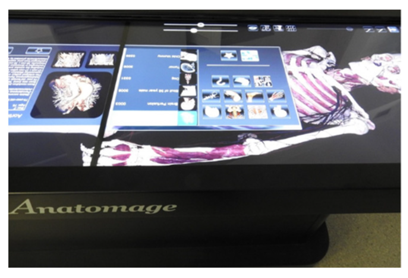
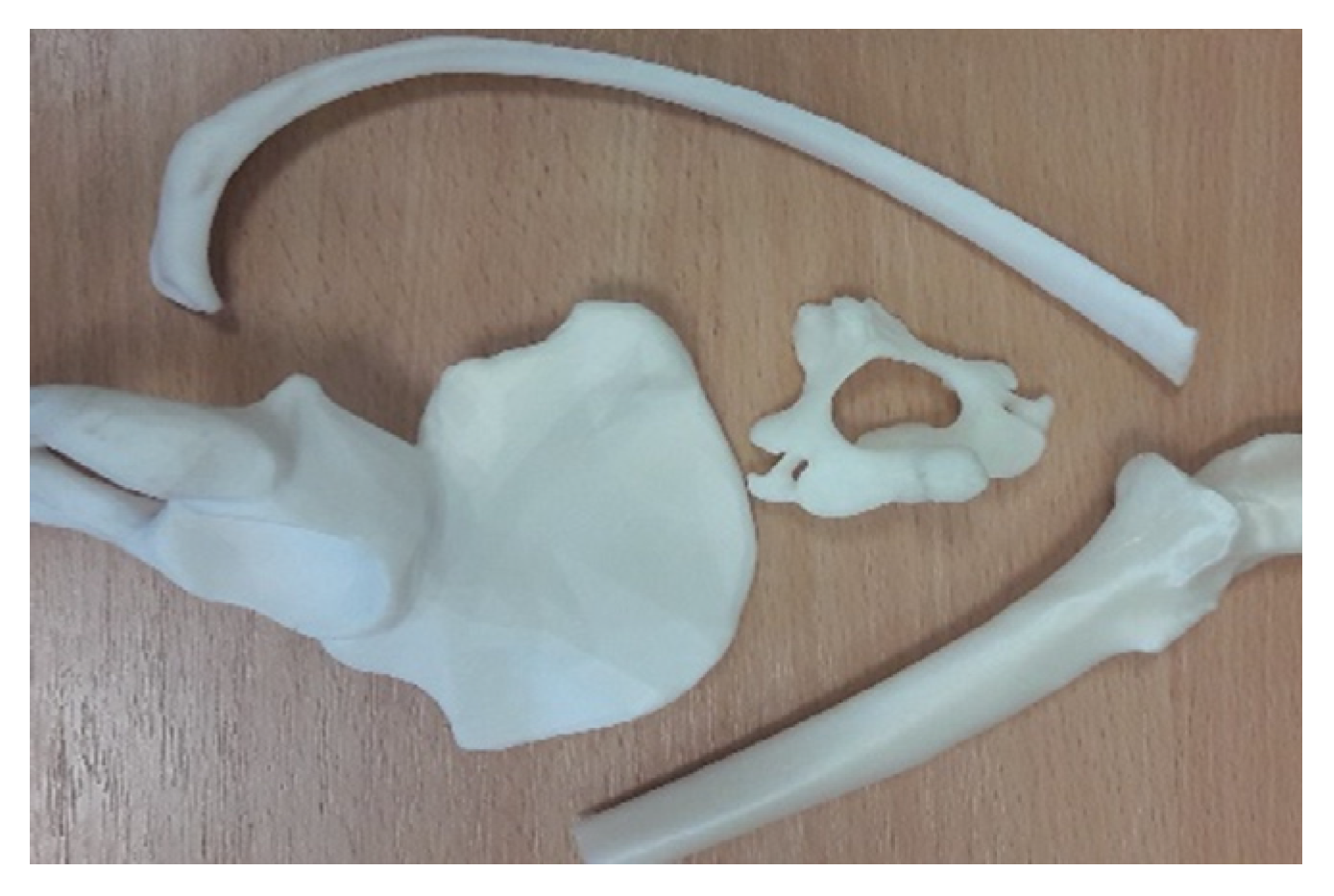
2.2.2. Teaching/Learning Environments with 3Dp Models

There are two ways of incorporating teaching tools such as 3D models in the education process. One of them is to order and buy them from specialist companies that produce these models [31].
The second way of obtaining educational 3D models is to create them on site, where is it is possible to adapt models for the needs of educators and students in specific educational institutions [32,33,34]. It is possible with access to 3D printers, some of which are safe and relatively easy to use for students and educators. Additionally, students find 3Dp very useful, interesting, and modern, and a creative way to learn human anatomy.
Printing files are publicly available from different free and open sources on the Internet. Some files can be downloaded and printed directly, while some 3D objects can be modified before printing. According to the preparation of 3D anatomical models, the dualization can be adjusted, and it is also possible to add or remove different anatomical structures from the final model. The possibility to use different materials during the printing process of anatomical modes makes a regular study of the human body more effective. Materials for 3D printing such as PLA (polylactic acid) are the easiest to use (especially for beginners), and this material is relatively cheap. It is clear that there exist many possibilities for printing time reduction. Some detailed models can be printed in a few minutes, while others require many hours to finish only one model. Fast printed models are useful for educators to better explain some anatomical structures, without any additional tools. More expensive materials can be useful for the printing of more complex structures of the human body [35].
The possibility to use the 3Dp method provides a lot of different benefits in the Human Anatomy course. Apart from medical plastic models that could be used daily in educators’ work, students can work on creating their 3D anatomical models for personal use during their practical lab.
However, more research needs to be performed measuring the effectiveness of the 3D model’s incorporation into the anatomy teaching processes and on assessment of the level of knowledge and skills of students. Young people will be able to precisely describe the quality of their model, its conditions, and develop an understanding of problems that can affect structures of the organs inside the human body. After acquiring such knowledge, students will be able to develop further steps for modification of their created models or to solve different clinical problems regarding a patient’s treatment.

2.3. Creation of Anatomical Models

There exist different strategies for creating 3D anatomical models [36,37]. Structures, shapes, and sizes of models can be prepared manually by using computer software.
However, it is up to the educators and/or students how detailed these models should be. For example, when creating a bone model, we could decide only to present the actual structures without any attachment places of muscles, or we could choose for the model to be more detailed and display major muscles on bone surfaces. A more complicated way of preparing realistic and highly detailed anatomical models is creating models from radiological medical images with the help of the computer segmentation technique, which allows identification of any anatomical structures and their variations, pathological processes, and conditions of separate organs. For this reason, for creating 3D models in our practical labs, we use medical digital images with the help of CT (Computed Tomography) and MRI (Magnetic Resonance Imaging) techniques.
Structures are identified and prepared step-by-step for segmentation with the help of specialized computer software, and afterward, they are converted into a special 3D file format, such as stereolithographic (STL) or OBJ file format. Created models can be printed in different sizes with different levels of detail [38]. It is clear that in medicine, a few millimeters can be very important in some cases.
This can provide a deeper understanding for students and our future specialists about structures and their conditions in the human body. Additionally, during lectures and practical labs, qualified educators can use real, increased, or decreased in size 3Dp models and combine their own or students’ knowledge and skills for exploration of anatomy during other basic study courses, such as embryology, histology, physiology, chemistry, and biology. These topics can be separated into several systems of the body: skeletal, muscular, respiratory, digestive, urinary, genital, nervous, circulatory, endocrine and immune, and sense organs.
According to our experience, the easiest system for 3Dp anatomical models is the skeletal system. Some bones and their structures are simple, and students can prepare these models very easily. Moreover, in the creation process of these models, the cross-sections and segmentation from any of the medical images can be used. Nonetheless, 3Dp technology can provide a lot of new options and possibilities, such as printing single bones or several bones in different sizes.
When students would like to try to prepare more complicated models, they can choose to print models from the muscular system. There are different possibilities, for example students can separate muscles one by one, or combine them into groups.
At the end of the first semester of the first study year, some of the students prepare for printing combinations of the bones together with muscles.
When it comes to the other systems of organs, students are able to prepare and print 3D models of the heart, brain, kidneys, or some specific structures that can be used for a better understanding of their location and functions. In later study semesters, some students come back to the 3Dp process, but in these cases, students have a strong motivation to prepare and print more complicated and specific things. At the Department of Morphology exists the possibility to study and obtain knowledge about bioprinting, a field of medicine that offers new ways to use human cells to print tissues, organs, or any other structures for research purposes.
3D models can help students’ understanding and evaluation of health status and risk factors, including pain location and irradiation, injuries, traumas, primary or secondary developed processes, variations, and levels of damages.

3. Materials and Methods

3.1. Participants and First Practical Procedures

This study was a small-scale study focusing on anatomy education processes, and it was conducted at the Department of Morphology, RSU, in Latvia. The department provides general education requirements for basic study as well as a few specific study courses. The study was carried out based on the Human Anatomy course that is compulsory and is taken by all students. For the time being, several modern, digital methods are implemented in our medical study courses, which are based on traditional as well as innovative educational models.
This study includes information about students’ knowledge and skills, regarding the incorporation of new educational instruments which are related to sustainable education.
Therefore, this study aimed to incorporate and use digital images and 3Dp models as innovative tools for medical students. Additionally, one of the objectives was to determine the level of knowledge and skills for students’ improvement and perspectives on SD.
In 2019–2020, 250 students of the first study year at the Faculty of Medicine, RSU, were included in this study. The mean age of participants ranged from 18 to 25 years. We chose this group because 3D printing is only part of the anatomy course at the beginning of studies at the Faculty of Medicine. There were no other students, study years, or Faculties with this option.
The selection of students was based on an open and voluntary invitation, and the study did not include any tests of significant importance on the subjects involved. According to this, the study did not require the Ethical committees’ permission. The participants were informed about their right to opt out from the study at any time. To ensure confidentiality and anonymity, the principle of secrecy was preserved. Besides this, the gathered data were intended for the purposes of this study.
In practical labs, educators used teacher- and student-centered methods.
Our selected students used 3D tools such as digital images and 3Dp models that were supported for learning about anatomy. All students were instructed on how to use the digital images and how to work with 3D printers to create models.
Students learned to identify several anatomical structures in digital images using the 3D Virtual Dissection Table “Anatomage” (USA). More than 100 cross-sections were used and studied from 4 prepared specimens of the “Anatomage” (Table Application software from Anatomage, Inc. (Table EDU 6.0)) database for 4 male and female cadavers, and over 50 clinical cases from a digital library with a variety of visualization options (X-rays, CT, MRI, etc.) of the body.
The basic procedures for the anatomical 3Dp model creation consisted of the following steps: introduction to 3Dp technology, an overview of 3D printing materials, preparation for the creation and downloading of the 3D file of the model, modeling, printing, and post-processing. We used 4 Fused Filament Fabrication (FFF) printers, and all anatomical models were produced using 2 “Prusa i3 MK2” (Prusa Research) and 2 “Ultimaker S5” 3D printers.

3.2. Data Collection Tool and Analysis

For gathering information, we used interviews as a qualitative data collection tool [39,40]. The open-ended questions were designed for the needs of this study to find out about students’ experiences (self-assessment) using digital images and 3Dp models, their perspectives for a level of knowledge, and skills after taking this course. Students answered these questions at the end of the practical lab. In the beginning, four structured questions were asked: Q1: What did you study during the course? Q2: What did you study from digital images and 3Dp models? Q3: How did you solve the problems or complicated situations during that time? Q4: How will the tools that you used help you in the future? At the end of the interview, a fifth question was asked: Q5: How satisfied were you with the use of the 3D tools during the course?
All answers were recorded and transcribed by one educator of the Human Anatomy course. Afterward, the data were processed, coded, and analyzed using content analysis [41,42]. Coding categories were derived directly from the answers. Categories were grouped into subcategories. In order to provide internal validity of the qualitative study, the texts of the interviews, codes, and reproduced categories were evaluated by the first author.
The data acquired were expressed with numbers and percentages. The approach used in this analysis was conventional [43].
Lastly, we also used content analysis of qualitative interview data, by categorizing and interpreting the data for assessing the level of participants’ satisfaction with the use of the 3D tools (digital images and 3Dp models) in the study course.

4. Results

In this section, we present the results of the study, aiming to answer the 4 questions mentioned above (Table 1).
For the question “What did you study during the course?”, 45 out of 250 (18%) students answered that they studied gross anatomy (dissections) and 3D visualizations of the structures (40, or 16%), followed by the use of 3Dp models (35 or 14%), while 5 out of 250 (2%) students answered that they studied only basic structures.
The question “What did you study from digital images and 3Dp models?” was asked to students, and 48 (19.2%) out of 250 students answered that they studied a deeper understanding of human anatomy, while 45 (18%) students indicated virtual dissection, followed by different variations/abnormalities (37 or 14.8%). Only 7 (2.8%) students mentioned the use of correct terminology in this question.
Regarding the question “How did you solve the problems or complicated situations during that time?”, 67 (26.8%) students stated using more visual aids and 52 (20.8%) students used repeating of material. Using more time and moving slower were solutions for 32 (12.8%) students. Very few students (10, or 4%) indicated that they used a simplification of information.
Finally, for the question “How will the tools that you used help you in the future?”, the answers varied from ideas about the relationship between basic and clinical study subjects (70, or 28%), the importance of anatomy for clinical studies (65, or 26%), and success on tests, exams (47, or 18.8%). For 13 (5.2%) students, these tools were indicated as related to scientific work.
It was discovered that the students were satisfied with the 3D tools that were used for their teaching during the course (Table 2).
The majority (51, or 20.4%) of students were stimulated and motivated to learn more materials and felt that this course was more intensive through the use of digital images and 3Dp models during these activities (Figure 2).

5. Discussion

Today, some classical educational methods are replaced, transformed, or modified by multiple digital technologies [44,45,46].
In the Human Anatomy course, dissection has been the primary teaching tool for a long time at RSU. With recent scientific progress, a lot of created technological innovations and their possibilities have developed new trends in medical education and led to curriculum changes. Nowadays, in our department and laboratory, we underline the fundamental necessity of cadaveric dissection and continuation of it for the future in combination with new and modern directions. We consider that students and tutors should use both traditional and progressive technological tools for anatomy education, theoretical knowledge, and practical skills. In this article, we analyzed the role of perspectives of digital images and 3Dp models, but we did not exclude or replace dissection from the anatomy curriculum. We also note that anatomy teaching excluded dissections in the time of COVID-19. To support the students and continue the teaching and learning processes of human anatomy in this situation, we implemented and used different digital tools and 3Dp models.
The transition from traditional anatomy to virtual anatomy presents certain challenges for both educators and students, and the methods of preparing and delivering the topics of lectures or practical labs change. Contents of lectures and practical labs can be prepared by educators, and students can learn all study materials at home on a personal computer. The accessibility of digital images makes it easier to present them in seminars, conferences, scientific works, and other activities, including the use of these tools in the distant education process. Finally, digital images can be utilized in other study courses and incorporated into online study platforms. In this new digital age, all parts of information and/or sources transform into digital language and format. Nowadays, a lot of educational resources are available in digital format and computers occupy a central place in the teaching and learning process [47,48,49]. Each institution and department have their own educational methods [50]. Some authors state that students must better understand the environments of their intended professions [51,52]. Educators should promote the students to understand their roles in SD and, according to this, collaborate in multi-disciplinary ways to share knowledge [53,54].
With increasing numbers of students, there is an increase in the need for different methods, materials, and resources. New methods of the educational process in the anatomy field have faced numerous challenges to their incorporation and widespread implementation. Different authors have written about curricular changes in the anatomical courses, many of which directly underlined the need to move away from the traditional educational styles [55,56]. Several authors described a re-thinking about how education should be implemented nowadays; however, they indicated to transition to more student-centered teaching [57,58].
Modern didactic processes should enable the use of different forms of teaching, and according to this, there is a need to revise the existing guidelines of the educational process. Teaching applies modern didactic methods. It is proposed that multi-methods are recommended for the modern medical anatomy study processes [59]. Different teaching techniques can be combined into Human Anatomy courses, depending on what teaching effects we expect. Nowadays, possibilities for the use of 3D tools may lead to better learning outcomes with more active roles for students during these types of practical labs. In our study process, active practical labs were perceived by students and educators as an effective study process.
Our traditional teaching methods include lectures, video lectures, practical labs, e-studies, cadaveric dissection, and anatomical models. Approaches concerning topographical anatomy are also included in the teaching/learning course, while digital simulation tools such as Virtual Dissection “Anatomage” Table and digital images are also used as major educational instruments. Highly accurate 3Dp models such as visual and interactive aids concerning normal and abnormal structures are used in the Human Anatomy study course, providing educational processes, clinical discussions, and training in some medical procedures.
All 3Dp models and digital images represent special anatomical information. According to this, an analysis of each structure and detail was very interesting from the detection of students’ knowledge and skills points of view.
The assessment process of the use of new innovations by students provides an opportunity to identify and address any field in which improvement may be made and to identify those directions that reflect effective educational practice in the Human Anatomy course. Evaluation of any innovations or changes in the study curriculum or teaching methods from the perspectives of the educators is very important for the development of the educational environment. In addition, in the education system, satisfaction of students is an indicator of the quality. The assessment of the current possibilities in Human Anatomy course education must provide educators with periodic information in order to continue, change, or correct the learning process accordingly. However, it is understood from various studies on teaching methodologies that the proper utilization of newer technologies along with the traditional teaching methods will certainly lead to better understanding of Human Anatomy and will eventually improve students’ performance.
The satisfaction of students with the use of 3D tools in the learning of Human Anatomy is demonstrated in several studies, though with some differences between them [60,61]. Students highly agreed that these tools helped them improve their ability to better study the Human Anatomy course and to develop their level of knowledge and skills for other basic and clinical studies. Students ranked teaching/learning with the use of digital images and 3Dp models as some of the best methods for understanding topics. Digitalization of different educational materials has a considerable impact on the environment in which medical students learn [62].
Thanks to advancements in technologies, 3D products have achieved great progress with new directions [63]. At present, the incorporation of digital images and 3D anatomical models into the Human Anatomy course offers new opportunities and possibilities for students and educators, for creative, innovative teaching/learning, and for increasing students’ results in their knowledge and skills. The assessment of the effectiveness of the anatomical education process is multi-factorial. As established by several studies, students’ obtainment of knowledge in the short-term and long-term retention must be evaluated [64,65].
It is widely mentioned that the use and role of medical images has increased drastically during recent years [66,67]. Medical education has rapidly upgraded under the influence of various factors, including this forced down-time during the COVID-19 pandemic [68]. According to the current needs, technologies have facilitated anatomists to develop several digital and creative solutions. It is a vital step to prepare the educators and students to cope with the modern technological innovations [69]. The world of anatomists has been disrupted by COVID-19, and these disruptions require that educators reimagine their role [70]. Virtual anatomy education is the only way to continue learning and teaching processes in the current pandemic situation [71].
Digital images and 3Dp models allow students to work on any topic in groups, create and exchange ideas between each other and the educator, and increase knowledge and show better results in a short period of time. Digital technologies with different possibilities help students and educators to reduce paperwork, developing new possibilities in the study process [72,73]. According to the literature sources, the technologies should also develop communication skills and logical thinking, including improvement of competencies [74,75]. In summary, we observed that this study can lead to modern anatomical education for SD.

6. Conclusions

There exist several directions and strategies that can develop the teaching and learning of the Anatomy course now and in the future. The usage of digital images and 3Dp models is becoming not only just a tool for anatomical lectures and practical labs, but it is also widely used by students in clinical studies. In the near future, these tools will be incorporated not only in the Human Anatomy curriculum but also in other courses requiring anatomical knowledge, as a new digital revolution in the educational sphere is approaching, changing the way that new things are learned and new skills are obtained.
During the 3D course, all of these challenges are related to SD, but more work is required to learn how to continue to improve SD skills in students’ education environment.

Author Contributions

Conceptualization, D.K. and M.P.; methodology, D.K. and M.P.; investigation, D.K. and E.E.; writing—original draft preparation, D.K.; writing—review and editing, D.K., M.P. and E.E. All authors have read and agreed to the published version of the manuscript.

Funding

This research received no external funding.

Institutional Review Board Statement

Not applicable.

Informed Consent Statement

Informed Consent was obtained from all subjects involved in the study.

Data Availability Statement

Data are contained within the article.

Acknowledgments

The authors are grateful to medical students of the Human Anatomy course for participating in this study and for the help, support, and guidance of the team of educators of 3Dp at the Department of Morphology.

Conflicts of Interest

The authors declare no conflict of interest.

References

  1. Mahlow, C.; Hediger, A. Digital Transformation in Higher Education—Buzzword or Opportunity? eLearn 2019, 5. [Google Scholar] [CrossRef]
  2. Abad-Segura, E.; Cortés-García, F.J.; Belmonte-Ureña, L.J. The Sustainable Approach to Corporate Social Responsibility: A Global Analysis and Future Trends. Sustainability 2019, 11, 5382. [Google Scholar] [CrossRef] [Green Version]
  3. Bell, S.; Douce, C.; Caeiro, S.; Teixeira, A.; Martín-Aranda, R.; Otto, D. Sustainability and Distance Learning: A Diverse European Experience? Open Learn. J. Open Distance e-Learn. 2017, 32, 95–102. [Google Scholar] [CrossRef] [Green Version]
  4. Nölting, B.; Molitor, H.; Reimann, J.; Skroblin, J.-H.; Dembski, N. Transfer for Sustainable Development at Higher Education Institutions-Untapped Potential for Education for Sustainable Development and for Societal Transformation. Sustainability 2020, 12, 2925. [Google Scholar] [CrossRef] [Green Version]
  5. Portuguez Castro, M.; Gómez Zermeño, M.G. Challenge Based Learning: Innovative Pedagogy for Sustainability through e-Learning in Higher Education. Sustainability 2020, 12, 4063. [Google Scholar] [CrossRef]
  6. Hasan, S.M.; Khan, E.A.; Nabi, M.N.U. Entrepreneurial Education at University Level and Entrepreneurship Development. Educ. Train. 2017, 59, 888–906. [Google Scholar] [CrossRef]
  7. Jeyakumar, A.; Dissanayake, B.; Dissabandara, L. Dissection in the Modern Medical Curriculum: An Exploration into Student Perception and Adaptions for the Future. Anat. Sci. Educ. 2020, 13, 366–380. [Google Scholar] [CrossRef]
  8. Memon, I. Cadaver Dissection Is Obsolete in Medical Training! A Misinterpreted Notion. Med. Princ. Pract. 2018, 27, 201–210. [Google Scholar] [CrossRef]
  9. Garas, M.; Vaccarezza, M.; Newland, G.; McVay-Doornbusch, K.; Hasani, J. 3D-Printed Specimens as a Valuable Tool in Anatomy Education: A Pilot Study. Ann. Anat. Anat. Anz. 2018, 219, 57–64. [Google Scholar] [CrossRef]
  10. Fasel, J.H.D.; Aguiar, D.; Kiss-Bodolay, D.; Montet, X.; Kalangos, A.; Stimec, B.V.; Ratib, O. Adapting Anatomy Teaching to Surgical Trends: A Combination of Classical Dissection, Medical Imaging, and 3D-Printing Technologies. Surg. Radiol. Anat. 2016, 38, 361–367. [Google Scholar] [CrossRef]
  11. Martín, J.G. Possibilities for the Use of Anatomage (the Anatomical Real Body-Size Table) for Teaching and Learning Anatomy with the Students. Biomed. J. Sci. Tech. Res. 2018, 4. [Google Scholar] [CrossRef] [Green Version]
  12. Bücking, T.M.; Hill, E.R.; Robertson, J.L.; Maneas, E.; Plumb, A.A.; Nikitichev, D.I. From Medical Imaging Data to 3D Printed Anatomical Models. PLoS ONE 2017, 12, e0178540. [Google Scholar] [CrossRef] [PubMed] [Green Version]
  13. Yakunina, G.E. Research of Digital Communications Models within Organizations and at the State Level in the Countries-Leaders in the Use of Digital Communication Technologies. E-Management 2020, 2, 41–50. [Google Scholar] [CrossRef]
  14. Lee, J.-Y.; An, J.; Chua, C.K. Fundamentals and Applications of 3D Printing for Novel Materials. Appl. Mater. Today 2017, 7, 120–133. [Google Scholar] [CrossRef]
  15. Jamróz, W.; Szafraniec, J.; Kurek, M.; Jachowicz, R. 3D Printing in Pharmaceutical and Medical Applications—Recent Achievements and Challenges. Pharm. Res. 2018, 35, 176. [Google Scholar] [CrossRef] [PubMed] [Green Version]
  16. Yuen, J. What Is the Role of 3D Printing in Undergraduate Anatomy Education? A Scoping Review of Current Literature and Recommendations. Med. Sci. Educ. 2020, 30, 1321–1329. [Google Scholar] [CrossRef]
  17. Backhouse, S.; Taylor, D.; Armitage, J.A. Is This Mine to Keep? Three-dimensional Printing Enables Active, Personalized Learning in Anatomy. Anat. Sci. Educ. 2019, 12, 518–528. [Google Scholar] [CrossRef]
  18. Dee, E.C.; Alty, I.G.; Agolia, J.P.; Torres-Quinones, C.; Houten, T.; Stearns, D.A.; Lillehei, C.W.; Shamberger, R.C. A Surgical View of Anatomy: Perspectives from Students and Instructors. Anat. Sci. Educ. 2021, 14, 110–116. [Google Scholar] [CrossRef]
  19. Verner, I.; Merksamer, A. Digital Design and 3D Printing in Technology Teacher Education. Procedia CIRP 2015, 36, 182–186. [Google Scholar] [CrossRef] [Green Version]
  20. Lee, N. The Lancet Technology: 3D Printing for Instruments, Models, and Organs? Lancet 2016, 388, 1368. [Google Scholar] [CrossRef]
  21. Marconi, S.; Pugliese, L.; Botti, M.; Peri, A.; Cavazzi, E.; Latteri, S.; Auricchio, F.; Pietrabissa, A. Value of 3D Printing for the Comprehension of Surgical Anatomy. Surg. Endosc. 2017, 31, 4102–4110. [Google Scholar] [CrossRef] [PubMed]
  22. Abou Hashem, Y.; Dayal, M.; Savanah, S.; Štrkalj, G. The Application of 3D Printing in Anatomy Education. Med. Educ. Online 2015, 20, 29847. [Google Scholar] [CrossRef]
  23. Smith, C.F.; Tollemache, N.; Covill, D.; Johnston, M. Take Away Body Parts! An Investigation into the Use of 3D-Printed Anatomical Models in Undergraduate Anatomy Education. Am. Assoc. Anat. 2018, 11, 44–53. [Google Scholar] [CrossRef] [PubMed]
  24. Balestrini, C.; Campo-Celaya, T. With the Advent of Domestic 3-Dimensional (3D) Printers and Their Associated Reduced Cost, Is It Now Time for Every Medical School to Have Their Own 3D Printer? Med. Teach. 2016, 38, 312–313. [Google Scholar] [CrossRef] [PubMed]
  25. Garcia, J.; Yang, Z.; Mongrain, R.; Leask, R.L.; Lachapelle, K. 3D Printing Materials and Their Use in Medical Education: A Review of Current Technology and Trends for the Future. BMJ STEL 2018, 4, 27–40. [Google Scholar] [CrossRef] [PubMed]
  26. Diment, L.E.; Thompson, M.S.; Bergmann, J.H.M. Clinical Efficacy and Effectiveness of 3D Printing: A Systematic Review. BMJ Open 2017, 7, e016891. [Google Scholar] [CrossRef] [PubMed] [Green Version]
  27. Ballard, D.H.; Trace, A.P.; Ali, S.; Hodgdon, T.; Zygmont, M.E.; DeBenedectis, C.M.; Smith, S.E.; Richardson, M.L.; Patel, M.J.; Decker, S.J.; et al. Clinical Applications of 3D Printing. Acad. Radiol. 2018, 25, 52–65. [Google Scholar] [CrossRef]
  28. Baratz, G.; Wilson-Delfosse, A.L.; Singelyn, B.M.; Allan, K.C.; Rieth, G.E.; Ratnaparkhi, R.; Jenks, B.P.; Carlton, C.; Freeman, B.K.; Wish-Baratz, S. Evaluating the Anatomage Table Compared to Cadaveric Dissection as a Learning Modality for Gross Anatomy. Med. Sci. Educ. 2019, 29, 499–506. [Google Scholar] [CrossRef]
  29. Bharati, A.S.; Rani, V.S. A Study on Student Perception of Virtual Dissection Table (Anatomage) at GSL Medical College, Rajahmundry. Acad. Anat. Int. 2018, 4. [Google Scholar] [CrossRef] [Green Version]
  30. Tsoucalas, G. Technology, Imaging, History and Anatomy the Future of Learning Techniques. Biomed. J. Sci. Tech. Res. 2018, 8. [Google Scholar] [CrossRef]
  31. Sheth, R.; Balesh, E.R.; Zhang, Y.S.; Hirsch, J.A.; Khademhosseini, A.; Oklu, R. Three-Dimensional Printing: An Enabling Technology for IR. J. Vasc. Interv. Radiol. 2016, 27, 859–865. [Google Scholar] [CrossRef] [PubMed]
  32. Lim, K.H.A.; Loo, Z.Y.; Goldie, S.J.; Adams, J.W.; McMenamin, P.G. Use of 3D Printed Models in Medical Education: A Randomized Control Trial Comparing 3D Prints versus Cadaveric Materials for Learning External Cardiac Anatomy: Use of 3D Prints in Medical Education. Am. Assoc. Anat. 2016, 9, 213–221. [Google Scholar] [CrossRef] [PubMed]
  33. Kong, X.; Nie, L.; Zhang, H.; Wang, Z.; Ye, Q.; Tang, L.; Huang, W.; Li, J. Do 3D Printing Models Improve Anatomical Teaching About Hepatic Segments to Medical Students? A Randomized Controlled Study. World J. Surg. 2016, 40, 1969–1976. [Google Scholar] [CrossRef] [PubMed]
  34. Li, C.; Cheung, T.F.; Fan, V.C.; Sin, K.M.; Wong, C.W.Y.; Leung, G.K.K. Applications of Three-Dimensional Printing in Surgery. Surg. Innov. 2017, 24, 82–88. [Google Scholar] [CrossRef] [PubMed]
  35. Powers, M.K.; Lee, B.R.; Silberstein, J. Three-Dimensional Printing of Surgical Anatomy. Curr. Opin. Urol. 2016, 26, 283–288. [Google Scholar] [CrossRef] [PubMed]
  36. Chen, S.; Pan, Z.; Wu, Y.; Gu, Z.; Li, M.; Liang, Z.; Zhu, H.; Yao, Y.; Shui, W.; Shen, Z.; et al. The Role of Three-Dimensional Printed Models of Skull in Anatomy Education: A Randomized Controlled Trail. Sci. Rep. 2017, 7, 575. [Google Scholar] [CrossRef]
  37. Nath, S.I.; Anuradha, B.; Dilip, B. A Study on Making Models in Anatomy. PIJR 2021, 85–87. [Google Scholar] [CrossRef]
  38. Mogali, S.R.; Yeong, W.Y.; Tan, H.K.J.; Tan, G.J.S.; Abrahams, P.H.; Zary, N.; Low-Beer, N.; Ferenczi, M.A. Evaluation by Medical Students of the Educational Value of Multi-Material and Multi-Colored Three-Dimensional Printed Models of the Upper Limb for Anatomical Education: 3D Printed Upper Limb in Anatomical Education. Am. Assoc. Anat. 2018, 11, 54–64. [Google Scholar] [CrossRef]
  39. Cohen, L.; Manion, L.; Morrison, K. Research Methods in Education, 6th ed.; Routledge: London, UK; New York, NY, USA, 2007; pp. 461–495. [Google Scholar]
  40. Strauss, A.L.; Corbin, J.M. Basics of Qualitative Research: Techniques and Procedures for Developing Grounded Theory, 4th ed.; Sage Publications: Thousand Oaks, CA, USA, 2015; pp. 85–105. [Google Scholar]
  41. Elo, S.; Kääriäinen, M.; Kanste, O.; Pölkki, T.; Utriainen, K.; Kyngäs, H. Qualitative Content Analysis: A Focus on Trustworthiness. SAGE Open 2014, 4, 215824401452263. [Google Scholar] [CrossRef]
  42. Ahmady, S.; Khajeali, N.; Kalantarion, M.; Amini, M. A Qualitative Content Analysis of “Problem Students”: How Can We Identify and Manage Them? BMC Res. Notes 2020, 13, 566. [Google Scholar] [CrossRef]
  43. Hsieh, H.-F.; Shannon, S.E. Three Approaches to Qualitative Content Analysis. Qual. Health Res. 2005, 15, 1277–1288. [Google Scholar] [CrossRef] [PubMed]
  44. Ghemawat, P. Strategies for Higher Education in the Digital Age. Calif. Manag. Rev. 2017, 59, 56–78. [Google Scholar] [CrossRef]
  45. Hill, C.; Lawton, W. Universities, the Digital Divide and Global Inequality. J. High. Educ. Policy Manag. 2018, 40, 598–610. [Google Scholar] [CrossRef]
  46. Beghetto, V.; Agostinis, L.; Taffarello, R.; Samiolo, R. Innovative Technology for Sustainable New Materials. Eur. J. Sustain. Dev. 2016, 5. [Google Scholar] [CrossRef]
  47. Maffey, G.; Homans, H.; Banks, K.; Arts, K. Digital Technology and Human Development: A Charter for Nature Conservation. Ambio 2015, 44 (Suppl. S4), 527–537. [Google Scholar] [CrossRef] [PubMed] [Green Version]
  48. Jääskelä, P.; Häkkinen, P.; Rasku-Puttonen, H. Teacher Beliefs Regarding Learning, Pedagogy, and the Use of Technology in Higher Education. J. Res. Technol. Educ. 2017, 49, 198–211. [Google Scholar] [CrossRef]
  49. Crittenden, W.F.; Biel, I.K.; Lovely, W.A. Embracing Digitalization: Student Learning and New Technologies. J. Mark. Educ. 2019, 41, 5–14. [Google Scholar] [CrossRef]
  50. Mintz, K.; Tal, T. The Place of Content and Pedagogy in Shaping Sustainability Learning Outcomes in Higher Education. Environ. Educ. Res. 2018, 24, 207–229. [Google Scholar] [CrossRef]
  51. González-Zamar, M.-D.; Ortiz Jiménez, L.; Sánchez Ayala, A.; Abad-Segura, E. The Impact of the University Classroom on Managing the Socio-Educational Well-Being: A Global Study. Int. J. Environ. Res. Public Health 2020, 17, 931. [Google Scholar] [CrossRef] [Green Version]
  52. López Meneses, E.; Vázquez-Cano, E.; Jaén Martínez, A. Los Portafolios Digitales Grupales: Un Estudio Diacrónico En La Universidad Pablo Olavide (2009–2015) = The Group e-Portfolio: A Diachronic Study at University Pablo de Olavide in Spain (2009–2015). Rev. Humanid. 2017, 123. [Google Scholar] [CrossRef] [Green Version]
  53. Testov, V.A. On Some Methodological Problems of Digital Transformation of Education. Inf. Obraz. 2019, 31–36. [Google Scholar] [CrossRef]
  54. Hilty, L.M.; Huber, P. Motivating Students on ICT-Related Study Programs to Engage with the Subject of Sustainable Development. Int. J. Sustain. High. Educ. 2018, 19, 642–656. [Google Scholar] [CrossRef] [Green Version]
  55. Mahmoud, A.; Bennett, M. Introducing 3-Dimensional Printing of a Human Anatomic Pathology Specimen: Potential Benefits for Undergraduate and Postgraduate Education and Anatomic Pathology Practice. Arch. Pathol. Lab. Med. 2015, 139, 1048–1051. [Google Scholar] [CrossRef]
  56. Moore, C.W.; Wilson, T.D.; Rice, C.L. Digital Preservation of Anatomical Variation: 3D-Modeling of Embalmed and Plastinated Cadaveric Specimens Using UCT and MRI. Ann. Anat. Anat. Anz. 2017, 209, 69–75. [Google Scholar] [CrossRef] [PubMed]
  57. Altomonte, S.; Logan, B.; Feisst, M.; Rutherford, P.; Wilson, R. Interactive and Situated Learning in Education for Sustainability. Int. J. Sustain. High. Educ. 2016, 17, 417–443. [Google Scholar] [CrossRef]
  58. Takala, A.; Korhonen-Yrjänheikki, K. A Decade of Finnish Engineering Education for Sustainable Development. Int. J. Sustain. High. Educ. 2019, 20, 170–186. [Google Scholar] [CrossRef]
  59. Sagun, L.; Arias, R. Digital Pathology: An Innovative Approach to Medical Education. Philipp. J. Pathol. 2018, 3, 7–11. [Google Scholar] [CrossRef] [Green Version]
  60. Berney, S.; Bétrancourt, M.; Molinari, G.; Hoyek, N. How Spatial Abilities and Dynamic Visualizations Interplay When Learning Functional Anatomy with 3D Anatomical Models: Interplay of Spatial Ability and Dynamic Visualization. Am. Assoc. Anat. 2015, 8, 452–462. [Google Scholar] [CrossRef] [Green Version]
  61. Hackett, M.; Proctor, M. Three-Dimensional Display Technologies for Anatomical Education: A Literature Review. J. Sci. Educ. Technol. 2016, 25, 641–654. [Google Scholar] [CrossRef]
  62. Alkhowailed, M.S.; Rasheed, Z.; Shariq, A.; Elzainy, A.; El Sadik, A.; Alkhamiss, A.; Alsolai, A.M.; Alduraibi, S.K.; Alduraibi, A.; Alamro, A.; et al. Digitalization Plan in Medical Education during COVID-19 Lockdown. Inform. Med. Unlocked 2020, 20, 100432. [Google Scholar] [CrossRef]
  63. Wright, N.; Wrigley, C. Broadening Design-Led Education Horizons: Conceptual Insights and Future Research Directions. Int. J. Technol. Des. Educ. 2019, 29, 1–23. [Google Scholar] [CrossRef]
  64. Biberhofer, P.; Lintner, C.; Bernhardt, J.; Rieckmann, M. Facilitating Work Performance of Sustainability-Driven Entrepreneurs through Higher Education: The Relevance of Competencies, Values, Worldviews and Opportunities. Int. J. Entrepr. Innov. 2019, 20, 21–38. [Google Scholar] [CrossRef]
  65. Brown, B.J.; Hanson, M.E.; Liverman, D.M.; Merideth, R.W. Global Sustainability: Toward Definition. Environ. Manag. 1987, 11, 713–719. [Google Scholar] [CrossRef]
  66. Ammanuel, S.; Brown, I.; Uribe, J.; Rehani, B. Creating 3D Models from Radiologic Images for Virtual Reality Medical Education Modules. J. Med. Syst. 2019, 43, 166. [Google Scholar] [CrossRef]
  67. Jang, H.W.; Oh, C.-S.; Choe, Y.H.; Jang, D.S. Use of Dynamic Images in Radiology Education: Movies of CT and MRI in the Anatomy Classroom. Am. Assoc. Anat. 2018, 11, 547–553. [Google Scholar] [CrossRef] [PubMed]
  68. Byrnes, K.G.; Kiely, P.A.; Dunne, C.P.; McDermott, K.W.; Coffey, J.C. Communication, Collaboration and Contagion: “Virtualisation” of Anatomy during COVID-19. Clin. Anat. 2021, 34, 82–89. [Google Scholar] [CrossRef] [PubMed]
  69. Singal, A.; Bansal, A.; Chaudhary, P.; Singh, H.; Patra, A. Anatomy Education of Medical and Dental Students during COVID-19 Pandemic: A Reality Check. Surg. Radiol. Anat. 2021, 43, 515–521. [Google Scholar] [CrossRef]
  70. Sadeesh, T.; Prabavathy, G.; Ganapathy, A. Evaluation of Undergraduate Medical Students’ Preference to Human Anatomy Practical Assessment Methodology: A Comparison between Online and Traditional Methods. Surg. Radiol. Anat. 2021, 43, 531–535. [Google Scholar] [CrossRef] [PubMed]
  71. Jones, D.G. Anatomy in a Post-Covid-19 World: Tracing a New Trajectory. Anat. Sci. Educ. 2021, 14, 148–153. [Google Scholar] [CrossRef]
  72. Frey, C.B.; Osborne, M.A. The Future of Employment: How Susceptible Are Jobs to Computerisation? Technol. Forecast. Soc. Chang. 2017, 114, 254–280. [Google Scholar] [CrossRef]
  73. Hara, C.Y.N.; Aredes, N.D.A.; Fonseca, L.M.M.; de Campos Pereira Silveira, R.C.; Camargo, R.A.A.; de Goes, F.S.N. Clinical Case in Digital Technology for Nursing Students’ Learning: An Integrative Review. Nurse Educ. Today 2016, 38, 119–125. [Google Scholar] [CrossRef] [PubMed]
  74. Hesrcu-Kluska, R. The Interaction between Learners and Learner-Facilitator in an Online Learning Environment. Creat. Educ. 2019, 10, 1713–1730. [Google Scholar] [CrossRef] [Green Version]
  75. Carter, J.; Bababekov, Y.J.; Majmudar, M.D. Training for Our Digital Future: A Human-Centered Design Approach to Graduate Medical Education for Aspiring Clinician-Innovators. NPJ Digit. Med. 2018, 1, 26. [Google Scholar] [CrossRef] [PubMed] [Green Version]
Figure 1. Prepared Virtual Dissection Table “Anatomage” with opened digital library in the practical lab.
Education 11 00380 g001
Figure 2. A few 3Dp models of the human skeletal system from plastic materials and in different sizes.
Education 11 00380 g002
Table 1. Students’ answers to the 4 study questions in the Human Anatomy course.
QuestionAttitudes and ViewsNumber of Students and %
What did you study during the course?anatomical terminology24 (10%)
basic structures5 (2%)
functions of structures25 (10%)
location of structures and organs21 (8.4%)
topography30 (12%)
radiological anatomy15 (6%)
3D visualizations of the
structures
40 (16%)
using 3Dp models and their
creation
35 (14%)
work in groups, teams10 (4%)
gross anatomy (dissection)45 (18%)
What did you study from the digital images and 3Dp models?more interesting learning and
education
25 (10%)
relationships between structures15 (6%)
analysis of clinical cases30 (12%)
virtual dissection45 (18%)
different variations and/or abnormalities37 (14.8%)
more deep understanding of anatomy48 (19.2%)
several simulations of clinical procedures10 (4%)
basics for clinical studies20 (8%)
overview of knowledge and skills13 (5.2%)
the use of correct anatomical
terminology
7 (2.8%)
How did you solve the
problems or complicated
situations during that time?
including more visual aids67 (26.8%)
repeating material52 (20.8%)
help from classmates and
educator
48 (19.2%)
using more time, moving slower32 (12.8%)
using basic concepts41 (16.4%)
simplification of the information10 (4%)
How will the tools that you used help you in the
future?
importance of anatomy for
clinical studies
65 (26%)
training of some procedures37 (14.8%)
relationship between basic and clinical study subjects70 (28%)
basics for scientific work13 (5.2%)
improving clinical skills18 (7.2%)
success in tests, exams47 (18.8%)
Table 2. The satisfaction of students with the 3D tools used in the study course.
The Use of 3D Tools for Students’ SatisfactionNumber of Students and %
reproduce taught/learned knowledge and skills46 (18.4%)
increase motivation and intensity for learning51 (20.4%)
develop knowledge and skills relevant to clinical needs48 (19.2%)
improve their thinking and solution of problems with
understanding of structures of the human body
28 (11.2%)
prepare for the assessment of knowledge and skills42 (16.8%)
provide a good background for the future35 (14%)
Publisher’s Note: MDPI stays neutral with regard to jurisdictional claims in published maps and institutional affiliations.

