Article

A DNA Methylation-Based Gene Signature Can Predict Triple-Negative Breast Cancer Diagnosis

by
Saioa Mendaza
1,*,
David Guerrero-Setas
1,2,
Iñaki Monreal-Santesteban
1,
Ane Ulazia-Garmendia
1,
Alicia Cordoba Iturriagagoitia
2,
Susana De la Cruz
3 and
Esperanza Martín-Sánchez
1,*
1
Molecular Pathology of Cancer Group, Navarrabiomed, Complejo Hospitalario de Navarra (CHN), Universidad Pública de Navarra (UPNA), Instituto de Investigación Sanitaria de Navarra (IdiSNA), Irunlarrea 3, 31008 Pamplona, Spain
2
Department of Pathology, Complejo Hospitalario de Navarra (CHN), Irunlarrea 3, 31008 Pamplona, Spain
3
Department of Medical Oncology, Complejo Hospitalario de Navarra (CHN), Irunlarrea 3, 31008 Pamplona, Spain
*
Authors to whom correspondence should be addressed.
Submission received: 19 July 2021 / Revised: 27 September 2021 / Accepted: 29 September 2021 / Published: 4 October 2021
(This article belongs to the Topic Cancer Biology and Therapy)

Abstract

:
Triple-negative breast cancer (TNBC) is the most aggressive breast cancer (BC) subtype and lacks targeted treatment. It is diagnosed by the absence of immunohistochemical expression of several biomarkers, but this method still displays some interlaboratory variability. DNA methylome aberrations are common in BC, thereby methylation profiling could provide the identification of accurate TNBC diagnosis biomarkers. Here, we generated a signature of differentially methylated probes with class prediction ability between 5 non-neoplastic breast and 7 TNBC tissues (error rate = 0.083). The robustness of this signature was corroborated in larger cohorts of additional 58 non-neoplastic breast, 93 TNBC, and 150 BC samples from the Gene Expression Omnibus repository, where it yielded an error rate of 0.006. Furthermore, we validated by pyrosequencing the hypomethylation of three out of 34 selected probes (FLJ43663, PBX Homeobox 1 (PBX1), and RAS P21 protein activator 3 (RASA3) in 51 TNBC, even at early stages of the disease. Finally, we found significantly lower methylation levels of FLJ43663 in cell free-DNA from the plasma of six TNBC patients than in 15 healthy donors. In conclusion, we report a novel DNA methylation signature with potential predictive value for TNBC diagnosis.

1. Introduction

Breast cancer (BC) has overtaken lung cancer as the most commonly diagnosed cancer globally in 2020 and is the top cause of deaths from cancer for women worldwide, becoming a public health major issue [1]. BC’s heterogeneity is reflected in its division into several molecular subtypes with a variety of pathological features, leading to diverse treatment options and prognoses. This categorisation relies on the differential expression of key genes in tumour initiation and progression [2]. As a consequence, current methods for BC subtype identification are based on the immunohistochemical expression of oestrogen receptor (ER), progesterone receptor (PR), and Ki-67, along with the expression and/or amplification of the gene encoding the human epidermal growth factor 2 receptor (HER2) [3]. Based on these biomarkers, BC can be classified into five intrinsic subtypes: Luminal A (ER+, PR+, HER2-, low Ki-67), Luminal B/HER2-negative (ER+, PR-/+, HER2-, low Ki-67), Luminal B/HER2-positive (ER+, PR-, HER2+, high Ki-67; or ER+, PR+, HER2+, low Ki-67), HER2-positive (ER-, PR-, HER2+, any Ki-67), and triple-negative BC (TNBC) (ER-, PR-, HER2-, any Ki-67) [4]. Besides diagnosis, these biomarkers have been shown to be suitable targets for targeted therapy, such as hormone therapy for luminal subtypes and anti-HER2 antibodies for HER2-overexpressing subtypes. Since TNBC is characterized by the lack of ER and PR expression, combined with the absence of both overexpression and amplification of the gene encoding HER2 [2,5], the targeted treatment that is effective in the other BC subtypes is futile for TNBC patients [6]. Thus, TNBC, which accounts for about 15% of all BC cases, is considered the most aggressive subgroup portrayed by early relapse, frequent distant metastasis, and poor overall survival [7]. Despite its malignancy, TNBC diagnosis remains a major concern worldwide [8], because it still relies on the subjective evaluation of immunostaining assays that are not 100% sensitive and specific [9], and show considerable interlaboratory variability [10]. Apart from immunohistochemistry, the development of other diagnosis methods has started for BC. Some investigations have been performed to identify gene-expression and epigenetic alterations as diagnosis biomarkers, including blood-based markers for non-invasive detection [11,12,13]. However, this field is poorly explored in TNBC. Therefore, a more reliable assessment based on additional molecular and quantifiable biomarkers is highly desirable [10,14].
Aberrations in DNA methylation patterns are known to be crucial players in cancer initiation [15,16]. Hence, DNA methylation signatures are being assessed as potential molecular biomarkers in cancer [17,18,19], and specifically, several DNA-methylation based biomarkers have been described to have significant diagnostic and prognostic potential in BC [15,20]. Remarkably, since DNA-methylation changes can also be detected in biological fluids, some of these biomarkers can be tracked in cell-free DNA (cfDNA) from BC patients’ plasma [21,22,23], which makes them really attractive from the translational point of view. However, few DNA-methylation alterations have been reported in TNBC [24,25,26,27,28], and nearly all of them have been proposed to have predictive value of prognosis and drug response, but not as diagnostic biomarkers. Thus, in this study, our aim was to identify a DNA-methylation-based diagnostic signature to establish new supplementary diagnostic tools for TNBC detection.

2. Materials and Methods

2.1. DNA Methylome Data Sets

In this study, we used seven data sets deposited in Gene Expression Omnibus (GEO, https://0-www-ncbi-nlm-nih-gov.brum.beds.ac.uk/geo/ assessed on 10 June 2019). All of them reported DNA methylation data profiled using the Illumina Infinium Methylation 450K BeadChip assay (Illumina, San Diego, CA, USA), which made them easily comparable. The signature was constructed from one of these data sets (GSE141338), generated by our group in a previous study [28], using non-neoplastic mammary tissues from 6 reduction mammoplasties, and tumours from 8 patients with TNBC. The remaining data sets, which were used to confirm the signature, included DNA methylation profiles from 58 non-neoplastic breast samples (GSE88883 [29] and GSE74214), 93 TNBC (GSE78751 [30] and GSE78754 [29]), 150 BC other than TNBC (GSE141338 [28] and GSE72245 [31]), and 30 non-neoplastic prostate and 30 prostate cancer samples (GSE76938).

2.2. Bioinformatics Analysis

2.2.1. Generation of a Diagnostic Signature for TNBC

With the aim of finding a signature with diagnostic potential for TNBC, we identified differentially methylated probes (DMPs) between six non-neoplastic mammary and eight TNBC tissues, with class prediction ability in our discovery data set. First, the methylation level of each of the 450,000 CpG sites interrogated in the array was estimated as normalized β values using the GenomeStudio program v2010.3 (Illumina, San Diego, CA, USA). β values ranged between 0 (fully unmethylated) and 1 (fully methylated). Thereafter, and without filtering by methylation fold change between TNBC and non-neoplastic tissues (Δβ) or genomic region, all 450,000 probes were subjected to a class prediction algorithm based on the K-nearest neighbours (KNN) method, and predictive probes were selected using the ANOVA F-ratio using the Tnasas tool (http://tnasas.iib.uam.es/ assessed on 20 June 2019). Briefly, KNN is a non-parametric analysis that predicts the samples of a test case as the majority vote among its k nearest neighbours. These ones are chosen based on the Euclidean distance, and their number (k) is selected by cross-validation. For probe selection, the program divided the whole series in 10 subsets and ranked the probes using ANOVA F-ratio, which provided a first set of ranked probes to further feed the predictor. The predictor was then built using different numbers of the best-ranked probes, and the left-out sample was predicted for each of them. The cross-validation error corresponding to each number of probes was computed, and the best predictor set was the one with the smallest cross-validation error and the smallest number of probes. Finally, the program run the probe-ranking method on the complete series and selected the top probes. To evaluate the error rate, the program divided again the whole series in 10 subsets, and for each of them, left aside one (the “out-of-bag” subset), found the best number of probes with the other 9 subsets (the “in-bag” ones), as just described, and predicted the out-of-bag samples with the predictor just found. Since at the end of the process, each sample was once in the “out-of-bag” set, the final error rate was obtained using all out-of-bag predictions. As the final output, the predictor set with the smallest error rate was returned.

2.2.2. Robustness of the Model

The potential of the signature was then assessed by applying its predictive power into larger cohorts. To do this, the methylation levels of the predictive probes were retrieved from data sets publicly available in the GEO repository, particularly, of non-neoplastic breast (n = 58) and TNBC samples (n = 93). Moreover, in order to assure whether these selected DMPs predicted TNBC specifically or were related to any type of BC, their methylation levels were interrogated in additional series of breast tumours not belonging to the TNBC subtype (n = 150). Furthermore, data from an unrelated-to-breast tissue (30 non-neoplastic prostate and 30 prostate cancer samples) were explored to test the accuracy of those probes as classifiers and therefore TNBC diagnostic biomarkers.
Finally, unsupervised clusterings were performed with the Babelomics 5 tool (http://babelomics.bioinfo.cipf.es/ assessed on 15 July 2019) [32], using the unweighted pair group method with arithmetic mean (UPGMA) method and the normal Euclidean distance.

2.3. Validation of the Signature

2.3.1. Patient Samples

Two cohorts of patients were used in this study to confirm the predictive diagnostic value of the signature. First, a series of formalin-fixed, paraffin-embedded (FFPE) samples from 51 patients with TNBC and 57 with BC of distinct subtypes other than TNBC, and 16 non-neoplastic breast tissues from reduction mammoplasties, was employed to validate the DNA methylation signature. Lastly, the methylation status of selected genes belonging to the signature was explored in cfDNA in a small series of plasma samples from six TNBC patients and 15 age-matched healthy women.
All patients were diagnosed with invasive ductal breast carcinoma in the Department of Pathology (Complejo Hospitalario de Navarra, Pamplona, Spain) in accordance with the criteria recommended by the St Gallen International Expert Consensus 2013 [4], considering specific Ki-67 threshold [33], grading according to the Nottingham system [34], and staging based on the AJCC (American Joint Committee on Cancer) system [35]. All cancer tissue samples harboured at least 70% of tumour cells. None of the patients had received radiotherapy or chemotherapy before surgery. Their pathological and clinical characteristics are summarised in Supplementary Table S1.

2.3.2. DNA and cfDNA Extraction and Bisulphite Conversion

To assess DNA methylation status, DNA and total cfDNA were extracted from cancer patients’ and healthy women’s mammary tissue and plasma samples using the QIAamp DNA FFPE Tissue kit and the QIAamp Circulating Nucleic Acid Kit (both from Qiagen, Hilden, Germany), respectively, and following the manufacturer’s instructions. After quantifying DNA concentration and purity in a NanoDrop spectrophotometer (Thermo Scientific, Waltham, MA), bisulphite conversion of 500 ng of DNA or 100 ng of cfDNA was performed using the EZ DNA Methylation-Gold kit (Zymo Research, Irvine, CA, USA) following the manufacturer’s recommendations.

2.3.3. Pyrosequencing

To confirm the methylation levels of selected genes, pyrosequencing was performed in bisulphite-converted DNA from FFPE tissues (51 TNBC, 57 BC of other subtypes and 16 non-neoplastic mammary tissues), and cfDNA from plasma (6 TNBC patients and 15 healthy donors). First, 2 μL of bisulphite-modified DNA or cfDNA were amplified by PCR using 0.5 μL Immolase DNA polymerase (BioLine, London, UK) in a final volume of 30 μL, and with the primers which amplified the same region recognized by the probe contained in the array (Table 1). Amplification conditions consisted of an initial DNA polymerase activation at 95 °C for 10 min, followed by 50 cycles at 95 °C for 30 s, specific melting temperature for each gene (Table 1) for 30 s and 72 °C for 30 s, and a final extension at 72 °C for 7 min. Then, pyrosequencing was carried out in a PyroMark q96 (Qiagen, Hilden, Germany) as previously described [36].

2.4. Statistical Analysis

Demographic, clinical, and pathological data were summarised as frequencies (and percentages) or means (and ranges), as appropriate. Medians of methylation in tumour and non-neoplastic tissues were compared using the Mann–Whitney U test. The optimal cut-off value identifying the hypomethylated or hypermethylated status of each selected probes measured by pyrosequencing was estimated by ROC curve analyses. Across several cut-off points, the largest positive likelihood ratio was chosen as the optimal value [37].

3. Results

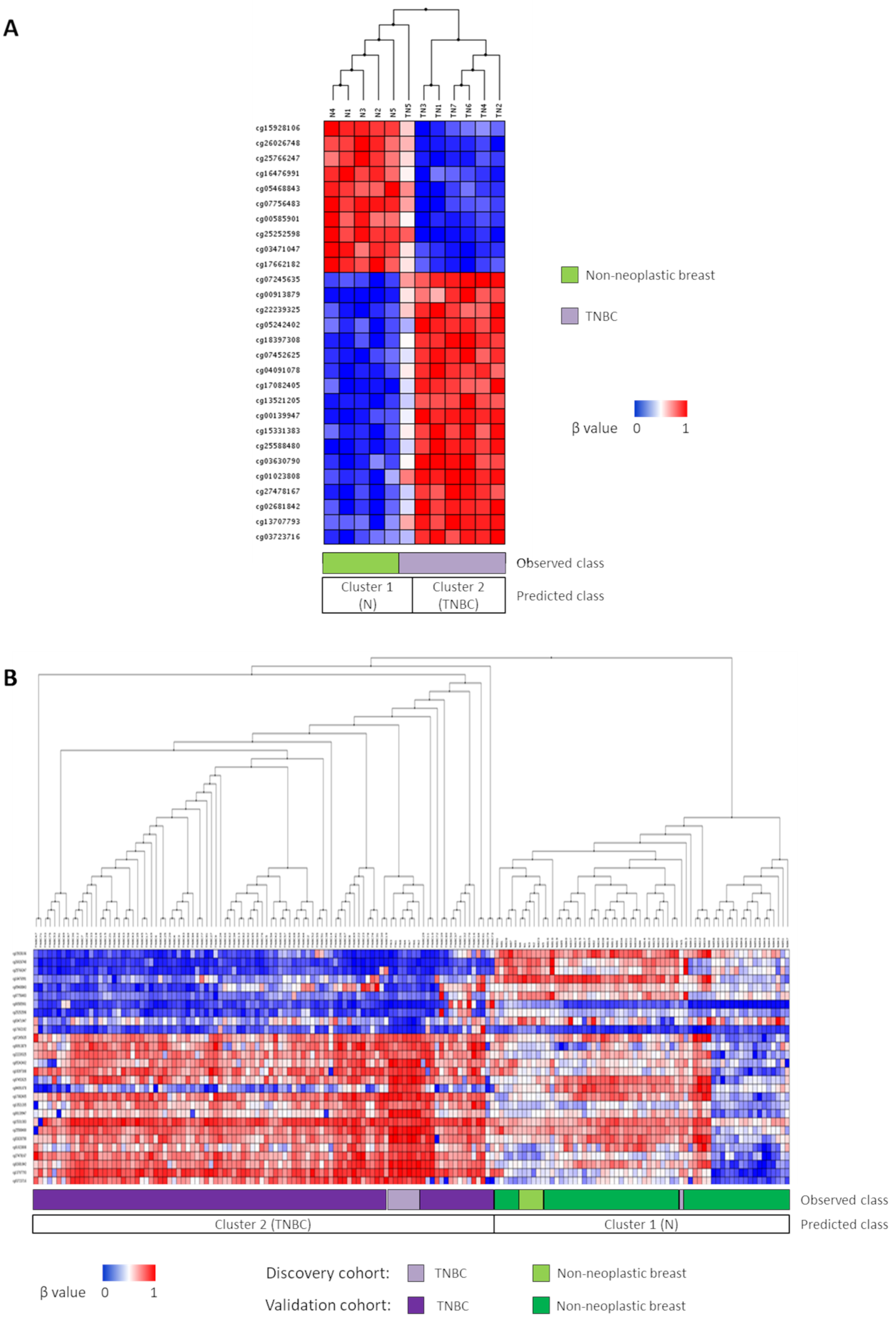
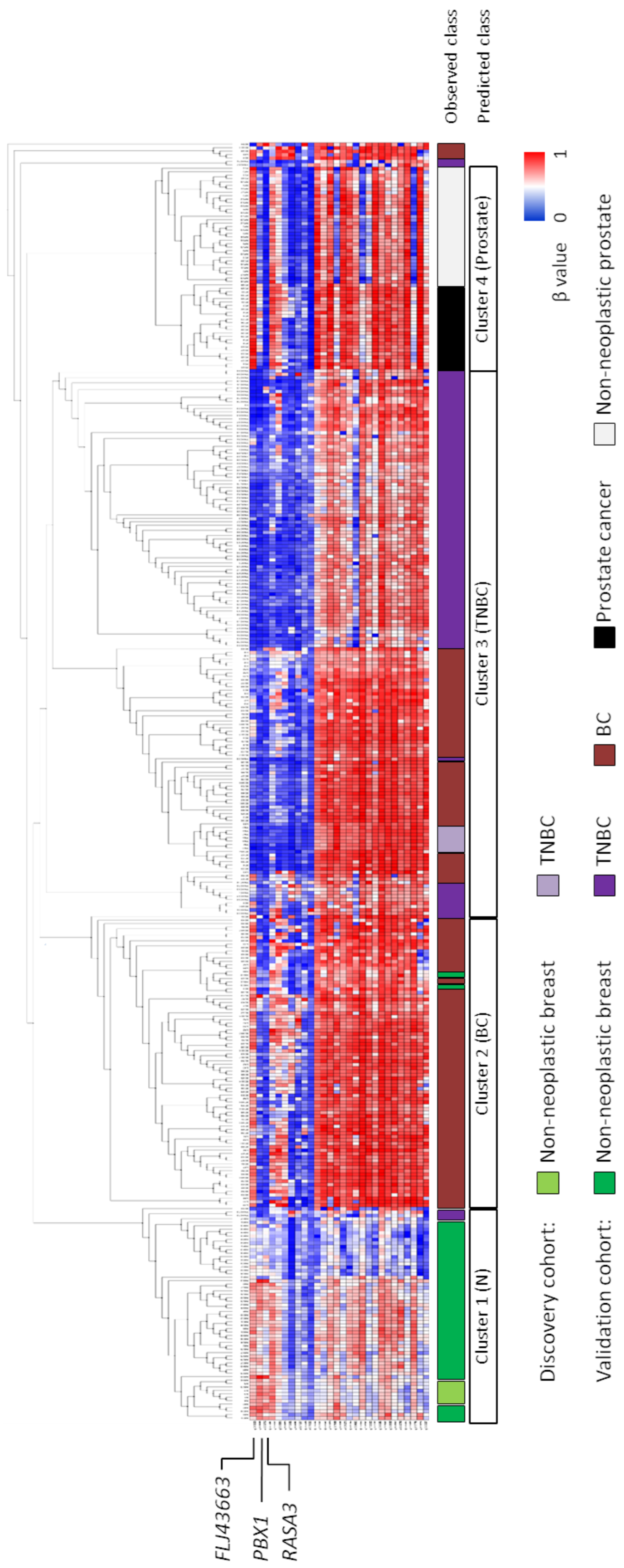
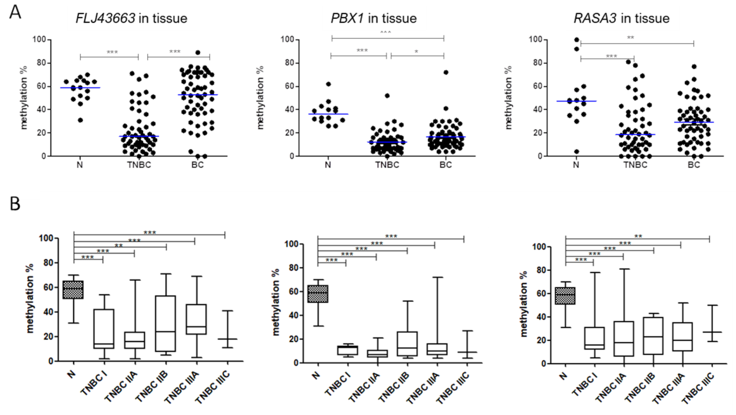
3.1. Novel Diagnostic DNA Methylation Signature for TNBC

To identify potential diagnostic biomarkers for TNBC, a signature of DMPs with class prediction ability between non-neoplastic breast (n = 6) and TNBC (n = 8) tissues was generated by comparing their DNA-methylation patterns. Based on our previous results [28], two samples, one non-neoplastic and one tumoural, had DNA methylation profiles quite different from those of the remaining samples in their groups, and were excluded from this analysis. Thus, the class-predictive signature with the minimum prediction error rate (0.083) that we found between 7 TNBC and 5 non-neoplastic samples was composed of 35 DMPs (Supplementary Table S2). Indeed, this signature accurately classified six out of seven TNBC and five out of five non-neoplastic breast samples in the TNBC and non-neoplastic groups, respectively (sensitivity = 83%; specificity = 100%) (Figure 1A).
In order to test the robustness of the class prediction model, our predictor signature was extended to larger cohorts of 58 non-neoplastic breast (GSE88883 and GSE74214) and 93 TNBC (GSE78751 and GSE78754) samples, whose methylation data were publicly available. Since the β value of one of the probes (cg15555527) could not be retrieved from those samples, the signature was then composed of 34 probes. Importantly, we found that this 34-predictor signature consistently made all TNBC samples cluster together, regardless of the data set to which they belonged (Figure 1B). Thus, the discriminative power of the diagnostic signature raised, categorizing 62 out of 63 non-neoplastic breast samples and 100 out of 100 TNBCs as the non-neoplastic and TNBC groups, respectively, and therefore, yielding a sensitivity of 98.4% and a specificity of 100% (error rate = 0.006) (Figure 1B).
The methylation levels of these 34 probes were also explored in a total of 150 samples diagnosed with BC of confirmed non-TNBC subtype (GSE141338) and unknown subtype (GSE72245). Above that, 30 non-neoplastic and 30 tumour prostate samples (GSE76938) were included as unrelated tissues to ensure that the observed differences in breast tissues were not due to slight differences in distinct subtypes from the same mammary origin. With the exception of a first small set of five non-TNBC cases and a second one with only two TNBC cases that segregated soon from the remaining patients, all samples derived from prostate tissues were, as expected, the least closely related to the breast ones (Figure 2). Regarding BC tissues, more than half of them clustered together and separated from non-neoplastic samples. Interestingly, a subset of BC cases of unknown subtype was located within the TNBC cluster. This finding could suggest their belonging to the TNBC subtype, but it cannot be confirmed, because information regarding molecular subtypes of BC from these public data sets was not available. Taken together, these results strengthen the robustness of our methylation signature to predict the TNBC diagnosis.

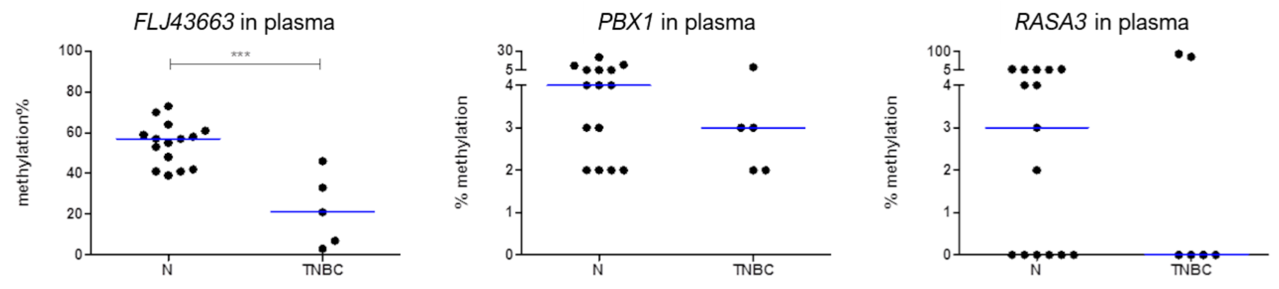
3.2. Methylation Status of FLJ43663, PBX1, and RASA3 in Breast Tissue and Plasma

Among the 34 predictor probes with the highest |Δβ| values in TNBC comparing to non-neoplastic tissues (Supplementary Table S2), three were selected to validate their usefulness as biomarkers for TNBC diagnosis: cg15928106, cg2626748, and cg16476991, which recognised FLJ43663, PBX Homeobox 1 (PBX1), and RAS P21 protein activator 3 (RASA3) genes, respectively (Figure 2). To do this, we measured by pyrosequencing the methylation levels of the genomic region recognized by each probe in a series of FFPE samples from 51 TNBC patients and 16 non-neoplastic breast tissues. We confirmed that TNBC tumours had significantly lower methylation levels of these selected genes than non-neoplastic samples (p ≤ 0.001) (Figure 3A). Moreover, those tumours even showed significantly lower methylation compared to other BC subtypes (n = 57, p ≤ 0.02) (Figure 3A). The optimal cut-off value distinguishing statistically the hypomethylated and hypermethylated status of each selected gene was estimated by ROC curve analysis (56% methylation for FLJ43663, 31% methylation for PBX1, and 36% methylation for RASA3). Based on these cut-offs, the simultaneous hypomethylation of the three selected probes can distinguish TNBC from non-neoplastic tissue with an error rate of 0.25. Therefore, the hypomethylation of FLJ43663, PBX1, and RASA3 could be a specific epigenetic biomarker predictive of TNBC diagnosis. Interestingly, the hypomethylation of these genes was maintained across all stages of TNBC, with no differences between them, but significantly lower than non-neoplastic breast tissues (p < 0.05), even at early stages (Figure 3B).
In order to determine whether these potential diagnosis biomarkers could also be detected by non-invasive methods, the methylation levels of FLJ43663, PBX1, and RASA3 in total cfDNA in plasma were preliminarily studied in a small series of six TNBC patients and 15 age-matched healthy women. As in tumour tissues, TNBC patients also displayed in cfDNA lower methylation levels of the three selected genes than did healthy women, reaching FLJ43663 to statistical significance (p < 0.0001) (Figure 4).

4. Discussion

Even if TNBC presents the most aggressive clinical behaviour among BC subtypes [6,38], its clinical management is characterized by the lack of biomarkers. Currently, in routine practice, a patient with clinical manifestations is diagnosed with TNBC when no expression of ER, PR, and HER2 is detected in her tumoural biopsy by immunohistochemistry. However, preanalytic variables, different thresholds for negativity and diverse interpretation criteria, along with interlaboratory discordances, persist and may generate inaccurate results [10,39]. For instance, the 2010 American Society of Clinical Oncology and the College of American pathologist guidelines suggest that up to 20% of ER and PR test results are false negative or false positive, and these misinterpretations can lead to the administration of ineffective therapies to inaccurately diagnosed patients [14,40]. Therefore, there is an unmet need for a robust method, complementary to immunohistochemisty, for TNBC diagnosis.
Since some genes seem to acquire a tissue-specific DNA methylation pattern [41], and aberrant DNA methylation is a common and early event in tumourigenesis [42,43], this epigenetic alteration is deemed as a promising biomarker for cancer diagnosis [44]. As DNA methylation is a stable modification, it can be profiled using small amounts of routinely collected DNA samples from biopsies, or even DNA released by tumour cells into biological fluids, such as peripheral blood, which can be detected through non-invasive methods [45,46]. All these features make DNA methylation an easily and readily applicable tool useful for the clinical practice.
In the last decades, whole genome strategies have dramatically expanded the identification of aberrantly methylated genes [15,47], allowing systematic approaches that seek for maximum accuracy by the generation of diagnostic signatures. Thus, in addition to classical biomarkers of diagnosis based on single methylated genes [43,48], some specific DNA-methylation-based signatures have been described for the detection of several tumours, such as prostate cancer [49,50], hepatocellular carcinoma [51], and glioma [52]. However, virtually no diagnostic biomarker or signature based on DNA methylation has been proposed for TNBC. Indeed, few methylation studies have been carried out in this specific BC subtype, and hardly any reports have profiled DNA methylome in TNBC. Those investigations have focused on TNBC subclassification, comparing DNA methylome among TNBC samples [24,25], or the identification of DNA methylation aberrations as potential prognosis biomarkers [26,27]. Only Stirzaker et al. [27] have used 282 TNBC-specific probes to classify TNBC and non-TNBC; however, this signature displayed a lower sensitivity compared with ours (72% vs 83%). Moreover, the authors compared TNBC samples with “normal” adjacent-to-tumour tissues, but, as we [28,53] and others have demonstrated [54,55,56,57,58,59,60], the latter should not be considered as such a good control group due to field cancerization phenomenon, by which adjacent-to-tumour breast tissue appears histologically normal, but contains changes in DNA methylation that may contribute to tumour initiation [54].
In the present study, we identified a novel DNA methylation signature with a high predictive power for distinguishing TNBC from purely non-neoplastic breast tissue and even from other BC subtypes. To the best of our knowledge, this is the first study focusing on DNA methylome aiming to generate a diagnostic signature in TNBC. Three probes with the highest Δβ values between TNBC and non-neoplastic samples, located in the FLJ43663, PBX1, and RASA3 genes, were selected from the predictive signature and further validated. First, FLJ43663 is a long non-coding RNA also known as long intergenic non-protein-coding RNA p53-induced transcript (LINC-PINT). Several studies have demonstrated the involvement of its down-regulation in cancer progression and tumour malignancy, and therefore, it has been suggested that it could act as a tumour suppressor in different types of cancer, such as lung, ovarian, pancreatic, and breast carcinomas, among others [61,62,63,64,65]. FLJ43663 has been also proposed as a diagnostic biomarker for non-small cell lung and pancreatic cancer [63,65]. Above that, a recent investigation illustrated a tumor suppressor role of FLJ43663 in sensitizing TNBC to chemotherapies [66]. In our research, the hypomethylation of FLJ43663 in TNBC compared with healthy women was confirmed for the first time. Further research is needed to depict the influence of this epigenetic aberration in TNBC tumourigenesis. Second, the protein encoded by the RASA3 gene functions as a negative regulator of the Ras signalling pathway [67]. Recently, RASA3 hypomethylation has been identified as a potential mechanism for hepatocellular carcinoma development, and therefore, as a useful biomarker for early detection [68]. Accordingly, here, we report the potential usefulness of RASA3 hypomethylation as a part of a signature for TNBC diagnosis. Lastly, the PBX1 gene encodes a nuclear protein that belongs to the PBX (Pre-B cell leukaemia transcription factor) homeobox family of transcriptional factors and is involved in a chromosomal translocation in human pre-B cell acute lymphoblastic leukaemia [69]. Its high expression has been related to BC aggressiveness [70], and proposed as a biomarker of poor prognosis in ER-positive BC [71], and also in ER-negative BC when coexpressed with EMP2 [72]. However, the epigenetic status of PBX1 in any BC has not been elucidated yet. Here, we provide the first description of PBX1 hypomethylation in TNBC compared to non-neoplastic breast tissue and other BC subtypes, which could explain its already described overexpression.
Of note, besides the hypomethylation of FLJ43663, PBX1, and RASA3 in TNBC compared to non-neoplastic breast samples, we also found that these epigenetic biomarkers were present at any stage of the disease, even at the earliest ones, which could make them potential candidates for TNBC screening, if these findings are confirmed in larger series of patients. In this regards, non-invasive approaches for the detection of cancer biomarkers, such as liquid biopsy, have acquired enormous momentum in the last few years [73]. Specifically, cfDNA in plasma from cancer patients has been explored as a diagnostic material, since it can be detected at early stages of the disease [74], and consistently mimics DNA methylation signatures present in tumoural DNA [74,75,76,77]. Consequently, some tumour-specific methylated genes in cfDNA have been described to have potential value for early detection of cancer, particularly in BC [21,22,23]. However, these promising evidences regarding altered DNA methylation in cfDNA have not been studied in TNBC yet. Newly, our group has reported the hypomethylation of the ADAM12 gene as the first change in methylation detected specifically in plasma of patients with TNBC comparing to age-matched healthy women [28]. In view of the usefulness of cfDNA as an informative material for the identification of epigenetic biomarkers with clinical value, here, we report the methylation status of the three selected probes in cfDNA. Even if the trend to lower methylation levels of PBX1 and RASA3 did not reach statistical significance, probably due to the limited number of samples, we demonstrate the significant hypomethylation of FLJ43663 also in cfDNA from TNBC patients compared to plasma from healthy women, mirroring the epigenetic alteration found in the tumoural tissue. This preliminary evidence opens the door to further investigations in larger and independent series of patients to confirm if our DNA methylation-based diagnostic signature could be also assessed not only in the tumour sample obtained by routine biopsy, but also in a non-invasive manner in plasma of patients with TNBC, even at earliest stages of the disease.
In summary, we identified a novel DNA methylation signature with predictive value for TNBC diagnosis, regardless of the stage, and proposed one surrogate, FLJ43663, whose hypomethylation may be detected in both tumour tissue and plasma from TNBC patients with potential predictive purposes for diagnosis.

Supplementary Materials

The following are available online at https://0-www-mdpi-com.brum.beds.ac.uk/article/10.3390/biomedicines9101394/s1, Table S1: Pathological and clinical characteristics of TNBC patients from the FFPE cohort used for validation (n = 51), Table S2: Description of the 35 differentially methylated probes with predictive value of TNBC diagnosis.

Author Contributions

Conceptualization, E.M.-S.; methodology, E.M.-S.; formal analysis E.M.-S. and S.M.; investigation, S.M., I.M.-S. and A.U.-G.; resources, A.C.I. and S.D.l.C.; data curation, S.M.; writing—original draft preparation, S.M.; writing—review and editing, E.M.-S. and D.G.-S.; visualization, S.M.; supervision, E.M.-S.; project administration, D.G.-S.; funding acquisition, D.G.-S. All authors have read and agreed to the published version of the manuscript.

Funding

This research was funded by a competitive call of the Interreg Sudoe European Regional Development Fund (ONCONET, GT 1.2.) and by the Breast Cancer Patients’ Association of Navarre (SARAY). E.M.-S. received a grant from the Spanish Ministry of Science, Innovation and Universities (PTA2015-11895-I). S.M. was supported by a postdoctoral grant from Public University of Navarre (UPNA).

Institutional Review Board Statement

This study was approved by the Regional Clinical Research Ethics Committee (2018/57) and samples were obtained in accordance with the current Spanish legislation regarding written informed consent. All procedures were performed in accordance with the Declaration of Helsinki.

Informed Consent Statement

Written informed consent has been obtained from the patient(s) to publish this paper.

Data Availability Statement

The data sets analysed in this study are publicly available in the Gene Expression Omnibus repository under the following accession numbers: GSE141338 (discovery series of 5 non-neoplastic and 7 TNBC samples), GSE88883 and GSE74214 (58 non-neoplastic breast samples), GSE78751 and GSE78754 (93 TNBC), GSE141338 and GSE72245 (150 BC), and GSE76938 (60 prostate-derived tissues).

Acknowledgments

We thank the Biobank (Navarrabiomed, Pamplona, Spain) for supplying plasma samples. We are also indebted to Francisco Vicente and Pedro Armendáriz (Department of Surgery, Complejo Hospitalario de Navarra, Pamplona, Spain) for providing the clinical database.

Conflicts of Interest

The authors declare no conflict of interest.

References

  1. Ferlay, J.; Colombet, M.; Soerjomataram, I.; Parkin, D.M.; Piñeros, M.; Znaor, A.; Bray, F. Cancer statistics for the year 2020: An overview. Int. J. Cancer 2021, 149, 778–789. [Google Scholar] [CrossRef]
  2. Perou, C.M.; Sorlie, T.; Eisen, M.B.; van de Rijn, M.; Jeffrey, S.S.; Rees, C.A.; Pollack, J.R.; Ross, D.T.; Johnsen, H.; Akslen, L.A.; et al. Molecular portraits of human breast tumours. Nature 2000, 406, 747–752. [Google Scholar] [CrossRef] [PubMed]
  3. Cardoso, F.; Kyriakides, S.; Ohno, S.; Penault-Llorca, F.; Poortmans, P.; Rubio, I.T.; Zackrisson, S.; Senkus, E. Early breast cancer: ESMO Clinical Practice Guidelines for diagnosis, treatment and follow-up. Ann. Oncol. 2019, 30, 1674. [Google Scholar] [CrossRef] [PubMed] [Green Version]
  4. Untch, M.; Gerber, B.; Harbeck, N.; Jackisch, C.; Marschner, N.; Mobus, V.; von Minckwitz, G.; Loibl, S.; Beckmann, M.W.; Blohmer, J.U.; et al. 13th st. Gallen international breast cancer conference 2013: Primary therapy of early breast cancer evidence, controversies, consensus—Opinion of a german team of experts (zurich 2013). Breast Care 2013, 8, 221–229. [Google Scholar] [CrossRef] [PubMed] [Green Version]
  5. Blows, F.M.; Driver, K.E.; Schmidt, M.K.; Broeks, A.; van Leeuwen, F.E.; Wesseling, J.; Cheang, M.C.; Gelmon, K.; Nielsen, T.O.; Blomqvist, C.; et al. Subtyping of breast cancer by immunohistochemistry to investigate a relationship between subtype and short and long term survival: A collaborative analysis of data for 10,159 cases from 12 studies. PLoS Med. 2010, 7, e1000279. [Google Scholar] [CrossRef]
  6. Foulkes, W.D.; Smith, I.E.; Reis-Filho, J.S. Triple-negative breast cancer. N. Engl. J. Med. 2010, 363, 1938–1948. [Google Scholar] [CrossRef] [Green Version]
  7. Bianchini, G.; Balko, J.M.; Mayer, I.A.; Sanders, M.E.; Gianni, L. Triple-negative breast cancer: Challenges and opportunities of a heterogeneous disease. Nat. Rev. Clin. Oncol. 2016, 13, 674–690. [Google Scholar] [CrossRef]
  8. Rhodes, A.; Jasani, B.; Balaton, A.J.; Miller, K.D. Immunohistochemical demonstration of oestrogen and progesterone receptors: Correlation of standards achieved on in house tumours with that achieved on external quality assessment material in over 150 laboratories from 26 countries. J. Clin. Pathol. 2000, 53, 292–301. [Google Scholar] [CrossRef] [PubMed] [Green Version]
  9. Cimino-Mathews, A. Novel uses of immunohistochemistry in breast pathology: Interpretation and pitfalls. Mod. Pathol. 2021, 34, 62–77. [Google Scholar] [CrossRef]
  10. Viale, G.; Slaets, L.; Bogaerts, J.; Rutgers, E.; Van’t Veer, L.; Piccart-Gebhart, M.J.; de Snoo, F.A.; Stork-Sloots, L.; Russo, L.; Dell’Orto, P.; et al. High concordance of protein (by IHC), gene (by FISH.; HER2 only), and microarray readout (by TargetPrint) of ER, PgR, and HER2: Results from the EORTC 10041/BIG 03-04 MINDACT trial. Ann. Oncol. 2014, 25, 816–823. [Google Scholar] [CrossRef]
  11. Constâncio, V.; Nunes, S.P.; Henrique, R.; Jerónimo, C. DNA Methylation-Based Testing in Liquid Biopsies as Detection and Prognostic Biomarkers for the Four Major Cancer Types. Cells 2020, 9, 624. [Google Scholar] [CrossRef] [Green Version]
  12. Du, Y.; Miao, Z.; Wang, K.; Lv, Y.; Qiu, L.; Guo, L. Expression levels and clinical values of miR-92b-3p in breast cancer. World J. Surg. Oncol. 2021, 19, 239. [Google Scholar] [CrossRef]
  13. Pasha, H.A.; Rezk, N.A.; Riad, M.A. Circulating Cell Free Nuclear DNA, Mitochondrial DNA and Global DNA Methylation: Potential Noninvasive Biomarkers for Breast Cancer Diagnosis. Cancer Investig. 2019, 37, 432–439. [Google Scholar] [CrossRef]
  14. Andre, F.; Ismaila, N.; Henry, N.L.; Somerfield, M.R.; Bast, R.C.; Barlow, W.; Collyar, D.E.; Hammond, M.E.; Kuderer, N.M.; Liu, M.C.; et al. Use of biomarkers to guide decisions on adjuvant systemic therapy for women with early-stage invasive breast cancer: ASCO clinical practice guideline update-integration of results from TAILORx. J. Clin. Oncol. 2019, 37, 1956–1964. [Google Scholar] [CrossRef] [PubMed] [Green Version]
  15. Davalos, V.; Martinez-Cardus, A.; Esteller, M. The Epigenomic Revolution in Breast Cancer: From Single-Gene to Genome-Wide Next-Generation Approaches. Am. J. Pathol. 2017, 187, 2163–2174. [Google Scholar] [CrossRef] [Green Version]
  16. Koch, A.; Joosten, S.C.; Feng, Z.; de Ruijter, T.C.; Draht, M.X.; Melotte, V.; Smits, K.M.; Veeck, J.; Herman, J.G.; Van Neste, L.; et al. Analysis of DNA methylation in cancer: Location revisited. Nat. Rev. Clin. Oncol. 2018, 15, 459–466. [Google Scholar] [CrossRef] [PubMed]
  17. Beltran-Garcia, J.; Osca-Verdegal, R.; Mena-Molla, S.; Garcia-Gimenez, J.L. Epigenetic IVD Tests for Personalized Precision Medicine in Cancer. Front. Genet. 2019, 10, 621. [Google Scholar] [CrossRef] [PubMed]
  18. Costa-Pinheiro, P.; Montezuma, D.; Henrique, R.; Jeronimo, C. Diagnostic and prognostic epigenetic biomarkers in cancer. Epigenomics 2015, 7, 1003–1015. [Google Scholar] [CrossRef]
  19. Pan, Y.; Liu, G.; Zhou, F.; Su, B.; Li, Y. DNA methylation profiles in cancer diagnosis and therapeutics. Clin. Exp. Med. 2018, 18, 1–14. [Google Scholar] [CrossRef]
  20. Bhat, S.A.; Majid, S.; Wani, H.A.; Rashid, S. Diagnostic utility of epigenetics in breast cancer—A review. Cancer Treat. Res. Commun. 2019, 19, 100125. [Google Scholar] [CrossRef]
  21. Kloten, V.; Becker, B.; Winner, K.; Schrauder, M.G.; Fasching, P.A.; Anzeneder, T.; Veeck, J.; Hartmann, A.; Knuchel, R.; Dahl, E. Promoter hypermethylation of the tumor-suppressor genes ITIH5, DKK3, and RASSF1A as novel biomarkers for blood-based breast cancer screening. Breast Cancer Res. 2013, 15, 1–11. [Google Scholar] [CrossRef] [PubMed] [Green Version]
  22. Shan, M.; Yin, H.; Li, J.; Li, X.; Wang, D.; Su, Y.; Niu, M.; Zhong, Z.; Wang, J.; Zhang, X.; et al. Detection of aberrant methylation of a six-gene panel in serum DNA for diagnosis of breast cancer. Oncotarget 2016, 7, 18485–18494. [Google Scholar] [CrossRef] [PubMed] [Green Version]
  23. Rahvar, F.; Salimi, M.; Mozdarani, H. Plasma GBP2 promoter methylation is associated with advanced stages in breast cancer. Genet. Mol. Biol. 2020, 43, e20190230. [Google Scholar] [CrossRef] [PubMed]
  24. DiNome, M.L.; Orozco, J.I.J.; Matsuba, C.; Manughian-Peter, A.O.; Ensenyat-Mendez, M.; Chang, S.C.; Jalas, J.R.; Salomon, M.P.; Marzese, D.M. Clinicopathological Features of Triple-Negative Breast Cancer Epigenetic Subtypes. Ann. Surg. Oncol. 2019, 26, 3344–3353. [Google Scholar] [CrossRef]
  25. Pineda, B.; Diaz-Lagares, A.; Perez-Fidalgo, J.A.; Burgues, O.; Gonzalez-Barrallo, I.; Crujeiras, A.B.; Sandoval, J.; Esteller, M.; Lluch, A.; Eroles, P. A two-gene epigenetic signature for the prediction of response to neoadjuvant chemotherapy in triple-negative breast cancer patients. Clin. Epigenetics 2019, 11, 33. [Google Scholar] [CrossRef]
  26. Mathe, A.; Wong-Brown, M.; Locke, W.J.; Stirzaker, C.; Braye, S.G.; Forbes, J.F.; Clark, S.J.; Avery-Kiejda, K.A.; Scott, R.J. DNA methylation profile of triple negative breast cancer-specific genes comparing lymph node positive patients to lymph node negative patients. Sci. Rep. 2016, 6, 33435. [Google Scholar] [CrossRef]
  27. Stirzaker, C.; Zotenko, E.; Song, J.Z.; Qu, W.; Nair, S.S.; Locke, W.J.; Stone, A.; Armstong, N.J.; Robinson, M.D.; Dobrovic, A.; et al. Methylome sequencing in triple-negative breast cancer reveals distinct methylation clusters with prognostic value. Nat. Commun. 2015, 6, 5899. [Google Scholar] [CrossRef] [Green Version]
  28. Mendaza, S.; Ulazia-Garmendia, A.; Monreal-Santesteban, I.; Córdoba, A.; de Azúa, Y.R.; Aguiar, B.; Beloqui, R.; Armendáriz, P.; Arriola, M.; Martín-Sánchez, E.; et al. ADAM12 is a potential therapeutic target regulated by hypomethylation in triple-negative breast cancer. Int. J. Mol. Sci. 2020, 21, 903. [Google Scholar] [CrossRef] [Green Version]
  29. Johnson, K.C.; Houseman, E.A.; King, J.E.; Christensen, B.C. Normal breast tissue DNA methylation differences at regulatory elements are associated with the cancer risk factor age. Breast Cancer Res. 2017, 19, 81. [Google Scholar] [CrossRef] [Green Version]
  30. Avery-Kiejda, K.A.; Mathe, A.; Scott, R.J. Genome-wide miRNA, gene and methylation analysis of triple negative breast cancer to identify changes associated with lymph node metastases. Genom. Data 2017, 14, 1–4. [Google Scholar] [CrossRef] [PubMed]
  31. Jeschke, J.; Bizet, M.; Desmedt, C.; Calonne, E.; Dedeurwaerder, S.; Garaud, S.; Koch, A.; Larsimont, D.; Salgado, R.; Van Den Eynden, G.; et al. DNA methylation-based immune response signature improves patient diagnosis in multiple cancers. J. Clin. Investig. 2017, 127, 3090–3102. [Google Scholar] [CrossRef] [PubMed] [Green Version]
  32. Alonso, R.; Salavert, F.; Garcia-Garcia, F.; Carbonell-Caballero, J.; Bleda, M.; Garcia-Alonso, L.; Sanchis-Juan, A.; Perez-Gil, D.; Marin-Garcia, P.; Sanchez, R.; et al. Babelomics 5.0: Functional interpretation for new generations of genomic data. Nucleic Acids Res. 2015, 43, W117–W121. [Google Scholar] [CrossRef] [Green Version]
  33. Cheang, M.C.U.; Chia, S.K.; Voduc, D.; Gao, D.X.; Leung, S.; Snider, J.; Watson, M.; Davies, S.; Bernard, P.S.; Parker, J.S.; et al. Ki67 Index, HER2 Status, and Prognosis of Patients with Luminal B Breast Cancer. J. Natl. Cancer Inst. 2009, 101, 736–750. [Google Scholar] [CrossRef] [Green Version]
  34. Elston, C.W.; Ellis, I.O. Pathological prognostic factors in breast cancer. I. The value of histological grade in breast cancer: Experience from a large study with long-term follow-up. Histopathology 2002, 41, 154–161. [Google Scholar] [CrossRef]
  35. Edge, S.B.; Compton, C.C. The American Joint Committee on Cancer: The 7th edition of the AJCC cancer staging manual and the future of TNM. Ann. Surg. Oncol. 2010, 17, 1471–1474. [Google Scholar] [CrossRef]
  36. Martin-Sanchez, E.; Mendaza, S.; Ulazia-Garmendia, A.; Monreal-Santesteban, I.; Blanco-Luquin, I.; Cordoba, A.; Vicente-Garcia, F.; Perez-Janices, N.; Escors, D.; Megias, D.; et al. CHL1 hypermethylation as a potential biomarker of poor prognosis in breast cancer. Oncotarget 2017, 8, 15789–15801. [Google Scholar] [CrossRef] [Green Version]
  37. Yuan, G.; Niu, L.; Zhang, Y.; Wang, X.; Ma, K.; Yin, H.; Dai, J.; Zhou, W.; Pan, Y. Defining optimal cutoff value of MGMT promoter methylation by ROC analysis for clinical setting in glioblastoma patients. J. Neurooncol. 2017, 133, 193–201. [Google Scholar] [CrossRef] [PubMed]
  38. Lehmann, B.D.; Jovanovic, B.; Chen, X.; Estrada, M.V.; Johnson, K.N.; Shyr, Y.; Moses, H.L.; Sanders, M.E.; Pietenpol, J.A. Refinement of Triple-Negative Breast Cancer Molecular Subtypes: Implications for Neoadjuvant Chemotherapy Selection. PLoS ONE 2016, 11, e0157368. [Google Scholar] [CrossRef]
  39. De Jesus, J.; Rosa, M. Suboptimal concordance in testing and retesting results of triple-negative breast carcinoma cases among laboratories: One institution experience. Cancer Cell Int. 2019, 19, 263. [Google Scholar] [CrossRef] [PubMed] [Green Version]
  40. Hammond, M.E.H.; Hayes, D.F.; Dowsett, M.; Allred, D.C.; Hagerty, K.L.; Badve, S.; Fitzgibbons, P.L.; Francis, G.; Goldstein, N.S.; Hayes, M.; et al. American Society of Clinical Oncology/College of American Pathologists guideline recommendations for immunohistochemical testing of estrogen and progesterone receptors in breast cancer. J. Clin. Oncol. Off. J. Am. Soc. Clin. Oncol. 2010, 28, 2784–2795. [Google Scholar] [CrossRef] [PubMed] [Green Version]
  41. Rodriguez-Paredes, M.; Esteller, M. Cancer epigenetics reaches mainstream oncology. Nat. Med. 2011, 17, 330–339. [Google Scholar] [CrossRef] [PubMed]
  42. Esteller, M. Epigenetics in cancer. N. Engl. J. Med. 2008, 358, 1148–1159. [Google Scholar] [CrossRef] [PubMed]
  43. Grützmann, R.; Molnar, B.; Pilarsky, C.; Habermann, J.K.; Schlag, P.M.; Saeger, H.D.; Miehlke, S.; Stolz, T.; Model, F.; Roblick, U.J.; et al. Sensitive detection of colorectal cancer in peripheral blood by septin 9 DNA methylation assay. PLoS ONE 2008, 3, e3759. [Google Scholar] [CrossRef] [PubMed] [Green Version]
  44. Kim, H.; Wang, X.; Jin, P. Developing DNA methylation-based diagnostic biomarkers. J. Genet. Genom. 2018, 45, 87–97. [Google Scholar] [CrossRef]
  45. Taryma-Leśniak, O.; Sokolowska, K.E.; Wojdacz, T.K. Correction to: Current status of development of methylation biomarkers for in vitro diagnostic IVD applications. Clin. Epigenetics 2020, 12, 107. [Google Scholar] [CrossRef]
  46. Siravegna, G.; Marsoni, S.; Siena, S.; Bardelli, A. Integrating liquid biopsies into the management of cancer. Nat. Rev. Clin. Oncol. 2017, 14, 531–548. [Google Scholar] [CrossRef]
  47. Baylin, S.B.; Jones, P.A. A decade of exploring the cancer epigenome-biological and translational implications. Nat. Rev. Cancer 2011, 11, 726–734. [Google Scholar] [CrossRef]
  48. Bernardini, S.; Miano, R.; Iori, R.; Finazzi-Agrò, E.; Palmieri, G.; Ballerini, S.; Angeloni, C.; Orlandi, A.; Bellincampi, L.; Cortese, C.; et al. Hypermethylation of the CpG islands in the promoter region of the GSTP1 gene in prostate cancer: A useful diagnostic and prognostic marker? Clin. Chim. Acta 2004, 350, 181–188. [Google Scholar] [CrossRef]
  49. Nikas, J.B.; Nikas, E.G. Genome-Wide DNA Methylation Model for the Diagnosis of Prostate Cancer. ACS Omega 2019, 4, 14895–14901. [Google Scholar] [CrossRef] [Green Version]
  50. Goh, L.K.; Liem, N.; Vijayaraghavan, A.; Chen, G.; Lim, P.L.; Tay, K.J.; Chang, M.; Low, J.S.W.; Joshi, A.; Huang, H.H.; et al. Diagnostic and prognostic utility of a DNA hypermethylated gene signature in prostate cancer. PLoS ONE 2014, 9, e91666. [Google Scholar] [CrossRef]
  51. Cheng, J.; Wei, D.; Ji, Y.; Chen, L.; Yang, L.; Li, G.; Wu, L.; Hou, T.; Xie, L.; Ding, G.; et al. Integrative analysis of DNA methylation and gene expression reveals hepatocellular carcinoma-specific diagnostic biomarkers. Genome Med. 2018, 10, 42. [Google Scholar] [CrossRef]
  52. Sabedot, T.; Malta, T.; Snyder, J.; Nelson, K.; Wells, M.; deCarvalho, A.; Mukherjee, A.; Chitale, D.; Mosella, M.; Sokolov, A.; et al. A serum-based DNA methylation assay provides accurate detection of glioma. Neuro-Oncology 2021, 23, 1494–1508. [Google Scholar] [CrossRef]
  53. Martin-Sanchez, E.; Mendaza, S.; Ulazia-Garmendia, A.; Monreal-Santesteban, I.; Cordoba, A.; Vicente-Garcia, F.; Blanco-Luquin, I.; De La Cruz, S.; Aramendia, A.; Guerrero-Setas, D. CDH22 hypermethylation is an independent prognostic biomarker in breast cancer. Clin. Epigenetics 2017, 9, 7. [Google Scholar] [CrossRef] [Green Version]
  54. Chai, H.; Brown, R.E. Field effect in cancer-an update. Ann. Clin. Lab. Sci. 2009, 39, 331–337. [Google Scholar]
  55. Baba, Y.; Ishimoto, T.; Kurashige, J.; Iwatsuki, M.; Sakamoto, Y.; Yoshida, N.; Watanabe, M.; Baba, H. Epigenetic field cancerization in gastrointestinal cancers. Cancer Lett. 2016, 375, 360–366. [Google Scholar] [CrossRef]
  56. Patel, A.; Tripathi, G.; Gopalakrishnan, K.; Williams, N.; Arasaradnam, R.P. Field cancerisation in colorectal cancer: A new frontier or pastures past? World J. Gastroenterol. 2015, 21, 3763–3772. [Google Scholar] [CrossRef]
  57. Dotto, G.P. Multifocal epithelial tumors and field cancerization: Stroma as a primary determinant. J. Clin. Investig. 2014, 124, 1446–1453. [Google Scholar] [CrossRef] [PubMed] [Green Version]
  58. Pereira, A.L.; Magalhaes, L.; Moreira, F.C.; Reis-das-Merces, L.; Vidal, A.F.; Ribeiro-Dos-Santos, A.M.; Demachki, S.; Anaissi, A.K.M.; Burbano, R.M.R.; Albuquerque, P.; et al. Epigenetic Field Cancerization in Gastric Cancer: microRNAs as Promising Biomarkers. J. Cancer 2019, 10, 1560–1569. [Google Scholar] [CrossRef] [PubMed]
  59. Spitzwieser, M.; Holzweber, E.; Pfeiler, G.; Hacker, S.; Cichna-Markl, M. Applicability of HIN-1, MGMT and RASSF1A promoter methylation as biomarkers for detecting field cancerization in breast cancer. Breast Cancer Res. 2015, 17, 125. [Google Scholar] [CrossRef] [PubMed] [Green Version]
  60. Spitzwieser, M.; Entfellner, E.; Werner, B.; Pulverer, W.; Pfeiler, G.; Hacker, S.; Cichna-Markl, M. Hypermethylation of CDKN2A exon 2 in tumor, tumor-adjacent and tumor-distant tissues from breast cancer patients. BMC Cancer 2017, 17, 260. [Google Scholar] [CrossRef] [Green Version]
  61. Marín-Béjar, O.; Mas, A.M.; González, J.; Martinez, D.; Athie, A.; Morales, X.; Galduroz, M.; Raimondi, I.; Grossi, E.; Guo, S.; et al. The human lncRNA LINC-PINT inhibits tumor cell invasion through a highly conserved sequence element. Genome Biol. 2017, 18, 202. [Google Scholar] [CrossRef] [PubMed]
  62. Gao, C.; Zhao, D.; Zhao, Q.; Dong, D.; Mu, L.; Zhao, X.; Guo, M.; Xu, A.; Fang, L.; Liu, Q.; et al. Microarray profiling and co-expression network analysis of lncRNAs and mRNAs in ovarian cancer. Cell Death Discov. 2019, 5, 93. [Google Scholar] [CrossRef] [PubMed] [Green Version]
  63. Li, L.; Zhang, G.-Q.; Chen, H.; Zhao, Z.-J.; Chen, H.-Z.; Liu, H.; Wang, G.; Jia, Y.-H.; Pan, S.-H.; Kong, R.; et al. Plasma and tumor levels of Linc-pint are diagnostic and prognostic biomarkers for pancreatic cancer. Oncotarget 2016, 7, 71773–71781. [Google Scholar] [CrossRef] [PubMed] [Green Version]
  64. Meissner, A.; Mikkelsen, T.S.; Gu, H.; Wernig, M.; Hanna, J.; Sivachenko, A.; Zhang, X.; Bernstein, B.E.; Nusbaum, C.; Jaffe, D.B.; et al. Genome-scale DNA methylation maps of pluripotent and differentiated cells. Nature 2008, 454, 766–770. [Google Scholar] [CrossRef] [PubMed] [Green Version]
  65. Zhang, C.; Gong, C.; Li, J.; Tang, J. Downregulation of long non-coding RNA LINC-PINT serves as a diagnostic and prognostic biomarker in patients with non-small cell lung cancer. Oncol. Lett. 2021, 21, 210. [Google Scholar] [CrossRef]
  66. Chen, J.; Zhu, M.; Zou, L.; Xia, J.; Huang, J.; Deng, Q.; Xu, R. Long non-coding RNA LINC-PINT attenuates paclitaxel resistance in triple-negative breast cancer cells via targeting the RNA-binding protein NONO. Acta Biochim. Biophys. Sin. 2020, 52, 801–809. [Google Scholar] [CrossRef]
  67. Schurmans, S.; Polizzi, S.; Scoumanne, A.; Sayyed, S.; Molina-Ortiz, P. The Ras/Rap GTPase activating protein RASA3: From gene structure to in vivo functions. Adv. Biol. Regul. 2015, 57, 153–161. [Google Scholar] [CrossRef] [PubMed]
  68. Lin, H.; Fan, X.; He, L.; Zhou, D. Methylation patterns of RASA3 associated with clinicopathological factors in hepatocellular carcinoma. J. Cancer 2018, 9, 2116–2122. [Google Scholar] [CrossRef]
  69. Nourse, J.; Mellentin, J.D.; Galili, N.; Wilkinson, J.; Stanbridge, E.; Smith, S.D.; Cleary, M.L. Chromosomal translocation t(1;19) results in synthesis of a homeobox fusion mRNA that codes for a potential chimeric transcription factor. Cell 1990, 60, 535–545. [Google Scholar] [CrossRef]
  70. Magnani, L.; Patten, D.K.; Nguyen, V.T.; Hong, S.P.; Steel, J.H.; Patel, N.; Lombardo, Y.; Faronato, M.; Gomes, A.R.; Woodley, L.; et al. The pioneer factor PBX1 is a novel driver of metastatic progression in ERα-positive breast cancer. Oncotarget 2015, 6, 21878–21891. [Google Scholar] [CrossRef]
  71. Ao, X.; Ding, W.; Ge, H.; Zhang, Y.; Ding, D.; Liu, Y. PBX1 is a valuable prognostic biomarker for patients with breast cancer. Exp. Ther. Med. 2020, 20, 385–394. [Google Scholar] [CrossRef]
  72. Qiu, Y.; Lu, G.; Wu, Y. Coexpression of PBX1 and EMP2 as Prognostic Biomarkers in Estrogen Receptor-Negative Breast Cancer via Data Mining. J. Comput. Biol. 2020, 27, 1509–1518. [Google Scholar] [CrossRef] [PubMed]
  73. Heitzer, E.; Haque, I.S.; Roberts, C.E.S.; Speicher, M.R. Current and future perspectives of liquid biopsies in genomics-driven oncology. Nat. Rev. Genet. 2019, 20, 71–88. [Google Scholar] [CrossRef]
  74. Eslami, S.Z.; Cortes-Hernandez, L.E.; Cayrefourcq, L.; Alix-Panabieres, C. The Different Facets of Liquid Biopsy: A Kaleidoscopic View. Cold Spring Harb. Perspect. Med. 2019, 10, a037333. [Google Scholar] [CrossRef]
  75. Chimonidou, M.; Tzitzira, A.; Strati, A.; Sotiropoulou, G.; Sfikas, C.; Malamos, N.; Georgoulias, V.; Lianidou, E. CST6 promoter methylation in circulating cell-free DNA of breast cancer patients. Clin. Biochem. 2013, 46, 235–240. [Google Scholar] [CrossRef] [PubMed]
  76. Konecny, M.; Markus, J.; Waczulikova, I.; Dolesova, L.; Kozlova, R.; Repiska, V.; Novosadova, H.; Majer, I. The value of SHOX2 methylation test in peripheral blood samples used for the differential diagnosis of lung cancer and other lung disorders. Neoplasma 2016, 63, 246–253. [Google Scholar] [CrossRef] [PubMed]
  77. Balgkouranidou, I.; Chimonidou, M.; Milaki, G.; Tsaroucha, E.; Kakolyris, S.; Georgoulias, V.; Lianidou, E. SOX17 promoter methylation in plasma circulating tumor DNA of patients with non-small cell lung cancer. Clin. Chem. Lab. Med. 2016, 54, 1385–1393. [Google Scholar] [CrossRef] [PubMed]
Figure 1. TNBC predictor signature. (A) A class prediction method provided a signature of 35 differentially methylated probes between non-neoplastic breast (N, n = 5) and TNBC samples (TN, n = 7) with a prediction error rate of 0.083. (B) Signature extended to larger independent cohorts of additional 58 non-neoplastic (GSE88883 and GSE74214) and 93 TNBC samples (GSE78751 and GSE78754) yielded an error rate of 0.006. Light colours represent discovery cohort. In both panels, the class is predicted based on the cluster segregation depicted in the dendrogram.
Biomedicines 09 01394 g001
Figure 2. Robustness of our DNA methylation signature as predictor for TNBC diagnosis. Methylation levels of the 34 predictor probes were explored in independent large cohorts from public repositories of 63 non-neoplastic breast (GSE141338, GSE88883, and GSE74214), 100 TNBC (GSE141338, GSE78751, and GSE78754), 150 BC (GSE141338, and GSE72245), and 60 prostate-derived tissues (GSE76938). Light colours represent discovery cohort. The class is predicted based on the cluster segregation depicted in the dendrogram. Genes corresponding to probes with the highest methylation fold change (|Δβ|) between TNBC and non-neoplastic mammary samples, and selected for further validation are pointed out.
Biomedicines 09 01394 g002
Figure 3. Methylation status of the FLJ43663, PBX1, and RASA3 genes in breast tissues. (A) Methylation levels of these genes were measured by pyrosequencing in FFPE samples from 16 non-neoplastic breast tissues (N), 51 TNBC, and 57 BC tissues other than TNBC subtype. The horizontal blue lines represent the median of the series. (B) Methylation percentages of the three selected genes in 16 non-neoplastic breast tissues (N), and tumours from patients with TNBC at stage I (n = 8), stage IIA (n = 21), stage IIB (n = 6), stage IIIA (n = 8), and stage IIIC (n = 3) (*, p < 0.05; **, p < 0.01; ***, p < 0.001).
Biomedicines 09 01394 g003
Figure 4. Methylation percentage of the FLJ43663, PBX1, and RASA3 genes in total circulating cell-free DNA. Methylation levels of these genes were measured by pyrosequencing in plasma samples from 15 healthy women (N) and 6 TNBC patients. The horizontal blue lines represent the median of the series (***, p < 0.0001).
Biomedicines 09 01394 g004
Table 1. Primer sequences used in PCR and pyrosequencing, resulting amplicon size, and specific melting temperatures (Tm). Primers were designed using PyroMark Assay Design 2.0 software (Qiagen, Hilden, Germany). Btn, biotin.
GeneForward Primer (5′-3′)Reverse Primer (5′-3′)Sequencing Primer (5′-3′)Size (bp)Tm (°C)
FLJ43663TTGTTTTGAAGGTGGTAAATTAGATTBtn-ATCCCCTTAATAAATAAAACTACACATCAAGGTGGTAAATTAGATTTT10858
PBX1AGAAGGAAGTGGTTTTGTTTAGABtn-CTATCAACCAAAAAAAACAAACAATAACATTGTTTAGAGGTTATATTTAGTG8360
RASA3ATAGATGGGGAGATTGAGGTTBtn-ATCTTCAAACCAAACCCAAAAACTCAATAAAGTTGTGAGTTTTAGTTTAG11860
Publisher’s Note: MDPI stays neutral with regard to jurisdictional claims in published maps and institutional affiliations.

