
Nutritional Aspects of Juvenile Idiopathic Arthritis: An A to Z for Dietitians
 , ,
, ,  , , and
Abstract
:1. Introduction
2. JIA Treatment
2.1. Medication-Induced Nutrition-Related Side Effects
2.1.1. MTX Adverse Events
2.1.2. Recombinant Human Growth Hormone (rhGH) Side-Effects
2.1.3. Glucocorticosteroid (GCC)-Related Adverse Events
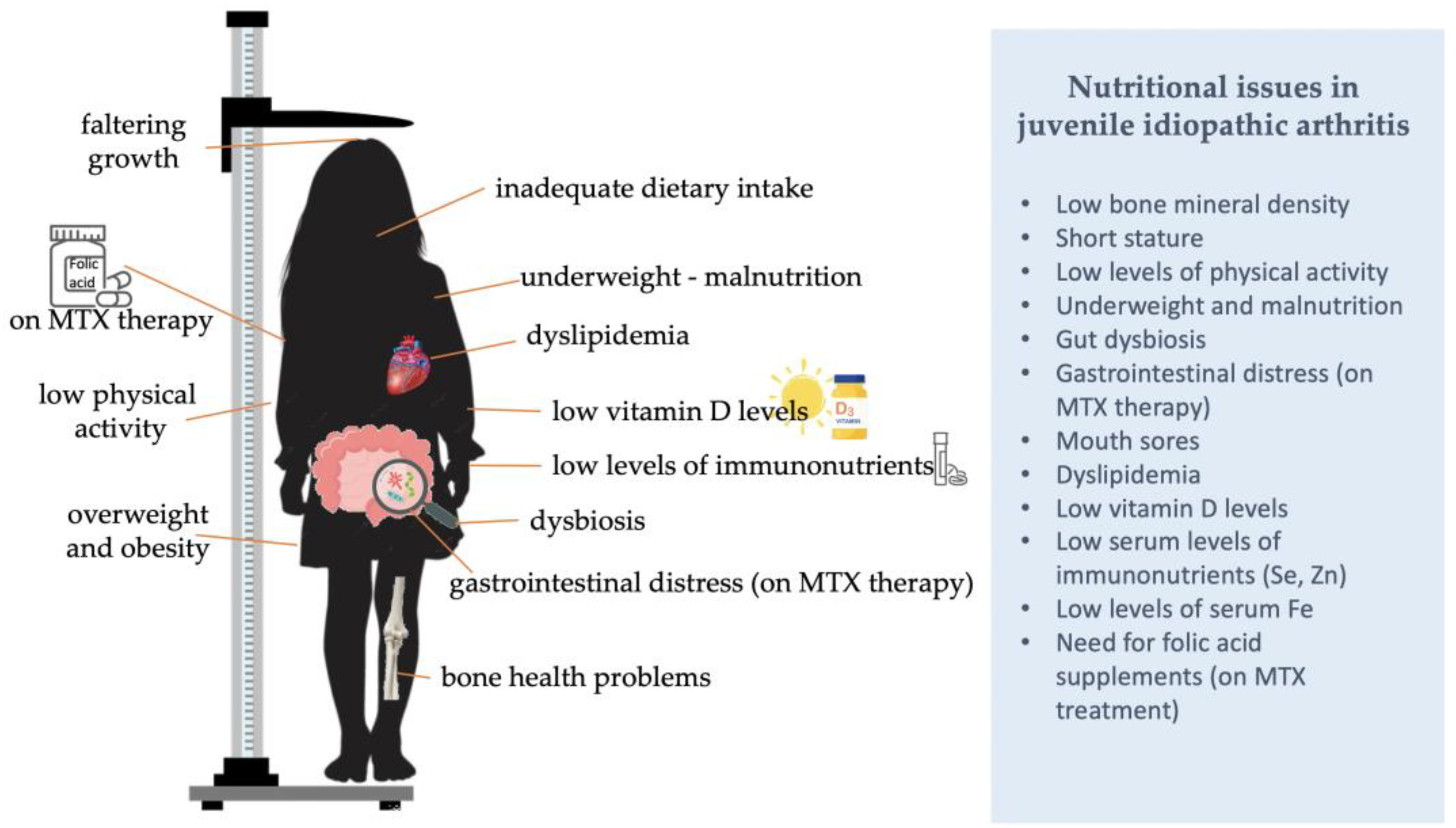
3. Malnutrition
3.1. Growth Delay in JIA
3.2. Undernutrition
3.3. Overweight and Obesity
4. Low Physical Activity (PA) Levels
5. Gastrointestinal Manifestations
6. Dyslipidemia
7. Bone Health
8. Energy Requirements and Dietary Intake
8.1. Studies Assessing Dietary Intake in Children with JIA

| First Author | Sample | Recruitment | Design | Dietary Assessment Method | Results |
|---|---|---|---|---|---|
| Amancio [139] | Cases: n = 41 patients with JRA Controls: n = 23 patients’ brothers | Pediatric Rheumatology Clinic, Universidade Federal de São Paulo/Escola Paulista de Medicina, Brazil | Case-control | Food Register | Cu and Zn intake were below the RDA. Compared to controls, serum Cu levels were higher, indicating that the number of inflamed joints is related to its variation. Zn levels did not differ in relation to control or disease characteristics. |
| Bacon [140] | Cases: n = 34 children with JRA Controls: n = 9 healthy controls | Rheumatology center of Chil- dren’s National Medical Center, Washington DC, USA | Case-control | Three-day dietary intake records | In patients with sJIA in particular, EI was suboptimal to a greater degree. The mean intake of vitamins and minerals for each subtype met the RDA, with the exceptions of Zn and vitamin E and Fe. |
| Caetano [69] | Cases: n = 48 pediatric patients with JIA Controls: n = 22 with JSLE | Pediatric Rheumatology Clinic, UNIFESPEPM, São Paulo, Brazil | Case-control | 24h diet recalls | An excessive intake of energy, protein, and lipids was observed in 12.5, 75, and 31.3% of the sample, respectively. On the other hand, 41.7, 8.3, and 31.3% of the patients reported consuming less energy, protein, and lipids than required. Fe intake was suboptimal in 29.2% of the sample, Ca in 62.5%, Zn and vitamin A in 87.5% and vitamin B6 in 64.6%. |
| Gorczyca [141] | Cases: n = 66 children with JIA Controls: n = 44 healthy controls | (i) Clinic of Paediatrics Rheumatology, Wroclaw Medical University, (ii) Department of Paediatric Pulmonology and Rheumatology, Medical University of Lublin, and (iii) John Paul II Paediatrics Centre, Poland | Case-control | Seven-day dietary record | No differences were noted in the EI from dietary fats and PUFA between children with JIA and controls. No differences were observed in the nutrient intake between sero-negative poly-JIA and oligo-JIA subtypes, or between those with active and inactive disease. |
| Grönlund [75] | Cases: n = 40 children with JIA Controls: n = 40 healthy controls | Department of Paediatrics, Turku University Hospital, Turku, Finland | Case-control | Seven-day food diaries | EI (kcal/day) was higher in patients with JIA. No differences were noted in the anthropometric measures, the energy and nutrient intakes between JIA subtypes, active and inactive disease. |
| Hari [142] | N = 33 patients with JIA | (i) Department of Rheumatology, El Ayachi University Hospital and (ii) Department of Pediatrics, University Hospital of children, Rabat-Sale, Morocco | Cross-sectional | 24h diet recall for seven consecutive days | A positive correlation was observed between LBM and dietary carbohydrate intake. |
| Haugen [143] | Cases: n = 15 patients with JIA Controls: n = 17 healthy controls | Oslo Sanitetsforening Rheumatism Hospital, Oslo, Norway | Case-control | Dietary records | Average daily intakes of protein, fat, vitamins and minerals were similar in all groups. EI/kg of body weight was higher in the polyarticular than in the pauciarticular type. The intake of thiamine, niacin, Fe, Ca and Zn was below the RDA in all groups. One patient with polyarticular and two with pauciarticular JIA abstained from milk, whereas all controls drank milk daily. |
| Lewis [144] | Cases: n = 10 teens with active JIA, n = 9 teens with JIA on clinical remission Controls: n = 20 healthy controls | Pediatric Rheumatology clinic, Texas TX, USA | Case-control | Three-day food records | Pain and carbohydrate intake were correlated among teens with active JIA. After controlling for sex, added sugars and carbohydrate intake predicted pain scores in active JIA. |
| López [43] | N = 91 children with JCA for at least a year | Unidad de Nutrición Infantil, Hospital Universitario Infantil “La Paz”, Madrid, Spain | Cohort (One year) | NR | In most cases, total EI suited WHO allowances and protein intake was high (15 ± 2% of total EI). Among the children who required an extra intake to reach an acceptable nutritional status, 54% did not suit their needs. |
| Mortensen [145] | N = 38 children with JCA | Children’s hospital, Camperdown, Australia | Cross-sectional | Seven day weighed food record | Mean EI was below the RDI among the systemic and polyarticular patients. Mean intakes of Ca and Zn were below the RDI in all of the polyarticular group. Thirteen (34%) children were taking self-prescribed vitamin and/or mineral ONS, with vitamin C being the most common. |
| Pereira [87] | Cases: n = 30 children with polyarticular JIA Controls: n = 41 children with JSLE and n = 20 children with DM and their parents | Department of Pediatrics, UNIFESPEPM, São Paulo, Brazil | Case-control | Three 24h dietary recalls | The average EI of children with JIA was 1721.3 ± 294.7 kcal/day. No difference was observed between the EI of parents and children. Regarding the consumption of protein, both parents and children consumed amounts exceeding the RDA, and a correlation was noted between parental and children’s consumption. |
| Rodrigues [68] | N = 62 children/adolescents with polyarticular or sJIA | Department of Pediatrics, UNIFESPEPM, São Paulo, Brazil | Cross-sectional | Three 24h food recalls | Median EI was 1933.6 kcal/day. None of the patients reported a carbohydrate intake exceeding RDA, although 4.9% had a protein intake above the RDA and 29% consumed more lipids than required. The median intake of SFA and trans fats was 24.4 g and 4.2 g, respectively. The majority (75.8%) of children exceeded the SFA recommendation and 79% exceeded the RDA for trans fats. |
8.1.1. Energy and Macronutrient Intake
8.1.2. Suboptimal Levels of Immunonutrients
8.1.3. The Curious Case of Ferritin
8.1.4. Fatty Acid Intake and Status
8.2. Considerations for the Assessment of Energy Requirements in JIA
8.3. Dietary Interventions for Children with JIA
8.3.1. Interventions Targeting Insufficient Vitamin D Levels
8.3.2. Interventions Delivering Supplementation/Dietary Intake with Fatty Acids
9. Conclusions
Author Contributions
Funding
Institutional Review Board Statement
Informed Consent Statement
Data Availability Statement
Conflicts of Interest
References
- Ravelli, A.; Martini, A. Juvenile idiopathic arthritis. Lancet 2007, 369, 767–778. [Google Scholar] [CrossRef] [PubMed] [Green Version]
- Barut, K.; Adrovic, A.; Şahin, S.; Kasapçopur, Ö. Juvenile Idiopathic Arthritis. Balk. Med. J. 2017, 34, 101. [Google Scholar] [CrossRef] [PubMed]
- McHugh, J. Global prevalence of JIA, JSLE and club foot. Nat. Rev. Rheumatol. 2020, 16, 408. [Google Scholar] [CrossRef]
- Thierry, S.; Fautrel, B.; Lemelle, I.; Guillemin, F. Prevalence and incidence of juvenile idiopathic arthritis: A systematic review. Jt. Bone Spine 2014, 81, 112–117. [Google Scholar] [CrossRef]
- Thatayatikom, A.; Modica, R.; Leucio, A. De Juvenile Idiopathic Arthritis; StatPearls: Tampa, FL, USA, 2022. [Google Scholar]
- Hayward, K.; Wallace, C.A. Recent developments in anti-rheumatic drugs in pediatrics: Treatment of juvenile idiopathic arthritis. Arthritis Res. Ther. 2009, 11, 216. [Google Scholar] [CrossRef] [Green Version]
- Giancane, G.; Ruperto, N. Treatment of juvenile idiopathic arthritis: What’s new? Curr. Opin. Rheumatol. 2019, 31, 428–435. [Google Scholar] [CrossRef]
- Warrier, K.C. A practical guide to using biologics in juvenile idiopathic arthritis. Paediatr. Child Health 2022, 32, 229–235. [Google Scholar] [CrossRef]
- Klein, A. Biologics in the treatment of juvenile idiopathic arthritis: A comparison of mono- and combination therapy with synthetic DMARDs. Z. Rheumatol. 2019, 78, 599–609. [Google Scholar] [CrossRef]
- Toquet, S.; Nguyen, Y.; Sabbagh, A.; Djerada, Z.; Boulagnon, C.; Bani-Sadr, F. Entéropathie sévère chez un patient traité par méthotrexate pour une polyarthrite rhumatoïde. Rev. Rhum. (Ed. Fr.) 2016, 83, 66–68. [Google Scholar] [CrossRef]
- Ozeki, T.; Fujita, Y. Asymptomatic colitis induced by low-dose methotrexate. BMJ Case Rep. 2016, 2016, bcr2016217771. [Google Scholar] [CrossRef]
- Asai, S.; Nagai, K.; Takahashi, N.; Watanabe, T.; Matsumoto, T.; Asai, N.; Sobue, Y.; Ishiguro, N.; Kojima, T. Influence of methotrexate on gastrointestinal symptoms in patients with rheumatoid arthritis. Int. J. Rheum. Dis. 2019, 22, 207–213. [Google Scholar] [CrossRef] [PubMed]
- Deller, D.J.; Urban, E.; Ibbotson, R.N.; Horwood, J.; Milazzo, S.; Robson, H.N. Folic-acid deficiency in rheumatoid arthritis: Relation of levels of serum folic-acid activity to treatment with phenylbutazone. Br. Med. J. 1966, 1, 765–767. [Google Scholar] [CrossRef] [PubMed] [Green Version]
- Morgan, S.L.; Baggott, J.E.; Lee, J.Y.; Alarcón, G.S. Folic acid supplementation prevents deficient blood folate levels and hyperhomocysteinemia during longterm, low dose methotrexate therapy for rheumatoid arthritis: Implications for cardiovascular disease prevention. J. Rheumatol. 1998, 25, 441–446. [Google Scholar]
- Bramley, D. What dose of folic acid to use with methotrexate in rheumatoid arthritis? Drug Ther. Bull. 2021, 59, 103–106. [Google Scholar] [CrossRef]
- Liu, L.; Liu, S.; Wang, C.; Guan, W.; Zhang, Y.; Hu, W.; Zhang, L.; He, Y.; Lu, J.; Li, T.; et al. Folate Supplementation for Methotrexate Therapy in Patients With Rheumatoid Arthritis: A Systematic Review. J. Clin. Rheumatol. 2019, 25, 197–202. [Google Scholar] [CrossRef]
- Grammatikopoulou, M.G.; Marakis, G.; Gkiouras, K.; Athanatou, D.; Maraki, M.I.; Bogdanos, D.P. Fly me to the immune: Immunonutrition in rheumatic diseases. Mediterr. J. Rheumatol. 2023, 33. [Google Scholar]
- Gonçalves, M.; D’Almeida, V.; Guerra-Shinohara, E.M.; Galdieri, L.C.; Len, C.A.; Hilário, M.O.E. Homocysteine and lipid profile in children with Juvenile Idiopathic Arthritis. Pediatr. Rheumatol. Online J. 2007, 5, 2. [Google Scholar] [CrossRef] [Green Version]
- Huemer, M.; Födinger, M.; Sailer-Höck, M.; Falger, J.; Rettenbacher, A.; Bernecker, M.; Artacker, G.; Kenzian, H.; Lang, T.; Stöckler-Ipsiroglu, S. Hyperhomocysteinemia in children with juvenile idiopathic arthritis is not influenced by methotrexate treatment and folic acid supplementation: A pilot study. Clin. Exp. Rheumatol. 2003, 21, 249–255. [Google Scholar]
- Alam, M.F.; Islam, M.M.; Haque, M.; Rahman, S.A. Serum homocysteine level in children with juvenile idiopathic arthritis. Bangladesh Med. Res. Counc. Bull. 2020, 46, 12–16. [Google Scholar] [CrossRef]
- Hayta, E.; Hizmetli, S.; Atalar, M.H.; Çinar, Z. Association of Plasma Homocysteine Level and Carotid Intima-Media Thickness in Rheumatoid Arthritis Patients Receiving Methotrexate. Arch. Rheumatol. 2015, 30, 214–220. [Google Scholar] [CrossRef]
- Głuszek, J.; Wierzowiecka, M.; Niklas, K.; Niklas, A. The importance of homocysteine in the development of cardiovascular complications in patients with rheumatoid arthritis. Reumatologia 2020, 58, 282. [Google Scholar] [CrossRef] [PubMed]
- Ringold, S.; Angeles-Han, S.T.; Beukelman, T.; Lovell, D.; Cuello, C.A.; Becker, M.L.; Colbert, R.A.; Feldman, B.M.; Ferguson, P.J.; Gewanter, H.; et al. 2019 American College of Rheumatology/Arthritis Foundation Guideline for the Treatment of Juvenile Idiopathic Arthritis: Therapeutic Approaches for Non-Systemic Polyarthritis, Sacroiliitis, and Enthesitis. Arthritis Care Res. 2019, 71, 717–734. [Google Scholar] [CrossRef] [Green Version]
- Ferrara, G.; Mastrangelo, G.; Barone, P.; La Torre, F.; Martino, S.; Pappagallo, G.; Ravelli, A.; Taddio, A.; Zulian, F.; Cimaz, R. Methotrexate in juvenile idiopathic arthritis: Advice and recommendations from the MARAJIA expert consensus meeting. Pediatr. Rheumatol. 2018, 16, 46. [Google Scholar] [CrossRef] [PubMed] [Green Version]
- Frittoli, R.B.; Longui, B.S.; Silva, A.M.; de Azevedo Barros Filho, A.; de Góes Monteiro, M.A.R.; Appenzeller, S. Effects of the use of growth hormone in children and adolescents with juvenile idiopathic arthritis: A systematic review. Rev. Bras. Reumatol. (Engl. Ed.) 2017, 57, 100–106. [Google Scholar] [CrossRef] [PubMed] [Green Version]
- Boros, C.; Whitehead, B. Juvenile idiopathic arthritis. Aust. Fam. Phycisian 2010, 39, 630–636. [Google Scholar]
- Kim, J.Y.; Kim, H.S.; Park, S.H. Disease flare after 7 year-remission of systemic type juvenile idiopathic arthritis: Is growth hormone therapy a culprit or innocent bystander? Int. J. Rheum. Dis. 2015, 3, 377–378. [Google Scholar] [CrossRef]
- Renier, G.; Clément, I.; Desfaits, A.C.; Lambert, A. Direct stimulatory effect of insulin-like growth factor-I on monocyte and macrophage tumor necrosis factor-alpha production. Endocrinology 1996, 137, 4611–4618. [Google Scholar] [CrossRef] [Green Version]
- Touati, G.; Prieur, A.M.; Ruiz, J.C.; Noel, M.; Czernichow, P. Beneficial Effects of One-Year Growth Hormone Administration to Children with Juvenile Chronic Arthritis on Chronic Steroid Therapy. I. Effects on Growth Velocity and Body Composition. J. Clin. Endocrinol. Metab. 1998, 83, 403–409. [Google Scholar] [CrossRef] [Green Version]
- Kim, S.-H.; Park, M.-J. Effects of growth hormone on glucose metabolism and insulin resistance in human. Ann. Pediatr. Endocrinol. Metab. 2017, 22, 145–152. [Google Scholar] [CrossRef] [Green Version]
- Vijayakumar, A.; Yakar, S.; LeRoith, D. The intricate role of growth hormone in metabolism. Front. Endocrinol. 2011, 2, 32. [Google Scholar] [CrossRef] [Green Version]
- Soybilgic, A.; Tesher, M.; Wagner-Weiner, L.; Onel, K.B. A survey of steroid-related osteoporosis diagnosis, prevention and treatment practices of pediatric rheumatologists in North America. Pediatr. Rheumatol. Online J. 2014, 12, 24. [Google Scholar] [CrossRef] [Green Version]
- Falcini, F.; Trapani, S.; Civinini, R.; Capone, A.; Ermini, M.; Bartolozzi, G. The primary role of steroids on the osteoporosis in juvenile rheumatoid patients evaluated by dual energy X-ray absorptiometry. J. Endocrinol. Investig. 1996, 19, 165–169. [Google Scholar] [CrossRef]
- Mehls, O.; Himmele, R.; Homme, M.; Kiepe, D.; Klaus, O. The interaction of glucocorticoids with the growth hormone-insulin-like growth factor axis and its effects on growth plate chondrocytes and bone cells. J. Pediatr. Endocrinol. Metab. 2001, 14, 1475–1482. [Google Scholar]
- Miyamae, T.; Yokoya, S.; Yamanaka, H.; Yokota, S. Effect of Tocilizumab on growth impairment in systemic juvenile idiopathic arthritis with long-term corticosteroid therapy. Mod. Rheumatol. 2014, 24, 567–571. [Google Scholar] [CrossRef]
- Simon, D.; Fernando, C.; Czernichow, P.; Prieur, A.-M. Linear growth and final height in patients with systemic juvenile idiopathic arthritis treated with longterm glucocorticoids. J. Rheumatol. 2002, 29, 1296–1300. [Google Scholar]
- Machado, S.H.; Xavier, R.M.; Lora, P.S.; Gonçalves, L.M.K.; Trindade, L.R.; Marostica, P.J.C. Height and sexual maturation in girls with juvenile idiopathic arthritis. J. Pediatr. 2020, 96, 100–107. [Google Scholar] [CrossRef]
- Li, J.X.; Cummins, C.L. Fresh insights into glucocorticoid-induced diabetes mellitus and new therapeutic directions. Nat. Rev. Endocrinol. 2022, 18, 540–557. [Google Scholar] [CrossRef]
- Szabłowski, M.; Okruszko, M.A.; Pochodowicz, K.; Abramowicz, P.; Konstantynowicz, J.; Bossowski, A.; Głowińska-Olszewska, B. Coincidence of juvenile idiopathic arthritis and type 1 diabetes: A case-based review. Rheumatol. Int. 2022, 42, 378. [Google Scholar] [CrossRef]
- Suppasit, P.; Vilaiyuk, S.; Poomthavorn, P.; Pongratanakul, S.; Khlairit, P.; Mahachoklertwattana, P. Glucose metabolism in systemic juvenile idiopathic arthritis. Pediatr. Rheumatol. 2022, 20, 55. [Google Scholar] [CrossRef]
- de Onis, M.; Garza, C.; Onyango, A.W.; Rolland-Cachera, M.F. [WHO growth standards for infants and young children]. Arch. Pediatr. 2009, 16, 47–53. [Google Scholar] [CrossRef]
- Knops, N.; Wulffraat, N.; Lodder, S.; Houwen, R.; de Meer, K. Resting energy expenditure and nutritional status in children with juvenile rheumatoid arthritis. J. Rheumatol. 1999, 26, 2039–2043. [Google Scholar] [PubMed]
- López, M.A.; More, L.R.A.; Molina, García-Consuegra, J.; Merino, M.R. Energy requirements and dietary intake in a pediatric population with juvenile chronic arthritis. J. Pediatr. Gastroenterol. Nutr. 2005, 40, 700. [Google Scholar] [CrossRef]
- Umławska, W.; Prusek-Dudkiewicz, A. Growth retardation and delayed puberty in children and adolescents with juvenile idiopathic arthritis. Arch. Med. Sci. 2010, 6, 19–23. [Google Scholar] [CrossRef] [PubMed]
- Zak, M.; Müller, J.; Karup Pedersen, F. Final Height, Armspan, Subischial Leg Length and Body Proportions in Juvenile Chronic Arthritis. Horm. Res. Paediatr. 1999, 52, 80–85. [Google Scholar] [CrossRef] [PubMed]
- Saha, M.T.; Verronen, P.; Laippala, P.; Lenko, H.L. Growth of prepubertal children with juvenile chronic arthritis. Acta Paediatr. Int. J. Paediatr. 1999, 88, 724–728. [Google Scholar] [CrossRef]
- d’Angelo, D.M.; Di Donato, G.; Breda, L.; Chiarelli, F. Growth and puberty in children with juvenile idiopathic arthritis. Pediatr. Rheumatol. 2021, 19, 28. [Google Scholar] [CrossRef]
- El Badri, D.; Rostom, S.; Bouaddi, I.; Hassani, A.; Chkirate, B.; Amine, B.; Hajjaj-Hassouni, N. Sexual maturation in Moroccan patients with juvenile idiopathic arthritis. Rheumatol. Int. 2014, 34, 665–668. [Google Scholar] [CrossRef]
- Umławska, W.; Prusek-Dudkiewicz, A. [Short stature of children suffering from certain chronic diseases]. Pediatr. Endocrinol. Diabetes Metab. 2007, 13, 135–138. [Google Scholar]
- Bechtold, S.; Beyerlein, A.; Ripperger, P.; Roeb, J.; Dalla Pozza, R.; Häfner, R.; Haas, J.P.; Schmidt, H. Total pubertal growth in patients with juvenile idiopathic arthritis treated with growth hormone: Analysis of a single center. Growth Horm. IGF Res. 2012, 22, 180–185. [Google Scholar] [CrossRef]
- David, H.; Aupiais, C.; Louveau, B.; Quartier, P.; Jacqz-Aigrain, E.; Carel, J.C.; Simon, D. Growth outcomes after GH therapy of patients given long-term corticosteroids for juvenile idiopathic arthritis. J. Clin. Endocrinol. Metab. 2017, 102, 4578–4587. [Google Scholar] [CrossRef] [Green Version]
- De Zegher, F.; Reynaert, N.; De Somer, L.; Wouters, C.; Roelants, M. Growth Failure in Children with Systemic Juvenile Idiopathic Arthritis and Prolonged Inflammation despite Treatment with Biologicals: Late Normalization of Height by Combined Hormonal Therapies. Horm. Res. Paediatr. 2019, 90, 337–343. [Google Scholar] [CrossRef]
- Kearsley-Fleet, L.; Hyrich, K.L.; Davies, R.; Lunt, M.; Southwood, T.R. Growth in children and adolescents with juvenile idiopathic arthritis over 2 years of treatment with etanercept: Results from the British Society for Paediatric and Adolescent Rheumatology Etanercept Cohort Study. Rheumatology 2015, 54, 1279–1285. [Google Scholar] [CrossRef] [Green Version]
- Lofthouse, C.M.; Azad, F.; Baildam, E.M.; Akobeng, A.K. Measuring the nutritional status of children with juvenile idiopathic arthritis using the bioelectrical impedance method. Rheumatology 2002, 41, 1172–1177. [Google Scholar] [CrossRef] [Green Version]
- McErlane, F.; Carrasco, R.; Kearsley-Fleet, L.; Baildam, E.M.; Wedderburn, L.R.; Foster, H.E.; Ioannou, Y.; Chieng, S.E.A.; Davidson, J.E.; Thomson, W.; et al. Growth patterns in early juvenile idiopathic arthritis: Results from the Childhood Arthritis Prospective Study (CAPS). Semin. Arthritis Rheum. 2018, 48, 53–60. [Google Scholar] [CrossRef]
- Polito, C.; Strano, C.G.; Olivieri, A.N.; Alessio, M.; Iammarrone, C.S.; Todisco, N.; Papale, M.R. Growth retardation in non-steroid treated juvenile rheumatoid arthritis. Scand. J. Rheumatol. 1997, 26, 99–103. [Google Scholar] [CrossRef]
- Simon, D.; Lucidarme, N.; Prieur, A.M.; Ruiz, J.C.; Czernichow, P. Linear growth in children suffering from juvenile idiopathic arthritis requiring steroid therapy: Natural history and effects of growth hormone treatment on linear growth. J. Pediatr. Endocrinol. Metab. 2001, 14, 1483–1486. [Google Scholar]
- Simon, D.; Lucidarme, N.; Prieur, A.-M.; Ruiz, J.C.; Czernichow, P. Effects on growth and body composition of growth hormone treatment in children with juvenile idiopathic arthritis requiring steroid therapy. J. Rheumatol. 2003, 30, 2492–2499. [Google Scholar]
- Uettwiller, F.; Perlbarg, J.; Pinto, G.; Bader-Meunier, B.; Mouy, R.; Compeyrot-Lacassagne, S.; Melki, I.; Wouters, C.; Prieur, A.M.; Landais, P.; et al. Effect of biologic treatments on growth in children with juvenile idiopathic arthritis. J. Rheumatol. 2014, 41, 128–135. [Google Scholar] [CrossRef] [Green Version]
- Uettwiller, F.; Pinto, G.; Polak, M.; Quartier, P. Impact of biologics on growth in children with juvenile idiopathic arthritis. Pediatr. Rheumatol. 2011, 9, P139. [Google Scholar] [CrossRef] [Green Version]
- Wang, S.J.; Yang, Y.H.; Lin, Y.T.; Yang, C.M.; Chiang, B.L. Attained adult height in juvenile rheumatoid arthritis with or without corticosteroid treatment. Clin. Rheumatol. 2002, 21, 363–368. [Google Scholar] [CrossRef]
- Simon, D. rhGH Treatment in Corticosteroid-Treated Patients. Horm. Res. Paediatr. 2007, 68, 38–45. [Google Scholar] [CrossRef] [PubMed]
- Świdrowska, J.; Zygmunt, A.; Biernacka-Zielińska, M.; Stańczyk, J.; Smolewska, E. Influence of biologic therapy on growth in children with chronic inflammatory connective tissue diseases. Reumatologia 2015, 53, 14–20. [Google Scholar] [CrossRef] [PubMed]
- Simon, D. Management of growth retardation in juvenile idiopathic arthritis. Horm. Res. 2007, 68 (Suppl. 5), 122–125. [Google Scholar] [CrossRef]
- Simon, D.; Bechtold, S. Effects of growth hormone treatment on growth in children with juvenile idiopathic arthritis. Horm. Res. 2009, 72, 55–59. [Google Scholar] [CrossRef] [PubMed]
- Neto, A.; Mourão, A.; Oliveira-Ramos, F.; Campanilho-Marques, R.; Estanqueiro, P.; Salgado, M.; Guedes, M.; Piotto, D.; Emi Aikawa, N.; Melo Gomes, J.; et al. Association of body mass index with Juvenile Idiopathic Arthritis disease activity: A Portuguese and Brazilian collaborative analysis. Acta Rheumatol. Port. 2021, 46, 7–14. [Google Scholar]
- Cleary, A.G. Nutritional impairment in juvenile idiopathic arthritis. Rheumatology 2004, 43, 1569–1573. [Google Scholar] [CrossRef] [Green Version]
- Rodrigues, W.D.R.; Sarni, R.O.S.; Fonseca, F.L.A.; Araújo, A.; Len, C.A.; Terreri, M.T. Biomarkers of lipid metabolism in patients with juvenile idiopathic arthritis: Relationship with disease subtype and inflammatory activity. Pediatr. Rheumatol. 2021, 19, 66. [Google Scholar] [CrossRef]
- Caetano, M.C.; Ortiz, T.T.; Terreri, M.T.S.L.R.; Sarni, R.O.S.; Silva, S.G.L.; Souza, F.I.S.; Hilário, M.O.E. Inadequate dietary intake of children and adolescents with juvenile idiopathic arthritis and systemic lupus erythematosus. J. Pediatr. 2009, 85, 509–515. [Google Scholar] [CrossRef] [Green Version]
- Wiȩch, P.; Sałacińska, I.; Bazaliński, D.; Dabrowski, M. Body composition and phase angle as an indicator of nutritional status in children with juvenile idiopathic arthritis. Pediatr. Rheumatol. 2018, 16, 82. [Google Scholar] [CrossRef] [Green Version]
- Kumar, S.; Dutt, A.; Hemraj, S.; Bhat, S.; Manipadybhima, B. Phase Angle Measurement in Healthy Human Subjects through Bio-Impedance Analysis. Iran. J. Basic Med. Sci. 2012, 15, 1184. [Google Scholar]
- Schulz, K.-H.; Patra, S.; Spielmann, H.; Klapdor, S.; Schlüter, K.; van Eckert, S. Physical condition, nutritional status, fatigue, and quality of life in oncological out-patients. SAGE Open Med. 2017, 5, 205031211774367. [Google Scholar] [CrossRef]
- Matrins, V.J.B.; Toledo Florêncio, T.M.M.; Grillo, L.P.; do Franco, M.C.P.; Martins, P.A.; Clemente, A.P.G.; Santos, C.D.L.; de Vieria, M.F.A.; Sawaya, A.L. Long-Lasting Effects of Undernutrition. Int. J. Environ. Res. Public Health 2011, 8, 1846. [Google Scholar] [CrossRef] [Green Version]
- Galler, J.R.; Bringas-Vega, M.L.; Tang, Q.; Rabinowitz, A.G.; Musa, K.I.; Chai, W.J.; Omar, H.; Abdul Rahman, M.R.; Abd Hamid, A.I.; Abdullah, J.M.; et al. Neurodevelopmental effects of childhood malnutrition: A neuroimaging perspective. Neuroimage 2021, 231, 117828. [Google Scholar] [CrossRef]
- Grönlund, M.M.; Kaartoaho, M.; Putto-Laurila, A.; Laitinen, K. Juvenile idiopathic arthritis patients with low inflammatory activity have increased adiposity. Scand. J. Rheumatol. 2014, 43, 488–492. [Google Scholar] [CrossRef]
- Caetano, M.C.; Sarni, R.O.S.; Terreri, M.T.L.; Ortiz, T.T.; Pinheiro, M.; De Souza, F.I.S.; Hilário, M.O. Excess of adiposity in female children and adolescents with juvenile idiopathic arthritis. Clin. Rheumatol. 2012, 31, 967–971. [Google Scholar] [CrossRef]
- Markula-Patjas, K.P.; Ivaska, K.K.; Pekkinen, M.; Andersson, S.; Moilanen, E.; Viljakainen, H.T.; Mäkitie, O. High adiposity and serum leptin accompanied by altered bone turnover markers in severe juvenile idiopathic arthritis. J. Rheumatol. 2014, 41, 2474–2481. [Google Scholar] [CrossRef]
- De Zanette, C.A.; Machado, S.H.; Brenol, J.C.T.; Xavier, R.M. Metabolic syndrome and juvenile idiopathic arthritis. Rev. Bras. Reumatol. 2010, 50, 190–204. [Google Scholar] [CrossRef] [Green Version]
- Held, M.; Sestan, M.; Jelusic, M. Obesity as a comorbidity in children and adolescents with autoimmune rheumatic diseases. Rheumatol. Int. 2022, in press. [Google Scholar] [CrossRef]
- Amine, B.; Ibn Yacoub, Y.; Rostom, S.; Hajjaj-Hassouni, N. Prevalence of overweight among Moroccan children and adolescents with juvenile idiopathic arthritis. Jt. Bone Spine 2011, 78, 584–586. [Google Scholar] [CrossRef]
- Giani, T.; De Masi, S.; Maccora, I.; Tirelli, F.; Simonini, G.; Falconi, M.; Cimaz, R. The Influence of Overweight and Obesity on Treatment Response in Juvenile Idiopathic Arthritis. Front. Pharmacol. 2019, 10, 637. [Google Scholar] [CrossRef]
- Milatz, F.; Klotsche, J.; Niewerth, M.; Hörstermann, J.; Windschall, D.; Weller-Heinemann, F.; Dressler, F.; Berendes, R.; Haas, J.P.; Horneff, G.; et al. POS1309 Prevalence and Correlates of underweight, overweight and obesity among patients with juvenile idiopathic arthritis (JIA): Evidence from the National Paediatric Rheumatologic Database (NPRD). Ann. Rheum. Dis. 2021, 80, 936–937. [Google Scholar] [CrossRef]
- Pelajo, C.F.; Lopez-Benitez, J.M.; Miller, L.C. Obesity and disease activity in juvenile idiopathic arthritis. Pediatr. Rheumatol. 2012, 10, 3. [Google Scholar] [CrossRef] [PubMed] [Green Version]
- Diaz-Cordovés Rego, G.; Núñez-cuadros, E.; Mena-vázquez, N.; Aguado Henche, S.; Galindo-Zavala, R.; Manrique-Arija, S.; Martín-Pedraz, L.; Redondo-Rodríguez, R.; Godoy-Navarrete, F.J.; Fernández-Nebro, A. Adiposity Is Related to Inflammatory Disease Activity in Juvenile Idiopathic Arthritis. J. Clin. Med. 2021, 10, 3949. [Google Scholar] [CrossRef] [PubMed]
- Schnabel, A.; Unger, E.; Bruck, N.; Holl-Wieden, A.; Morbach, H.; Berner, R.; Leszczynska, A.; Hedrich, C.; Sellami, M.; Fazaa, A.; et al. FRI0575 Prevalence of overweight and obesity in children with juvenile idiopathic arthritis. Ann. Rheum. Dis. 2019, 78, 983. [Google Scholar] [CrossRef] [Green Version]
- Schenck, S.; Niewerth, M.; Sengler, C.; Trauzeddel, R.; Thon, A.; Minden, K.; Klotsche, J. Prevalence of overweight in children and adolescents with juvenile idiopathic arthritis. Scand. J. Rheumatol. 2015, 44, 288–295. [Google Scholar] [CrossRef]
- Pereira, L.; Previdelli, Á.N.; Rossi, R.G.D.T.; Rodrigues, W.D.; Fonseca, F.L.A.; Len, C.A.; Terreri, M.T.; Saccardo Sarni, R.O. Anthropometric Evaluation and Assessment of Food Intake of Parents of Pediatric Patients with Chronic Rheumatic Diseases. Ann. Nutr. Metab. 2020, 76, 387–395. [Google Scholar] [CrossRef]
- Głowińska-Olszewska, B.; Dobreńko, E.; Hryniewicz, A.; Tołwińska, J.; Żelazowska-Rutkowska, B.; Wojciuk, M.; Łuczyński, W.; Piotrowska-Jastrzębska, J.; Bossowski, A.; Pediatrii, K.; et al. Influence of obesity on the early atherosclerosis in children with juvenile idiopathic arthritis. Reumatologia/Rheumatology 2011, 49, 231–238. [Google Scholar]
- Nesbitt, C.; Kuntze, G.; Toomey, C.; Esau, S.; Brooks, J.; Mosher, D.; Twilt, M.; Nettel-Aguirre, A.; Palacios-Derflingher, L.M.; Ronsky, J.; et al. Secondary consequences of juvenile idiopathic arthritis in children and adolescents with knee involvement: Physical activity, adiposity, fitness, and functional performance. Rheumatol. Int. 2022, 42, 319–327. [Google Scholar] [CrossRef]
- Risum, K.; Edvardsen, E.; Godang, K.; Selvaag, A.M.; Hansen, B.H.; Molberg, Ø.; Bollerslev, J.; Holm, I.; Dagfinrud, H.; Sanner, H. Physical Fitness in Patients With Oligoarticular and Polyarticular Juvenile Idiopathic Arthritis Diagnosed in the Era of Biologics: A Controlled Cross-Sectional Study. Arthritis Care Res. 2019, 71, 1611–1620. [Google Scholar] [CrossRef]
- Rochette, E.; Bourdier, P.; Pereira, B.; Echaubard, S.; Borderon, C.; Caron, N.; Chausset, A.; Courteix, D.; Fel, S.; Kanold, J.; et al. Impaired muscular fat metabolism in juvenile idiopathic arthritis in inactive disease. Front. Physiol. 2019, 10, 528. [Google Scholar] [CrossRef]
- Kuntze, G.; Nesbitt, C.; Nettel-Aguirre, A.; Esau, S.; Scholz, R.; Brooks, J.; Twilt, M.; Toomey, C.; Mosher, D.; Ronsky, J.L.; et al. Gait Adaptations in Youth With Juvenile Idiopathic Arthritis. Arthritis Care Res. 2020, 72, 917–924. [Google Scholar] [CrossRef]
- Booth, F.W.; Roberts, C.K.; Laye, M.J. Lack of exercise is a major cause of chronic diseases. Compr. Physiol. 2012, 2, 1211. [Google Scholar] [CrossRef] [Green Version]
- Posadzki, P.; Pieper, D.; Bajpai, R.; Makaruk, H.; Könsgen, N.; Neuhaus, A.L.; Semwal, M. Exercise/physical activity and health outcomes: An overview of Cochrane systematic reviews. BMC Public Health 2020, 20, 1724. [Google Scholar] [CrossRef]
- Bohr, A.H.; Fuhlbrigge, R.C.; Pedersen, F.K.; de Ferranti, S.D.; Müller, K. Premature subclinical atherosclerosis in children and young adults with juvenile idiopathic arthritis. A review considering preventive measures. Pediatr. Rheumatol. Online J. 2016, 14, 3. [Google Scholar] [CrossRef] [Green Version]
- De Filippo, C.; Di Paola, M.; Giani, T.; Tirelli, F.; Cimaz, R. Gut microbiota in children and altered profiles in juvenile idiopathic arthritis. J. Autoimmun. 2019, 98, 1–12. [Google Scholar] [CrossRef]
- Pichler, J.; Ong, C.; Shah, N.; Sebire, N.; Kiparrissi, F.; Borrelli, O.; Pilkington, C.; Elawad, M. Histopathological features of gastrointestinal mucosal biopsies in children with juvenile idiopathic arthritis. Pediatr. Res. 2016, 79, 895–901. [Google Scholar] [CrossRef] [Green Version]
- Gonzalez, T.; Malagon, C.; Guarnizo, P.; Mosquera, A.C.; Chila-Moreno, L.; Romero-Sanchez, C. Autoantibodies and Gastrointestinal Symptoms in Colombian Children with Juvenile Idiopathic Arthritis. Curr. Rheumatol. Rev. 2018, 14, 163–171. [Google Scholar] [CrossRef]
- Weber, P.; Brune, T.; Ganser, G.; Zimmer, K.P. Gastrointestinal symptoms and permeability in patients with juvenile idiopathic arthritis. Clin. Exp. Rheumatol. 2003, 21, 657–662. [Google Scholar]
- Nuñez, E.; Galindo, R.; Lopez, E.; Diaz, G.; Vicioso, M.I.; Sierra, C. Coeliac disease and intestinal inflammation in juvenile idiopathic arthritis. Pediatr. Rheumatol. 2011, 9, P168. [Google Scholar] [CrossRef] [Green Version]
- Rebane, K.; Tuomi, A.-K.; Kautiainen, H.; Peltoniemi, S.; Glerup, M.; Aalto, K. Abdominal pain in Finnish young adults with juvenile idiopathic arthritis. Scand. J. Gastroenterol. 2022, 57, 1189–1194. [Google Scholar] [CrossRef]
- Matei, D.E.; Menon, M.; Alber, D.G.; Smith, A.M.; Nedjat-Shokouhi, B.; Fasano, A.; Magill, L.; Duhlin, A.; Bitoun, S.; Gleizes, A.; et al. Intestinal barrier dysfunction plays an integral role in arthritis pathology and can be targeted to ameliorate disease. Med 2021, 2, 864–883.e9. [Google Scholar] [CrossRef] [PubMed]
- Aalto, K.; Lahdenne, P.; Kolho, K.L. Fecal calprotectin in juvenile idiopathic arthritis patients related to drug use. Pediatr. Rheumatol. 2017, 15, 9. [Google Scholar] [CrossRef] [PubMed] [Green Version]
- Tibble, J.A.; Sigthorsson, G.; Foster, R.; Scott, D.; Fagerhol, M.K.; Roseth, A.; Bjarnason, I. High prevalence of NSAID enteropathy as shown by a simple faecal test. Gut 1999, 45, 362–366. [Google Scholar] [CrossRef] [PubMed]
- Lamot, L.; Miler, M.; Vukojević, R.; Vidović, M.; Lamot, M.; Trutin, I.; Gabaj, N.N.; Harjaček, M. The Increased Levels of Fecal Calprotectin in Children With Active Enthesitis Related Arthritis and MRI Signs of Sacroiliitis: The Results of a Single Center Cross-Sectional Exploratory Study in Juvenile Idiopathic Arthritis Patients. Front. Med. 2021, 8, 650619. [Google Scholar] [CrossRef] [PubMed]
- Maller, J.; Fox, E.; Park, K.T.; Paul, S.S.; Baszis, K.; Borocco, C.; Prahalad, S.; Quartier, P.; Reinhardt, A.; Schonenberg-Meinema, D.; et al. Inflammatory bowel disease in children with systemic juvenile idiopathic arthritis. J. Rheumatol. 2021, 48, 567–574. [Google Scholar] [CrossRef]
- Zeng, L.; Deng, Y.; He, Q.; Yang, K.; Li, J.; Xiang, W.; Liu, H.; Zhu, X.; Chen, H. Safety and efficacy of probiotic supplementation in 8 types of inflammatory arthritis: A systematic review and meta-analysis of 34 randomized controlled trials. Front. Immunol. 2022, 13, 5186. [Google Scholar] [CrossRef]
- Zaiss, M.M.; Joyce Wu, H.J.; Mauro, D.; Schett, G.; Ciccia, F. The gut–joint axis in rheumatoid arthritis. Nat. Rev. Rheumatol. 2021, 17, 224–237. [Google Scholar] [CrossRef]
- Barsalou, J.; Bradley, T.J.; Silverman, E.D. Cardiovascular risk in pediatric-onset rheumatological diseases. Arthritis Res. Ther. 2013, 15, 212. [Google Scholar] [CrossRef] [Green Version]
- Yeh, K.W.; Lee, C.M.; Chang, C.J.; Lin, Y.J.; Huang, J.L. Lipid Profiles Alter from Pro-Atherogenic into Less Atherogenic and Proinflammatory in Juvenile Idiopathic Arthritis Patients Responding to Anti TNF-α Treatment. PLoS ONE 2014, 9, e90757. [Google Scholar] [CrossRef] [Green Version]
- Bogmat, L.F.; Nikonova, V.V.; Shevchenko, N.S.; Bessonova, I.M. Dyslipidemia as a comorbid condition in children with juvenile idiopathic arthritis. Child’s Health 2020, 15, 279–286. [Google Scholar] [CrossRef]
- Marangoni, R.G.; Hayata, A.L.; Borba, E.F.; Azevedo, P.M.; Bonfá, E.; Goldenstein-Schainberg, C. Decreased high-density lipoprotein cholesterol levels in polyarticular juvenile idiopathic arthritis. Clinics 2011, 66, 1549–1552. [Google Scholar] [CrossRef] [Green Version]
- Krasnopolskaya, A.; Balykova, L.; Shekina, N.; Soldatov, Y.; Pomerantceva, S. Dyslipidemia as an atherogenic factor in patients with different forms of juvenile arthritis. Russ. Pediatr. J. 2021, 2, 31. [Google Scholar]
- Rodrigues, W.D.R.; Sarni, R.O.S.; Abad, T.T.O.; da Silva, S.G.L.; de Souza, F.I.S.; Len, C.A.; Terreri, M.T. Lipid profile of pediatric patients with chronic rheumatic diseases—A retrospective analysis. Rev. Assoc. Med. Bras. 2020, 66, 1093–1099. [Google Scholar] [CrossRef]
- Sun, D.M.; Ding, Y.; Zhnag, Y.; Xia, K. [Serum lipid profile in children with different subtypes of juvenile idiopathic arthritis]. Zhongguo Dang Dai Er Ke Za Zhi 2019, 21, 547–551. [Google Scholar] [CrossRef]
- Gicchino, M.F.; Marzuillo, P.; Zarrilli, S.; Melone, R.; Guarino, S.; Miraglia del Giudice, E.; Olivieri, A.N.; Di Sessa, A. Uric acid could be a marker of cardiometabolic risk and disease severity in children with juvenile idiopathic arthritis. Eur. J. Pediatr. 2022, 182, 149–154. [Google Scholar] [CrossRef]
- Mani, P.; Uno, K.; Duong, M.; Wolski, K.; Spalding, S.; Elaine Husni, M.; Nicholls, S.J. HDL function and subclinical atherosclerosis in juvenile idiopathic arthritis. Cardiovasc. Diagn. Ther. 2016, 6, 343. [Google Scholar] [CrossRef]
- Torelli, W.; Ross, J.; Quinn, T.; Erickson, K.; Soliman, A.; Harit, A. Lehigh Valley Health Network Screening and Treating Hyperlipidemia in Patient’s on Tofacitinib, Tocilizumab, Sarilumab, and Baricitinib. Arthritis Rheumatol 2019, 71. Available online: https://acrabstracts.org/abstract/screening-and-treating-hyperlipidemia-in-patients-on-tofacitinib-tocilizumab-sarilumab-and-baricitinib/ (accessed on 10 December 2022).
- Agca, R.; Heslinga, S.C.; Rollefstad, S.; Heslinga, M.; McInnes, I.B.; Peters, M.J.L.; Kvien, T.K.; Dougados, M.; Radner, H.; Atzeni, F.; et al. EULAR recommendations for cardiovascular disease risk management in patients with rheumatoid arthritis and other forms of inflammatory joint disorders: 2015/2016 update. Ann. Rheum. Dis. 2017, 76, 17–28. [Google Scholar] [CrossRef] [Green Version]
- Di Marcello, F.; Di Donato, G.; D’angelo, D.M.; Breda, L.; Chiarelli, F. Bone Health in Children with Rheumatic Disorders: Focus on Molecular Mechanisms, Diagnosis, and Management. Int. J. Mol. Sci. 2022, 23, 5725. [Google Scholar] [CrossRef]
- Sušić, G.; Pilipović, N.; Stojanović, R. [Bone mineral density in patients with juvenile idiopathic arthritis]. Srp. Arh. Celok. Lek. 2009, 137, 396–401. [Google Scholar] [CrossRef]
- Ota, Y.; Kaneko, Y.; Takeuchi, T.; Schmiedl, A.; Reuter, S.; Lerner, A.; Brunner, S.; Schwarting, A.; Matthias, T. AB1356 Age peculiarities of bone mineral density in young female with juvenile idiopathic arthritis. Ann. Rheum. Dis. 2018, 77, 1765–1766. [Google Scholar] [CrossRef]
- Shin, J.; Kang, M.J.; Kim, K.N. Prevalence of Lower Bone Mineral Density and Its Associated Factors in Korean Children and Adolescents with Juvenile Idiopathic Arthritis. J. Rheum. Dis. 2018, 25, 248–254. [Google Scholar] [CrossRef] [Green Version]
- Burnham, J.M.; Shults, J.; Dubner, S.E.; Sembhi, H.; Zemel, B.S.; Leonard, M.B. Bone Density, Structure, and Strength in Juvenile Idiopathic Arthritis: Importance of Disease Severity and Muscle Deficits. Arthritis Rheum. 2008, 58, 2527. [Google Scholar] [CrossRef] [PubMed] [Green Version]
- Stagi, S.; Cavalli, L.; Signorini, C.; Bertini, F.; Cerinic, M.M.; Brandi, M.L.; Falcini, F. Bone mass and quality in patients with juvenile idiopathic arthritis: Longitudinal evaluation of bone-mass determinants by using dual-energy x-ray absorptiometry, peripheral quantitative computed tomography, and quantitative ultrasonography. Arthritis Res. Ther. 2014, 16, R83. [Google Scholar] [CrossRef] [PubMed] [Green Version]
- Rooney, M.; Davies, U.M.; Reeve, J.; Preece, M.; Ansell, B.M.; Woo, P.M.M. Bone mineral content and bone mineral metabolism: Changes after growth hormone treatment in juvenile chronic arthritis. J. Rheumatol. 2000, 27, 1073–1081. [Google Scholar]
- Carrasco, R.; Lovell, D.J.; Giannini, E.H.; Henderson, C.J.; Huang, B.; Kramer, S.; Ranz, J.; Heubi, J.; Glass, D. Biochemical markers of bone turnover associated with calcium supplementation in children with juvenile rheumatoid arthritis: Results of a double-blind, placebo-controlled intervention trial. Arthritis Rheum. 2008, 58, 3932–3940. [Google Scholar] [CrossRef] [Green Version]
- Hillman, L.S.; Cassidy, J.T.; Chanetsa, F.; Hewett, J.E.; Higgins, B.J.; Robertson, J.D. Percent true calcium absorption, mineral metabolism, and bone mass in children with arthritis: Effect of supplementation with vitamin D3 and calcium. Arthritis Rheum. 2008, 58, 3255–3263. [Google Scholar] [CrossRef]
- Lovell, D.J.; Glass, D.; Ranz, J.; Kramer, S.; Huang, B.; Sierra, R.I.; Henderson, C.J.; Passo, M.; Graham, B.; Bowyer, S.; et al. A randomized controlled trial of calcium supplementation to increase bone mineral density in children with juvenile rheumatoid arthritis. Arthritis Rheum. 2006, 54, 2235–2242. [Google Scholar] [CrossRef]
- Stark, L.J.; Davis, A.M.G.; Janicke, D.M.; Mackner, L.M.; Hommel, K.A.; Bean, J.A.; Lovell, D.; Heubi, J.E.; Kalkwarf, H.J. A randomized clinical trial of dietary calcium to improve bone accretion in children with juvenile rheumatoid arthritis. J. Pediatr. 2006, 148, 501–507. [Google Scholar] [CrossRef]
- Tang, T.; Zhang, Y.; Luo, C.; Liu, M.; Xu, L.; Tang, X. Adjunctive vitamin D for the treatment of active juvenile idiopathic arthritis: An open-label, prospective, randomized controlled trial. Exp. Ther. Med. 2019, 18, 4921–4926. [Google Scholar] [CrossRef] [Green Version]
- Warady, B.D.; Lindsley, C.B.; Robinson, D.G.; Lukert, B.P. Effects of nutritional supplementation on bone mineral status of children with rheumatic diseases receiving corticosteroid therapy—PubMed. J. Rheumatol. 1994, 21, 530–535. [Google Scholar]
- Wey, H.E.; Binkley, T.L.; Beare, T.M.; Wey, C.L.; Specker, B.L. Cross-Sectional versus Longitudinal Associations of Lean and Fat Mass with pQCT Bone Outcomes in Children. J. Clin. Endocrinol. Metab. 2011, 96, 114. [Google Scholar] [CrossRef] [Green Version]
- Armbrust, W.; Lelieveld, O.H.T.M.; Tuinstra, J.; Wulffraat, N.M.; Bos, G.J.F.J.; Cappon, J.; van Rossum, M.A.J.; Sauer, P.J.J.; Hagedoorn, M. Fatigue in patients with Juvenile Idiopathic Arthritis: Relationship to perceived health, physical health, self-efficacy, and participation. Pediatr. Rheumatol. 2016, 14, 65. [Google Scholar] [CrossRef] [Green Version]
- Norheim, K.B.; Jonsson, G.; Omdal, R. Biological mechanisms of chronic fatigue. Rheumatology 2011, 50, 1009–1018. [Google Scholar] [CrossRef] [Green Version]
- Korte-Bouws, G.A.H.; Albers, E.; Voskamp, M.; Hendriksen, H.; de Leeuw, L.R.; Güntürkün, O.; de Roock, S.; Vastert, S.J.; Korte, S.M. Juvenile arthritis patients suffering from chronic inflammation have increased activity of both IDO and GTP-CH1 pathways but decreased BH4 efficacy: Implications for well-being, including fatigue, cognitive impairment, anxiety, and depression. Pharmaceuticals 2019, 12, 9. [Google Scholar] [CrossRef] [Green Version]
- Henderson, C.J.; Lovell, D.J. Assessment of protein-energy malnutrition in children and adolescents with juvenile rheumatoid arthritis. Arthritis Rheum. 1989, 2, 108–113. [Google Scholar] [CrossRef]
- Henderson, C.J.; Lovell, D.J. Nutritional aspects of juvenile rheumatoid arthritis. Rheum. Dis. Clin. N. Am. 1991, 17, 403–413. [Google Scholar] [CrossRef]
- Silverio Amancio, O.M.; Alves Chaud, D.M.; Yanaguibashi, G.; Esteves Hilário, M.O. Copper and zinc intake and serum levels in patients with juvenile rheumatoid arthritis. Eur. J. Clin. Nutr. 2003, 57, 706–712. [Google Scholar] [CrossRef] [Green Version]
- Bacon, M.C.; White, P.H.; Raiten, D.J.; Craft, N.; Margolis, S.; Levander, O.A.; Taylor, M.L.; Lipnick, R.N.; Sami, S. Nutritional status and growth in juvenile rheumatoid arthritis. Semin. Arthritis Rheum. 1990, 20, 97–106. [Google Scholar] [CrossRef]
- Gorczyca, D.; Postępski, J.; Czajkowska, A.; Paściak, M.; Prescha, A.; Olesińska, E.; Gruenpeter, A.; Lachór-Motyka, I.; Szponar, B. The profile of polyunsaturated fatty acids in juvenile idiopathic arthritis and association with disease activity. Clin. Rheumatol. 2017, 36, 1269. [Google Scholar] [CrossRef] [Green Version]
- Hari, A.; Rostom, S.; Hassani, A.; El Badri, D.; Bouaadi, I.; Barakat, A.; Chkirat, B.; Elkari, K.; Amine, B.; Hajjaj-Hassouni, N. Body composition in children with juvenile idiopathic arthritis: Effect of dietary intake of macronutrient: Results from a cross sectional study. PAMJ 2015, 20, 244. [Google Scholar] [CrossRef] [PubMed]
- Haugen, M.A.; Høeraal, H.M.; Larsed, S.; Gilboe, I.M.; Trygg, K. Nutrient Intake and Nutritional Status in Children with Juvenile Chronic Arthritis. Scand. J. Rheumatol. 2009, 21, 165–170. [Google Scholar] [CrossRef] [PubMed]
- Lewis, K.; Osier, N.; Carter, P.; Nguyen, C.; Carrasco, R.; Brooks, S.; Garcia, A.; Tiziani, S. Carbohydrate and Sugar Intake Predict Pain in Teens with Active JIA Disease but Not in Teens in Remission or Controls: A Cross-Sectional, Case-Control Metabolomics Pilot Study. Arthritis Rheumatol. 2020, 72. Available online: https://acrabstracts.org/abstract/carbohydrate-and-sugar-intake-predict-pain-in-teens-with-active-jia-disease-but-not-in-teens-in-remission-or-controls-a-cross-sectional-case-control-metabolomics-pilot-study/ (accessed on 10 December 2022).
- Mortensen, A.L.; Allen, J.R.; Allen, R.C. Nutritional assessment of children with juvenile chronic arthritis. J. Paediatr. Child Health 1990, 26, 335–338. [Google Scholar] [CrossRef] [PubMed]
- Johansson, U.; Portinsson, S.; Akesson, A.; Svantesson, H.; Ockerman, P.A.; Akesson, B. Nutritional status in girls with juvenile chronic arthritis. Hum. Nutr. Clin. Nutr. 1986, 40, 57–67. [Google Scholar]
- Frangos, T.; Maret, W. Zinc and Cadmium in the Aetiology and Pathogenesis of Osteoarthritis and Rheumatoid Arthritis. Nutrients 2020, 13, 53. [Google Scholar] [CrossRef]
- Ma, Y.; Zhang, X.; Fan, D.; Xia, Q.; Wang, M.; Pan, F. Common trace metals in rheumatoid arthritis: A systematic review and meta-analysis. J. Trace Elem. Med. Biol. 2019, 56, 81–89. [Google Scholar] [CrossRef]
- Qamar, N.; John, P.; Bhatti, A. Emerging role of selenium in treatment of rheumatoid arthritis: An insight on its antioxidant properties. J. Trace Elem. Med. Biol. 2021, 66, 126737. [Google Scholar] [CrossRef]
- Kirel, B.; Yetgin, S.; Saatci, U.; Ozen, S.; Bakkaloglu, A.; Besbas, N. Anaemia in juvenile chronic arthritis. Clin. Rheumatol. 1996, 15, 236–241. [Google Scholar] [CrossRef]
- Kivivuori, S.M.; Pelkonen, P.; Ylijoki, H.; Verronen, P.; Siimes, M.A. Elevated serum transferrin receptor concentration in children with juvenile chronic arthritis as evidence of iron deficiency. Rheumatology 2000, 39, 193–197. [Google Scholar] [CrossRef] [Green Version]
- Recant Martin Colman, W.; Karayalcin, G.; Khanijou, A.; Aballi, A.J.; Lanzkowsky, P.; Koerper, M.A.; Dallman, P.R.; Koerper, A.; Stempel, D.A.; Miller, I.J.J. Iron deficiency anemia in children with Juvenile Rheumatoid Arthritis (JRA). Pediatr. Res. 1977, 11, 473. [Google Scholar] [CrossRef] [Green Version]
- Craft, A.W.; Eastham, E.J.; Bell, J.I.; Brigham, K. Serum ferritin in juvenile chronic polyarthritis. Ann. Rheum. Dis. 1977, 36, 271–273. [Google Scholar] [CrossRef] [Green Version]
- Eraso, R.; Benítez, C.P.; Jaramillo, S.; Acosta-Reyes, J.; Aristizábal, B.H.; Quevedo, A. Ferritin levels in children with juvenile idiopathic arthritis of systemic onset and children with other causes of fever of unknown origin: A multicenter study of diagnostic tests. Biomedica 2021, 41, 787–802. [Google Scholar] [CrossRef]
- Albokhari, S.M.; Muzaffer, M.; Albokhari, S.M.; Muzaffer, M. Anemia in Juvenile Idiopathic Arthritis (JIA) and Other Pediatric Rheumatologic Diseases: A Retrospective Study. Open J. Rheumatol. Autoimmune Dis. 2021, 11, 188–202. [Google Scholar] [CrossRef]
- Aalto, K.; Honkanen, V.; Lahdenne, P. Iron status during anti-TNF therapy in children with juvenile idiopathic arthritis. Clin. Rheumatol. 2011, 30, 115–119. [Google Scholar] [CrossRef]
- Kell, D.B.; Pretorius, E. Serum ferritin is an important inflammatory disease marker, as it is mainly a leakage product from damaged cells. Metallomics 2014, 6, 748–773. [Google Scholar] [CrossRef] [Green Version]
- Maeno, N.; Takei, S.; Nomura, Y.; Imanaka, H.; Hokonohara, M.; Miyata, K.; Yamamura, M.; Kawashima, M.; Makino, H. Highly elevated serum levels of interleukin-18 in systemic juvenile idiopathic arthritis but not in other juvenile idiopathic arthritis subtypes or in Kawasaki disease: Comment on the article by Kawashima et al. Arthritis Rheum. 2002, 46, 2539–2541. [Google Scholar] [CrossRef]
- Nirmala, N.; Grom, A.; Gram, H. Biomarkers in Systemic Juvenile Idiopathic Arthritis: A comparison with biomarkers in Cryopyrin Associated Periodic Syndromes. Curr. Opin. Rheumatol. 2014, 26, 552. [Google Scholar] [CrossRef]
- Das, G.; Sarkar, S.; Datta, S. Evaluation of serum ferritin and its correlation with disease activity in non-systemic juvenile idiopathic arthritis. Int. J. Contemp. Pediatr. 2021, 8, 625–630. [Google Scholar] [CrossRef]
- Zandman-Goddard, G.; Shoenfeld, Y. Ferritin in autoimmune diseases. Autoimmun. Rev. 2007, 6, 457–463. [Google Scholar] [CrossRef]
- Fall, N.; Barnes, M.; Thornton, S.; Luyrink, L.; Olson, J.; Ilowite, N.T.; Gottlieb, B.S.; Griffin, T.; Sherry, D.D.; Thompson, S.; et al. Gene expression profiling of peripheral blood from patients with untreated new-onset systemic juvenile idiopathic arthritis reveals molecular heterogeneity that may predict macrophage activation syndrome. Arthritis Rheum. 2007, 56, 3793–3804. [Google Scholar] [CrossRef] [PubMed]
- Eloseily, E.M.A.; Minoia, F.; Crayne, C.B.; Beukelman, T.; Ravelli, A.; Cron, R.Q. Ferritin to Erythrocyte Sedimentation Rate Ratio: Simple Measure to Identify Macrophage Activation Syndrome in Systemic Juvenile Idiopathic Arthritis. ACR Open Rheumatol. 2019, 1, 345–349. [Google Scholar] [CrossRef] [PubMed] [Green Version]
- Berntson, L. A pilot study of possible anti-inflammatory effects of the specific carbohydrate diet in children with juvenile idiopathic arthritis. Pediatr. Rheumatol. 2021, 19, 88. [Google Scholar] [CrossRef] [PubMed]
- Berntson, L. Anti-inflammatory effect by exclusive enteral nutrition (EEN) in a patient with juvenile idiopathic arthritis (JIA): Brief report. Clin. Rheumatol. 2014, 33, 1173–1175. [Google Scholar] [CrossRef]
- Little, E.M.I.; Grevich, S.; Huber, J.L.; Suskind, D.L.; Bradford, M.C.; Stevens, A.M.; Zhao, Y. Parental Perception of Dietary Intervention in Juvenile Idiopathic Arthritis. J. Altern. Complement. Med. 2019, 25, 643–647. [Google Scholar] [CrossRef] [Green Version]
- Wu, C.Y.; Yang, H.Y.; Luo, S.F.; Huang, J.L.; Lai, J.H. Vitamin D Supplementation in Patients with Juvenile Idiopathic Arthritis. Nutrients 2022, 14, 1538. [Google Scholar] [CrossRef]
- Zou, J.; Thornton, C.; Chambers, E.S.; Rosser, E.C.; Ciurtin, C. Exploring the Evidence for an Immunomodulatory Role of Vitamin D in Juvenile and Adult Rheumatic Disease. Front. Immunol. 2021, 11, 616483. [Google Scholar] [CrossRef]
- Çomak, E.; Serpil Doğan, C.; Uslu-Gökçeoğlu, A.; Akbaş, H.; Özdem, S.; Koyun, M.; Akman, S. Association between vitamin D deficiency and disease activity in juvenile idiopathic arthritis—PubMed. Turk. J. Pediatr. 2014, 56, 626–631. [Google Scholar]
- Cetrelli, L.; Bletsa, A.; Lundestad, A.; Gil, E.G.; Fischer, J.; Halbig, J.; Frid, P.; Angenete, O.; Lillevoll, I.; Rosén, A.; et al. Vitamin D, oral health, and disease characteristics in juvenile idiopathic arthritis: A multicenter cross-sectional study. BMC Oral Health 2022, 22, 333. [Google Scholar] [CrossRef]
- Stawicki, M.K.; Abramowicz, P.; Góralczyk, A.; Młyńczyk, J.; Kondratiuk, A.; Konstantynowicz, J. Prevalence of Vitamin D Deficiency in Patients Treated for Juvenile Idiopathic Arthritis and Potential Role of Methotrexate: A Preliminary Study. Nutrients 2022, 14, 1645. [Google Scholar] [CrossRef]
- Finch, S.L.; Rosenberg, A.M.; Vatanparast, H. Vitamin D and juvenile idiopathic arthritis. Pediatr. Rheumatol. 2018, 16, 34. [Google Scholar] [CrossRef] [Green Version]
- Nisar, M.K.; Masood, F.; Cookson, P.; Sansome, A.; Östör, A.J.K. What do we know about juvenile idiopathic arthritis and vitamin D? A systematic literature review and meta-analysis of current evidence. Clin. Rheumatol. 2013, 32, 729–734. [Google Scholar] [CrossRef]
- de Sousa Studart, S.A.; Leite, A.C.R.M.; Marinho, A.L.L.F.; Pinto, A.C.M.D.; Rabelo Júnior, C.N.; de Melo Nunes, R.; Rocha, H.A.L.; Rocha, F.A.C. Vitamin D levels in juvenile idiopathic arthritis from an equatorial region. Rheumatol. Int. 2015, 35, 1717–1723. [Google Scholar] [CrossRef] [Green Version]
- Bouaddi, I.; Rostom, S.; El Badri, D.; Hassani, A.; Chkirate, B.; Abouqal, R.; Amine, B.; Hajjaj-Hassouni, N. Vitamin D concentrations and disease activity in Moroccan children with juvenile idiopathic arthritis. BMC Musculoskelet. Disord. 2014, 15, 115. [Google Scholar] [CrossRef] [Green Version]
- Sengler, C.; Zink, J.; Klotsche, J.; Niewerth, M.; Liedmann, I.; Horneff, G.; Kessel, C.; Ganser, G.; Thon, A.; Haas, J.P.; et al. Vitamin D deficiency is associated with higher disease activity and the risk for uveitis in juvenile idiopathic arthritis—Data from a German inception cohort. Arthritis Res. Ther. 2018, 20, 276. [Google Scholar] [CrossRef] [Green Version]
- Sumi, S.K.; Rahman, S.A.; Islam, M.I.; Islam, M.M.; Talukder, M.K. Vitamin D Profile in Juvenile Idiopathic Arthritis Patients in a Tertiary Care Hospital in Bangladesh—PubMed. Mymensingh Med. J. 2020, 29, 311–316. [Google Scholar]
- Dağdeviren-Çakır, A.; Arvas, A.; Barut, K.; Gür, E.; Kasapçopur, Ö. Serum vitamin D levels during activation and remission periods of patients with juvenile idiopathic arthritis and familial Mediterranean fever. Turk. J. Pediatr. 2016, 58, 125–131. [Google Scholar] [CrossRef]
- Marini, F.; Falcini, F.; Stagi, S.; Fabbri, S.; Ciuffi, S.; Rigante, D.; Cerinic, M.M.; Brandi, M.L. Study of vitamin D status and vitamin D receptor polymorphisms in a cohort of Italian patients with juvenile idiopathic arthritis. Sci. Rep. 2020, 10, 17550. [Google Scholar] [CrossRef]
- Wang, Y.; Lu, M.-P.; Teng, L.-P.; Xu, Y.-P.; Zou, L.-X.; Tong, M.-Q. [Association of vitamin D concentrations with juvenile idiopathic arthritis]. Zhongguo Dang Dai Er Ke Za Zhi 2015, 17, 375–378. [Google Scholar]
- Rosiles, V.H.; Salazar, C.D.; Velazquez, R.M.; Ruiz, R.R.; Clark, P. [Determination of 25(OH)D serum levels in children with systemic lupus erythematosus and juvenile idiopathic arthritis]. Bol. Med. Hosp. Infant. Mex. 2015, 72, 99–105. [Google Scholar] [CrossRef] [Green Version]
- Szymańska-Kałuża, J.; Biernacka-Zielińska, M.; Stańczyk, J.; Smolewska, E. Vitamin D level in children with juvenile idiopathic arthritis and its correlation with clinical picture of the disease. Reumatologia/Rheumatology 2013, 51, 271–276. [Google Scholar] [CrossRef]
- Nandi, M.; Mullick, M.A.S.; Nandy, A.; Samanta, M.; Sarkar, S.; Sabui, T.K. Evaluation of vitamin D profile in juvenile idiopathic arthritis. Mod. Rheumatol. 2022, 32, 792–796. [Google Scholar] [CrossRef] [PubMed]
- Kondratyeva, E.I.; Odinaeva, N.D.; Klimov, L.Y.; Podchernyaeva, N.S.; Ilenkova, N.I.; Dolbnya, S.V.; Zhekaite, E.K.; Kuryaninova, V.A.; Kotova, Y.V.; Tikhaya, M.I.; et al. Vitamin D Status Among Children With Juvenile Idiopathic Arthritis: A Multicenter Prospective, Non-randomized, Comparative Study. Front. Pediatr. 2022, 10, 1028. [Google Scholar] [CrossRef] [PubMed]
- Shevchenko, N.; Khadzhynova, Y. Juvenile idiopathic arthritis and vitamin D status in Ukrainian patients. Georgian Med. News 2019, 294, 88–91. [Google Scholar] [CrossRef]
- Stagi, S.; Bertini, F.; Cavalli, L.; Matucci-Cerinic, M.; Brandi, M.L.; Falcini, F. Determinants of vitamin D levels in children, adolescents, and young adults with juvenile idiopathic arthritis. J. Rheumatol. 2014, 41, 1884–1892. [Google Scholar] [CrossRef] [PubMed]
- Pelajo, C.F.; Lopez-Benitez, J.M.; Kent, D.M.; Price, L.L.; Miller, L.C.; Dawson-Hughes, B. 25-hydroxyvitamin D levels and juvenile idiopathic arthritis: Is there an association with disease activity? Rheumatol. Int. 2012, 32, 3923–3929. [Google Scholar] [CrossRef] [Green Version]
- Finch, S.L.; Rosenberg, A.M.; Kusalik, A.J.; Maleki, F.; Rezaei, E.; Baxter-Jones, A.; Benseler, S.; Boire, G.; Cabral, D.; Campillo, S.; et al. Higher concentrations of vitamin D in Canadian children with juvenile idiopathic arthritis compared to healthy controls are associated with more frequent use of vitamin D supplements and season of birth. Nutr. Res. 2021, 92, 139–149. [Google Scholar] [CrossRef]
- Bašić, J.; Vojinović, J.; Jevtović-Stoimenov, T.; Despotović, M.; Sušić, G.; Lazarević, D.; Milošević, V.; Cvetković, M.; Pavlović, D. Vitamin D receptor gene polymorphism influences lipid profile in patients with juvenile idiopathic arthritis. Clin. Rheumatol. 2019, 38, 117–124. [Google Scholar] [CrossRef]
- Abu-Zaid, M.; El-Barbary, A.; Gaber, R.; Abdelnabi, H. THU0524 Vitamin d deficiency and risk for cardiovascular disease in juvenile idiopathic arthritis. Ann. Rheum. Dis. 2017, 76, 404. [Google Scholar] [CrossRef] [Green Version]
- Munekata, R.V.; Terreri, M.T.R.A.; Peracchi, O.A.B.; Len, C.; Lazaretti-Castro, M.; Sarni, R.O.S.; Hilário, M.O.E. Serum 25-hydroxyvitamin D and biochemical markers of bone metabolism in patients with juvenile idiopathic arthritis. Braz. J. Med. Biol. Res. Rev. Bras. Pesqui. Med. Biol. 2013, 46, 98–102. [Google Scholar] [CrossRef] [Green Version]
- Goralczyk, A.; Konstantynowicz, J.; Abramowicz, P.; Dobrenko, E.; Babinska-Malec, E. Deficits of vitamin D are strongly associated with methotrexate treatment in patients with juvenile idiopathic arthritis. Bone Abstr. 2015, 4, P183. [Google Scholar] [CrossRef]
- Oosterom, N.; Dirks, N.F.; Heil, S.G.; de Jonge, R.; Tissing, W.J.E.; Pieters, R.; van den Heuvel-Eibrink, M.M.; Heijboer, A.C.; Pluijm, S.M.F. A decrease in vitamin D levels is associated with methotrexate-induced oral mucositis in children with acute lymphoblastic leukemia. Support. Care Cancer 2019, 27, 183–190. [Google Scholar] [CrossRef] [Green Version]
- Royal Australian College of General Practitioners (RACGP). Clinical Guideline for the Diagnosis and Management of Juvenile Idiopathic Arthritis; Royal Australian College of General Practitioners (RACGP): Melbourne, VIC, Australia, 2009. [Google Scholar]
- Gkiouras, K.; Grammatikopoulou, M.G.; Myrogiannis, I.; Papamitsou, T.; Rigopoulou, E.I.; Sakkas, L.I.; Bogdanos, D.P. Efficacy of n-3 fatty acid supplementation on rheumatoid arthritis’ disease activity indicators: A systematic review and meta-analysis of randomized placebo-controlled trials. Crit. Rev. Food Sci. Nutr. 2022, 1–15. [Google Scholar] [CrossRef]
- Yarema, N.; Fedortsiv, O.; Palasiuk, B. Influence of ω-3 polyunsaturated fatty acids on the structure of immunocompetent cell membranes and the parameters of cellular and humoral immunity in children with rheumatoid arthritis. Fam. Med. Prim. Care Rev. 2018, 20, 78–82. [Google Scholar] [CrossRef]
- Golini, J.; Jones, W. Lou Kre-Celazine(®) as a viable treatment for juvenile rheumatoid arthritis/juvenile idiopathic arthritis—A pilot study. J. Med. Food 2014, 17, 1022–1026. [Google Scholar] [CrossRef] [Green Version]
- Gheita, T.; Kamel, S.; Helmy, N.; El-Laithy, N.; Monir, A. Omega-3 fatty acids in juvenile idiopathic arthritis: Effect on cytokines (IL-1 and TNF-α), disease activity and response criteria. Clin. Rheumatol. 2012, 31, 363–366. [Google Scholar] [CrossRef]
- Vargova, V.; Vesely, R.; Sasinka, M.; Torok, C. Will administration of omega-3 unsaturated fatty acids reduce the use of nonsteroidal antirheumatic agents in children with chronic juvenile arthritis? Cas. Lek. Ceskych 1998, 137, 651–653. [Google Scholar]
- Zandonadi, R.P. An Overview of Nutritional Aspects in Juvenile Idiopathic Arthritis. Nutrients 2022, 14, 4412. [Google Scholar] [CrossRef]
- Zare, N.; Mansoubi, M.; Coe, S.; Najafi, A.A.; Bailey, K.; Harrison, K.; Sheehan, J.; Dawes, H.; Barker, K. An investigation into the relationship between nutritional status, dietary intake, symptoms and health-related quality of life in children and young people with juvenile idiopathic arthritis: A systematic review and meta-analysis. BMC Pediatr. 2023, 23, 3. [Google Scholar] [CrossRef]
- Rondanelli, M.; Perdoni, F.; Peroni, G.; Caporali, R.; Gasparri, C.; Riva, A.; Petrangolini, G.; Faliva, M.A.; Infantino, V.; Naso, M.; et al. Ideal food pyramid for patients with rheumatoid arthritis: A narrative review. Clin. Nutr. 2021, 40, 661–689. [Google Scholar] [CrossRef]
- Guyatt, G.; Oxman, A.D.; Akl, E.A.; Kunz, R.; Vist, G.; Brozek, J.; Norris, S.; Falck-Ytter, Y.; Glasziou, P.; deBeer, H.; et al. GRADE guidelines: 1. Introduction—GRADE evidence profiles and summary of findings tables. J. Clin. Epidemiol. 2011, 64, 383–394. [Google Scholar] [CrossRef] [PubMed]

| First Author | Origin | Design | Duration | Participants | Male/ Female Ratio | Results |
|---|---|---|---|---|---|---|
| Bechtold [50] | Germany | Case- control | Until final height | Cases: n = 39 children on rhGH and Controls: n = 24 not on rhGH | 29/35 | Greater final height was achieved with rhGH treatment. Final HAZ was determined by age, difference to target stature at puberty onset, and height gain during puberty. |
| David [51] | France | Prospective cohort | NR | N = 58 patients with JIA (53 on rhGH) | NR | Adult height was available for 48 patients, 8.6 years post-rhGH initiation, however, median adult height was still below target stature. Determinants of growth included age and height at rhGH initiation, and mean CRP levels during follow up. |
| de Zegher [52] | Belgium | Prospective case series | Until adulthood | N = 2 patients with JIA on biologics | 1/1 | When rhGH therapy (≈50 μg/kg/day) was added in an on-biological, post-GCC, inactive phase, marked prepubertal growth accelerations were observed, comparable to the “catch-up” responses observed in GH-deficient children. |
| Kearsley-Fleet [53] | UK | Prospective cohort | 2 years | N = 191 ETN-treated patients with JIA (median age 11.0 years) | 35/65 | Baseline mean HAZ was −0.74 ± 1.4. After 2 years HAZ increased to −0.45 ± 1.4. HAZ improvement was associated with lower baseline HAZ and no use of oral CCS at baseline. |
| Lofthouse [54] | UK | Case- control | N/A | Cases: n = 22 children, consecutive patients with JIA (n = 7 pauciarticular, n = 15 polyarticular) Controls: n = 22 age- and sex-matched controls | 5/17 | 22.7% were below the third pc for height, with children with polyarthritis being more affected. |
| Machado [37] | Brazil | Case- control | N/A | Cases: n = 44 girls with JIA Controls: n = 59 healthy controls aged between 8 and 18 years | 0/44 | HAZ was lower in girls with JIA compared to controls. These values differed significantly in Tanner stage II. Three (6.8%) girls with JIA had HAZ <−2SD. Girls with polyarticular JIA and higher cumulative GCC doses were more likely to have short stature. |
| McErlane [55] | UK | Retrospective cohort | 3 years | N = 568 children with JIA (median age 7.4 years) | 35/65 | Height at 3 years was within the population norm, however, as a cohort, children with JIA showed a growth reduction in height over the first 3 years of diagnosis. Patients with the lowest HAZ at diagnosis were more likely to show improvement at 3 years. |
| Miyamae [35] | Japan | Prospective cohort | NR | N = 45 patients with sJIA (8.1 ± 4.2 years) on TCZ | 17/28 | Baseline HAZ was inversely related to disease duration. Improvement in Δ HAZ velocity was seen from 1 year pre- to 1 year post-baseline. Reduction in CCS use was associated with improvement in HAZ velocity. |
| Moráis [43] | Spain | Prospective cohort | 1 year | N = 91 children with JCA (43% pauciarticular, 26% polyarticular, 20% systemic and 11% others) | NR | In 18.7% of the participants, growth rate was retarded, especially in the sJIA and CCS-treated groups. Within the sample, 11% of children had a low Waterloo index. |
| Polito [56] | Italy | Prospective cohort | 4.9 +/- 2.8 years | N = 58 patients with JRA, who had never received CCS | 15/43 | In systemic and polyarticular JRA a negative association was noted between disease duration and the Δ in HAZ, and between the latter and cumulative periods of active disease. The longer the disease duration, the higher the degree of functional joint involvement. In systemic and polyarticular JRA age of puberty is risk factor for stature impairment. |
| Saha [46] | Finland | Prospective cohort | 4 years | N = 64 prepubertal children with mild-to-moderate JIA | 21/43 | Preceding the diagnosis, children were slightly taller than their healthy peers, growing at a faster rate. During the 1st year of treatment growth velocity decreased, but at further follow-up, it returned to the pretreatment levels. Growth was aggravated among patients with polyarticular JIA. The cumulative total GCC dose influenced growth. |
| Simon [36,57] | France | Prospective cohort | NR | N = 24 patients with JIA on steroid therapy | NR | A height loss exceeding 2SD during the first years of the disease was noted, correlated with the duration of PRED therapy. After remission and discontinuation of PRED, 70% achieved catch-up growth, but 30% showed a persistent loss of stature. Mean final height correlated with mean stature post-CCS treatment and was different between those attaining catch-up growth, or not. |
| Simon [58] | France | Prospective cohort | 3 years | N = 13 patients with JIA on rhGH (0.46 mg/kg/week) | NR | Median growth velocity increased from 2.1 to 6.0 cm/year in the 1st year of rhGH therapy and remained above baseline in the 2nd year. HAZ did not change, but growth response varied across patients. |
| Touati [29] | France | Prospective cohort | 2 years | N = 14 patients with severe systemic and/or polyarticular JCA, treated with rhGH (1.4 U/kg/week) for a year | NR | rhGH therapy increased IGF-1 and IGFBP3 levels above normal. All patients showed an increase in growth velocity. In the children who were monitored for a year post-rhGH cessation, growth velocity fell to pretreatment levels, and HAZ at the end of the 2nd year was lower than before treatment. |
| Uettwiller [59] | France | Retrospective cohort | 6 months | N = 100 rhGH-naïve children with JIA (7.1M years (range: 1.6–15.7) at the onset of biologic treatment) | 33/77 | Patients who required several biologics and those with sJIA had a lower growth velocity post-initiation of biologic treatment. At the last follow-up, 18% of the patients had low growth velocities and 19% were below −2SD, or shorter than genetically programmed. |
| Uettwiller [60] | France | Prospective cohort | NR | N = 100 prepubescent patients with JIA at onset of biologic treatment (4.26 ± 0.31 years) | NR | More than one biological agent was linked with poor growth. At last follow-up, 10 children had a growth delay and 6 were treated with rhGH. |
| Wang [61] | Taiwan | Prospective cohort | NR | N = 33 patients with JRA (n = 14 with <1 week of systemic CCS therapy, n = 13 on CCS therapy for >1 week but never continuously for > 12 months, and n = 6 on long-term CCS) | NR | The greatest difference between adult height and target height was observed in those on long-term CCS therapy. |
| Zak [45] | Denmark | Prospective cohort | 18.7 to 46.9 years | N = 65 patients with JIA (32.2 years) | 13/52 | A HAZ < −2SD was present in 10.7% of patients, all with polyarticular JIA. Polyarticular and sJIA, systemic CCS use and Steinbro-cker functional class II–IV in 1979, were associated with short stature. Disease severity predicted low armspan and final height. |
| First Author | Recruitment | Population | Weight Status Classification | Results |
|---|---|---|---|---|
| Amine [80] | Department of Rheumatology, University Hospital of Rabat-Sale, Morocco | N = 58 patients with JIA | IOTF | Twenty-four patients (41.4%) were overweight and 13 (22.4%) were obese. Overweight/obesity were more prevalent in older patients with more functional impairment and active disease (higher VAS). No relationships were observed between adiposity, JIA subtype or CCS treatment. |
| Caetano [76] | Paediatric Rheumatology Division (Unifesp) | Cases: n = 42 girls with JIA Controls: n = 35 healthy controls | WHO z-scores | An equal proportion of the girls with JIA were overweight and obese (11.9%). Girls with JIA had higher median BMIz scores. |
| Giani [81] | Rheumatology Unit, A. Meyer Children’s Hospital, University of Florence, Florence, Italy | N = 110 patients with JIA | CDC | Baseline BMI was ≥5th and ≤84th pc in 80 patients, 85–94th in 27, and ≥95th in 3. No associations were noted between BMI and ESR, CRP, or number of active joints at baseline. Involvement of the joints of lower limbs was greater in patients with overweight/obesity. |
| Grönlund [75] | Department of Paediatrics, Turku University Hospital, Finland | Cases: n = 40 children with JIA Controls: n = 40 healthy children | NR | Obesity/overweight was more common in JIA (30% vs. 12.5%). |
| Milatz [82] | Centre of Reference for Paediatric and Adolescent Rheumatology, University Hospital Centre Zagreb, Croatia | Cases: n = 3334 children-adolescents with JIA Controls: n = 3334 sex- and age-matched pairs | German reference system | Overweight and obesity rates were 8.8% (vs. 8.5%) and 6.1% (vs. 5.7%), respectively. Higher rates of overweight were observed in adolescent patients than affected children. Patients with ERA (22%), PsA (21%) and sJIA (20%) had the highest overweight/obesity rates. |
| Neto [66] | Rheumatic Diseases Portuguese Register | N = 275 patients with JIA | WHO pc | The prevalence of overweight and obesity was 15.3% and 10.5%, respectively. |
| Pelajo [83] | Department of Pediatric Rheumatology, Floating Hospital for Children, Tufts Medical Center, USA | N = 154 patients with JIA | 2000 CDC growth charts | Obesity was found in 18% of the patients, whereas 12% were overweight. There was no association between obesity and JADAS-27, physician’s assessment of disease activity, parent’s assessment of child’s well-being, ESR, active joint count, or CRP. |
| Rego [84] | Pediatric Rheumatology Department, Hospital Regional Universitario de Málaga, Spain | Cases: n = 80 children with JIA Controls: n = 80 healthy age- and sex-matched volunteers | WHO z-scores | Patients with a high inflammatory activity (JADAS-27 > 4.2 for oligoarticular JIA or >8.5 for polyarticular disease) had higher values of BMI. Overweight/obesity was associated the duration of biological therapy. |
| Rodrigues [68] | Department of Pediatrics, Universidade Federal de São Paulo, Brasil | N = 62 children with JIA | WHO pc | 21% of the patients were classified as overweight/obese. |
| Sellami [85] | Mongi Slim Hospital, Rheumatology, Tunisia | N = 55 patients with JIA | French curves | Twenty-two patients (40%) were overweight and 15 (27%) were obese. Severe functional limitation, sJIA, and active disease were the parameters most correlated with obesity. |
| Schenck [86] | National Paediatric Rheumatological Database (Germany) | n = 2951 children with JIA (2003), n = 3903 (2005) and n = 5667 (2012) | Curves for German children | The rate of overweight decreased from 14.2% in 2003 to 8.3% in 2012. sJIA and ERA were more likely associated with overweight. The use of high-dose CCS, lower functional limitations, and a low level of school sports participation were predictors of overweight. |
| First Author | Origin | Population | Intervention(s) and Comparator(s) | Intervention Duration | Outcomes | Results |
|---|---|---|---|---|---|---|
| Carrasco [127] | USA | N = 198 patients with JRA |
| 24 months | Serum PTH, Ca, OC, 25(OH)D, urine P levels | Patients with ≤4 affected joints had higher serum levels of Ca and PTH. Ca-treated patients with ≤4 joints with active disease had lower levels of OC. At follow-up, levels of 1,25(OH)2D, OC, PTH, and urine P were lower in the Ca-receiving arm. |
| Hillman [128] | USA | N = 18 children with JRA |
| 6 months each arm | BMC, bone turnover, PTH, 25(OH)D, 1,25(OH)2D levels | Percent true Ca absorption was in the lower-normal range and did change by treatment. ONS with vitamin D3 and vitamin D3 + Ca increased 25(OH)D levels. Serum Ca levels were improved only in the vitamin D3 and vitamin D3 + Ca arm. BMC or bone turnover markers did not differ. |
| Lovell [129] | USA | N = 198 children with JRA |
| 24 months | BMD | At 24 months, BMD was improved in the Ca-receiving arm compared with the placebo. Baseline BMD, sex, Tanner stage, treatment adherence and body composition were factors explaining greater BMD in the Ca-arm compared with the placebo. |
| Stark [130] | USA | N = 49 children with JRA |
| 6 months | Ca intake, total BMC, arms and legs BMC, lumbar spine BMD (DXA) | BI maintained an average Ca intake of 1500 mg/d at 6 and 12 months. This was greater than the baseline level, but not greater than the intake maintained by usual care. The BI induced a 4% and 2.9% greater gain in total BMC than usual care at 6 and 12 months, and a 7.1% and 5.3% greater gain in arms and legs BMC. |
| Tang [131] | China | N = 36 treatment-naive patients with JIA (pp) |
| 24 weeks | JADAS-27BMDz serum levels of 25(OH)D | No differences were noted between the two arms regarding the change in BMD or JADAS-27 score. |
| Warady [132] | USA | N = 10 children with RMD and OP (n = 6 with JRA), on CCS |
| 6 months | OC, BMD | Spinal BMD improved with supplementation but OC levels remained low throughout the study. |
| First Author | Recruitment | Population | Results |
|---|---|---|---|
| Abu-Zaid [190] | Rheumatology and Rehabilitation, Faculty of Medicine Tanta University, Egypt | Cases: n = 30 patients with JIA Controls: n = 30 healthy age and sex-matched volunteers | Only 23.3% of the patients had adequate vitamin D levels. Vitamin D insufficiency and deficiency were observed in 30% and 46.7%, of the patients, respectively. JIA patients had lower levels as compared to controls. |
| Bouaddi [173] | Department of Rheumatology, El Ayachi Hospital, Morocco | N = 40 children with JIA | Hypovitaminosis D was observed in 75% of the sample. Levels of 25(OH)D were negatively related to the DAS28 for polyarticular and oligoarticular JIA. |
| Cetrelli [188] | From three geographically spread regions in Norway | N = 223 participants with JIA | Mean serum vitamin D concentrations were 61.4 nmol/L and 29.6% had insufficient levels. Vitamin D levels differed between regions, age groups, BMI tiers, and seasons for blood sampling. Insufficient vitamin D levels were associated with dentin caries and gingival bleeding. No associations were observed with active or more severe disease. |
| Çomak [185] | Division of Pediatric Nephrology and Rheumatology, Department of Pediatrics, Akdeniz University, Faculty of Medicine, Turkey | N = 47 patients with JIA | Vitamin D insufficiency and deficiency were observed in 19.1% and 53.2% of the sample, respectively. Overall, the levels of 25(OH)D were <20 ng/mL in 72.3% of the children. There was a negative correlation between vitamin D concentrations and disease activity. |
| Dağdeviren-Çakır [176] | Division of Pediatric Endocrinology, İstanbul University Cerrahpasa Faculty of Medicine, Turkey | Cases: n = 64 patients with JIA, n = 36 patients with FMF Controls: n = 100 healthy children | There was no significant difference between vitamin D levels during activation and remission periods in the patients with JIA. No significant relationship was found between disease activity and serum vitamin D levels. The levels of vitamin D in JIA and FMF were lower compared to those of healthy children. |
| de Sousa Studart [172] | Fortaleza, Brazil | Cases: n = 50 patients with JIA Controls: n = 20 age- and sex-matched controls | Levels of 25(OH)D were similar regardless of JIA type, disease activity/severity (JADAS-27, CHAQ, or presence of joint deformities). Most (52%) patients had optimal vitamin D levels, 40% had sufficient and 8% were deficient in vitamin D. Ethnicity, BMI, season and CCS use did not influence 25(OH)D levels. |
| Finch [187] | Alberta Children’s Hospital, Cumming School of Medicine, University of Calgary, Canada | Cases: n = 164 patients with JIA Controls: n = 4027 same-age general population data | Mean 25(OH)D concentrations were higher in JIA than controls, as the first reported more frequent use of vitamin D containing supplements (50% vs. 7%). The prevalence of 25(OH)D deficiency was 6% in both groups. Children with JIA and 25(OH)D deficiency/insufficiency had higher CRP levels and were more often born in the fall/winter. |
| Kondratyeva [182] | Multicenter in Russia | Cases: n = 150 patients with various JIA variants Controls: n = 277 healthy children | A high prevalence of low vitamin D was observed among children with JIA (66%) and dietary intake was below expected norms. Prophylactic vitamin D doses of 500–1000 IU/day and 1500–2000 IU/day do not seem to meet the needs of children with JIA. Vitamin D status among children with JIA was not affected by clinical therapies or the presence of VDR genetic variants. |
| Marini [177] | Anna Meyer Children’s University Hospital, Italy | N = 103 patients with JIA | The majority of patients (84.5%) had suboptimal levels, in many cases (84.1%) not solved by ONS. Vitamin D status was independent of VDR genotypes. ApaI genotypes were more prevalent in JIA with both the TT genotype and the T allele more frequent. |
| Munekata [191] | Unidade de Reumatologia Pediátrica, Departamento de Pediatria, Brasil | Cases: n = 30 patients with polyarticular JIA Controls: n = 30 healthy age and gender-matched individuals | No differences were observed in the 25(OH)D, PTH or serum P levels between JIA and control arms. These values were not associated with disease activity, use of medications or BMD. A high prevalence of 25(OH)D insufficiency/deficiency was observed. |
| Nandi [181] | Department of Pediatrics, Nilratan Sircar Medical College and Hospital, India | N = 40 patients with JIA | A negative correlation was observed between the JADAS-27 score and serum vitamin D levels. |
| Pelajo [186] | Division of Pediatric Rheumatology, Floating Hospital for Children, Tufts Medical Center, USA | N = 154 patients with JIA | Levels of 25(OH)D were not associated with JADAS-27, or its individual components. Age, ethnicity, BMI and season were related to serum 25(OH)D levels. |
| Rosiles [179] | Servicio de Reumatología, Instituto Nacional de Rehabilitación, México | Cases: n = 37 patients with JIA Controls: n = 37 patients with SLE, n = 79 healthy controls | Approximately 35.1% of patients with JIA and 31.6% of healthy controls had deficient levels of vitamin D. |
| Sengler [174] | German Rheumatism Research Center, Leibniz Institute, Germany | N = 360 patients with JIA | Nearly half of the patients had a deficient 25(OH)D status (< 20 ng/mL) in the first sample and ¼ had a deficient level in both samples. Disease activity and the risk of developing JIA-associated uveitis were inversely related to the concentrations of 25(OH)D. |
| Shevchenko [183] | Department of Cardio-Rheumatology, Institute of Children and Adolescents Health Care, Ukraine | Cases: n = 69 patients with JIA Controls: n = 15 healthy children | Although reduced vitamin D status was observed in both groups, healthy children demonstrated higher 25(OH)D levels. A significant relationship was observed between the number of active joints and the age of patients, duration of disease, the level of vitamin D in serum, the number of injured joints, and disease activity (JADAS-27). |
| Stagi [184] | Department of BioMedicine, Section of Rheumatology, Transition Clinic, University of Florence, Italy | Cases: n = 152 patients with JIA Controls: n = 152 age and sex-matched controls | Patients with JIA exhibit lower 25(OH)D and higher PTH levels. |
| Stawicki [169] | Department of Pediatrics, Rheumatology, Immunology and Metabolic Bone Diseases, Poland | N = 189 patients with JIA | Vitamin D deficiency was observed in 67.2% of the sample and was independent of sex, disease manifestation, CRP, ESR, ALP, or PO4 concentrations. Greater MTX doses corresponded to lower 25(OH)D levels. |
| Sumi [175] | Pediatric Rheumatology clinic of BSMMU, Bangladesh | Cases: n = 30 newly diagnosed cases of JIA Controls: n = 30 age and sex-matched controls | Among JIA patients 60%, and among controls 33% showed hypovitaminosis D. Serum 25(OH)D levels were lower in JIA compared to controls and the result was significant in cases of poly-articular JIA and sJIA. Level of serum 25(OH)D significantly decreased as disease duration continue increased. |
| Szymańska-Kałuża [180] | Department of Pediatric Cardiology and Rheumatology, Medical University of Lodz, Poland | Cases: n = 50 children with JIA Controls: n = 28 healthy controls | The concentration of serum 1,25(OH)2D in JIA was lower compared to that of children in the control arm. |
| Wang [178] | Department of Rheumatology Immunology and Allergy, Children’s Hospital, Zhejiang University, China | Cases: n = 53 children confirmed as having JIA Controls: n = 106 healthy children | Compared to controls, children with JIA had lower serum 25(OH)D3 levels. The percentage of subjects with severe deficiency in the JIA group was higher than that of controls (17.0% vs. 6.6%). Serum 25(OH)D3 levels were not related to JIA subtypes, ACR Pediatric 30 Score, CRP or ESR. |
| First Author | Origin | Population | Intervention(s) and Comparator(s) | Intervention Duration | Outcomes | Results |
| Gheita [196] | Egypt | N = 27 patients with JIA | ONS with n-3 FA (2 g/day) | 12 weeks | JADAS-27, ACR response criteria, CHAQ, IL-1, TNF-α, NSAIDs use | An improvement was noted in active joint count, number of swollen joints, JADAS-27, CHAQ, TNF-α, and IL-1 levels. The pediatric ACR response criteria improved in 92.6% of the sample. The daily need for NSAIDs was decreased. |
| Gollini [197] | USA | N = 16 youngsters with JIA with unremitting pain and inflammation | ONS with Kre-Celazine (alkali-buffered creatine monohydrate and cetylated FA blend) | 30 days | Inflammation markers, ROM, pain, CRP and ESR levels | Treatment with Kre-Celazine improved and, in some cases, resolved symptoms among youngsters who had not achieved satisfactory relief from the use of NSAIDs. |
| Vargová [199] | Slovakia | N = 23 children with JCA |
| 5 months | Ibuprofen dose reduction | In the ibuprofen + diet arm, in the course of treatment the original ibuprofen dose declined by 17.3%, while in the control arm there was a decline equal to 6.5%. |
| Yarema [198] | Ukraine | Cases: n = 53 children JRA Controls: n = 15 healthy controls |
| 24 weeks | CD3, CD4, CD8, CD16, CD22, IgA, IgM, IgG, IL-1, IL-4, IL-6, CIC | A greater improvement of immune parameters was demonstrated in the n-3 PUFA receiving arm. Between the 2 treatment arms improvements were noted in IgA, IgM, CD8, CD22, IL-1, and IL-4 levels. |
Disclaimer/Publisher’s Note: The statements, opinions and data contained in all publications are solely those of the individual author(s) and contributor(s) and not of MDPI and/or the editor(s). MDPI and/or the editor(s) disclaim responsibility for any injury to people or property resulting from any ideas, methods, instructions or products referred to in the content. |
© 2023 by the authors. Licensee MDPI, Basel, Switzerland. This article is an open access article distributed under the terms and conditions of the Creative Commons Attribution (CC BY) license (https://creativecommons.org/licenses/by/4.0/).
Share and Cite
Grammatikopoulou, M.G.; Gkiouras, K.; Syrmou, V.; Vassilakou, T.; Simopoulou, T.; Katsiari, C.G.; Goulis, D.G.; Bogdanos, D.P. Nutritional Aspects of Juvenile Idiopathic Arthritis: An A to Z for Dietitians. Children 2023, 10, 203. https://0-doi-org.brum.beds.ac.uk/10.3390/children10020203
Grammatikopoulou MG, Gkiouras K, Syrmou V, Vassilakou T, Simopoulou T, Katsiari CG, Goulis DG, Bogdanos DP. Nutritional Aspects of Juvenile Idiopathic Arthritis: An A to Z for Dietitians. Children. 2023; 10(2):203. https://0-doi-org.brum.beds.ac.uk/10.3390/children10020203
Chicago/Turabian StyleGrammatikopoulou, Maria G., Konstantinos Gkiouras, Vasiliki Syrmou, Tonia Vassilakou, Theodora Simopoulou, Chistina G. Katsiari, Dimitrios G. Goulis, and Dimitrios P. Bogdanos. 2023. "Nutritional Aspects of Juvenile Idiopathic Arthritis: An A to Z for Dietitians" Children 10, no. 2: 203. https://0-doi-org.brum.beds.ac.uk/10.3390/children10020203