Article

Dental and Dental Hygiene Students’ Knowledge and Capacity to Discriminate the Developmental Defects of Enamel: A Self-Submitted Questionnaire Survey

by
Maria Grazia Cagetti
1,
Claudia Salerno
1,
Giuliana Bontà
1,
Anna Bisanti
1,
Cinzia Maspero
1,2,
Gianluca Martino Tartaglia
1,2 and
Guglielmo Campus
3,4,*
1
Department of Biomedical, Surgical and Dental Sciences, University of Milan, Via Beldiletto 1, 20142 Milan, Italy
2
UOC Maxillo-Facial Surgery and Dentistry Fondazione IRCCS Cà Granda, Ospedale Maggiore Policlinico, University of Milan, 20100 Milan, Italy
3
Department of Restorative, Preventive and Pediatric Dentistry, University of Bern, Freiburgstrasse 7, 3012 Bern, Switzerland
4
Department of Surgery, Microsurgery and Medicine Sciences, School of Dentistry, University of Sassari, Viale San Pietro 3/c, 07100 Sassari, Italy
*
Author to whom correspondence should be addressed.
Submission received: 26 October 2022 / Revised: 10 November 2022 / Accepted: 14 November 2022 / Published: 16 November 2022
(This article belongs to the Special Issue Advance Research in Pediatric Dental Disease)

Abstract

:
Background: A prompt and accurate diagnosis of developmental defects of enamel (DDE) is mandatory for proper treatment management. This cross-sectional survey, designed and carried out using anonymous self-administered questionnaires, aimed to assess dental and dental hygiene students’ knowledge and their capability to identify different enamel development defects. Methods: The questionnaire consisted of twenty-eight closed-ended questions. Two different samples of undergraduate students were selected and enrolled: a group of dental hygiene (GDH) students and a group of dental (GD) students. A multivariate logistic regression was performed by adopting the correct answers as explanatory variables to assess the difference between the two groups. Results: Overall, 301 completed questionnaires were analyzed: 157 from the GDH and 144 from the GD. The dental student group showed better knowledge than the GDH of enamel hypomineralization and hypoplasia (p = 0.03 for both). A quarter (25.25%) of the total sample correctly identified the period of development of dental fluorosis with a statistically significant difference between the groups (p < 0.01). Amelogenesis imperfecta (AI) was identified as a genetic disease by 64.45% of the sample, with a better performance from the GD (p = 0.01), while no statistical differences were found between the groups regarding molar incisor hypomineralization. Multivariate analysis showed that AI (OR = 0.40, [0.23;0.69], p < 0.01) and caries lesion (OR = 0.58, [0.34;0.94], p = 0.03) were better recognized by the GD. Conclusions: Disparities exist in the knowledge and management of DDE among dental and dental hygiene students in Italy; however, significant knowledge gaps were found in both groups. Education on the diagnosis and treatment of DDE during the training for dental and dental hygiene students needs to be strongly implemented.

Graphical Abstract

1. Introduction

Developmental defects of the enamel (DDEs) are non-carious enamel conditions that comprise a heterogeneous group of defects with varying severity and etiology. These disorders occur during amelogenesis due to many potential damaging agents [1].
The prevalence of DDE is reportedly increasing worldwide, although is difficult to accurately assess their frequency as the high prevalence of dental caries can mask their presence [2]. The characteristics of the defects vary widely in terms of size, color, and shape, affecting both primary and permanent dentitions and being localized to one or more teeth. In affected teeth, the structure of the enamel differs greatly; it may consist of a quantitative defect of the enamel, i.e., hypoplasia, or a qualitative defect, i.e., hypomineralization that appears as an opacity, or a combination of the two types of defects. The degree of the defect is related to the time of amelogenesis. During the period of enamel growth, several conditions can adversely affect the enamel-forming cells [3]. If the condition acts in the secretory phase, hypoplasia occurs; if in the maturation phase, hypomineralization occurs [2]. Since enamel is unable to remodel itself, any aberration that occurs during its formation is permanently recorded on the surface. The defects’ location indicates the timing of the events that disrupted the tissue formation. A good knowledge of the chronology of tooth development can be helpful in estimating the approximate timing of the insult [4].
Defects in enamel development are clinically significant. Defects can have an affect either from an aesthetic point of view, or by causing severe hypersensitivity or impairing the masticatory function [5]. In addition, lesions located in posterior teeth are recognized as an important risk factor for caries development [6]. The etiology of DDE lesions often remains unknown, and their management is not consistently supported by evidence-based treatments [7].
Affected teeth might be discolored, and they are often sensitive and prone to fracture or wear easily (post-eruptive breakdown). Furthermore, the restoration of affected teeth is frequently very problematic as conventional dental materials do not bond effectively to the defective enamel. In addition, affected individuals may present high levels of dental anxiety, making their management challenging and stressful for the patient, as well as for the parents and clinicians [2].
Several indices have been proposed over the past 50 years to classify enamel defects, resulting in a lack of comparability between studies on enamel defects [8]. The Working Group of the FDI Commission on Oral Health, Research and Epidemiology was established in 1977. The Group recommended the use of a descriptive index called the Developmental Defects of Enamel (DDE) Index in which the type (opacity, hypoplasia, discoloration), number (single or multiple), demarcation (demarcated or diffuse), and the location of defects on the buccal and lingual surfaces of teeth are recorded [8]. Several versions of the original DDE index were later proposed [9,10,11]. The DDE index can provide valuable support to clinicians in framing defects; however, an etiologic diagnosis can only be postulated by combining the clinical aspect with a historical investigation, for example, molar incisor hypomineralization or amelogenesis imperfecta [10].
Not all DDE require treatment. If the defect does not create problems with sensitivity, occlusion, mastication, and aesthetics, simple monitoring is the most appropriate treatment choice [12]. Unless the defect is located in an aesthetic area and does not cause sensitivity, treatment can be delayed; otherwise, if the defect causes pain and progresses rapidly towards a post-eruptive breakdown and caries development, effective and timely treatment must be implemented [13]. For a proper treatment management plan, an accurate diagnosis of the type of defect is mandatory, including the accurate collection of anamnestic data and a meticulous clinical evaluation of the defect’s appearance.
During undergraduate courses in Dentistry and Dental Hygiene, students should acquire knowledge as well as several practical skills, ranging from laboratory work to diagnostic measures and a variety of therapeutic interventions. During pre-clinical classes, students are expected to acquire the knowledge needed to diagnose the different pathologies that enamel can present. In Italy, the undergraduate degree in Dentistry lasts for 6 years with five courses in which knowledge about DDE is provided with regard to their preventive and therapeutic management. The Dental Hygiene degree lasts for 3 years with about three courses in which knowledge about DDE is provided [14].
Thus, the aim of this survey was to investigate dental and dental hygiene students’ knowledge and their ability to differentiate different developmental defects of enamel. To achieve this goal, a self-submitted anonymous questionnaire was developed and distributed online.

2. Materials and Methods

2.1. Study Sample

This online-based cross-sectional survey was conducted throughout Italy with students attending the last two years of the degree in Dental Hygiene and the last three years of the degree in Dentistry. The STROBE guidelines for cross-sectional studies and the guidelines of the Declaration of Helsinki were carefully observed during the research [15,16]. The study protocol was approved by the Ethic Committee of the University of Sassari, Italy (N°AOU_SS 26) on 11 December 2020. The research was conducted at the Department of Surgery, Microsurgery and Medicine Sciences, School of Dentistry, University of Sassari.
Only students who had already attended DDE courses in accordance with their curriculum were considered eligible, therefore students attending the last two years of the degree in Dental Hygiene and the last three years of the degree in Dentistry were included. Before being enrolled, all students had to sign an informed consent form allowing the researchers to use their questionnaire data.
The minimum sample size appraised was 258 subjects, calculated with a statistical power of 90% within a significance level of 5%, considering the entire sample of student in the last two years of Dental Hygiene degrees and the last three years of the Dentistry degrees in Italy was about 5500 students [17,18].
The sample was divided according to the type of school attended: a group of dental hygiene (GDH) students and a group of students in dentistry (GD). Failure to sign informed consent, failure to submit the questionnaire and failure to answer all items were the exclusion criteria. Data were collected between March and April 2022.

2.2. Questionnaire

The anonymous questionnaire consisted of 28 closed-ended questions in the form of dichotomous, multiple-choice or Likert scales (Supplementary Table S1). The items investigated the basic knowledge acquired about DDE and the ability to distinguish among different developmental defects of enamel through the presentation of five clinical images (Figure 1).
Prior to the actual survey, a pre-test and the standardization of the questionnaire were performed to check its comprehensibility and quality. The questionnaire was pre-tested for comprehensibility on a small sample of 20 students (10 Dental students and 10 Dental Hygiene students) not included in the study sample. After completion, the subjects were contacted to find out whether they had experienced any difficulty in understanding the questions, assigning a comprehension score from 1 (extreme difficulty) to 5 (no difficulty) with a result of 4.6 ± 0.34.
The only demographic data required were sex, degree studied and year of study.
The first clinical image showed molars affected by severe molar incisor hypomineralization (MIH), the second image showed an entire permanent dentition affected by amelogenesis imperfecta (AI) (hypomineralized type), the third image showed upper canines and premolars affected by dental fluorosis (moderate grade following Dean classification [19]) and the last two images showed two different caries lesions (ICDAS score of 2 and 4 [20], respectively). After each image, the participant had to answer whether the defect presented a low, medium or high risk of caries and which treatment was appropriate for the type of defect depicted.
A link to the questionnaire hosted on Google Forms (Google, Mountain View, CA, USA) was sent to the students by the faculty’s secretaries to ensure the anonymity of the survey. Responses were automatically collected in Google Forms and a data sheet was generated and exported in Microsoft Excel® (Microsoft, Redmond, WA, USA). The data was then cleaned, coded and the quantitative data obtained were tabulated and used for statistical analysis.

2.3. Statistical Analysis

For items with only one correct response, the remaining incorrect answers were merged for analysis. Discrete variables were expressed as absolute and relative frequencies (%) and compared with Pearson’s chi-squared test or Fisher’s exact test. Alpha risk was set to 5% and two-tailed tests were used. Multivariate logistic regression was performed to assess whether dental students were better at identifying different clinical images than dental hygiene students. For multivariate logistic regression analysis, the two groups were used as the dependent variable and the correct responses related to the framing of different clinical images as explanatory variables. The data were checked for multicollinearity using the Belsley–Kuh–Welsch technique. Heteroskedasticity and normality of the residuals were assessed with the White test and the Shapiro–Wilk test, respectively. A p-value < 0.05 was considered statistically significant. Due to the nature of the self-administered and cluster-distributed questionnaire in the different faculties, no analysis on response adherence was conducted. Statistical analysis was performed with EasyMedStat (EasyMedStat, Levallois-Perret, France) [21].

3. Results

The questionnaire link was opened by 323 students; 22 questionnaires were not completed and were excluded. In the end, 301 completed questionnaires were analyzed: 157 from the GDH and 144 from the GD.
Most of the questionnaires (70.43%) were filled in by women. Almost all of the sample (92.69%) claimed to have received information about DDE (91.72% in GDH and 93.75% in GD). A statistically significant difference between the two groups was found regarding how the information on DDE where obtained (p < 0.05). Most of the GDH (43.95 %) received information about DDE only from university courses, while the majority of the GD (55.56%) received information from both university courses and non-university activities.
Enamel hypomineralization as a qualitative defect was indicated by almost two thirds of participants (64.78%; 58.60% of GDH and 71.53% of GD, p = 0.03) while the percentage of correct answers rose to 69.44 % (63.69% of GDH and 75.69% of GD, p = 0.03) in recognizing enamel hypoplasia as a quantitative defect. Only 25.25% of the entire sample (33.76% of GDH and 15.97% of GD, p < 0.01) was aware that dental fluorosis is a DDE that occurs in the pre-eruptive phase, and of these, 61.84% believed that fluorosis could be easily confused with plaque demineralization. Amelogenesis imperfecta was recognized as a genetic disease by 64.45% of the entire sample (57.32% of GDH and 72.22% of GD, p = 0.01) and 77.41% of the total students (74.52% of GDH and 80.56% of GD) were aware that the disease develops in the pre-eruptive phase with no statistical differences among the two groups. Only 53.49 % of the whole sample (48.41% in GDH and 59.93% in GD) knew that molar incisor hypomineralization is a qualitative enamel defect and a smaller percentage (47.51%; 56.05% of GDH and 48.61% of GD) knew that it develops during the pre-eruptive phase. Both questions posed on molar incisor hypomineralization showed no statistically significant differences between the two groups. When asked how confident the students were in their ability to distinguish between DDE, only 39.53% of the sample said they were absolutely sure or fairly sure. The answers to the above questions for the two groups are presented in Table 1.
Regarding the five clinical images, only 52.81% of the entire sample correctly recognized molar incisor hypomineralization; 55.15% of the sample felt that the teeth shown had a high risk of caries and 72.43% said that fluoride or other remineralizing agents should be an appropriate treatment. Only 31.23% felt that professional whitening was an appropriate treatment to improve the aesthetic of the affected teeth (Table 2).
The students performed better (66.78%) in recognizing the second clinical picture in which teeth affected by amelogenesis imperfecta were shown. Almost all students (90.37%) believed that the affected teeth had a high caries risk and 66.11% claimed that the use of fluoride or other remineralizing agents was a desirable treatment. More than 30% of the sample would use sealants, 43.85% would use a glass-ionomer cement and 33.89% a resin-based material. Only 74.09% of the sample agreed that professional bleaching was not a suitable treatment to improve the aesthetics of the affected teeth.
Only 35.55 % of the sample correctly identified the third clinical case (dental fluorosis). A high or medium caries risk was claimed by 81.06 % of the sample; 64.12% chose the use of fluoride or other remineralizing products to treat affected teeth. Only 20.27% of the entire sample would use professional whitening to improve the aesthetics.
About one-third of the sample (32.88%) correctly identified the fourth clinical picture (white spot lesion). However, most students (75.74%) judged the affected teeth to be at high or medium caries risk, but only 61.13% stated that fluoride or other remineralizing products would be an appropriate treatment. Professional bleaching was chosen by 21.93% to improve the aesthetics.
Only 37.54 % of the sample correctly recognized the last clinical picture (caries lesion). More than half of the entire sample (63.79%) stated that fluoride or other remineralizing agents would be an appropriate treatment. More than half of the sample would use sealants, 57.48% would use a glass-ionomer cement and 55.81%, a resin-based material. Professional bleaching to improve the aesthetics was proposed by 10.63% of the sample. The results of the comparison between the two groups along with the response rates are shown in Table 2.
Multivariate logistic regression was performed to assess whether dental students were better at identifying different clinical images than dental hygiene students. Multivariate analysis (Table 3) shows that amelogenesis imperfecta (OR = 0.40, [0.23;0.69], p < 0.01) and caries lesion (OR = 0.58, [0.34;0.94], p = 0.03) were recognized more frequently by dental students than dental hygiene students, while no statistically significant differences were found for the other clinical images showing molar incisor hypomineralization, dental fluorosis and white spot lesion.

4. Discussion

This is the first survey to assess dental and dental hygiene students’ knowledge about DDE and their ability to correctly identify different developmental defects of enamel and confounding clinical images.
Students showed the least competence in regard to dental fluorosis and MIH, with a significant difference between dental and dental hygiene students in favor of hygiene students for fluorosis. About a quarter of the sample knew that fluorosis falls under DDE classification and is formed in the pre-eruptive phase. Regarding the concept of enamel hypomineralization and hypoplasia, dental students were more capable than hygiene students in correctly classifying these conditions. Although most of the sample recognized enamel hypomineralization as a quality defect, about half of the sample failed to recognize MIH. It is quite unexpected that the results obtained are still far from satisfactory in terms of identifying DDEs as pre-defined defect conditions [22]. Thus, these findings highlight important gaps in both groups regarding their basic knowledge about DDE. Given the high prevalence of DDE in the primary dentition (about 30%) and in the permanent dentition (from 9 to 36%) [23,24,25,26], dental professionals should be aware of these enamel defects as they are a frequent occurrence in daily clinical practice.
To avoid bias in the analysis of the comparison between the two groups, it was decided to analyze only those answers that provided a diagnostic hypothesis, thus the “don’t know” answers were excluded. A statistically significant difference was found between the two groups; the dental students performed better on the ability to distinguish between different clinical images showing MIH, amelogenesis imperfecta and caries lesions. In contrast, a statistically significant difference was found for fluorosis knowledge, with the dental hygiene students performing better. The treatment options chosen by all students also reflected a lack of knowledge about this EDD. This could be explained by the low prevalence of fluorosis in Italy, which could lead teachers to neglect this topic during courses [26].
The ability to identify severe MIH lesions was higher in the GD group compared to that described in the literature [27], where the participants were students and not professional dentists [27]. Appropriate treatments (i.e., the use of remineralizing agents and glass-ionomer sealants) were correctly selected by more hygiene students than dental students, underlying the importance of dental staff (dentists and dental hygienists) being well prepared to promote oral health through oral hygiene recommendations. Various treatment options are available and recommended, such as dietary advice, the prescription of fluoride products and other remineralizing products such as casein phosphopeptide-amorphous calcium phosphate [28]. The recommendation on the use of GIC versus resin-based materials is dependent on the chemical bonding of GIC and the capacity to release fluoride [29,30].
Amelogenesis imperfecta was recognized more frequently by dental students, although hygiene students were more likely to consider individuals with AI at high risk of caries. No statistically significant differences were found on the type of treatment proposed, with the exception of remineralizing agents, which were strongly recommended by the hygiene students. Surprisingly, a small percentage of students in both groups suggested professional bleaching for this DDE. This cosmetic treatment should not be performed on heavily hypomineralized enamel as it can severely worsen sensitivity and contribute to tissue demineralization [31].
Most of the sample confused dental fluorosis with plaque demineralization. Although the two conditions share some traits, fluorosis is a symmetrical defect, potentially affecting all tooth surfaces, unlike plaque demineralization which is mainly localized to the sites of plaque accumulation [32]. Although hygiene students were more able to recognize this type of DDE, they were more likely to incorrectly consider the subject at high risk of caries. Again, remineralizing agents were more strongly recommended by dental hygiene students.
Disappointing results were found for plaque demineralization lesions (white spots), which were correctly identified by only one-third of the sample, and almost half of the hygiene students rated the caries risk as high, while most dental students rated the caries risk as medium with a statistically significant difference. This finding also reflects the deficiencies of most students with regard to caries classification indices and treatment. The last image, “a cavitated occlusal carious lesion” was most frequently recognized by dental students, but overall, only one-third of the total sample made a correct assessment. Regarding the type of treatment recommended, most of the sample suggested potentially suitable strategies, with the exception of bleaching, which was considered useful by only a small percentage of students.
Because early diagnosis is essential to establish effective treatment and to avoid the risk of rapid lesion progression, a multivariate analysis was performed to assess whether either group had a higher chance of avoiding misdiagnosis [13]. With regard to amelogenesis imperfecta and caries lesion, dental students demonstrated a significantly lower OR than hygiene students in the multivariate analysis. This result suggests that dental students, despite studying for more years and taking more courses on the topic, did not always show significantly better knowledge about DDE than dental hygiene students. This finding underlines the importance of the role that dental hygienists, as competent and qualified health professionals, play within a dental team,, as previously reported [33].
One of the main strengths of this study, to the best of the authors’ knowledge, is the novelty of the subject of this survey, which assessed dental and dental hygiene students’ knowledge of EDDs and their ability to distinguish between different defects. The results fill a gap in the literature, and provide an indication of which areas of student preparation need to be improved. One limitation of the study is the imbalance regarding sex, as the sample included a high prevalence of females. This reflects the fact that dental hygiene is a female-dominated profession, and also the difference between the two sexes in their compliance to this kind of investigation [34]. A further limitation could be the different size and type of electronic devices that were used to fill in the questionnaire, which could affect the captioning of the details of the images shown and thereby their recognition.

5. Conclusions

Disparities exist in the knowledge and management of DDE among dental and dental hygiene students in Italy, with significant knowledge gaps found in both groups. Education on the diagnosis and treatment of DDE during the training of students in both courses need to be strongly implemented. The results of this survey highlight the urgent need to improve students’ basic knowledge of DDEs and their management. Since the students surveyed showed particular difficulties in diagnosing the clinical conditions in the images they were shown, it follows that the teaching of DDEs would benefit from training based on clinical images that facilitate the recognition of different conditions so as to improve the ability to make differential diagnoses between developmental and acquired enamel defects.

Supplementary Materials

The following supporting information can be downloaded at: https://0-www-mdpi-com.brum.beds.ac.uk/article/10.3390/children9111759/s1, Table S1: Questionnaire items and type of answers.

Author Contributions

Conceptualization, M.G.C. and C.S.; methodology, G.C.; validation, G.B. and C.M.; formal analysis, C.S. and G.C.; investigation, A.B. and G.M.T.; data curation, C.S. and G.B.; writing—original draft preparation, C.S.; writing—M.G.C. and G.C.; visualization, M.G.C.; supervision, M.G.C. All authors have read and agreed to the published version of the manuscript.

Funding

This research received no external funding.

Institutional Review Board Statement

The study was conducted in accordance with the Declaration of Helsinki, and approved by the Ethics Committee of the University of Sassari, Italy (N°AOU_SS 26) on 11 December 2020.

Informed Consent Statement

Informed consent was obtained from all subjects involved in the study.

Data Availability Statement

Not applicable.

Conflicts of Interest

The authors declare no conflict of interest.

References

  1. Seow, W.K. Developmental Defects of Enamel and Dentine: Challenges for Basic Science Research and Clinical Management. Aust. Dent. J. 2014, 59 (Suppl. 1), 143–154. [Google Scholar] [CrossRef] [PubMed]
  2. Drummond, B.K.; Kilpatrick, N. Planning and Care for Children and Adolescents with Dental Enamel Defects: Etiology, Research and Contemporary Management; Springer: Berlin/Heidelberg, Germany, 2015; ISBN 9783662447994. [Google Scholar]
  3. Butera, A.; Maiorani, C.; Morandini, A.; Simonini, M.; Morittu, S.; Barbieri, S.; Bruni, A.; Sinesi, A.; Ricci, M.; Trombini, J.; et al. Assessment of Genetical, Pre, Peri and Post Natal Risk Factors of Deciduous Molar Hypomineralization (DMH), Hypomineralized Second Primary Molar (HSPM) and Molar Incisor Hypomineralization (MIH): A Narrative Review. Children 2021, 8, 432. [Google Scholar] [CrossRef]
  4. Sunderland, E.P.; Smith, C.J.; Sunderland, R. A Histological Study of the Chronology of Initial Mineralization in the Human Deciduous Dentition. Arch. Oral. Biol. 1987, 32, 167–174. [Google Scholar] [CrossRef]
  5. Salanitri, S.; Seow, W.K. Developmental Enamel Defects in the Primary Dentition: Aetiology and Clinical Management. Aust. Dent. J. 2013, 58, 133–140. [Google Scholar] [CrossRef]
  6. Caufield, P.W.; Li, Y.; Bromage, T.G. Hypoplasia-Associated Severe Early Childhood Caries-a Proposed Definition. J. Dent. Res. 2012, 91, 544–550. [Google Scholar] [CrossRef] [PubMed]
  7. Clarkson, J.J.; O’Mullane, D.M. Prevalence of Enamel Defects/Fluorosis in Fluoridated and Non-Fluoridated Areas in Ireland. Commun. Dent. Oral. Epidemiol. 1992, 20, 196–199. [Google Scholar] [CrossRef] [PubMed]
  8. Clarkson, J.; O’Mullane, D.M. An Epidemiological Index of Developmental Defects of Dental Enamel (DDE Index). Commission on Oral Health, Research and Epidemiology. Int. Dent. J. 1982, 32, 159–167. [Google Scholar]
  9. Mohamed, A.R.; Thomson, W.M.; MacKay, T.D. An Epidemiological Comparison of Dean’s Index and the Developmental Defects of Enamel (DDE) Index. J. Public Health Dent. 2010, 70, 344–347. [Google Scholar] [CrossRef] [PubMed]
  10. Clarkson, J.; O’mullane, D. A Modified DDE Index for Use in Epidemiological Studies of Enamel Defects. J. Dent. Res. 1989, 68, 445–450. [Google Scholar] [CrossRef]
  11. Banks, P.A.; Richmond, S. Enamel Sealants: A Clinical Evaluation of Their Value during Fixed Appliance Therapy. Eur. J. Orthod. 1994, 16, 19–25. [Google Scholar] [CrossRef] [PubMed]
  12. Dabiri, D.; Eckert, G.J.; Li, Y.; Seow, K.; Schroth, R.J.; Warren, J.; Wright, J.T.; Zhao, S.; Fontana, M. Diagnosing Developmental Defects of Enamel: Pilot Study of Online Training and Accuracy. Pediatr. Dent. 2018, 40, 105–109. [Google Scholar] [PubMed]
  13. Negre-Barber, A.; Montiel-Company, J.M.; Catalá-Pizarro, M.; Almerich-Silla, J.M. Degree of Severity of Molar Incisor Hypomineralization and Its Relation to Dental Caries. Sci. Rep. 2018, 8, 1248. [Google Scholar] [CrossRef] [PubMed] [Green Version]
  14. MIUR Ministerial Acts. Available online: http://attiministeriali.miur.it/media/155598/dmcdl_magistrale.pdf (accessed on 22 May 2022).
  15. von Elm, E.; Altman, D.G.; Egger, M.; Pocock, S.J.; Gøtzsche, P.C.; Vandenbroucke, J.P. The Strengthening the Reporting of Observational Studies in Epidemiology (STROBE) Statement: Guidelines for Reporting Observational Studies. Int. J. Surg. 2014, 12, 1495–1499. [Google Scholar] [CrossRef] [Green Version]
  16. World Medical Association Declaration of Helsinki: Ethical Principles for Medical Research Involving Human Subjects. JAMA 2013, 310, 2191–2194. [CrossRef] [Green Version]
  17. MIUR Ministerial Act N236/26062020. Available online: https://www.miur.gov.it/-/decreto-ministeriale-n-236-del-26-06-2020-definizione-dei-posti-disponibili-per-le-immatricolazioni-ai-corsi-di-laurea-magistrale-a-ciclo-unico-in-odo (accessed on 21 April 2022).
  18. MIUR Ministerial Act 616/08072019. Available online: https://www.miur.gov.it/-/definizione-dei-posti-disponibili-per-le-immatricolazioni-ai-corsi-di-laurea-triennale-delle-professioni-sanitarie-a-a-2019-2020 (accessed on 21 April 2022).
  19. Rozier, R.G. Epidemiologic Indices for Measuring the Clinical Manifestations of Dental Fluorosis: Overview and Critique. Adv. Dent. Res. 1994, 8, 39–55. [Google Scholar] [CrossRef]
  20. Ekstrand, K.R.; Gimenez, T.; Ferreira, F.R.; Mendes, F.M.; Braga, M.M. The International Caries Detection and Assessment System—ICDAS: A Systematic Review. Caries Res. 2018, 52, 406–419. [Google Scholar] [CrossRef]
  21. EasyMedStat—Give Wings to Your Clinical Research. Available online: https://www.easymedstat.com/ (accessed on 24 May 2022).
  22. Suckling, G.W. Developmental Defects of Enamel—Historical and Present-Day Perspectives of Their Pathogenesis. Adv. Dent. Res. 1989, 3, 87–94. [Google Scholar] [CrossRef]
  23. Massignan, C.; Ximenes, M.; da Silva Pereira, C.; Dias, L.; Bolan, M.; Cardoso, M. Prevalence of Enamel Defects and Association with Dental Caries in Preschool Children. Eur. Arch. Paediatr. Dent. 2016, 17, 461–466. [Google Scholar] [CrossRef]
  24. Jälevik, B.; Szigyarto-Matei, A.; Robertson, A. The Prevalence of Developmental Defects of Enamel, a Prospective Cohort Study of Adolescents in Western Sweden: A Barn I TAnadvarden (BITA, Children in Dental Care) Study. Eur. Arch. Paediatr. Dent. 2018, 19, 187–195. [Google Scholar] [CrossRef] [Green Version]
  25. Rugg-Gunn, A.J.; Al-Mohammadi, S.M.; Butler, T.J. Effects of Fluoride Level in Drinking Water, Nutritional Status, and Socio-Economic Status on the Prevalence of Developmental Defects of Dental Enamel in Permanent Teeth in Saudi 14-Year-Old Boys. Caries Res. 1997, 31, 259–267. [Google Scholar] [CrossRef] [PubMed]
  26. Angelillo, I.F.; Anfosso, R.; Nobile, C.G.A.; Pavia, M. Prevalence of Dental Caries in Schoolchildren in Italy. Eur. J. Epidemiol. 1998, 14, 351–357. [Google Scholar] [CrossRef]
  27. Craveia, J.; Rouas, P.; Carat, T.; Manton, D.J.; Boileau, M.J.; Garot, E. Knowledge and Management of First Permanent Molars with Enamel Hypomineralization among Dentists and Orthodontists. J. Clin. Pediatr. Dent. 2020, 44, 20–27. [Google Scholar] [CrossRef]
  28. Baroni, C.; Marchionni, S. MIH Supplementation Strategies: Prospective Clinical and Laboratory Trial. J. Dent. Res. 2011, 90, 371–376. [Google Scholar] [CrossRef]
  29. Chay, P.L.; Manton, D.J.; Palamara, J.E.A. The Effect of Resin Infiltration and Oxidative Pre-Treatment on Microshear Bond Strength of Resin Composite to Hypomineralised Enamel. Int. J. Paediatr. Dent. 2014, 24, 252–267. [Google Scholar] [CrossRef]
  30. Fragelli, C.M.B.; de Souza, J.F.; Jeremias, F.; de Cássia Loiola Cordeiro, R.; Santos-Pinto, L. Molar Incisor Hypomineralization (MIH): Conservative Treatment Management to Restore Affected Teeth. Braz. Oral Res. 2015, 29, 1–7. [Google Scholar] [CrossRef] [Green Version]
  31. Patel, M.; McDonnell, S.T.; Iram, S.; Chan, M.F.W.Y. Amelogenesis Imperfecta—Lifelong Management. Restorative Management of the Adult Patient. Br. Dent. J. 2013, 215, 449–457. [Google Scholar] [CrossRef]
  32. Alvarez, J.A.; Rezende, K.M.P.C.; Marocho, S.M.S.; Alves, F.B.T.; Celiberti, P.; Ciamponi, A.L. Dental Fluorosis: Exposure, Prevention and Management. Med. Oral. Patol. Oral. Cir. Bucal. 2009, 1, 14–18. [Google Scholar]
  33. Lupi, S.M.; Pascadopoli, M.; Maiorani, C.; Preda, C.; Trapani, B.; Chiesa, A.; Esposito, F.; Scribante, A.; Butera, A. Oral Hygiene Practice among Hospitalized Patients: An Assessment by Dental Hygiene Students. Healthcare 2022, 10, 115. [Google Scholar] [CrossRef] [PubMed]
  34. Bontà, G.; Campus, G.; Cagetti, M.G. COVID-19 Pandemic and Dental Hygienists in Italy: A Questionnaire Survey. BMC Health Serv. Res. 2020, 20, 994. [Google Scholar] [CrossRef]
Figure 1. Clinical images showed in the questionnaire: (a), molar incisor hypomineralization; (b), amelogenesis imperfecta; (c), dental fluorosis; (d), white spot lesion; (e), caries lesion.
Children 09 01759 g001
Table 1. DDE knowledge among GDH and GD (expressed as number and percentage). The difference between groups was analyzed by Pearson’s chi-square test.
ItemsGDH
N = 157
GD
N = 144
p-Value
Enamel hypomineralization is a …… defect:
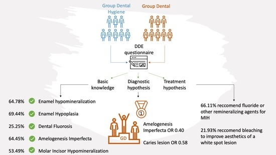
Qualitative92 (58.60%)103 (71.53%)0.03
Quantitative/Both/I don’t know65 (41.4%)41 (28.47%)
Enamel hypoplasia is a ……. defect:
Quantitative100 (63.69%)109 (75.69%)0.03
Qualitative/Both/I don’t know57 (36.31%)35 (24.31%)
When does Dental Fluorosis develop?
In the pre-eruptive phase53 (33.76%)23 (15.97%)<0.01
In the post-eruptive phase/any age/I don’t know104 (66.24%)121 (84.03%)
Amelogenesis Imperfecta is a condition caused by:
Genetic factors90 (57.32%)104 (72.22%)0.01
Systemic factors/Multifactorial/viral or bacterial infection/Don’t know67 (42.68%)40 (27.78%)
When does Amelogenesis Imperfecta develop?
In the pre-eruptive phase117 (74.52%)116 (80.56%)0.26
In the post-eruptive phase/any age/I don’t know40 (25.48%)28 (19.44%)
Molar Incisor Hypomineralization is a …… defect:
Qualitative76 (48.41%)85 (59.03%)0.08
Quantitative/Both/Don’t know81 (51.59%)59 (40.97%)
When does Molar Incisor Hypomineralization develop?
In the pre-eruptive phase88 (56.05%)70 (48.61%)0.24
In the post-eruptive phase/any age/I don’t know69 (43.95%)74 (51.39%)
Table 2. Comparison of GDH and GD answers in regard to the clinical images (expressed as number and percentage). Differences between the groups were analyzed by Pearson’s chi-square test or Fisher’s exact test.
ItemsGDHGDp-Value
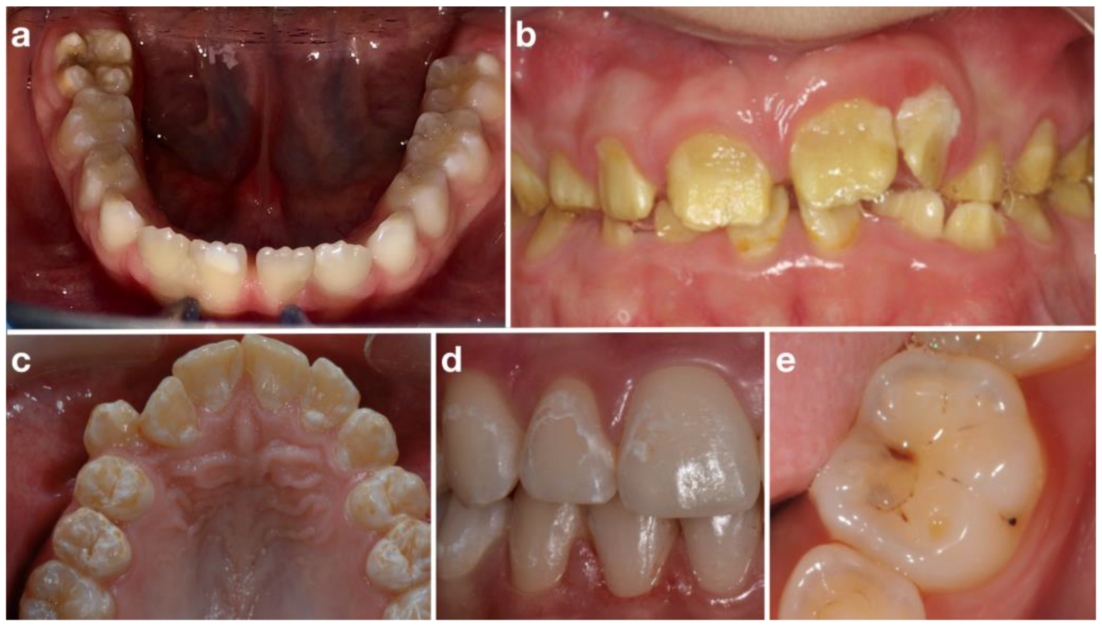
Picture 1: Molar Incisor HypomineralizationN = 137N = 121
Possible choices
Molar Incisor Hypomineralization83 (60.58%)91 (75.21%)0.04
Amelogenesis Imperfecta24 (17.52%)9 (7.44%)
Plaque demineralization17 (12.41%)13 (10.74%)
Dental Fluorosis13 (9.49%)8 (6.61%)
I don’t know20/157 (12.74%)23/144 (15.97%)
The caries risk in this situation is generally:
High98 (71.53%)48 (39.67%)<0.01
Medium38 (27.74%)68 (56.20%)
Low1 (0.73%)5 (4.13%)
Which of the following treatments would you recommend?
Remineralizing products and/or fluoride-based varnish or gel119 (86.86%)84 (69.42%)0.01
Glass-ionomer sealants63(45.99%)52 (42.98%)0.72
Resin-based sealants51 (37.23%)49 (40.50%)0.68
Professional bleaching28 (20.44%)15 (12.40%)0.12
Picture 2: Amelogenesis ImperfectaN = 131N = 135
Possible choices
Amelogenesis Imperfecta89 (67.94%)112 (82.96%)0.02
Plaque demineralization25 (19.08%)10 (7.41%)
Molar Incisor Hypomineralization12 (9.16%)10 (67.41%)
Dental Fluorosis5 (3.82%)3 (2.22%)
I don’t know26/157 (16.56%)9/144(6.25%)
The caries risk in this situation is generally:
High125 (95.42%)117(86.67%)0.02
Medium6 (4.58%)16 (11.85%)
Low0 (0.00%)2 (1.48%)
Which of the following treatments would you recommend?
Remineralizing products and/or fluoride-based varnish or gel100 (76.34%)86 (63.7%)0.03
Glass-ionomer sealants66 (50.38%)55 (40.74%)0.16
Resin-based sealants53 (40.46%)41(30.37%)0.11
Professional bleaching14 (10.69%)15 (11.11%)>0.99
Picture 3: Dental FluorosisN = 146N = 133
Possible choices
Dental Fluorosis61 (41.78%)46 (34.59%)0.01
Plaque demineralization46 (31.51%)66 (49.62%)
Molar Incisor Hypomineralization33 (22.60%)18 (13.53%)
Amelogenesis Imperfecta6 (4.11%)3 (2.26%)
I don’t know11/157 (7.01%)11/144 (7.64%)
The caries risk in this situation is generally:
High53 (36.30%)25 (18.80%)<0.01
Medium71 (48.63%)83 (62.41%)
Low22 (15.07%)25 (18.80%)
Which of the following treatments would you recommend?
Remineralizing products and/or fluoride-based varnish or gel104 (71.23%)78 (58.65%)0.04
Glass-ionomer sealants74 (50.68%)70 (52.63%)0.84
Resin-based sealants88 (60.27%)72 (54.14%)0.36
Professional bleaching31 (21.23%)24 (18.05%)0.60
Picture 4: White spot lesionN = 133N = 112
Possible choices
Plaque demineralization54 (40.60%)45 (40.18%)0.22
Dental Fluorosis61 (45.86%)60 (53.57%)
Amelogenesis Imperfecta11 (8.27%)3 (2.68%)
Molar Incisor Hypomineralization7 (5.26%)4 (3.57%)
I don’t know24/157 (15.29%)32/144 (22.22%)
The caries risk in this situation is generally:
High62 (46.62%)17 (15.18%)<0.01
Medium53 (39.85%)62 (55.36%)
Low18 (13.53%)33 (29.46%)
Which of the following treatment would you recommend?
Remineralizing products and/or fluoride-based varnish or gel91 (68.42%)73 (65.18%)0.69
Glass-ionomer sealants57 (42.86%)56 (50.00%)0.32
Resin-based sealants58 (43.61%)68 (60.71%)0.01
Professional bleaching35 (26.32%)21 (18.75%)0.21
Picture 5: Caries lesionN = 121N = 104
Possible choices
Plaque demineralization48 (39.67%)65 (62.50%)<0.01
Molar Incisor Hypomineralization67 (55.37%)37 (35.58%)
Dental Fluorosis4 (3.31%)1 (0.96%)
Amelogenesis Imperfecta2 (1.65%)1 (0.96%)
I don’t know36/157 (22.93%)40/104 (27.78%)
Which of the following treatments would you recommend?
Remineralizing products and/or fluoride-based varnish or gel91 (75.21%)76 (73.08%)0.83
Glass-ionomer sealants86 (71.07%)66 (63.46%)0.28
Resin-based sealants71 (58.68%)71 (68.27%)0.18
Professional bleaching9 (7.44%)15 (14.42%)0.14
For each clinical pictures’ comparison analysis, the answer “I don’t know” was excluded
Table 3. Multivariate logistic regression analysis between GD and GDH on their recognition of clinical images. The correct answer was chosen as a reference.
Ref: Group of students in Dentistry (GD)
Clinical PicturesOdds Ratiop-Value
  • Molar Incisor Hypomineralization
0.93 [0.56;1.54]0.77
2.
Amelogenesis Imperfecta
0.40 [0.23;0.69]<0.01
3.
Dental Fluorosis
1.38 [0.81;2.35]0.23
4.
White spot lesion
1.30 [0.75;2.24]0.35
5.
Caries lesion
0.58 [0.34;0.94]0.03
Publisher’s Note: MDPI stays neutral with regard to jurisdictional claims in published maps and institutional affiliations.

