
Pediatric Stroke due to Thoracic Outlet Syndrome Treated with Thrombolysis and Thrombectomy: A Case Report

 ,
, 
Abstract
:1. Introduction
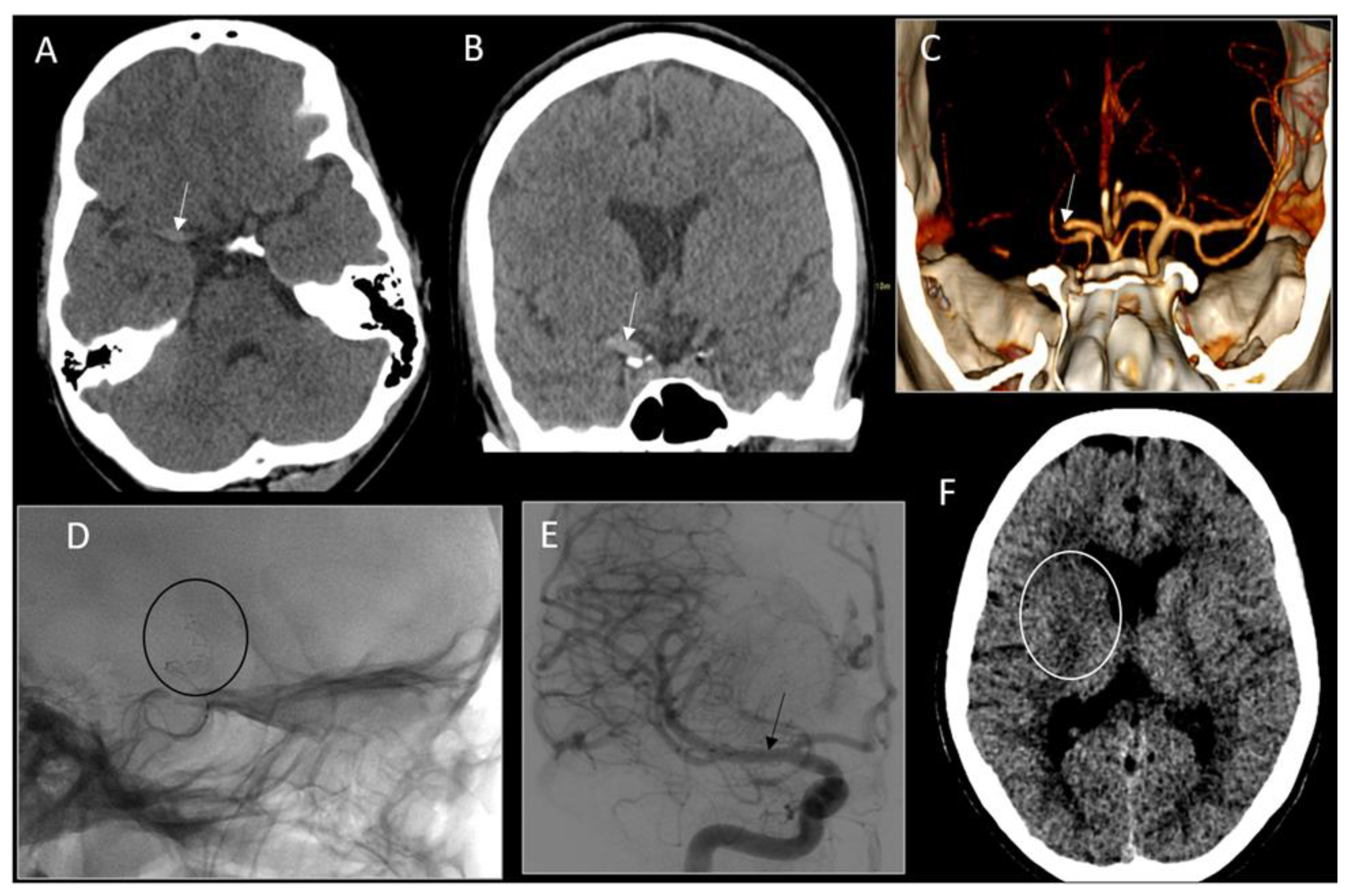
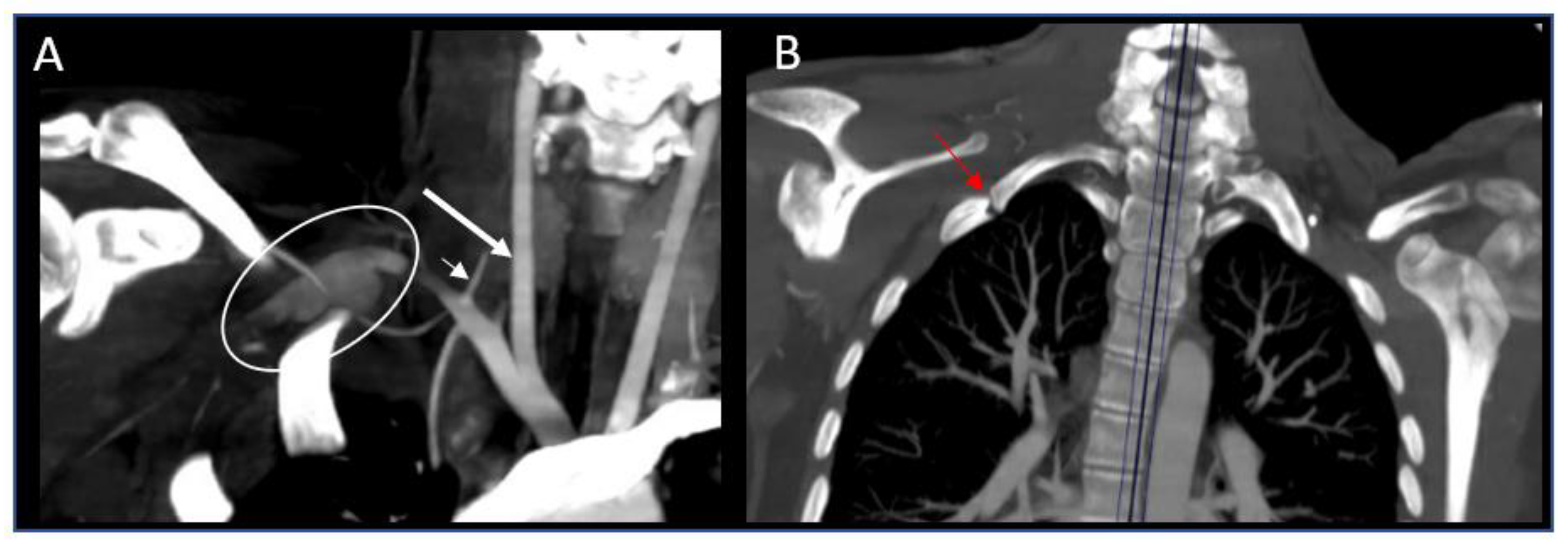
2. Case Report
3. Discussion
4. Conclusions
Author Contributions
Funding
Institutional Review Board Statement
Informed Consent Statement
Data Availability Statement
Conflicts of Interest
References
- Fullerton, H.J.; Wu, Y.W.; Sidney, S.; Johnston, S.C. Risk of recurrent childhood arterial ischemic stroke in a population-based cohort: The importance of cerebrovascular imaging. Pediatrics 2007, 119, 495–501. [Google Scholar] [CrossRef] [PubMed]
- Lehman, L.L.; Khoury, J.C.; Taylor, J.M.; Yeramaneni, S.; Sucharew, H.; Alwell, K.; Moomaw, C.J.; Peariso, K.; Flaherty, M.; Khatri, P.; et al. Pediatric Stroke Rates Over 17 Years: Report from a Population-Based Study. J. Child Neurol. 2018, 33, 463–467. [Google Scholar] [CrossRef] [PubMed]
- Greenham, M.; Gordon, A.; Anderson, V.; Mackay, M.T. Outcome in Childhood Stroke. Stroke 2016, 47, 1159–1164. [Google Scholar] [CrossRef] [PubMed] [Green Version]
- Kochanek, K.D.; Xu, J.; Murphy, S.L.; Miniño, A.M.; Kung, H.C. Deaths: Final data for 2009. Natl. Vital Stat. Rep. 2011, 60, 1–116. [Google Scholar]
- Mackay, M.T.; Wiznitzer, M.; Benedict, S.L.; Lee, K.J.; Deveber, G.A.; Ganesan, V. Arterial Ischemic Stroke Risk Factors: The International Pediatric Stroke Study. Ann. Neurol. 2011, 69, 130–140. [Google Scholar] [CrossRef]
- Sanders, R.J.; Hammond, S.L.; Rao, N.M. Diagnosis of thoracic outlet syndrome. J. Vasc. Surg. 2007, 46, 601–604. [Google Scholar] [CrossRef] [Green Version]
- Meumann, E.M.; Chuen, J.; Fitt, G.; Perchyonok, Y.; Pond, F.; Dewey, H.M. Thromboembolic stroke associated with thoracic outlet syndrome. J. Clin. Neurosci. 2014, 21, 886–889. [Google Scholar] [CrossRef]
- Bains, R.D.; Platt, J.; MacGregor, D.; Borschel, G.H. Atypical thoracic outlet syndrome and reverse flow thromboembolism. Pediatr. Neurol. 2014, 51, 453–456. [Google Scholar] [CrossRef]
- Kataria, R.; Sharma, A.; Srivastava, T.; Bagaria, H.; Sharma, A. Cervical rib, a rare cause of recurrent stroke in the young: Case report. Neurologist 2012, 18, 321–323. [Google Scholar] [CrossRef]
- Kuril, S.; Chopade, P.R.; Mandava, M.; Bhatia, S. A Rare Case of Stroke in an Adolescent Violinist Due to Thoracic Outlet Syndrome. Neurol. India 2021, 69, 1777–1780. [Google Scholar] [CrossRef]
- Lee, T.S.; Hines, G.L. Cerebral embolic stroke and arm ischemia in a teenager with arterial thoracic outlet syndrome: A case report. Vasc. Endovasc. Surg. 2007, 41, 254–257. [Google Scholar] [CrossRef] [PubMed]
- Strzelecka, J.; Skadorwa, T.; Franckiewicz, M.; Jóźwiak, S. A case of symmetric retrograde thromboembolic cerebral infarction in an 8-year-old child due to arterial thoracic outlet syndrome. Childs Nerv. Syst. 2018, 34, 2503–2507. [Google Scholar] [CrossRef] [PubMed] [Green Version]
- Aghamiri, S.H.; Assarzadegan, F.; Ghaffari, M.; Khorasani, N.M.; Lima, B.S.; Sepehrirad, A.; Azimi, B.; Delkash, P. Recurrent middle cerebral artery stroke caused by arterial thoracic outlet syndrome and coagulopathy. Radiol. Case Rep. 2022, 17, 1665–1669. [Google Scholar] [CrossRef]
- Celier, A.; Chabay, S.; Maurizot, A.; Cochennec, F.; Stanciu, D.; Pico, F. Posterior cerebral artery stroke by reverse flow embolism in thoracic outlet syndrome—A case report. BMC Neurol. 2020, 20, 229. [Google Scholar] [CrossRef] [PubMed]
- Sharma, S.; Kumar, S.; Joseph, L.; Singhal, V. Cervical rib with stroke as the initial presentation. Neurol. India 2010, 58, 645–647. [Google Scholar] [CrossRef] [PubMed]
- Blank, R.H.; Connar, R.G. Arterial complications associated with thoracic outlet compression syndrome. Ann. Thorac. Surg. 1974, 17, 315–324. [Google Scholar] [CrossRef]
- Saxton, E.H.; Miller, T.Q.; Collins, J.D. Migraine complicated by brachial plexopathy as displayed by MRI and MRA: Aberrant subclavian artery and cervical ribs. J. Natl. Med. Assoc. 1999, 91, 333–341. [Google Scholar]
- Chahwala, V.; Tashiro, J.; Li, X.; Baqai, A.; Rey, J.; Robinson, H.R. Venous Thoracic Outlet Syndrome as a Cause of Intractable Migraines. Ann. Vasc. Surg. 2017, 39, 285.e5–285.e8. [Google Scholar] [CrossRef]
- Goeggel Simonetti, B.; Rafay, M.F.; Chung, M.; Lo, W.D.; Beslow, L.A.; Billinghurst, L.L.; Fox, C.K.; Pagnamenta, A.; Steinlin, M.; Mackay, M.T. Comparative Study of Posterior and Anterior Circulation Stroke in Childhood: Results from the International Pediatric Stroke Study. Neurology 2020, 94, e337–e344. [Google Scholar] [CrossRef]
- Hussain, M.A.; Aljabri, B.; Al-Omran, M. Vascular Thoracic Outlet Syndrome. Semin. Thorac. Cardiovasc. Surg. 2016, 28, 151–157. [Google Scholar] [CrossRef] [Green Version]
- Powers, W.J.; Rabinstein, A.A.; Ackerson, T.; Adeoye, O.M.; Bambakidis, N.C.; Becker, K.; Biller, J.; Brown, M.; Demaerschalk, B.M.; Hoh, B.; et al. Guidelines for the Early Management of Patients with Acute Ischemic Stroke: 2019 Update to the 2018 Guidelines for the Early Management of Acute Ischemic Stroke: A Guideline for Healthcare Professionals from the American Heart Association/American Stroke Association. Stroke 2019, 50, e344–e418. [Google Scholar] [CrossRef] [PubMed]
- Roach, E.S.; Golomb, M.R.; Adams, R.; Biller, J.; Daniels, S.; Deveber, G.; Ferriero, D.; Jones, B.V.; Kirkham, F.J.; Scott, R.M.; et al. Management of stroke in infants and children: A scientific statement from a Special Writing Group of the American Heart Association Stroke Council and the Council on Cardiovascular Disease in the Young. Stroke 2008, 39, 2644–2691. [Google Scholar] [CrossRef] [PubMed] [Green Version]
- Amlie-Lefond, C.; Shaw, D.W.W.; Cooper, A.; Wainwright, M.S.; Kirton, A.; Felling, R.J.; Abraham, M.G.; Mackay, M.T.; Dowling, M.M.; Torres, M.; et al. Risk of Intracranial Hemorrhage Following Intravenous tPA (Tissue-Type Plasminogen Activator) for Acute Stroke Is Low in Children. Stroke 2020, 51, 542–548. [Google Scholar] [CrossRef] [PubMed]
- Harrar, D.B.; Benedetti, G.M.; Jayakar, A.; Carpenter, J.L.; Mangum, T.K.; Chung, M.; Appavu, B. Pediatric Acute Stroke Protocols in the United States and Canada. J. Pediatr. 2022, 242, 220–227.e7. [Google Scholar] [CrossRef]
- Rivkin, M.J.; Bernard, T.J.; Dowling, M.M.; Amlie-Lefond, C. Guidelines for Urgent Management of Stroke in Children. Pediatr. Neurol. 2016, 56, 8–17. [Google Scholar] [CrossRef] [Green Version]
- Goyal, M.; Menon, B.K.; van Zwam, W.H.; Dippel, D.W.; Mitchell, P.J.; Demchuk, A.M.; Dávalos, A.; Majoie, C.B.; van der Lugt, A.; de Miquel, M.A.; et al. Endovascular thrombectomy after large-vessel ischaemic stroke: A meta-analysis of individual patient data from five randomised trials. Lancet 2016, 387, 1723–1731. [Google Scholar] [CrossRef]
- Sporns, P.B.; Sträter, R.; Minnerup, J.; Wiendl, H.; Hanning, U.; Chapot, R.; Henkes, H.; Henkes, E.; Grams, A.; Dorn, F.; et al. Feasibility, Safety, and Outcome of Endovascular Recanalization in Childhood Stroke: The Save ChildS Study. JAMA Neurol. 2020, 77, 25–34. [Google Scholar] [CrossRef]
- Bhatia, K.D.; Briest, R.; Goetti, R.; Webster, R.; Troedson, C.; Dale, R.C.; Muthusami, P.; Miteff, C.; Miteff, F.; Worthington, J.; et al. Incidence and Natural History of Pediatric Large Vessel Occlusion Stroke: A Population Study. JAMA Neurol. 2022, 79, 488–497. [Google Scholar] [CrossRef]
- Ferriero, D.M.; Fullerton, H.J.; Bernard, T.J.; Billinghurst, L.; Daniels, S.R.; DeBaun, M.R.; deVeber, G.; Ichord, R.N.; Jordan, L.C.; Massicotte, P.; et al. Management of Stroke in Neonates and Children: A Scientific Statement from the American Heart Association/American Stroke Association. Stroke 2019, 50, e51–e96. [Google Scholar] [CrossRef] [Green Version]
- Sun, L.R.; Harrar, D.; Drocton, G.; Castillo-Pinto, C.; Felling, R.; Carpenter, J.L.; Wernovsky, G.; McDougall, C.G.; Gailloud, P.; Pearl, M.S. Mechanical Thrombectomy for Acute Ischemic Stroke: Considerations in Children. Stroke 2020, 51, 3174–3181. [Google Scholar] [CrossRef]


| Study | Age/ Sex | Risk Factors | Preceding Symptoms | Stroke Distribution | Treatment | Outcome |
|---|---|---|---|---|---|---|
| Current study | 15 y F | Partially fused R 1st and 2nd ribs |
| R MCA |
| Mild L foot dystonia |
| Aghamiri et al., 2022 [13] | 15 y F | Heterozygous for plasminogen activator inhibitor and MTHFR |
| R MCA |
| Not reported |
| Kuril et al., 2021 [10] | 14 y M | Violinist and B cervical ribs |
| B posterior circulation |
| Not reported |
| Strzelecka et al., 2018 [12] | 8 y F | R cervical rib and R bifid 1strib | None | Posterior circulation |
| R-sided weakness, intention tremor |
| Bains et al., 2014 [8] | 12 y M | B hypoplastic 1st vs. cervical ribs |
| Posterior circulation | Rib excision | Mild dyscoordination, arm shakiness |
| Meumann et al., 2013 [7] | 16 y F | B cervical ribs |
| R MCA |
| Not reported |
| Kataria et al., 2012 [9] | 14 y F | R cervical rib | Prior stroke | R MCA (old), posterior circulation (acute) |
| Minimal L-sided weakness not impacting function |
| Sharma et al., 2010 [15] | 18 y M | R cervical rib | none | R MCA | Rib excision | Not reported |
| Lee et al., 2007 [11] | 15 y F | B cervical ribs | R arm sensory changes | R MCA |
| L arm weakness |
| Blank et al., 1974 [16] | 18 y F | B cervical ribs | R hand pain | Posterior circulation | None reported | Resolution of neurologic symptoms |
Publisher’s Note: MDPI stays neutral with regard to jurisdictional claims in published maps and institutional affiliations. |
© 2022 by the authors. Licensee MDPI, Basel, Switzerland. This article is an open access article distributed under the terms and conditions of the Creative Commons Attribution (CC BY) license (https://creativecommons.org/licenses/by/4.0/).
Share and Cite
Angappan, D.; Garrett, M.; Henry, C.; Riddle, A.; Wilson, J.L. Pediatric Stroke due to Thoracic Outlet Syndrome Treated with Thrombolysis and Thrombectomy: A Case Report. Children 2022, 9, 875. https://0-doi-org.brum.beds.ac.uk/10.3390/children9060875
Angappan D, Garrett M, Henry C, Riddle A, Wilson JL. Pediatric Stroke due to Thoracic Outlet Syndrome Treated with Thrombolysis and Thrombectomy: A Case Report. Children. 2022; 9(6):875. https://0-doi-org.brum.beds.ac.uk/10.3390/children9060875
Chicago/Turabian StyleAngappan, Dhanalakshmi, McKinnon Garrett, Candice Henry, Art Riddle, and Jenny L. Wilson. 2022. "Pediatric Stroke due to Thoracic Outlet Syndrome Treated with Thrombolysis and Thrombectomy: A Case Report" Children 9, no. 6: 875. https://0-doi-org.brum.beds.ac.uk/10.3390/children9060875