

Aflatoxins: Source, Detection, Clinical Features and Prevention
by
 ,
,
Solomon Abrehame
1,2,3,4,
Valsa Remony Manoj
1,
Merry Hailu
1,2,3,4,
Yu-Yi Chen
3,4,
Yu-Chun Lin
5 and
Yen-Po Chen
3,4,* 1
Department of Biotechnology, Vel Tech Rangarajan Dr. Sagunthala R & D Institute of Science and Technology, 400 Feet Outer Ring Road, Avadi, Chennai 600062, India
2
Veterinary Drug and Feed Administration and Control Authority of Ethiopia [VDFACA], MoA, P.O. Box 62347, Addis Ababa 1000, Ethiopia
3
Department of Animal Science, National Chung Hsing University, 145 Xingda Road, South Dist., Taichung City 402, Taiwan
4
The iEGG and Animal Biotechnology Center, National Chung Hsing University, Taichung City 402, Taiwan
5
Livestock Research Institute, Council of Agriculture, Executive Yuan, No. 112, Farm Road, HsinHua District, Tainan 712, Taiwan
*
Author to whom correspondence should be addressed.
Processes 2023, 11(1), 204; https://0-doi-org.brum.beds.ac.uk/10.3390/pr11010204
Submission received: 31 August 2022
/
Revised: 1 January 2023
/
Accepted: 2 January 2023
/
Published: 9 January 2023
(This article belongs to the Special Issue Application and Development of Disease Diagnosis Microbiology Technology)
Abstract
:The most potent mycotoxin, aflatoxins are the secondary metabolite produced by fungi, especially Aspergillus, and have been found to be ubiquitous, contaminating cereals, crops, and even milk and causing major health and economic issues in some countries due to poor storage, substandard management, and lack of awareness. Different aspects of the toxin are reviewed here, including its structural biochemistry, occurrence, factors conducive to its contamination and intoxication and related clinical features, as well as suggested preventive and control strategies and detection methods.
1. Introduction
Mycotoxins, including trichothecenes, fumonisins, zearalenone, and ochratoxin A, are secondary metabolites of different fungus species which are mostly soil-borne and produced by species within the genera Aspergillus, Fusarium, and Penicillium [1], with aflatoxins causing major health issues for humans and animals, even leading to serious economic problems [2,3]. Mainly produced by Aspergillus flavus and Aspergillus parasiticus, common fungi in nature [4,5,6], aflatoxins are common contaminants for various foods, feeds, and crops, such as groundnuts [7], maize [1,8], rice [9,10], sorghum [11], and oilseeds [12], which are major foods for both humans and animals. Moreover, their secondary biological metabolites are often found in milk and milk products and other animal products [6,13,14]. The aflatoxin problem is especially serious in some geographical locations and agricultural practices due to higher humidity and temperature, in addition to poor storage conditions [15,16].
Some of the various types of aflatoxins have been identified and derived from nature, others are their metabolites, with the former including four major types, namely AFB1, AFB2, AFG1, and AFG2, all produced by Aspergillus species. Their major metabolic products include AFM1, AFM2, AFP1, AFQ1, and AFB1-8,9-epoxide [17,18]. It is known that aflatoxins and their metabolites are a major cause for mutagenicity, carcinogenicity, hepatotoxicity, and immunosuppression in both humans and animals, whose biotransformation with physiological enzymes may make them more pathogenic [18,19]. One of the most pathogenic and carcinogenic aflatoxins is AFB1, which has been classified as a group I human carcinogen by the International Agency for Research on Cancer (IARC) [9].
In addition to health issues, contamination of feeds and foods has resulted in serious economic loss, since it is unsalvageable, although it is preventable via measures before and after harvest [15,19,20]. Moreover, aflatoxins are highly heat-resistant, unaffected by conventional thermal treatment for foods [21]. This review looked into key findings on aflatoxins in various aspects, including structural biochemistry, contamination and intoxication, and related clinical features, as well as suggested preventive and control strategies and detection methods.
2. Prevalence and Occurrence of Aflatoxins
Aflatoxins were first discovered in the UK in 1960, being confirmed as the causative agent for the outbreak of turkey X diseases, which killed over 100,000 turkeys and other kinds of poultry [22]. The disease, with major symptoms of liver necrosis and bile duct hyperplasia, was caused by toxic feeds, resulting from contamination of its ingredients, mainly groundnuts and cottonseeds. The culprit was later identified as A-fla-toxin, produced mainly by fungi, with “A” and “fla” denoting Aspergillus and flavus, respectively [23,24].
The event sparked a so-called “mycotoxin gold rush” from 1960 through 1975, when many chemical researchers joined the search for mycotoxins [23], leading to the discovery of several hundreds of fungal metabolites with toxic properties, along with their structures and chemical profiles.
Aflatoxins are a ubiquitous food and feed contaminant, often found in agricultural products and byproducts, even including those from manufacturers with good manufacturing practices [25,26,27]. According to the Food and Agriculture Organization (FAO) of the United Nations, 25% of the world’s crop harvest is contaminated by mycotoxins during growth or storage, perhaps due to the fact that conventional farming practices are conducive to fungal growth. Moreover, fungi can persist in feed and food from processing through prepared meal due to their ability to endure stresses from harsh environmental conditions, as well as various decontamination methods during processing [5,25,28,29].
The problem prevails especially in tropical and subtropical regions, such as Sub-Saharan Africa and Southeast Asian and South American countries with substandard agricultural operations, hot temperature, and high humidity that favors growth of the fungus [21,27,30,31,32,33]. It has also emerged in North America and Europe due to global warming [8,26].
3. Structures and Biochemistry of Different Types of Aflatoxins
There are at least 18 types of aflatoxins identified so far, including such important ones from the perspective of public health as aflatoxin B1 (AFB1), B2 (AFB2), G1 (AFG1), G2 (AFG2), and M1 (AFM1), of which AFB1, AFB2, AFG1, and AFG2 derive from nature and AFM1 from mammals’ milk after the biotransformation of the previous toxins in their liver [2,18,28]. When exposing aflatoxins to UV light, blue and green lights will be exhibited, reasons for the detonations of “B” and “G” aflatoxins, while “M” refers to aflatoxins found in milk and dairy products (Table 1) [3,28].
Aflatoxins are biosynthesized in fungi with type II polyketide synthase in a processing starting with the generation of norsolorinic acid, followed by a series of complex post-polyketide synthase steps, resulting in progressively more toxigenic anthraquinone and difurocoumarin metabolites (Table 1) [2,36,37].
Aflatoxins are derivatives of dihydrofurancoumarins that are divided into two broad groups according to their chemical structure, namely the difurocoumarocyclopentenone series (AFB1, AFB2, AFB2A, AFM1, AFM2, AFM2A, and aflatoxicol) and the difurocoumarolactone series (AFG1, AFG2, AFG2A, AFGM1, AFGM2, AFGM2A, and AFQ1) [2,34,36]. They have a polycyclic structure deriving from a coumarin nucleus linked to a bifurano system, while aflatoxins of type B are connected to a pentanone, and G-type aflatoxins are connected to a 6-member lactone. Structurally AFB1, AFG1, and AFM1 have double bonds in 8–9 positions, while AFB2, AFG2, and AFM2 do not have the double bond (Table 1) [34,35,36]. With low molecular weight, aflatoxins are soluble in solvents such as methanol, benzene, chloroform, and acetonitrile, with a wide spectrum of toxicity. Although they are stable in a wide range of pH (3 to 10) and have a melting point above 250 °C, they are unstable in UV light exposure with no immunogenicity [21,35]. When exposed to UV light, different aflatoxins with different crystal types have different melting points with varying fluorescence (Table 1) [6,21,35].
4. Sources of Intoxication
4.1. Factors Favoring the Presence of Aflatoxins
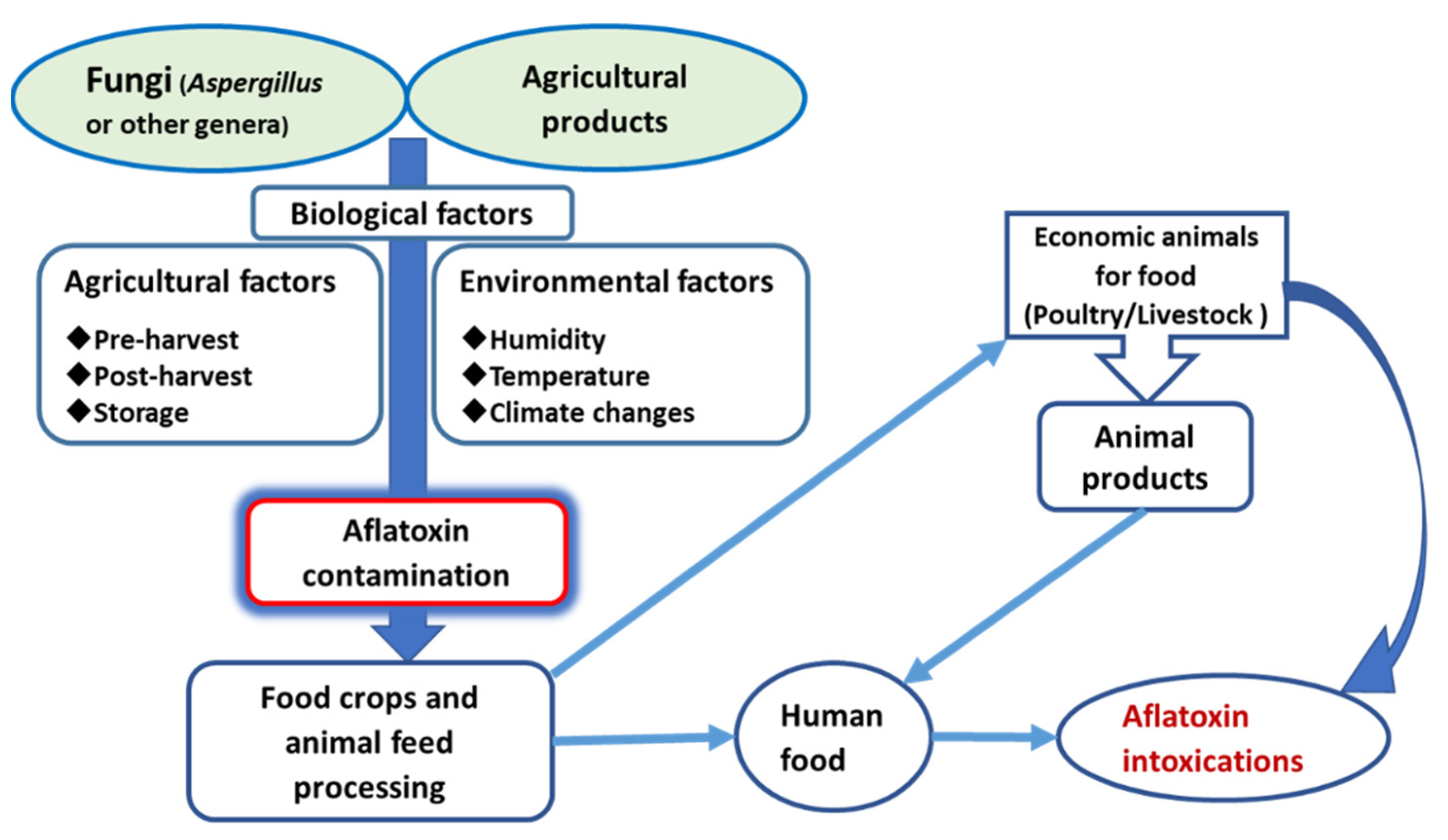
Since the discovery of aflatoxins, there have been various reports on contaminations of agricultural products and animal products, such as milk and meat products [15,26,31,38,39,40]. With a favorable environment, aflatoxins are certain to appear with the presence of relevant fungi. Mycotoxin-producing fungi can access cereals during transportation or storage. They can derive from post-harvest wastes on the field, including grains undergoing microbiological deterioration and decaying vegetation (Figure 1) [10,26,41].
The occurrence of aflatoxins in food and feed varies with geographic location and agricultural/agronomic practices. Moreover, the toxin has high ability to contaminate commodities and persist throughout storage, transportation, and processing. It is noteworthy that absence of visible mold does not guarantee the absence of mycotoxins and aflatoxins, since they often cannot be removed even by cooking or processing of food products [5,9,15,19].
There are several factors, some extrinsic and others intrinsic, in pre- and post-harvest practices, which are conducive to the occurrence of aflatoxins in feed and food chains. Extrinsic factors include temperature, humidity, water or heat stress, soil conditions/properties, mechanical injury on food commodity, and insects and rodent infestations, while intrinsic factors include genotype, pH, nutrient composition, and moisture content/water activity [42]. While these factors are key to the frequency and severity of contamination, a combination of two or more factors may be needed to foster fungal growth and cause corresponding toxin production [7,10,41,43].
Pre-harvest: Fungi can reside on various parts of plants, with most cases in seeds. Many studies have found that crops and crop residues can harbor fungi in spore and mycelial forms [44]. Fungi contaminate crops during harvest as spores or mycelium and are then transferred to harvest crops as an organism or its metabolites (i.e., aflatoxins), which will remain in the crops up to storage [10,26].
Post-harvest: A major factor for the development of aflatoxins and other mycotoxins in feed are storage conditions in the post-harvest period, including moisture, humidity, and temperature, mostly with interrelated effects [41,43,44,45,46].
Temperatures: While temperatures between 25 and 37 °C are most favorable for fungal growth and toxin production, fungi can reproduce even in temperature as low as 12 °C and as high as 48 °C. Generally, fungi need a suboptimal temperature for the production of aflatoxins. Studies on A. flavus have shown that the optimal temperature for its growth is 25–37 °C, with maximum growth and aflatoxin production at 35–37 °C and no production at temperatures below 13 °C or above 42 °C, indicating that temperature increase is conducive to fungal growth and aflatoxin production. Other conditions, like moisture and humidity, are inseparable from temperature when it is considered as a factor [8,21,41,42,45,47,48].
Humidity and moisture: Environmental humidity and crop moisture content are also important factors, as they determine the accumulation of aflatoxins in harvested crops for food and feed. As water is indispensable for the metabolism and growth of microorganism, it is a key determinant for the formation of aflatoxins in crops [42,47]. A minimum of 13% moisture or water activity of 0.65 is necessary for the growth, proliferation, and toxin production of fungi (e.g., Aspergillus). While in storage conditions, with 77% moisture and above being optimal, the maximum growth and toxin production take place at 90–95% moisture and 25 °C [15,27,41,45,46,47]. Moreover, prolonged storage time, improper storage arrangement, and lack of adequate drying facilities, which affect the water activity of crops during the post-harvest period, promote aflatoxin production as well. Drying is an effective method to inhibit aflatoxin production, while reduction of moisture level to 10% via quick and proper drying of post-harvested cereals and legumes can suppress A. flavus proliferation and aflatoxin production [16].
Damaged crops and grains: Aflatoxins produced by fungi could contaminate seeds but cannot enter and infect crop/seed husks unless there are favorable conditions, such as damage of crop husk, that facilitate infection by fungi and entry of pathogenic fugal toxins. Many studies have shown that seeds with damaged husks are more vulnerable to infection by mycotoxin than seeds with intact husks. Damage to the seed husk may be caused by improper harvesting processes, insect bites, and inadequate food processing [15,30,41].
4.2. Source of Aflatoxin Intoxication
Various studies have found that aflatoxins could be isolated from raw milk and dairy products, cereals, coffee, grapevine, dried fruits (e.g., figs and raisins), spices, peanuts, groundnuts, and nuts such as almonds, pistachio, walnuts, and coconut [2,49,50]. The following are some important sources of aflatoxin intoxication.
4.2.1. Cereals
Various cereals are vulnerable to aflatoxin contamination starting from the blooming stage, including maize, wheat, barley, oat, sorghum, pearl millet, rice, and oilseeds such as groundnut, soybean, sunflower, and cotton [15,48,51]. Fungal intoxication of feed starts from infestation of crops with fungal spores at farms through storage facility. A common subject for study in this field is corn farms, whose crop contains high nutrients facilitating aflatoxin contamination [44]. Oilseeds and their cakes (i.e., byproducts) are also a common source of aflatoxin intoxication affecting both humans and animals due to their nutritious contents that are conducive to fungal growth [7,15,49,51].
4.2.2. Spices
Ground spices and herbs have been evaluated to be substrates for mycelial growth, sporulation, and eventually aflatoxin production. Spices that could potentially harbor aflatoxins include chili, black/red pepper, coriander, turmeric, and ginger [50,52,53,54]. Spices are exposed to a wide range of fungal contamination from production through marketing, in agricultural fields, post-harvesting practices, and processing. Factors include environmental conditions, such as high temperature and humidity, poor collection conditions, inadequate production process, substandard drying, and poorly sheltered transport and storage, which can exacerbate contamination [31,50,53,54].
Previous studies have demonstrated that toxigenic molds and various microorganisms, especially Aspergillus species, could contaminate spices at different levels of production. Consequently, spices on the market are often contaminated with unacceptable concentrations of aflatoxins and pose public health hazards since they usually are not cooked or treated with heat during processing [54,55,56]. For instance, a study on Sri Lankan spices found that 63% and 67% of tested AFB1-positive black pepper and dry chili samples, respectively, exceeded the EU minimum levels of 5 µg/kg, [50]. Another study carried out in Turkey also showed that 9 out of 13 red-scaled, 3 of 11 red pepper, and 2 out of 8 black pepper positive samples contained AFB1 (5 µg/kg) and total aflatoxins (10 µg/kg) exceeding the limits set by the EU and Turkish Food Codex [52].
4.2.3. Milk Products
The occurrence of aflatoxins, including AFB1 and AFM1, in raw milk and milk products, either of human or animal origin, is a major health issue, especially for infants and toddlers, for whom milk products provide vitamins, minerals, fatty acids, and proteins essential for their growth [57,58]. There have been numerous reports on AFM1 poisoning incidents related to various milk products, including cheese, yogurt, cream, butter, ice cream, and milk powder [2,28,38,59,60].
Aflatoxin contamination of milk products can be caused by the ingestion of contaminated feeds by dairy cattle, contaminated additives, or accidents [28,59]. In the first case, usually 0.3–6.2% of consumed AFB1 is metabolized to AFM1, which is then contained in secreted milk after 12–24 h. The content of AFM1 declines gradually, reaching an undetectable level within 72 h after ingestion [28,59,60,61].
5. Intoxications, Metabolism, and Clinical Features of Aflatoxins
5.1. Routes of Intoxications
Ingestion is the main route of aflatoxin intoxication. Inhalation of contaminated grain dust is also a major path for AFB1 exposure for people in certain occupations [62], and slow skin absorption is another route, though in rare cases [63]. Animals or humans often consume aflatoxin-contaminated feeds/foods unaware, due to lack of unusual features on their appearances in many cases [30,64,65].
5.2. Absorption
Aflatoxins can be easily assimilated into our digestive tract. ADME (absorption, distribution, metabolism, and excretion) analysis in a study showed that 13 aflatoxin analogues have high gastrointestinal (GI) absorption properties [34]. AFB1 can be efficiently absorbed by the intestinal tract, mainly the duodenum. AFB1, a light molecular compound, is absorbed into enterocytes via a passive diffusion mechanism [35]. In line with the findings, in vitro studies have shown that AFB1 is transported via a differentiated monolayer of intestinal epithelial cell line Caco2, from the apical to the basolateral site and vice versa at similar rates [66,67]. The absorption of aflatoxins starts from the mucosal surface at the upper section of the GI tract, and the main absorption site is at the lower section of the intestinal lumen. AFB1 is detected in plasma immediately after oral administration of a single dose of 4.8 mg to calves, reaching the peak after four hours, which suggests that absorption starts at the rumen [68]. Moschini et al. (2007) also demonstrated that AFB1 is detected in the plasma of lactating dairy cows 15 min after administration of 4.9 mg of AFB1 in contaminated corn [69].
5.3. Metabolism and Mechanism of Action
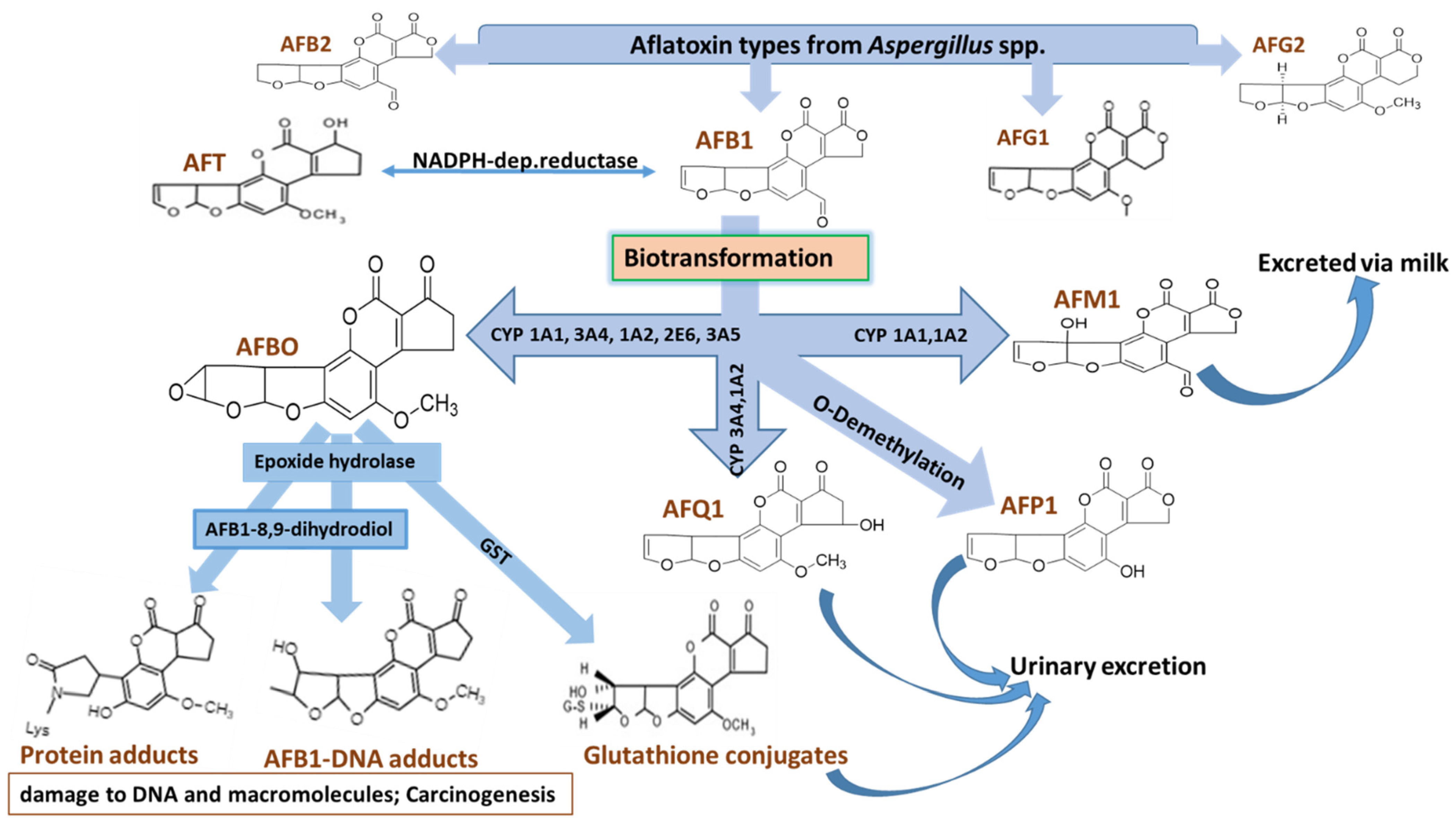
Metabolism of most ingested aflatoxins follows similar pathways in the human body. After absorption, first-pass metabolism occurs in intestinal and respiratory epithelium, although their roles remain to be evaluated [35,70,71]. From the absorption site, AFB1 enters the bloodstream and is transported to the liver, the principal site of accumulation and metabolism. This is due to the fact that aflatoxins are lipophilic molecules and the liver is predominantly a lipophilic organ as well as the destination of all molecules in circulation for processing [2,72]. AFB1 by itself is not such a potent toxin. However, via a detoxification process, the liver and some other organs bio-transform it into a more potent form that damages the liver itself, causing hepatotoxicity and carcinogenicity. Other biotransformation byproducts are excreted via different routes, with urine being the principal excretion means (Figure 2).
Metabolism of aflatoxins is carried out in three phases: (1) oxidation of AFB1; (2) detoxification; (3) bacterial deconjugation.
In phase I, AFB1 is oxidized into hydroxylated metabolites, such as AFM1, AFQ1, and AFP1, as well as highly reactive aflatoxin-8,9-exo-epoxide. In the liver, AFB1 is metabolized into aflatoxin B1-8,9-exo-epoxide by cytochrome P450 enzymes (CYPs), which can be abundantly found in the liver and are also present in respiratory and intestinal tissues, prompting the latter to also bio-activate aflatoxins and causing them to suffer from their toxicity [35,73,74,75]. CYP3A4 and CYP1A2 are the two principal human CYP450 isoforms, responsible for the biotransformation of AFB1 into highly reactive aflatoxin B1-8,9-exo-epoxide. [74,75,76]. CYP1A2 can also catalyze the epoxidation of AFB1 to yield a high proportion of endo-epoxide and the hydroxylation of AFB1 to form AFM1, which is excreted via milk and can cause similar intoxication of any host via ingestion.
These CYPs also metabolize AFB1 into AFP1 and AFQ1, both less toxic because they are not good substrates for epoxidation. AFP1 derives from O-demethylation [21]. Aflatoxin B1 epoxide can occur in two isomers, the endo- and the exo-form, but only the exo-isomer (aflatoxin B1-8,9-exo-epoxide) is related to toxicity and carcinogenicity (Figure 2). The formed epoxide is highly unstable, ready to be bound with biological nucleophiles, such as nucleic acids, to form stable adducts with RNA and DNA. Its covalent binding with DNA induces point mutations and DNA strand breaks and is linked to the carcinogenic effects of AFB1 exposure. With the presence of water, epoxide will be rapidly and non-enzymatically hydrolyzed into AFB1 8,9-dihydrodiole [17,18,71,77]. Meanwhile, cytosolic reductase enzyme can reduce AFB1 to aflatoxicol, a detoxification product which can be re-oxidized back to AFB1 by a microsomal dehydrogenase, thereby increasing the half-life of AFB1 and posing as an in vivo reservoir of AFB1 [17,21,77,78]. AFM1 will continue their pathogenicity by excretion through milk, followed by metabolization in liver of other organisms which ingest the contaminated milk, while others (i.e., AFP1 and AFQ1), with little reaction in the body, are removed. AFM1 can exert toxicity without causing metabolic activation, with children being especially vulnerable to its toxicity [14,79]. There exists a proportionally linear relationship between AFB1 concentration in feed and AFM1 in milk. It is estimated that about 0.3–6.2% of AFB1 consumed by dairy cattle is metabolized into AFM1 [80,81,82].
Phase II aflatoxin metabolism in the human body mainly involves detoxification, when the water solubility and excretion of phase I metabolites is increased by conjugating them with glutathione or glucuronic acid with the help of glutathione-S-transferase enzyme. Conjugated epoxides and hydroxylated AFB1 metabolites are then freely excreted into the intestinal tract via bile, where they might be subject to bacterial deconjugation as a phase III reaction [35,78,83].
5.4. Excretion
The major excretion route of AFB1 and its metabolites is the biliary pathway via feces, followed by the urinary system. AFM1 and some other metabolites are also excreted via milk in lactating mammals. After phase II metabolism and conjugation, the metabolites are mostly removed through the biliary pathway via feces, with some being reabsorbed by the intestines due to intestinal microbial de-conjugation of the metabolites [78,83]. Urine is another excretion way of aflatoxins, especially for AFM1, on top of milk. A regular pattern of excretion of AFB1 (in feces) and AFM1 (in urine) was demonstrated by a study on calves fed with a single dose of 4.8 mg of AFB1 [68]. Hence, many studies have employed urine as a biomarker for aflatoxin determination [80,84].
The metabolites of aflatoxins can be accumulated in the liver, kidney, and other body parts before excretion. It can pose a public health hazard when the metabolites are accumulated in the edible parts of economic animals in the food supply chain [85,86]. However, the most serious public health hazard of aflatoxicosis is excretion via milk, since it affects infants and young children. AFM1, which is mostly excreted via milk, is responsible for carcinogenicity, mutagenicity, and stunted growth in children. Because of its semi-polar characteristics, AFM1 binds strongly to casein, a major nutritious protein in milk [28,60].
5.5. Pathogenesis
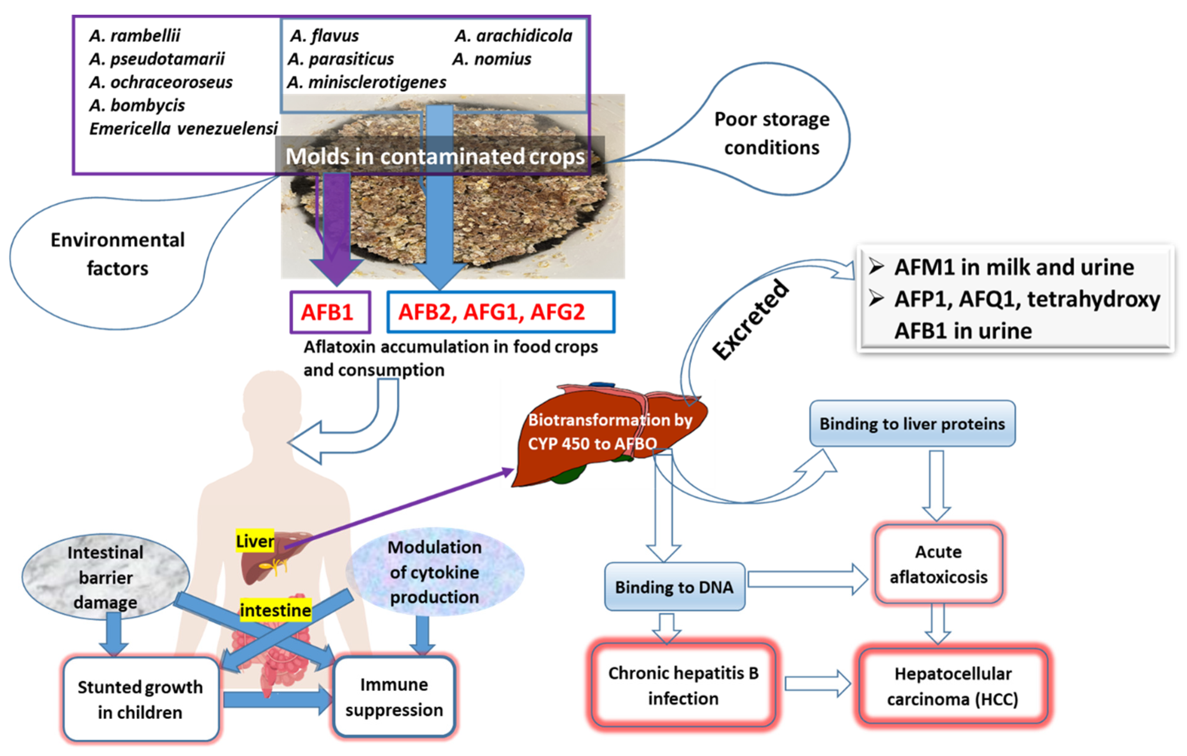
Consumption of feed or food contaminated by aflatoxins can cause various health problems due to their potent carcinogenicity that may affect all the organs and systems, especially the liver and kidneys, resulting in teratogenic, carcinogenic, immunosuppressive or genotoxic and mutagenic consequences [2,18,87,88]. Intake of large amounts of aflatoxins in the diet leads to an acute type of aflatoxicosis, while long-term exposure to aflatoxins results in the chronic type (Figure 3). The IARC has defined four groups for classification of mycotoxins as carcinogenic or potentially carcinogenic to humans, and classified AFB1 as a Group 1 carcinogen to humans [9,18].
Among all types of aflatoxins, AFB1 is the most pathogenic and potent one, with its median lethal dose (LD50) ranging from 1 to 50 mg/kg for most species and less than 1 mg/kg for highly susceptible animals like poultry, rainbow trout, and rats, with the susceptibility of a given species also varying according to genetic background, age, nutritional factors, and detoxification ability of its liver [23,76].
Aflatoxin B1-8,9-exo-epoxide, a reactive form that binds to DNA and albumin in blood serum, derived from AFB1 via liver biotransformation, forms adducts and hence causes DNA damage. Aflatoxin B1-8,9-exo-epoxide either binds to liver proteins, resulting in acute aflatoxicosis, or to DNA, causing hepatocellular carcinoma. Binding of these metabolites to nucleic acid and nucleoproteins essential to cellular viability (e.g., noticeably, the p53 tumor suppressor gene) in the liver leads to an excessive build-up of hepatic lipids, which enlarges the liver, proliferation of bile duct epithelium, necrosis, and hepatocellular carcinoma [18,74,88,89,90,91]. The binding of exo-epoxide to DNA results in the formation of the predominant pro-mutagenic 8,9-dihydro-8-(N7 guanyl)-9-hydroxy AFB1 adduct (AFB1-N7-Gua). Then, AFB1-N7-Gua will be transformed into the AFB1-formamidopyrimidine (AFB1-FABY) adduct, which may result in transversion mutations from guanine (G) to thymine (T). In addition, these adducts might trigger mutation of arginine to serine at codon 249 of the p53 tumor suppressor gene, abrogating the function of the tumor suppressor gene and contributing to carcinogenesis (Figure 3) [18,90,91,92]. Although AFM1 has lesser mutagenic and carcinogenic activity than AFB1, it exhibits a high level of genotoxic activity, posing a health hazard because of its possible accumulation and linkage to DNA [92,93,94].
Aflatoxicosis may cause symptoms such as vomiting, abdominal pain, pulmonary edema, and fatty infiltration and necrosis of the liver [64,95,96]. Aflatoxicosis may affect the intestines first, especially in infants and toddlers, given their dependence on milk products and multigrain cereal products and susceptibility to any disease. Aflatoxin intake might result in altered intestinal integrity and modulation of cytokine expression, both of which might cause stunted growth in young children and infants, as well as immune suppression [14,15,43,95,97]. Aflatoxins may also adversely affect various human organs and systems, including the kidney, heart, lungs, brain, epididymis, testis, ovary, many endocrine and exocrine organs, and skeletal muscles [2,89,95,98]. The largest risk for humans derives from long-term dietary aflatoxin exposure, associated with hepatocellular carcinomas, which may be synergistic with hepatitis B virus infection and hepatitis B [89,96].
A reduced level of secretory immunoglobulin A (IgA) is also associated with aflatoxicosis, impairing human cellular immunity against infections [98,99]. Aflatoxicosis is also the cause of other diseases, like Kwashiorkor and Reye’s syndrome in young children [73]. Kwashiorkor, a form of protein-energy malnutrition, and Reye’s syndrome, featuring encephalopathy and visceral deterioration, result in liver and kidney enlargement and cerebral edema. Some pathological characteristics of both diseases are linked to aflatoxin intoxication [86,95,100].
6. Detection and Analysis of Aflatoxins
Aflatoxins can contaminate cereals/crops intended for food and feed, as well as milk and animal products. Therefore, several studies focused their sampling on animal farms, food markets, food and feed processing facilities, and storage facilities in order to assess their toxic levels for comparison with toxins for samples from other sources [62,81,85,101]. Determination of aflatoxin intoxication and toxicity is difficult due to the need of a suitable analytical method and technique for the detection and quantification of the toxins in various samples. Moreover, the possibility of aflatoxins contaminating various foods throughout the food chain complicates the work of accurately determining the intoxication level in humans and animals. Studies have employed different biomarkers for determining intoxication in humans and animals [62,80,84].
Various analytical methods have been developed for the analysis and detection of aflatoxins from different sources. Those analytical methods are meant for different types of samples (in terms of physicochemical properties and sample matrix). Other factors include sample preparation techniques, presence of skilled experts, and availability of equipped facilities. Moreover, the intended type of result from detection methods (whether qualitative or quantitative), duration of analysis, and limits of detection and/or quantification cannot exceed the regulatory limits set by the relative authority [61,102].
6.1. Sample Preparation
Sample preparation is critical for aflatoxin detection, accounting for almost two-thirds of the analytical process. Most methods involve extraction of the toxin from matrices and using appropriate clean-up methods. Hence, the most commonly applied clean-up methods are liquid–liquid (LLE) or liquid–solid extractions (LSE) and solid-phase extraction (SPE) methods [81]. LLE and LSE are cheap and simple methods based on the solubility of aflatoxins in aqueous phases and matrices in a liquid solvent in solid phases, respectively. An appropriate extraction solvent is critical in the process, with acetonitrile/water and methanol/water solutions exhibiting their effectiveness in extracting aflatoxins from different matrices [62,81,103,104]. SPE, when used before the use of high-performance liquid chromatography (HPLC), has been successfully carried out with C18 cartridges for cleaning and extraction of aflatoxins [103]. However, these column-based extractions are time-consuming for elution of the retained analytes and have to use a significant amount of toxic organic solvents [105,106]. In contrast, another study used the magnetic solid phase extraction method, attaining 89.0% extraction yield for AFB1 with good repeatability when coupled with HPLC MS/MS [104].
6.2. Analytic Methods
Several factors should be considered in choosing an aflatoxin detection method, including limit of detection (LOD), sample type, type of extraction and sample processing, sensitivity, and repeatability of the result’s intended use [106,107,108]. Existing aflatoxin detection and analysis methods in common use include the chromatographic method, immunological method, and biosensors, plus recently developed mycotoxin detection, such as the use of nanomaterials and aptamers, which are better options.
6.2.1. Chromatographic Techniques
Aflatoxins have usually been detected with their photophysical properties, such as absorption and emission spectra, as they show characteristic absorption at 360 nm, which is the absorption maximum of the aflatoxin ring [108]. According to its characterization, different chromatographic and spectrophotometric methods have been developed.
As a conventional method for aflatoxin detection, thin layer chromatography (TLC) has been largely replaced by other chromatographic techniques, since TLC often produces inaccurate results due to errors in analysis procedures and inadequate laboratory conditions, such as moisture [108]. An advanced method is high-performance thin-layer chromatography (HPTLC), which has been validated by Ramesh et al. (2013), with LOD reaching 0.5 ppb [109,110].
Another new method is HPLC, boasting high detection sensitivity and adaptability, according to a suitable sample and analyte with or without both pre-column and post-column derivatizations [110,111,112]. It has been designated as an official analytical method for aflatoxins, due to its sensitivity, selectivity and reputability [108,113]. However, it needs qualified technicians to operate expensive instruments.
With higher detection sensitivity than HPLC, liquid chromatography-mass spectrometry (LC-MS/MS) can detect all the four types of aflatoxins at the same time from a sample [80,104]. Mass spectrophotometry technology augments the detection level, with LOD less than 1 ng/mL [114]. Despite reliance of its sensitivity on ionization and matrix-assisted calibration curves for quantitative analysis, this method is considered the most advanced technique for aflatoxin analysis [108]. Moreover, a recent addition of some supportive technical procedures has further enhanced the detection speed, sensitivity, and reputability of various detection methods. For instance, combined with a triple quadrupole (QqQ) mass analyzer, ultra-performance liquid chromatography-tandem mass spectrometry (UPLC MS/MS) has become one of the best solutions with hyphenated techniques for the quantitative determination of aflatoxins and other derived organic compounds [81,115].
6.2.2. Immunological Techniques
Among various immunological techniques, enzyme-linked immunosorbent assay (ELISA) has been applied in detecting and quantifying different aflatoxin analogues from liquid or liquefied samples [38,52,61]. Other immunological methods include immune-affinity column immune-enzymatic and immunochemical methods, which are usually applied for amplification of aflatoxin extraction and cleaning before chromatographic analysis [116,117]. These analytical and immunological methods, usually considered rapid techniques, are highly sensitive, specific, reproducible, and feature detection limits lower than those of biological detection methods, which are qualitative and often nonspecific in nature, time-consuming, and less sensitive compared to other methods [61,64,108,118,119].
The most extensively applied immunological screening technique for aflatoxin detection is competitive ELISA [38,120,121], whose detection sensitivity for aflatoxins varies with the kits of various manufacturers. It has been applied in detecting and quantifying aflatoxins from milk [38,61,119,121], crops, and food stuffs [7,52], due to the merits of simplicity, quickness, safety, as well as an LOD lower than that of other methods. However, they are not the best aflatoxin detection methods, given their cross reactivity of results, magnified false positive results, and lower accuracy [81,108]. However, a recently improved version has been introduced as tumor-specific antigen (TSA)-ELISA, which can increase the sensitivity of ELISA by amplifying the signal. Zhang et al. (2018), for instance, employed tyramine signal amplification to raise the sensitivity of ELISA several-fold in detecting AFB1 in edible oil samples [120]. Xu et al. (2021) also developed a novel indirect competitive metal-organic framework (MOF)-linked immunosorbent assay (MOFLISA) method with higher sensitivity, with recoveries and relative standard deviations (RSD) ranging from 86.41% to 99.74% and from 2.38% to 9.04%, respectively [122].
Another less known immunological technique for aflatoxin detection is radio-immunoassays (RIA), which, despite having risk of radiation for the analyst, have showed excellent recovery for application in crops [81,123]. Other immunological methods for aflatoxin detection include fluoroimmunoassays, flow-cytometry-based competitive fluorescent microsphere immunoassay (CFIA), and chemiluminescence immunoassay (CLIA), although they need sophisticated instruments for application [106,124].
6.2.3. Newer Detection Methods
Apart from those mentioned above, different analytical techniques identifying aflatoxins as hazardous xenobiotics have been applied. Another approach is the use of simple detection kits with sophisticated instruments. These other methods are mainly meant to quicken the analysis process of aflatoxins due to on-spot simple application, especially suitable for rural areas in developing countries to improve the detection process [81,102,105]. Most of these rapid techniques have the problem of insufficient sensitivity and limited detection [106]. Development of other analytical methods with high sensitivity is in the works, including the use of nanoparticles for ultradetection [6,61,118,125], aptamer-based methods (i.e., aptasensors) [126,127], and methods using immunosensors and other biosensors [61,128].
A common approach for developing these more effective and efficient methods is the combination of different techniques by replacing the drawbacks of one technique with another better procedure and making the analysis more robust. For instance, immune-chromatographic assays show remarkable analytic performance when using immuno-analysis as the extraction step to reduce analyte loss of samples. Additionally, the combination of an immunoaffinity column and high-performance liquid chromatography (IAC-HPLC) turns out to be highly effective and has become one of the widely used techniques for various sample types [105,116,129].
Nanotechnology has also been applied to detect and analyze aflatoxins with nanomaterials. Adányi et al. (2018) developed an immune-sensing method for the analysis of aflatoxins in a paprika matrix, using gold nanoparticles in a competitive immunoassay and optical waveguide light mode spectroscopy. It reaches a dynamic AFB1 detecting range of 0.01–10 ng/mL with EC50 of 0.044 ± 0.005 ng/mL [130]. Gold nanoparticles have also been used for total aflatoxin determination by labeling monoclonal antibodies of aflatoxins in the nanomaterials and applying an ultrasensitive immunochromatographic assay [117]. In addition, they have been combined with immunochromatographic analysis for rapid screening of aflatoxins [131].
7. Prevention and Control
The World Health Organization (WHO) reported in February 2018 that aflatoxins and associated problems can be controlled only by an integrated approach, calling for implementation of different strategies and monitoring in a “stable to table” manner, so as to minimize that risk of aflatoxin intoxication [28,96,100,102]. Applicable approaches include pre-harvest and post-harvest blockage of the toxin. Pre-harvest immunization preventing plants from harboring fungi can be applied via targeted plant breeding, enhancement of host plant resistance, and biological control [10,132,133]. Moreover, it is even more effective for these methods to be applied in conjunction with post-harvest technologies, such as proper drying and storage of susceptible crops, as well as methods contributive to the recovery of the lost value of contaminated crops [96,132,133,134].
Therefore, there are some ways for a competent authority to contribute to the control of aflatoxin contamination, including [96]:
- Removing the sources of contamination.
- Promoting better agricultural and storage techniques.
- Ensuring adequate resources that are available for testing and early diagnosis.
- Enforcing strict food safety standards.
- Informing and educating consumers and small/subsistence farmers.
- Promoting better livestock feeding and management.
- Creating public awareness of aflatoxin-contamination prevention.
Rules and Regulations on Aflatoxin Control in Various Countries
As it is impossible to remove aflatoxin contamination of human and animal diets entirely, the primary target, especially in more vulnerable countries, should be prevention and control via regulations and legislation to strictly monitor the trade of food and feed and transfer/migration of aflatoxins and the fungus from place to place, ultimately lowering intoxication occurrence [43,135,136].
At least 199 countries have instituted such regulations and legislations, specifying maximum acceptable aflatoxin level in feeds and foods, which vary for different commodities in various countries, often set according to specific economic and social factors, with acceptable AFM1 limits invariably much lower than food/feed levels, especially considering the effect of intoxication issues on children [137]. Such aflatoxin limits, thus, often vary for different food (Table 2) or feed types in a country [64,136,137]. The absence of such limits or even relevant regulations and legislation have brought grave consequences to some countries [102].
Sub-Saharan Africa and other developing countries are victims of aflatoxin intoxication, which causes grave economical and public health consequences. In addition to climatic, economical, and agronomical factors, another major cause of the ravage of aflatoxins in those countries is the absence of rigorous regulation, such as that on food and feed import. The Codex Alimentarius has formulated standards as reference for regulation on imported feed and food, suggesting setting the maximum levels for aflatoxins in various nuts, grains, dried figs, and milk in the range of 0.5 to 15 ppb (µg/kg) [24,43,64].
The United States Food and Drug Administration (USFDA) has set different limits of aflatoxins for various types of commodities and food and feed products, including a maximum level of 20 ppb for food, 0.5 ppb AFM1 for milk, and 100 ppb and 300 ppb for feeds for breeding and feedlot animals, respectively [138,139]. The Commission Regulation of the European Union (EU) No. 165/2010 has set the maximum level of 0.05 ppb for AFM1 in milk. In addition, the EU set the total acceptable levels of total aflatoxins in food to be 4 ppb and 20 ppb for animals feed, which is somehow different and more stringent [24,33,144].
8. Conclusions
Aflatoxin intoxication is a universal food problem worldwide due to the ubiquitous presence of fungi, as well as in view of global warming and climate change, which enhance moisture and temperature everywhere. Tropical countries are susceptible to aflatoxin intoxication of feed and food products. Aflatoxin detection and screening is not easy, leading to the prevalence of aflatoxin contamination of foods. The best strategy dealing with the problem is prevention via pre- and post-harvest inspections. Developing a robust, fast, and more accurate detection and analytical method for aflatoxins is, therefore, crucial in mitigating the devastating consequences of intoxication. As the pathogenesis and potency of aflatoxins is closely associated with their biotransformation, which makes them hazardous, how to reduce these harmful alterations is a worthwhile subject for study in this field. As potent carcinogens, aflatoxins have been on the radar of governments worldwide, many of which have instituted stringent regulations for their prevention and control. Moreover, the industrial sector has also been striving to solve the problem in order to reduce economic losses caused by aflatoxin contamination.
Author Contributions
Conceptualization, S.A., V.R.M. and M.H.; writing—original draft preparation, S.A., V.R.M. and M.H.; writing—review and editing, Y.-Y.C., Y.-C.L. and Y.-P.C.; supervision, V.R.M. and Y.-P.C.; funding acquisition, Y.-P.C. All authors have read and agreed to the published version of the manuscript.
Funding
The study was funded by the iEGG and Animal Biotechnology Center from The Feature Area Research Center Program within the framework of the Higher Education Sprout Project by the Ministry of Education (MOE) in Taiwan.
Institutional Review Board Statement
Not applicable.
Informed Consent Statement
Not applicable.
Data Availability Statement
Not applicable.
Acknowledgments
The authors acknowledge the joint agreement between NCHU and Vel Tech Rangarajan Dr. Sagunthala R&D Institute of Science and Technology. In addition, we would like to pass our gratitude to both Universities for their support.
Conflicts of Interest
The authors declare no conflict of interest.
References
- Chauhan, N.M.; Washe, A.P.; Minota, T. Fungal infection and aflatoxin contamination in maize collected from Gedeo zone, Ethiopia. Springerplus 2016, 5, 753. [Google Scholar] [CrossRef] [PubMed] [Green Version]
- Bbosa, G.S.; Kitya, D.; Lubega, A.; Ogwal-Okeng, J.; Anokbonggo, W.W.; Kyegombe, B.D. Review of the Biological and Health Effects of Aflatoxins on Body Organs and Body Systems. In Aflatoxins—Recent Adv Futur Prospect; Razzaghi-Abyaneh, M., Ed.; IntechOpen: London, UK, 2013; pp. 240–265. [Google Scholar]
- Balina, A.; Kebede, A.; Tamiru, Y. Review on Aflatoxin and its Impacts on Livestock. JOJ Sci. Publ. 2018, 1, 1–7. [Google Scholar]
- Probst, C.; Schulthess, F.; Cotty, P.J. Impact of Aspergillus section Flavi community structure on the development of lethal levels of aflatoxins in Kenyan maize (Zea mays). J. Appl. Microbiol. 2010, 108, 600–610. [Google Scholar] [CrossRef]
- Pankaj, S.; Shi, H.; Keener, K.M. A review of novel physical and chemical decontamination technologies for aflatoxin in food. Trends Food Sci. Technol. 2018, 71, 73–83. [Google Scholar] [CrossRef]
- Alshannaq, A.; Yu, J.-H. Occurrence, Toxicity, and Analysis of Major Mycotoxins in Food. Int. J. Environ. Res. Public Health 2017, 14, 632. [Google Scholar] [CrossRef] [Green Version]
- Chala, A.; Mohammed, A.; Ayalew, A.; Skinnes, H. Natural occurrence of aflatoxins in groundnut (Arachis hypogaea L.) from eastern Ethiopia. Food Control 2013, 30, 602–605. [Google Scholar] [CrossRef]
- Battilani, P.; Toscano, P.; Van Der Fels-Klerx, H.J.; Moretti, A.; Leggieri, M.C.; Brera, C.; Rortais, A.; Goumperis, T.; Robinson, T. Aflatoxin B1 contamination in maize in Europe increases due to climate change. Sci. Rep. 2016, 6, 24328. [Google Scholar] [CrossRef] [Green Version]
- Al-Zoreky, N.S.; Saleh, F.A. Limited survey on aflatoxin contamination in rice. Saudi J. Biol. Sci. 2017, 26, 225–231. [Google Scholar] [CrossRef]
- Gonçalves, A.; Gkrillas, J.L.A.; Dorne, C.J.L.; Dall’Asta, R.C.; Palumbo, R.; Lima, N.R.; Battilani, P.; Venâncio, A.; Giorni, V.P. Pre- and Postharvest Strategies to Minimize Mycotoxin Contamination in the Rice Food Chain. Compr. Rev. Food Sci. Food Saf. 2019, 18, 441–454. [Google Scholar] [CrossRef]
- Chala, A.; Taye, W.; Ayalew, A.; Krska, R.; Sulyok, M.; Logrieco, A. Multimycotoxin analysis of sorghum (Sorghum bicolor L. Moench) and finger millet (Eleusine coracana L. Garten) from Ethiopia. Food Control 2014, 45, 29–35. [Google Scholar] [CrossRef]
- Heshmati, A.; Khorshidi, M.; Khaneghah, A.M. The prevalence and risk assessment of aflatoxin in sesame based products. Ital. J. Food Sci. 2021, 33, 92–102. [Google Scholar] [CrossRef]
- Min, L.; Li, D.; Tong, X.; Sun, H.; Chen, W.; Wang, G.; Zheng, N.; Wang, J. The challenges of global occurrence of aflatoxin M1 contamination and the reduction of aflatoxin M1 in milk over the past decade. Food Control 2020, 117, 107352. [Google Scholar] [CrossRef]
- Fakhri, Y.; Rahmani, J.; Oliveira, C.A.F.; Franco, L.T.; Corassin, C.H.; Saba, S.; Rafique, J.; Khaneghah, A.M. Aflatoxin M1 in human breast milk: A global systematic review, meta-analysis, and risk assessment study (Monte Carlo simulation). Trends Food Sci. Technol. 2019, 88, 333–342. [Google Scholar] [CrossRef]
- Achaglinkame, M.A.; Opoku, N.; Amagloh, F.K. Aflatoxin contamination in cereals and legumes to reconsider usage as complementary food ingredients for Ghanaian infants: A review. J. Nutr. Intermed. Metab. 2017, 10, 1–7. [Google Scholar] [CrossRef]
- Hassane, A.M.A.; El-Shanawany, A.A.; Abo-Dahab, N.F.; Abdel-Hadi, A.M.; Abdul-Raouf, U.M.; Mwanza, M. Influence of Different Moisture Contents and Temperature on Growth and Production of Aflatoxin B1 by a Toxigenic Aspergillus flavus Isolate in Wheat Flour. J. Ecol. Health Environ. 2017, 5, 77–83. [Google Scholar] [CrossRef]
- Wu, J.; Chen, R.; Zhang, C.; Li, K.; Xu, W.; Wang, L.; Chen, Q.; Mu, P.; Jiang, J.; Wen, J.; et al. Bioactivation and Regioselectivity of Pig Cytochrome P450 3A29 towards Aflatoxin B1. Toxins 2016, 8, 267. [Google Scholar] [CrossRef] [Green Version]
- Dai, Y.; Huang, K.; Zhang, B.; Zhu, L.; Xu, W. Aflatoxin B1-induced epigenetic alterations: An overview. Food Chem. Toxicol. 2017, 109, 683–689. [Google Scholar] [CrossRef]
- Pickova, D.; Ostry, V.; Toman, J.; Malir, F. Aflatoxins: History, Significant Milestones, Recent Data on Their Toxicity and Ways to Mitigation. Toxins 2021, 13, 399. [Google Scholar] [CrossRef]
- Prietto, L.; Moraes, P.S.; Kraus, R.B.; Meneghetti, V.; Fagundes, C.A.A.; Furlong, E.B. Post-harvest operations and aflatoxin levels in rice (Oryza sativa). Crop Prot. 2015, 78, 172–177. [Google Scholar] [CrossRef]
- Benkerroum, N. Aflatoxins: Producing-molds, structure, health issues and incidence in southeast asian and sub-saharan african countries. Int. J. Environ. Res. Public Health 2020, 17, 1215. [Google Scholar] [CrossRef] [Green Version]
- Blount, W.P. Turkey “X” disease. J. Br. Turkey Fed. 1961, 9, 52–77. [Google Scholar]
- Bennett, J.W.; Kale, S.; Yu, J. Aflatoxins: Background, toxicology, and molecular biology. In Foodborne Diseases, Infectious Disease, 1st ed.; Simjee, S., Ed.; Humana Press Inc.: Totowa, NJ, USA, 2007; pp. 355–373. [Google Scholar]
- Lizárraga-Paulín, E.G.; Moreno-Martínez, E.; Miranda-Castro, S.P. Aflatoxins and their impact on human and animal health: An emerging problem. In Aflatoxins—Biochemistry and Molecular Biology, 1st ed.; Guevara-Gonzalez, R.G., Ed.; IntechOpen: London, UK, 2011; pp. 255–282. [Google Scholar]
- Wacoo, A.P.; Wendiro, D.; Nanyonga, S.; Hawumba, J.F.; Sybesma, W.; Kort, R. Feasibility of A Novel On-Site Detection Method for Aflatoxin in Maize Flour from Markets and Selected Households in Kampala, Uganda. Toxins 2018, 10, 327. [Google Scholar] [CrossRef] [PubMed]
- Pinotti, L.; Ottoboni, M.; Giromini, C.; Dell’Orto, V.; Cheli, F. Mycotoxin Contamination in the EU Feed Supply Chain: A Focus on Cereal Byproducts. Toxins 2016, 8, 45. [Google Scholar] [CrossRef] [PubMed] [Green Version]
- Guchi, E. Aflatoxin contamination in groundnut (Arachis hypogaea L.) Caused by Aspergillus Species in Ethiopia. J. Appl. Environ. Microbiol. 2015, 3, 11–19. [Google Scholar]
- Campagnollo, F.B.; Ganev, K.C.; Khaneghah, A.M.; Portela, J.B.; Cruz, A.G.; Granato, D.; Corassin, C.H.; Oliveira, C.A.F.; Sant’Ana, A.S. The occurrence and effect of unit operations for dairy products processing on the fate of aflatoxin M1: A review. Food Control 2016, 68, 310–329. [Google Scholar] [CrossRef]
- Peng, Z.; Chen, L.; Zhu, Y.; Huang, Y.; Hu, X.; Wu, Q.; Nüssler, A.K.; Liu, L.; Yang, W. Current major degradation methods for aflatoxins: A review. Trends Food Sci. Technol. 2018, 80, 155–166. [Google Scholar] [CrossRef]
- Fandohan, P.; Zoumenou, D.; Hounhouigan, D.J.; Marasas, W.F.O.; Wingfield, M.J.; Hell, K. Fate of aflatoxins and fumonisins during the processing of maize into food products in Benin. Int. J. Food Microbiol. 2005, 98, 249–259. [Google Scholar] [CrossRef]
- Sirma, A.; Lindahl, J.; Makita, K.; Senerwa, D.; Mtimet, N.; Kang’Ethe, E.; Grace, D. The impacts of aflatoxin standards on health and nutrition in sub-Saharan Africa: The case of Kenya. Glob. Food Secur. 2018, 18, 57–61. [Google Scholar] [CrossRef]
- Warnatzsch, E.A.; Reay, D.S.; Leggieri, M.C.; Battilani, P. Climate Change Impact on Aflatoxin Contamination Risk in Malawi’s Maize Crops. Front. Sustain. Food Syst. 2020, 4, 1–13. [Google Scholar] [CrossRef]
- Rajarajan, P.N.; Rajasekaran, K.M.; Devi, N.K.A. Aflatoxin Contamination in Agricultural Commodities. Indian J. Pharm. Biol. Res. 2013, 1, 148–151. [Google Scholar] [CrossRef]
- Hailu, M.; Narayanaswamy, R.; Abrehame, S.; Remony, M.V. In Silico analysis of glycogen synthase kinase 3-beta (GSK-3β) as aflatoxin binder. Biomed. Pharmacol. J. 2019, 12, 1449–1456. [Google Scholar]
- Gratz, S. Aflatoxin binding by probiotics: Experimental studies on intestinal aflatoxin transport, metabolism and toxicity. Kuopio Univ. Publ. D Med. Sci. 2007, 404, 1–100. [Google Scholar]
- Abrehame, S.; Hailu, M.; Manoj, V.R.; Chen, Y.-P.; Narayanaswamy, R. Molecular docking analysis of Azadirachta indica constituents as inhibitors of aflatoxin polyketide synthase (APKS). Rasayan J. Chem. 2021, 14, 920–929. [Google Scholar] [CrossRef]
- Korman, T.P.; Crawford, J.M.; Labonte, J.W.; Newman, A.G.; Wong, J.; Townsend, C.A.; Tsai, S.-C. Structure and function of an iterative polyketide synthase thioesterase domain catalyzing Claisen cyclization in aflatoxin biosynthesis. Proc. Natl. Acad. Sci. USA 2010, 107, 6246–6251. [Google Scholar] [CrossRef] [Green Version]
- Gizachew, D.; Szonyi, B.; Tegegne, A.; Hanson, J.; Grace, D. Aflatoxin contamination of milk and dairy feeds in the Greater Addis Ababa milk shed, Ethiopia. Food Control 2016, 59, 773–779. [Google Scholar] [CrossRef]
- Meijer, N.; Kleter, G.; de Nijs, M.; Rau, M.-L.; Derkx, R.; van der Fels-Klerx, H.J. The aflatoxin situation in Africa: Systematic literature review. Compr. Rev. Food Sci. Food Saf. 2021, 20, 2286–2304. [Google Scholar] [CrossRef]
- Yu, J. Current Understanding on Aflatoxin Biosynthesis and Future Perspective in Reducing Aflatoxin Contamination. Toxins 2012, 4, 1024–1057. [Google Scholar] [CrossRef] [Green Version]
- Lahouar, A.; Marin, S.; Crespo-Sempere, A.; Saïd, S.; Sanchis, V. Effects of temperature, water activity and incubation time on fungal growth and aflatoxin B1 production by toxinogenic Aspergillus flavus isolates on sorghum seeds. Rev. Argent Microbiol. 2016, 48, 78–85. [Google Scholar] [CrossRef] [Green Version]
- Mannaa, M.; Kim, K.D. Influence of Temperature and Water Activity on Deleterious Fungi and Mycotoxin Production during Grain Storage. Mycobiology 2017, 45, 240–254. [Google Scholar] [CrossRef]
- Wagacha, J.; Muthomi, J. Mycotoxin problem in Africa: Current status, implications to food safety and health and possible management strategies. Int. J. Food Microbiol. 2008, 124, 1–12. [Google Scholar] [CrossRef]
- Abbas, H.; Wilkinson, J.; Zablotowicz, R.; Accinelli, C.; Abel, C.; Bruns, H.; Weaver, M. Ecology of Aspergillus flavus, regulation of aflatoxin production, and management strategies to reduce aflatoxin contamination of corn. Toxin Rev. 2009, 28, 142–153. [Google Scholar] [CrossRef]
- Hedayati, M.T.; Pasqualotto, A.C.; Warn, P.A.; Bowyer, P.; Denning, D.W. Aspergillus flavus: Human pathogen, allergen and mycotoxin producer. Microbiology 2007, 153, 1677–1692. [Google Scholar] [CrossRef] [PubMed] [Green Version]
- Atanda, S.A. Fungi and mycotoxins in stored foods. Afr. J Microbiol. Res. 2011, 5, 4373–4382. [Google Scholar] [CrossRef]
- Donato, C.J.R.; Cendoya, E.; Demonte, L.D.; Repetti, M.R.; Chulze, S.N.; Ramirez, M.L. Influence of abiotic factors (water activity and temperature) on growth and aflatoxin production by Aspergillus flavus in a chickpea-based medium. Int. J. Food Microbiol. 2022, 379, 109841. [Google Scholar] [CrossRef]
- Sharma, K.K.; Pothana, A.; Prasad, K.; Shah, D.; Kaur, J.; Bhatnagar, D.; Chen, Z.-Y.; Raruang, Y.; Cary, J.W.; Rajasekaran, K.; et al. Peanuts that keep aflatoxin at bay: A threshold that matters. Plant Biotechnol. J. 2017, 16, 1024–1033. [Google Scholar] [CrossRef]
- Hlashwayo, D.F. Aflatoxin B1 contamination in raw peanuts sold in Maputo City, Mozambique and associated factors. J. Stored Prod. Postharvest Res. 2018, 9, 58–67. [Google Scholar]
- Jacxsens, L.; Pratheeb, Y.; De Meulenaer, B. Risk Assessment of Mycotoxins and Predictive Mycology in Sri Lankan Spices: Chilli and Pepper. Procedia Food Sci. 2016, 6, 326–330. [Google Scholar] [CrossRef] [Green Version]
- Abdullah, E.; Idris, A.; Saparon, A. Peanuts that keep aflatoxin at bay: A threshold that matters. ARPN J. Eng. Appl. Sci. 2017, 12, 3218–3221. [Google Scholar]
- Colak, H.; Bingol, E.B.; Hampikyan, H.; Nazli, B. Determination of aflatoxin contamination in red-scaled, red and black pepper by ELISA and HPLC. J. Food Drug Anal. 2005, 14, 12. [Google Scholar] [CrossRef]
- Kilic, S.; Cam, I.B.; Tongur, T.; Kilic, M. Health Risk Assessment of Exposure to Heavy Metals and Aflatoxins via Dietary Intake of Dried Red Pepper from Marketplaces in Antalya, Southern Turkey. J. Food Sci. 2018, 83, 2675–2681. [Google Scholar] [CrossRef]
- Naz, F.; Verpoort, F.; Iqbal, S.Z.; Naheed, N.; Asi, M.R. Seasonal Variation of Aflatoxin Levels in Selected Spices Available in Retail Markets: Estimation of Exposure and Risk Assessment. Toxins 2022, 14, 597. [Google Scholar] [CrossRef] [PubMed]
- Singh, P.; Cotty, P.J. Aflatoxin contamination of dried red chilies: Contrasts between the United States and Nigeria, two markets differing in regulation enforcement. Food Control 2017, 80, 374–379. [Google Scholar] [CrossRef]
- El Mahgubi, A.; Puel, O.; Bailly, S.; Tadrist, S.; Querin, A.; Ouadia, A.; Oswald, I.; Bailly, J. Distribution and toxigenicity of Aspergillus section Flavi in spices marketed in Morocco. Food Control 2013, 32, 143–148. [Google Scholar] [CrossRef]
- Maleki, F.; Abdi, S.; Davodian, E.; Haghani, K.; Bakhtiyari, S. Exposure of infants to aflatoxin M1 from mother’s breast milk in Ilam, Western Iran. Osong Public Health Res. Perspect. 2015, 6, 283–287. [Google Scholar] [CrossRef] [Green Version]
- Scaglioni, P.; Becker-Algeri, T.; Drunkler, D.; Badiale-Furlong, E. Aflatoxin B1 and M1 in milk. Anal. Chim. Acta 2014, 829, 68–74. [Google Scholar] [CrossRef]
- Shigute, T.; Washe, A.P. Reduction of Aflatoxin M1 Levels during Ethiopian Traditional Fermented Milk (Ergo) Production. J. Food Qual. 2018, 2018, 4570238. [Google Scholar] [CrossRef] [Green Version]
- Mohammadi, H. A review of aflatoxin M 1, milk and milk products. Aflatoxins—Biochem. Mol. Biol. 2001, 1, 397–414. [Google Scholar]
- Gurban, A.-M.; Epure, P.; Oancea, F.; Doni, M. Achievements and Prospects in Electrochemical-Based Biosensing Platforms for Aflatoxin M1 Detection in Milk and Dairy Products. Sensors 2017, 17, 2951. [Google Scholar] [CrossRef] [PubMed]
- Viegas, S.; Assunção, R.; Nunes, C.; Osteresch, B.; Twarużek, M.; Kosicki, R.; Grajewski, J.; Martins, C.; Alvito, P.; Almeida, A.; et al. Exposure assessment to mycotoxins in a Portuguese fresh bread dough company by using a multi-biomarker approach. Toxins 2018, 10, 342. [Google Scholar] [CrossRef] [Green Version]
- Kemppainen, B.W.; Riley, R.T.; Pace, J.G. Skin Absorption as a Route of Exposure for Aflatoxin and Trichothecenes. J. Toxicol. Toxin Rev. 1988, 7, 95–120. [Google Scholar] [CrossRef]
- Ajani, A.J.; Chakravarthy, D.V.S.; Tanuja, P.; Pasha, K.V. Aflatoxins. Indian J. Adv. Chem. Sci. 2014, 3, 49–60. [Google Scholar]
- Prabakaran, J.J.; Dhanapal, S. Analysis of Proximate Composition and Aflatoxins of Some Poultry Feeds. Asian J. Biotechnol. 2009, 1, 104–110. [Google Scholar] [CrossRef] [Green Version]
- Zhang, J.; Zheng, N.; Liu, J.; Li, F.; Li, S.; Wang, J. Aflatoxin B1 and aflatoxin M1 induced cytotoxicity and DNA damage in differentiated and undifferentiated Caco-2 cells. Food Chem. Toxicol. 2015, 83, 54–60. [Google Scholar] [CrossRef] [PubMed]
- Gao, Y.; Wang, J.; Li, S.; Zhang, Y.; Zheng, N. Aflatoxin M1 cytotoxicity against human intestinal Caco-2 cells is enhanced in the presence of other mycotoxins. Food Chem. Toxicol. 2016, 96, 79–89. [Google Scholar] [CrossRef]
- Zhang, L.Y.; Liu, S.; Zhao, X.J.; Wang, N.; Jiang, X.; Xin, H.S.; Zhang, Y.G. Lactobacillus rhamnosus GG modulates gastrointestinal absorption, excretion patterns, and toxicity in Holstein calves fed a single dose of aflatoxin B1. J. Dairy Sci. 2019, 102, 1330–1340. [Google Scholar] [CrossRef] [PubMed] [Green Version]
- Moschini, M.; Masoero, F.; Gallo, A.; Díaz, D. Mucosal absorption of aflatoxin B1 in lactating dairy cows. Ital. J. Anim. Sci. 2007, 6, 324–326. [Google Scholar] [CrossRef]
- Zhu, Q.; Ma, Y.; Liang, J.; Wei, Z.; Li, M.; Zhang, Y.; Liu, M.; He, H.; Qu, C.; Cai, J.; et al. AHR mediates the aflatoxin B1 toxicity associated with hepatocellular carcinoma. Signal Transduct. Target. Ther. 2021, 6, 299. [Google Scholar] [CrossRef]
- Van Vleet, T.R.; Klein, P.J.; Coulombe, R.A., Jr. Metabolism of aflatoxin B1 by normal human bronchial epithelial cells. J. Toxicol. Environ. Health Part A 2001, 63, 525–540. [Google Scholar] [CrossRef]
- Wilson, R.; Ziprin, R.; Ragsdale, S.; Busbee, D. Uptake and vascular transport of ingested aflatoxin. Toxicol. Lett. 1985, 29, 169–176. [Google Scholar] [CrossRef]
- Costanzo, P.; Santini, A.; Fattore, L.; Novellino, E.; Ritieni, A. Toxicity of aflatoxin B1 towards the vitamin D receptor (VDR). Food Chem. Toxicol. 2015, 76, 77–79. [Google Scholar] [CrossRef] [Green Version]
- Mace, K.; Aguilar, F.; Wang, J.S.; Vautravers, P.; Gómez-Lechón, M.; Gonzalez, F.J.; Groopman, J.; Harris, C.C.; Pfeifer, A.M. Aflatoxin B1-induced DNA adduct formation and p53 mutations in CYP450- expressing human liver cell lines. Carcinog. 1997, 18, 1291–1297. [Google Scholar] [CrossRef] [PubMed] [Green Version]
- Diaz, G.J.; Murcia, H.W.; Cepeda, S.M. Cytochrome P450 enzymes involved in the metabolism of aflatoxin B1 in chickens and quail. Poult. Sci. 2010, 89, 2461–2469. [Google Scholar] [CrossRef] [PubMed]
- Seid, A.; Mama, A. Aflatoxicosis and occurrence of aflatoxin M1 (AFM1) in milk and dairy products: A Review. Austin. J. Vet. Sci. Anim Husb. 2019, 1, 1–12. [Google Scholar]
- Gacem, M.A.; El Hadj-Khelil, A.O. Toxicology, biosynthesis, bio-control of aflatoxin and new methods of detection. Asian Pac. J. Trop. Biomed. 2016, 6, 808–814. [Google Scholar] [CrossRef] [Green Version]
- Tran, V.N.; Viktorová, J.; Ruml, T. Mycotoxins: Biotransformation and Bioavailability Assessment Using Caco-2 Cell Monolayer. Toxins 2020, 12, 628. [Google Scholar] [CrossRef]
- Tadesse, S.; Berhanu, T.; Woldegiorgis, A.Z. Aflatoxin M1 in milk and milk products marketed by local and industrial producers in Bishoftu town of Ethiopia. Food Control 2020, 118, 107386. [Google Scholar] [CrossRef]
- Ayelign, A.; Woldegiorgis, A.Z.; Adish, A.; De Boevre, M.; Heyndrickx, E.; De Saeger, S. Assessment of aflatoxin exposure among young children in Ethiopia using urinary biomarkers. Food Addit. Contam. Part A 2017, 34, 1606–1616. [Google Scholar] [CrossRef]
- Miklós, G.; Angeli, C.; Ambrus, Á.; Nagy, A.; Kardos, V.; Zentai, A.; Kerekes, K.; Farkas, Z.; Jó´zwiak, Á.; Bartók, T. Detection of aflatoxins in different matrices and food-chain positions. Front Microbiol. 2020, 11, 1916. [Google Scholar] [CrossRef]
- Battacone, G.; Nudda, A.; Cannas, A.; Borlino, A.C.; Bomboi, G.; Pulina, G. Excretion of Aflatoxin M1 in Milk of Dairy Ewes Treated with Different Doses of Aflatoxin B1. J. Dairy Sci. 2003, 86, 2667–2675. [Google Scholar] [CrossRef] [Green Version]
- Crudo, F.; Barilli, A.; Mena, P.; Rotoli, B.M.; Del Rio, D.; Dall’Asta, C.; Dellafiora, L. An in vitro study on the transport and phase II metabolism of the mycotoxin alternariol in combination with the structurally related gut microbial metabolite urolithin C. Toxicol. Lett. 2021, 340, 15–22. [Google Scholar] [CrossRef]
- Franco, L.T.; Khaneghah, A.M.; Lee, S.H.I.; Oliveira, C.A.F. Biomonitoring of mycotoxin exposure using urinary biomarker approaches: A review. Toxin Rev. 2019, 40, 383–403. [Google Scholar] [CrossRef]
- Hassan, F.F. Detection of aflatoxin B1 in some canned foods and reduction of toxin by ultraviolet radiation. Iraqi. J. Sci. 2018, 58, 2343–2349. [Google Scholar]
- Magnussen, A.; Parsi, M.A. Aflatoxins, hepatocellular carcinoma and public health. World J. Gastroenterol. 2013, 19, 1508–1512. [Google Scholar] [CrossRef] [PubMed]
- Ghasemi-Kebria, F.; Joshaghani, H.R.; Taheri, N.S.; Semnani, S.; Aarabi, M.; Salamat, F.; Roshandel, G. Aflatoxin contamination of wheat flour and the risk of esophageal cancer in a high risk area in Iran. Cancer Epidemiol. 2013, 37, 290–293. [Google Scholar] [CrossRef] [PubMed] [Green Version]
- Wu, H.-C.; Santella, R. The Role of Aflatoxins in Hepatocellular Carcinoma. Hepat. Mon. 2012, 12, e7238. [Google Scholar] [CrossRef] [Green Version]
- Qi, L.N.; Bai, T.; Chen, Z.S.; Wu, F.X.; Chen, Y.Y.; De Xiang, B.; Peng, T.; Han, Z.G.; Li, L.Q. The p53 mutation spectrum in hepatocellular carcinoma from Guangxi, China: Role of chronic hepatitis B virus infection and aflatoxin B1 exposure. Liver Int. 2015, 35, 999–1009. [Google Scholar] [CrossRef] [PubMed]
- Marchese, S.; Polo, A.; Ariano, A.; Velotto, S.; Costantini, S.; Severino, L. Aflatoxin B1 and M1: Biological Properties and Their Involvement in Cancer Development. Toxins 2018, 10, 214. [Google Scholar] [CrossRef] [Green Version]
- Kew, M.C. Aflatoxins as a cause of hepatocellular carcinoma. J. Gastrointest. Liver Dis. 2013, 22, 305–310. [Google Scholar]
- Yilmaz, S.; Baga, H. Afl atoxin B1: Mechanism, oxidative stress, and eff ects on animal health. Insights Vet. Sci. 2022, 6, 17–18. [Google Scholar]
- Yang, X.; Lv, Y.; Huang, K.; Luo, Y.; Xu, W. Zinc inhibits aflatoxin B1-induced cytotoxicity and genotoxicity in human hepatocytes (HepG2 cells). Food Chem. Toxicol. 2016, 92, 17–25. [Google Scholar] [CrossRef]
- Gross-Steinmeyer, K.; Eaton, D.L. Dietary modulation of the biotransformation and genotoxicity of aflatoxin B1. Toxicology 2012, 299, 69–79. [Google Scholar] [CrossRef] [PubMed]
- Kumar, P.; Mahato, D.K.; Kamle, M.; Mohanta, T.K.; Kang, S.G. Aflatoxins: A Global Concern for Food Safety, Human Health and Their Management. Front. Microbiol. 2017, 7, 2170. [Google Scholar] [CrossRef] [PubMed] [Green Version]
- WHO. Aflatoxins. Food Safety Digest, Department of Food Safety and Zoonoses; WHO Technical Report Series; WHO: Geneva, Switzerland, 2018.
- Khlangwiset, P.; Shephard, G.S.; Wu, F. Aflatoxins and growth impairment: A review. Crit. Rev. Toxicol. 2011, 41, 740–755. [Google Scholar] [CrossRef] [PubMed]
- Nabi, H.; Hussain, I.; Adil, M.; Nasir, A.; Sikandar, A.; Khan, S.; Khan, N. Impact of Mycotoxin Binders on Humoral Immunity, Lymphoid Organs and Growth Performance of Broilers. Pak. J. Zoöl. 2018, 50, 1611–1618. [Google Scholar] [CrossRef]
- Genevieve, S.; Bondy, J.; Pestka, J. Immunomodulation by fungal toxins. J. Toxicol. Environ. Health Part B Crit. Rev. 2000, 12, 2004–2007. [Google Scholar]
- Bennett, J.W.; Klich, M. Mycotoxins. Clin. Microbiol. Rev. 2003, 16, 497–516. [Google Scholar] [CrossRef] [Green Version]
- Nguyen, T.; Flint, S.; Palmer, J. Control of aflatoxin M1 in milk by novel methods: A review. Food Chem. 2019, 311, 125984. [Google Scholar] [CrossRef]
- Jallow, A.; Xie, H.; Tang, X.; Qi, Z.; Li, P. Worldwide aflatoxin contamination of agricultural products and foods: From occurrence to control. Compr. Rev. Food Sci. Food Saf. 2021, 20, 2332–2381. [Google Scholar] [CrossRef]
- Khayoon, W.S.; Saad, B.; Yan, C.B.; Hashim, N.H.; Ali, A.S.M.; Salleh, M.I.; Salleh, B. Determination of aflatoxins in animal feeds by HPLC with multifunctional column clean-up. Food Chem. 2010, 118, 882–886. [Google Scholar] [CrossRef]
- Mccullum, C.; Tchounwou, P.; Ding, L.; Sheng, L.X.; Liu, Y.M. Extraction of aflatoxins from liquid foodstu of samples with polydopamine-coated superparamagnetic nanoparticles for HPLC-MS/MS Analysis. J. Agri. Food Chem. 2014, 62, 4261–4267. [Google Scholar] [CrossRef]
- Yao, H.; Hruska, Z.; Di Mavungu, J.D. Developments in detection and determination of aflatoxins. World Mycotoxin J. 2015, 8, 181–191. [Google Scholar] [CrossRef]
- Matabaro, E.; Ishimwe, N.; Uwimbabazi, E.; Lee, B.H. Current Immunoassay Methods for the Rapid Detection of Aflatoxin in Milk and Dairy Products. Compr. Rev. Food Sci. Food Saf. 2017, 16, 808–820. [Google Scholar] [CrossRef] [PubMed]
- Nyangi, C.J. Quantitative risk assessment for aflatoxin and fumonisin from maize consumption in Northern Tanzania. Int. J. Innov. Res. Dev. 2018, 7, 478–487. [Google Scholar]
- Mahfuz, M.; Gazi, A.; Hossain, M.; Islam, R.; Fahim, S.M.; Ahmed, T. General and advanced methods for the detection and measurement of aflatoxins and aflatoxin metabolites: A review. Toxin Rev. 2018, 39, 123–137. [Google Scholar] [CrossRef]
- Ramesh, J.; Sarathchandra, G.; Sureshkumar, V. Analysis of feed samples for aflatoxin B1 contamination by HPTLC: A validated method. Int. J. Curr. Microbiol. App. Sci. 2013, 2, 373–377. [Google Scholar]
- Kotinagu, K.; Mohanamba, T.; Kumari, L.R. Assessment of aflatoxin B1 in livestock feed and feed ingredients by high-performance thin layer chromatography. Vet. World 2015, 8, 1396–1399. [Google Scholar] [CrossRef] [PubMed]
- Nile, S.H.; Park, S.W.; Khobragade, C.N. Occurrence and analysis of aflatoxin M1 in milk produced by Indian dairy species. Food Agric. Immunol. 2016, 27, 358–366. [Google Scholar] [CrossRef] [Green Version]
- Jangampalli, A.; Matcha, B. Analysis of aflatoxin B1 in contaminated feed, media, and serum samples of Cyprinus carpio L. by high-performance liquid chromatography. Food Qual. Saf. 2018, 2, 199–204. [Google Scholar] [CrossRef] [Green Version]
- Sprynskyy, M.; Krzemień-Konieczka, I.; Gadzała-Kopciuch, R.; Buszewski, B. Separation of aflatoxin B1 from synthetic physiological fluids using talc and diatomite: Kinetic and isotherm aspects. J. Chromatogr. B Anal. Technol. Biomed. Life Sci. 2018, 1072, 1–8. [Google Scholar] [CrossRef]
- Takino, M.; Tanaka, T. Determination of Aflatoxins in food by LC/MS/MS Application. Food Saf. 2008, 1–8. Available online: www.agilent.com/chem (accessed on 25 September 2020).
- Soleimany, F.; Jinap, S.; Faridah, A.; Khatib, A. A UPLC-MS/MS for simultaneous determination of aflatoxins, ochratoxin A, zearalenone, DON, fumonisins, T-2 toxin and HT-2 toxin, in cereals. Food Control 2012, 25, 647–653. [Google Scholar] [CrossRef]
- Kim, H.J.; Lee, M.J.; Kim, H.J.; Cho, S.K.; Park, H.J.; Jeong, M.H. Analytical method development and monitoring of aflatoxin B1, B2, G1, G2 and ochratoxin A in animal feed using HPLC with fluorescence detector and photochemical reaction device. Cogent. Food Agric. 2017, 3, 1419788. [Google Scholar] [CrossRef]
- Zhang, D.; Li, P.; Zhang, Q.; Zhang, W. Ultrasensitive nanogold probe-based immunochromatographic assay for simultaneous detection of total aflatoxins in peanuts. Biosens. Bioelectron. 2011, 26, 2877–2882. [Google Scholar] [CrossRef] [PubMed]
- Zhao, M.; Wang, P.; Guo, Y.; Wang, L.; Luo, F.; Qiu, B.; Guo, L.; Su, X.; Lin, Z.; Chen, G. Detection of aflatoxin B1 in food samples based on target-responsive aptamer-cross-linked hydrogel using a handheld pH meter as readout. Talanta 2018, 176, 34–39. [Google Scholar] [CrossRef]
- Vaz, A.; Cabral Silva, A.C.; Rodrigues, P.; Venâncio, A. Detection Methods for Aflatoxin M1 in Dairy Products. Microorganisms 2020, 8, 246. [Google Scholar] [CrossRef] [Green Version]
- Zhang, Y.; Li, M.; Cui, Y.; Hong, X.; Du, D. Using of Tyramine Signal Amplification to Improve the Sensitivity of ELISA for Aflatoxin B1 in Edible Oil Samples. Food Anal. Methods 2018, 11, 2553–2560. [Google Scholar] [CrossRef]
- Mohamadi Sani, A.; Khezri, M.; Moradnia, H. Determination of aflatoxin M1 in milk by ELISA Technique in Mashad (Northeast of Iran). ISRN Toxicol. 2012, 2012, 1–4. [Google Scholar] [CrossRef]
- Xu, Z.; Long, L.-L.; Chen, Y.-Q.; Chen, M.-L.; Cheng, Y.-H. A nanozyme-linked immunosorbent assay based on metal–organic frameworks (MOFs) for sensitive detection of aflatoxin B1. Food Chem. 2020, 338, 128039. [Google Scholar] [CrossRef]
- Korde, A.; Pandey, U.; Banerjee, S.; Sarma, H.D.; Hajare, S.; Venkatesh, M.; Sharma, A.K.; Pillai, M.R.A. Development of a Radioimmunoassay Procedure for Aflatoxin B1 Measurement. J. Agric. Food Chem. 2003, 51, 843–846. [Google Scholar] [CrossRef]
- Sun, L.; Zhao, Q. Competitive horseradish peroxidase-linked aptamer assay for sensitive detection of Aflatoxin B1. Talanta 2018, 179, 344–349. [Google Scholar] [CrossRef]
- Xue, Z.; Zhang, Y.; Yu, W.; Zhang, J.; Wang, J.; Wan, F.; Kim, Y.; Liu, Y.; Kou, X. Recent advances in a fl aflatoxin B1 detection based on nanotechnology and nanomaterials—A review. Anal. Chim. Acta. 2019, 1069, 1–27. [Google Scholar] [CrossRef] [PubMed]
- Danesh, N.M.; Bostan, H.B.; Abnous, K.; Ramezani, M.; Youssefi, K.; Taghdisi, S.M.; Karimi, G. Ultrasensitive detection of aflatoxin B1 and its major metabolite aflatoxin M1 using aptasensors: A review. TrAC Trends Anal. Chem. 2018, 99, 117–128. [Google Scholar] [CrossRef]
- Geleta, G.S.; Zhao, Z.; Wang, Z. A sensitive electrochemical aptasensor for detection of Aflatoxin B2 based on a polyacrylamide/phytic acid/polydopamine hydrogel modified screen printed carbon electrode. Anal. Methods 2018, 10, 4689–4694. [Google Scholar] [CrossRef]
- Uludag, Y.; Esen, E.; Kokturk, G.; Ozer, H.; Muhammad, T.; Olcer, Z.; Basegmez, H.I.O.; Simsek, S.; Barut, S.; Gok, M.Y.; et al. Lab-on-a-chip based biosensor for the real-time detection of aflatoxin. Talanta 2016, 160, 381–388. [Google Scholar] [CrossRef]
- Alvito, P.C.; Sizoo, E.A.; Almeida, C.M.M.; Van Egmond, H.P. Occurrence of Aflatoxins and Ochratoxin A in Baby Foods in Portugal. Food Anal. Methods 2010, 3, 22–30. [Google Scholar] [CrossRef]
- Adányi, N.; Nagy, Á.G.; Takács, B.; Szendrő, I.; Szakacs, G.; Szűcs, R.; Tóth-Szeles, E.; Lagzi, I.; Weiser, D.; Bódai, V.; et al. Sensitivity enhancement for mycotoxin determination by optical waveguide lightmode spectroscopy using gold nanoparticles of different size and origin. Food Chem. 2018, 267, 10–14. [Google Scholar] [CrossRef]
- Tang, X.; Li, P.; Zhang, Q.; Zhang, Z.; Zhang, W.; Jiang, J. Time-Resolved Fluorescence Immunochromatographic Assay Developed Using Two Idiotypic Nanobodies for Rapid, Quantitative, and Simultaneous Detection of Aflatoxin and Zearalenone in Maize and Its Products. Anal. Chem. 2017, 89, 11520–11528. [Google Scholar] [CrossRef]
- Torres, A.M.; Barros, G.G.; Palacios, S.A.; Chulze, S.N.; Battilani, P. Review on pre- and post-harvest management of peanuts to minimize aflatoxin contamination. Food Res. Int. 2014, 62, 11–19. [Google Scholar] [CrossRef]
- Luo, Y.; Liu, X.; Li, J. Updating techniques on controlling mycotoxins—A review. Food Control 2018, 89, 123–132. [Google Scholar] [CrossRef]
- Kebede, H.; Liu, X.; Jin, J.; Xing, F. Current status of major mycotoxins contamination in food and feed in Africa. Food Control 2019, 110, 106975. [Google Scholar] [CrossRef]
- FAO. Mycotoxin Regulations in 2003 and Current Developments; FAO: Rome, Italy, 2004. [Google Scholar]
- Van Egmond, H.P.; Jonker, M.A. Worldwide Regulations on Aflatoxins—The Situation in 2002. J. Toxicol. Toxin Rev. 2004, 23, 273–293. [Google Scholar] [CrossRef]
- de Freitas, C.H.; Gonçalves, C.L.; da Silva, N.P. Aflatoxins B1 and M1: Risks related to milk produced in Brazil. Ann. Microbiol. 2018, 68, 793–802. [Google Scholar] [CrossRef]
- FDA, (US Food and Drug Administration). Compliance Policy Guides–CPG Sec. 683.100. In Action Levels for Aflatoxins in Animal Food: Guidance for FDA Staff; 2019. Available online: https://www.fda.gov/ICECI/ComplianceManuals/CompliancePolicyGuidanceManual/default.htm (accessed on 3 April 2022).
- FDA, (US Food and Drug Administration). Compliance Policy Guide Sec. 555.400. In Aflatoxins in Human Food: Guidance for FDA Staff; 2021. Available online: https://www.fda.gov/media/149666/download (accessed on 15 April 2022).
- Canadian Food Inspection Agency. 2016 Children’s Food Project—Annual Report Summary; Canadian Food Inspection Agency: Ottawa, ON, Canada, 2016.
- Variane, A.C.F.; Santos, F.C.; Castro, F.F.; Barbosa-Tessmann, I.P.; Santos, G.T.; Pozza, M.S.S. The occurrence of aflatoxigenic Aspergillus spp. in dairy cattle feed in Southern Brazil. Braz J. Microbiol. 2018, 49, 919–928. [Google Scholar] [CrossRef]
- Sibaja, K.V.M.; Garcia, S.D.O.; Nogueira, W.V.; de Oliveira, F.K.; Badiale-Furlong, E.; Garda-Buffon, J. Dietary Exposure Assessment of Aflatoxin M1 in Milk and Dairy Products of Latin America. Food Rev. Int. 2021, 38, 669–682. [Google Scholar] [CrossRef]
- Foerster, C.; Muñoz, K.; Delgado-Rivera, L.; Rivera, A.; Cortés, S.; Müller, A.; Arriagada, G.; Ferreccio, C.; Rios, G. Occurrence of relevant mycotoxins in food commodities consumed in Chile. Mycotoxin Res. 2019, 36, 63–72. [Google Scholar] [CrossRef]
- The European Commission. Commission Regulation (EC) No 165/2010 of 26 February 2010, amending Regulation (EC) No 1881/2006setting maximum levels for certain contaminants in foodstuffs as regards aflatoxins. Off. J. Eur. Union. 2010, 50, 8–12. [Google Scholar]
- China NHFPC Issued Maximum Levels of Mycotoxins in Food (GB 2761-2017) and Maximum Levels of Contaminants in Food (GB 2762-2017). Food & Food Contact Materials, CIRS. 2017. Available online: https://www.cirs-group.com/en/food/china-nhfpc-issued-maximum-levels-of-mycotoxins-in-food-gb-2761-2017-and-maximum-levels-of-contaminants-in-food-gb-2762-2017 (accessed on 5 April 2022).
- Lin, L.-C.; Liu, F.-M.; Fu, Y.-M.; Shih, D.-C. Survey of aflatoxin M1 contamination of dairy products in Taiwan. J. Food Drug Anal. 2004, 12, 8. [Google Scholar] [CrossRef]
- Kurai, T.; Mariya, R. Overview of Aflatoxin-Related Import Regulations in Japan. In USDA/United States Department of Agriculture Foreign Agricultural Service: Global Agricultural Information Network (GAIN), Report No. JA2020-0177; 2020. Available online: https://apps.fas.usda.gov/newgainapi/api/Report/DownloadReportByFileName?fileName=Overview%20of%20Aflatoxin-related%20Import%20Regulations%20in%20Japan_Tokyo_Japan_10-19-2020 (accessed on 3 April 2022).
- Chilaka, C.A.; Obidiegwu, J.E.; Chilaka, A.C.; Atanda, O.O.; Mally, A. Mycotoxin Regulatory Status in Africa: A Decade of Weak Institutional Efforts. Toxins 2022, 14, 442. [Google Scholar] [CrossRef]
Figure 1.
Aflatoxin intoxication via feed and food supply chain.

Figure 2.
Major metabolic pathways and biotransformation of AFB1 (CYP: cytochromes P450).

Figure 3.
Sources, exposure, and pathogenesis pathway of aflatoxins (A.: Aspergillus).


Table 1.
Summary of structure and biochemical characterizations of aflatoxins.
| Structural Classification of Aflatoxins | Source | Chemical Structure | Melting Points (°C) | Color of Fluorescence 1 | References |
|---|---|---|---|---|---|
| Difurocoumarocyclopentenone Series | |||||
| Aflatoxin B1 (AFB1) | Naturally produced by Aspergillus spp. 2 |  | 268–269 | Blue | [2,21,24,34] |
| Aflatoxin B2 (AFB2) | Naturally produced by Aspergillus spp. 2 |  | 286–289 | Blue | [2,21,24,34] |
| Aflatoxin B2a (AFB2a) | Hydroxylated metabolite of AFB1. Naturally produced by A. flavus and A. parasiticus |  | 286–289 | Blue | [2,3,21,24,34] |
| Aflatoxin M1 (AFM1) | Hydroxylated metabolite of aflatoxin B1 by hepatic microsomal mixed-function oxidase system (MFO) Excreted via milk in lactating animals and humans |  | 299 | Blue-violet | [2,21,28,34] |
| Aflatoxin M2 (AFM2) | Hydroxylated metabolite of AFB2 by MFO Excreted via milk in lactating animals and humans |  | 293 | Violet | [2,21,28,34] |
| Aflatoxicol (AFL) | Reduced metabolite of AFB1, catalyzed by soluble NADPH-dependent reductases in the liver |  | N/A 3 | N/A 3 | [2,21,34,35] |
| Aflatoxin B1-8,9-epoxide | Metabolite of AFB1 by CYP 1A1, CYP 3A4, CYP 1A2, CYP 2E6, and CYP 3A5 |  | N/A 3 | N/A 3 | [2,21,28,34,35] |
| Aflatoxin P1 (AFP1) | Demethylated metabolite of AFB1 by O-demethylase in liver microsome |  | N/A 3 | N/A 3 | [2,21,28,34,35] |
| Aflatoxin Q1 (AFQ1) | Hydroxylated metabolite of AFB1 by microsomal enzymes of the liver |  | 268 | N/A 3 | [2,21,28,34,35] |
| Difurocoumarolactone Series | |||||
| Aflatoxin G1 (AFG1) | Naturally produced by Aspergillus spp. 2 |  | 244–246 | Green | [2,21,24,34] |
| Aflatoxin G2 (AFG2) | Naturally produced by Aspergillus spp. 2 |  | 237–240 | Green | [2,21,24,34] |
| Aflatoxin G2a (AFG2a) | Hydroxylated metabolite of AFG1 Naturally produced by A. flavus |  | 190 | Green | [2,3,21,24,34] |
| Aflatoxin GM1 (AFGM1) | Hydroxylated metabolite of AFG1 by MFO in the liver of mammals |  | 276 | N/A 3 | [2,21,24,34] |
| Aflatoxin GM2 (AFGM2) | Hydroxylated derivative of AFG2 by MFO in the liver of mammals |  | 270–272 | N/A 3 | [2,21,24,34] |
1 Color of emitted fluorescence after the indicated aflatoxin is exposed to UV light. 2 This manuscript lists all of the responsible species in Figure 3. 3 N/A, not available.
Table 2.
The maximum permissible level of aflatoxins in food/feed according to the regulations of each country.
Table 2.
The maximum permissible level of aflatoxins in food/feed according to the regulations of each country.
| Continents | Nations | Limits of Total AFB1 in Food Commodities (ppb) | Limits of Total AFM1 in Milk (ppb) | Reference |
|---|---|---|---|---|
| North America | USA | 20 | 0.5 | [138,139] |
| Canada | 15 (nuts) | - | [140] | |
| South America | Guatemala | 20 | - | [64] |
| Brazil | 5–20 | 0.5 | [137,141] | |
| Argentina | 20 | 0.05 | [142] | |
| Chile | 5 | 0.05 | [142,143] | |
| Colombia | 10 | 0.5 | [142] | |
| Europe | EU | 4 | 0.05 | [144] |
| Germany | 4 | 0.05 | [64] | |
| Ireland | 30 | 0.05 | [64] | |
| Australia | Australia | 5 | 0.05 | [142] |
| Asia | India | 30 | 0.5 | [64,142] |
| China | 5–20 | 0.5 | [142,145] | |
| Taiwan | 50 | 0.5 | [64,146] | |
| Japan | 10 | 0.5 | [142,147] | |
| Africa | Kenya | 20 | - | [64] |
| Egypt | 5–20 | 0.5 | [148] | |
| South Africa | 5–10 | 0.05 | [148] | |
| Nigeria | 2–20 | 0.05 | [148] |
Disclaimer/Publisher’s Note: The statements, opinions and data contained in all publications are solely those of the individual author(s) and contributor(s) and not of MDPI and/or the editor(s). MDPI and/or the editor(s) disclaim responsibility for any injury to people or property resulting from any ideas, methods, instructions or products referred to in the content. |
© 2023 by the authors. Licensee MDPI, Basel, Switzerland. This article is an open access article distributed under the terms and conditions of the Creative Commons Attribution (CC BY) license (https://creativecommons.org/licenses/by/4.0/).
Share and Cite
MDPI and ACS Style
Abrehame, S.; Manoj, V.R.; Hailu, M.; Chen, Y.-Y.; Lin, Y.-C.; Chen, Y.-P. Aflatoxins: Source, Detection, Clinical Features and Prevention. Processes 2023, 11, 204. https://0-doi-org.brum.beds.ac.uk/10.3390/pr11010204
AMA Style
Abrehame S, Manoj VR, Hailu M, Chen Y-Y, Lin Y-C, Chen Y-P. Aflatoxins: Source, Detection, Clinical Features and Prevention. Processes. 2023; 11(1):204. https://0-doi-org.brum.beds.ac.uk/10.3390/pr11010204
Chicago/Turabian StyleAbrehame, Solomon, Valsa Remony Manoj, Merry Hailu, Yu-Yi Chen, Yu-Chun Lin, and Yen-Po Chen. 2023. "Aflatoxins: Source, Detection, Clinical Features and Prevention" Processes 11, no. 1: 204. https://0-doi-org.brum.beds.ac.uk/10.3390/pr11010204
Note that from the first issue of 2016, this journal uses article numbers instead of page numbers. See further details here.