Article

Correlation between Antibacterial Activity and Free-Radical Scavenging: In-Vitro Evaluation of Polar/Non-Polar Extracts from 25 Plants

1
Faculty of Medicine and Health Sciences, An-Najah National University, P.O. Box 7, Nablus, Palestine
2
Faculty of Pharmacy, Al-Quds University, P.O. Box 20002 Abu-Dies, Palestine
*
Author to whom correspondence should be addressed.
Submission received: 18 November 2019 / Revised: 4 January 2020 / Accepted: 15 January 2020 / Published: 17 January 2020
(This article belongs to the Special Issue Extraction, Characterization and Pharmacology of Natural Products)

Abstract

:
Objectives: The current study aimed to measure the antioxidant and antibacterial activities of 25 wild Palestinian edible plants, which were subjected to extraction by polar and non-polar solvents. Correlations between free radical scavenging activity and antibacterial activity of the extracts were assessed for both polar and non-polar fractions. Materials: Twenty-five wild edible plant species that are frequently consumed by people in Palestine (mainly in a rural area) were examined. Among them, 10 plant species were among those with the highest mean cultural importance values, according to an ethnobotanical survey that was conducted in the West Bank, Palestine, a few years ago. Method: The protocol of the DPPH assay for testing free-radical scavenging was utilized for determining EC50 values, while microdilution tests were conducted to determine the 50% inhibitory concentration (IC50) of the extracts for the microorganism Staphylococcus mutans. Results and Discussion: Eight extracts (non-polar fractions) were found to possess an antibacterial IC50 of less than 20 ppm, such as Foeniculum vulgare, Salvia palaestinafruticose, Micromeria fruticose, Trigonella foenum-graecum, Cichorium pumilum jacq, Salvia hierosolymitana boiss, Ruta chalepensis, and Chrysanthemum coronarium. The polar fractions possess higher antioxidant activity, while non-polar fraction possess higher antibacterial activity. Looking at all the results together can deceive and lead to the conclusion that there is no correlation between antibacterial activity against S. mutans and free radical scavenging (R2 equals 0.0538). However, in-depth analysis revealed that non-polar plant extracts with an EC50 of free radical scavenging ≤100 ppm have a four-fold order of enrichment toward more activity against S. mutans. These findings are of high importance for screening projects. A four-fold order of enrichment could save plenty of time and many in screening projects. The antibacterial active extracts marked by low-medium free radical scavenging might act through a mechanism of action other than that of highly active, free radical scavenging extracts. Conclusion: The screening of antioxidant and antimicrobial activity performed on 25 selected wild plant extracts revealed a satisfactory free radical scavenging and antimicrobial potential that could be of value in the management of oxidative stress. Further studies are recommended to explore novel and highly active natural antibacterial products.

1. Introduction

Many current commercial drugs and cosmetics are derived from natural sources. Naturally derived products are inherently better tolerated in the human body as opposed to synthetic chemicals, and, therefore, have a greater chance of being approved as drugs [1,2]. Modern therapeutics derived from natural products, particularly herbal-based drugs, averaged more than 70% of all drugs in use by the year 1990 [3]. However, natural products have become a less significant source of leads and drugs over the past couple of decades. A dramatic decrease of about 50% in the number of new drug entities approved is observed. However, the global expenditures on drug research have doubled since 1991 [4]. This situation has changed with a shift back to natural-based products, which are believed to be better tolerated by the human body [5]. Nutritional factors are also recognized to play a key role in the prevention of, and therapy for, many ailments [6,7].
Palestine possesses a wide array of biodiversity, despite its limited area [8]. Although exploration for medicinal plants from Palestine for their antioxidant and antimicrobial benefits has improved over the last decade, a huge number of medicinal plants remain to be investigated [9,10]. Certain wild plants have acquired a reputation among Palestinians, since they are an integral component of the culture. A few years ago, Ali-Shtayah et al. [11] documented 100 wild edible plant species in an ethnobotanical survey of plant foods in the Northern West Bank. They identified 10 wild edible plants of notable cultural importance, which were further investigated in this study. The free radical scavenging activity of polar and non-polar fractions that were extracted from 25 wild edible plants (see Table 1) were investigated. Since free radicals of reactive oxygen species (ROS) are produced by the body to facilitate the killing of cancer cells [12] and microbes [13], assessing correlations between free radical scavenging activity and antimicrobial activity for the wild edible plants tested in this study was of great importance.
Free radicals are defined as chemical species possessing an unpaired active electron in the valence shell (hydroxyl HO, nitric oxide NO, etc.). Free radicals, which result from aerobic metabolic processes, are connected to a series of regulatory processes such as cell proliferation, apoptosis, and gene expression. They are ideally compensated for by an elaborate endogenous antioxidant defense system, so that normal cell functioning is characterized by an oxidant-antioxidant balance (redox homeostasis) [14]. However, the excessive generation of free radicals, or the acquisition of more from outside sources, can neutralize the defensive capability of the antioxidant systems to a large extent, which leads to damage in many vital biological components, such as DNA, cell proteins, enzymes, carbohydrates, and cellular membranes [15].
Oxidative stress is produced by the presence of unnecessary free radicals that are not neutralized by the antioxidant systems. It plays a vital pathological role in many chronic diseases and metabolic disorders, such as cancer [12,16], diabetes [17], cardiovascular disease [18], arthritis [19], ulcerative colitis [20], neural disorders [21], Alzheimer’s disease [22], Parkinson’s disease [23], cirrhosis [24], and aging [21]. Strengthening the antioxidant systems of the body by introducing antioxidants from outside sources can help eliminate excessive free radicals that cause oxidative stress, which impedes the progress of many oxidative stress-linked diseases. Free radicals are generated in the body by certain reactions that occur during normal cellular metabolism or by ionizing radiation (γ, X, and UV) [25], the biotransformation of dietary chemicals, and some diet-related components such as transient metal ions. Two approaches are recommended for restoring the free radical/antioxidant balance. The first is to remove factors that stimulate the production of free radicals. The second is to supply the body with antioxidants from outside sources. Recently, we are witnessing a growing interest in natural antioxidants that can be found plentifully in edible plants [26].
Thus, plants can serve as an excellent source of natural antioxidants and can offer therapeutic benefits for the treatment of oxidative stress through combinations of the wide range of active compounds found in them [27]. Natural products have been optimized to act on biological targets through a long natural selection process [28]. Thus, nature is rich in bioactive ingredients in general [28,29], and antioxidants are rich in particular [30]. Nature has been considered the best source of medicines for millennia [31], and most marketed drugs have natural origins [5].
Since the publication of the paper “The Seven Counties Study” by Keys et al. [32], interest in the Mediterranean diet has grown exponentially [33]. Despite its tiny size, Palestine, as part of the Mediterranian, enjoys a vast diversity of unique wild plants that have never been investigated. This uniqueness is due to its geographical location and its climate, which provides rich and generous soil. This study tested the health benefits of wild edible plants by screening polar/non-polar fractions from 25 plants for their antioxidant and antibacterial activity. Correlations between the antioxidant and antibacterial activities of the extracts were assessed.

2. Materials and Methods

2.1. Chemicals and Instrumentation

Gallic acid standard and the 2,2-diphenyl-1-picrylhydrazyl radical (DPPH) were purchased from Sigma Aldrich (Milwaukee, WI, USA).

2.2. Plants

The 25 species of wild edible plants were carefully selected. Ten of them possessed among the highest mean values for cultural importance include [11]: Majorana syriaca, Foeniculum vulgare, Malvasylvestris, Salvia palaestina, Cyclamen persicum, Micromeria fruticosa, Arum palaestinum, Trigonella foenum-graecum, Gundelia tournefortii, and Matricaria aurea. The other 15 plant species were Centaurea dumulosa boiss, Cichorium pumilum jacq, Salvia hierosolymitana boiss, Rumex patientia L., Ruta chalepensis, Ceratonia siliqua, Urtica urens L., Portulaca oleracea L., Eryngium creticum lam, Chrysanthemum coronarium L., Sinapis alba, Asparagus aphyllus L., Rumex acetosa, Teucrium chamaedrys, and Ephedra foeminea forssk.

2.3. Plant Extraction

The plants were collected from the mountains of the Nablus region and Kabul mountain (north Galilee) during the period May 2018 to April 2019. They were identified and authenticated by Prof. Khaled Sawalha of the Biology Department at Al-Quds University. Three batches from each species were collected and mixed together to prepare the representative material of each plant. The specimens were washed with distilled water and dried in the shade for one month. Then, they were ground up and packed in the tubes of Soxhlet (Corning, Inc., NY, USA). In a round-bottomed flask, a mixture of solvents composed of 60 mL of water, 30 mL of ethanol, 30 mL of ethyl acetate, and 30 mL of hexane was introduced. The aim of using a mixture of solvents is maximizing natural products extraction into polar and non-polar layers. After the extraction was completed (following 3 cycles), the polar and non-polar phases were separated with a separation funnel. Both the polar and non-polar extracts were filtered by passing the solutions through 0.2-µm disposable filters. The solvents from each extract were evaporated in an oven under reduced pressure. The polar extracts (free of solvents) were dissolved in water, while the non-polar extracts were dissolved in dimethyl sulfoxide (DMSO) both at a concentration of 20,000 ppm. The prepared solutions were subjected to free radical scavenging tests using the DPPH assay. Microdilution tests were also conducted to determine the IC50s of the extracts against S. mutans.

2.4. Free Radical Scavenging Activity

There are several assays that are used to determine antioxidant activity of plant extracts. The most used one is based on free radical scavenging of the antioxidant to the stable chromogenic radical scavenge DPPH (2,2-diphenyl-1-picryl hydrazyl). The reduction of DPPH by the hydrogen donor antioxidants cause decolorization of the dark purple color of DPPH and, as a result, the absorption decreases. The method is simple, rapid, accurate, and inexpensive. Currently, more than 17,000 articles were cited using this simple and straightforward method [34]. The method was first invented by Blois [35] and then modified by many investigators [36]. In the current study, the free radical scavenging activity of the various extracts was measured by microdiluting the DPPH assay, with some modifications. The microdilution of the DPPH assay was performed using two-fold serial dilution in ethanol. The tests were carried out in 96-well, flat-bottomed microtitration plates. The absorbance of the solution was measured at 517 nm and converted into a percentage of free radical scavenging activity using Equation (1).
Scavenging activity % = 100 × {1 − [(Asample − Ablank_1)/(Acontrol − Ablank_2)]}
where the Asample is the absorbance of the mixture (plant extract and DPPH), Ablank_1 is the absorbance of the plant extract, Acontrol is the absorbance of the ethanolic solution of DPPH, and Ablank_2 is the absorbance of the blank (solvent).
Gallic acid was used as a positive control. The free-radical scavenging was expressed in terms of EC50 (the amount of antioxidant necessary to decrease the initial DPPH absorbance by 50%). The EC50 value for each plant extract was determined by extracting the value from the equation for the linear part of the graph. We substitute 50% for the y value while calculating the concentration value of the x-axis.

2.5. Antibacterial Activity

The microdilution test was used to determine the 50% inhibitory concentration (IC50) of the different extracted plant samples. A broth microdilution assay was performed using two-fold serial dilution in brain heart infusion (BHI) broth. The test was carried out in 96-well, flat-bottomed microtitration plates. The cell suspension was prepared in BHI broth with an optical density equivalent to the 0.5 McFarland standard and diluted 1:100 in BHI broth to obtain a final concentration of 5 × 10⁵ clone-forming units per milliliter (CFU/mL). Controls of broth only and broth with bacteria without any of the antibacterial agents were also included in each plate. One hundred µL of antibacterial agent was put in the first microplate well and serially diluted in BHI broth. One hundred µL, corresponding to 5 × 10⁵ CFU/mL, was added to all the wells. The plates were incubated at 37 °C for 24 h. For more details regarding the cultivation condition, see our recent publications [1,37]. Tetracycline was used as a positive control for S. mutans. The IC50 was defined as the concentration able to inhibit the growth of bacteria in the quadruplicate wells. The absorbance of the solution was measured at 620 nm and converted into a percentage of bacterial growth inhibition, using the Equation (2).
Antibacterial activity% = 100 × {1 − [(Asample_24 − Asample_0)/(Acontrol_24 − Acontrol_0)]}
where Asample_24 is the absorbance of the mixture (plant extract and bacteria) after 24 h, Asample_0 is the absorbance of the mixture (plant extract and bacteria) at time 0, Acontrol_24 is the absorbance of the untreated bacteria wells after 24 h, and Acontrol_0 is the absorbance of the untreated bacteria wells at time 0.
The antibacterial IC50 for each plant extract was determined by extracting the value from the equation of the linear part of the graph. The negative control was DMSO solvent and, as reported in previous publications [1,37], its minimum inhibitory concentration (MIC) value is 25% (v/v).

2.6. Model Assessments

Parameters such as Matthew’s correlation coefficient (MCC) [38], accuracy, precision, enrichment factors, and the area under the ROC curve (AUC) were used to assess the quality of the antibacterial/ free radical scavenging correlation models.
Equation (3): Matthew’s correlation coefficient (MCC)
M C C = ( P N ) ( P N ) ( N + N f ) ( N + P f ) ( P + N f ) ( P + P f )
Equation (4): Accuracy
Accuracy = (P + N)/(P + N + Pf + Nf)
Equation (5): Precision
Precision = P/(P + Pf)
Equation (6): Enrichment factor
Enrichment factor = TFRS/TRS
where P, N, Pf, and Nf are the numbers of true positive, true negative, false positive, and false negative predictions, respectively. TFRS is the percentage of actives when using the free radical scavenging threshold criterion and TRS is the percentage of actives by random selection.

2.7. Statistical Analysis

All data are presented as the average of three to four experiments, and error bars display the standard errors of deviations. All statistical analyses were conducted using Microsoft EXCEL 2013 with its Data Analysis add-in (v16.0, Microsoft, Redmond, WA, USA). The dose-response curves for scavenging activity were plotted based on the percentage of scavenging and the concentration. The EC50 values for free radical scavenging were determined with linear regression analysis. Reliability lowers with a decrease in the R2 value. Differences between the groups were evaluated by applying a one-way analysis of variance (ANOVA). A p-value of less than 0.05 is considered statistically significant.

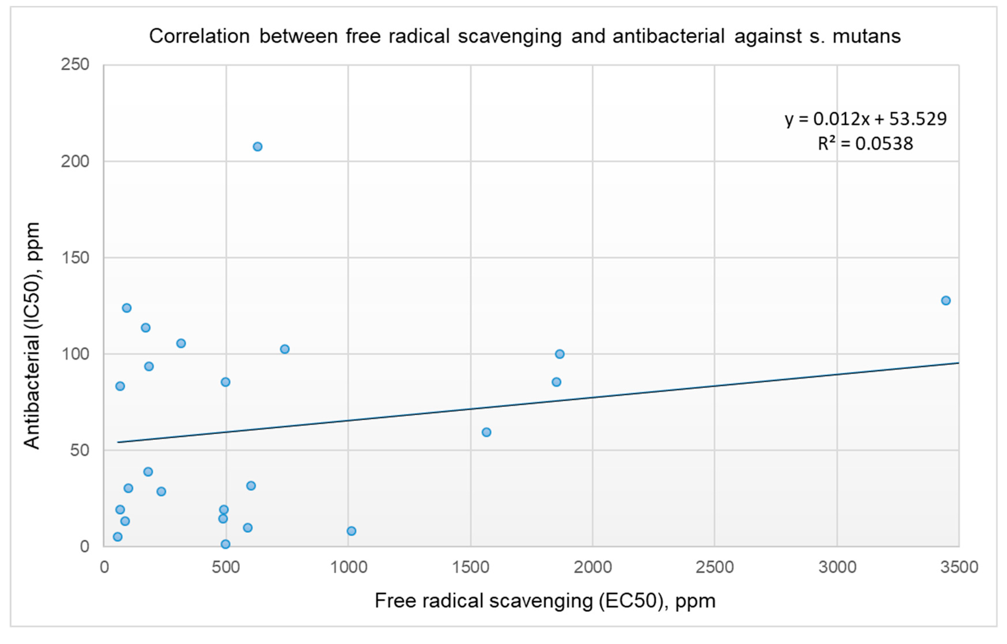
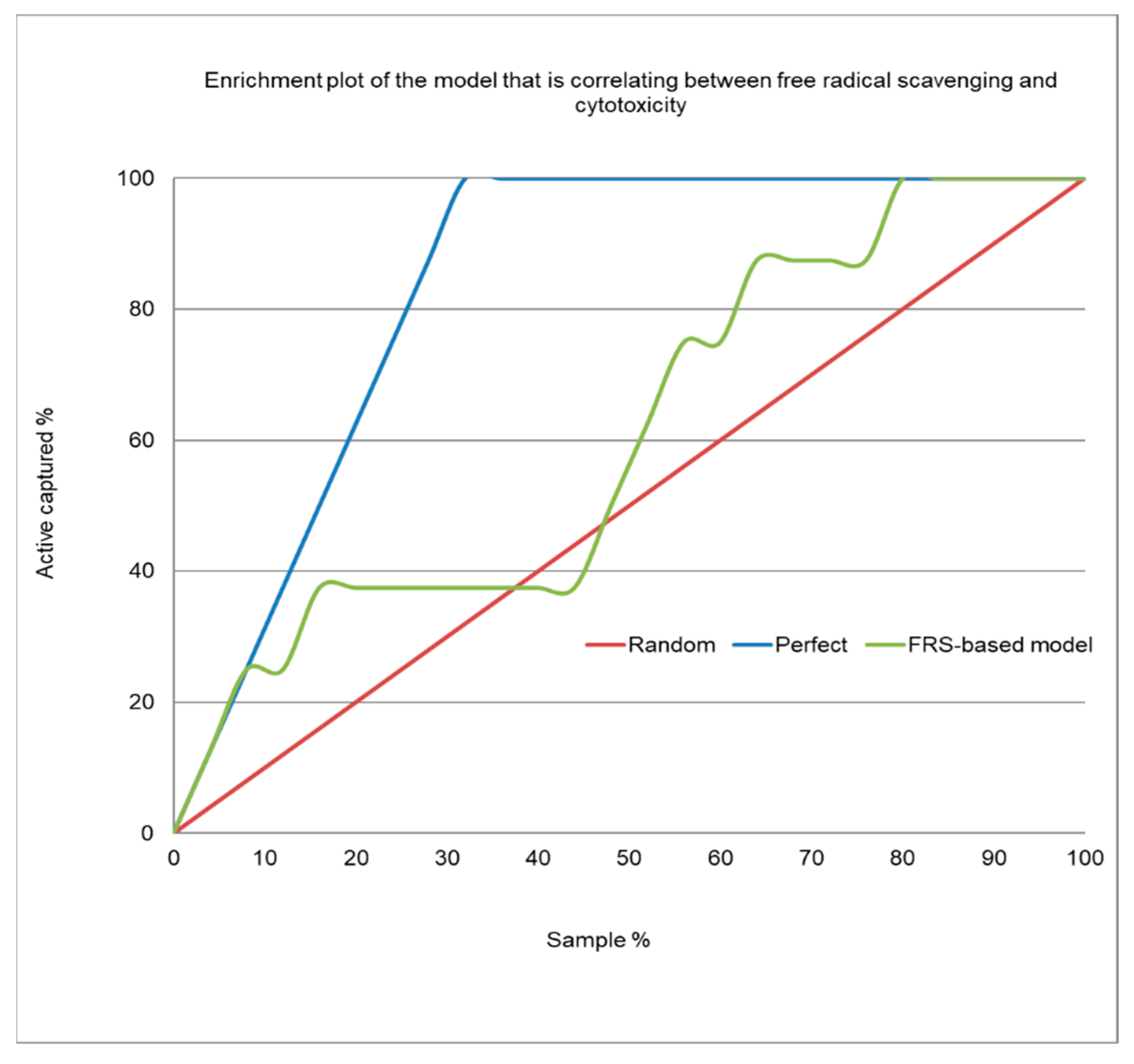
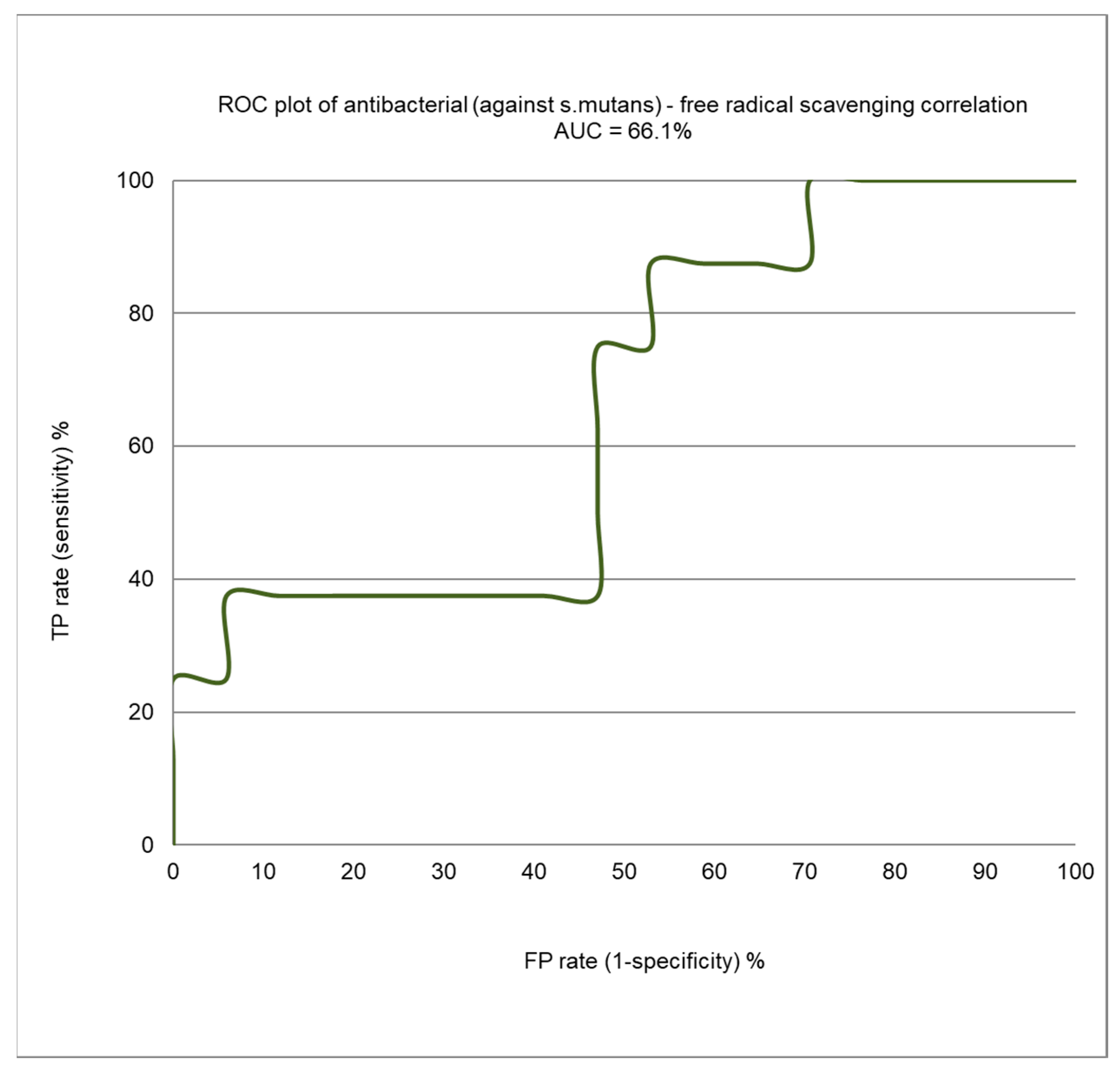
3. Results and Discussion

Twenty-five wild edible plants were studied for their free radical scavenging and antibacterial activity against S. mutans. Antibacterial potential was first verified by screening all the extracts (polar and non-polar fractions) for their activity, using one concentration of 250 ppm. The polar fractions were less active than the non-polar fractions. Column 3 of Table 1 depicts the EC50 of the free radical scavenging and the percentage of inhibition against S. mutans at a 5000-ppm concentration of the polar fraction. The extracts that exhibited a percentage of bacterial inhibition above 60% were tested in the second round at lower concentrations in a dose-response manner to extract their IC50 values. The antibacterial IC50 values of the polar fraction of the three plants Ruta chalepensis, Rumex patientia L., and Portulaca oleracea L. are 1750 ppm, 3925 ppm, and 4133 ppm, respectively. Column 4 of Table 1 depicts the EC50 of the free radical scavenging and the IC50 against S. mutans. Eight extracts (non-polar fractions) were found to possess relatively high antibacterial IC50 values of less than 20 ppm such as Foeniculum vulgare, Salvia palaestina, Micromeria fruticose, Trigonella foenum-graecum, Cichorium pumilum jacq, Salvia hierosolymitana boiss, Ruta chalepensis, and Chrysanthemum coronarium.
Rules-based analysis using Matthew’s correlation coefficient (MCC) scores and enrichment factors as criteria for evaluating the models’ efficiency revealed that the plant extracts whose EC50 for free radical scavenging <100 ppm showed some degree of enrichment toward being more active as an antibacterial agent against S. mutans (see Table 2). The values for the enrichment factor, the MCC, accuracy, and precision were 4.1, 0.217, 0.68, and 0.5, respectively. These findings are of high importance for screening projects. A four-fold order of enrichment toward more activity against S. mutans for the extracts with an EC50 of free radical scavenging ≤100 ppm could save plenty of time and could save many in screening projects. Considering all the results can lead to the conclusion that there is no correlation between antibacterial activity against S. mutans and free radical scavenging (as shown in Figure 1, R2 equals 0.0538), and also for the eight most active non-polar plant extracts (as shown in Figure 2, R2 equals 0.0469). Three of the eight active plant extracts (Salvia palaestina, Micromeria fruticose, and Salvia hierosolymitana boiss) show relatively high free radical scavenging, and four others (Foeniculum vulgare, Cichorium pumilum jacq, Ruta chalepensis, and Chrysanthemum coronarium) show medium free radical scavenging, while the remaining extract (Trigonella foenum-graecum) shows relatively low free radical scavenging. The results show that differences in antibacterial activity among the non-polar extracts cannot always be attributed to antioxidant levels but could be associated with inhibitory effects via other mechanisms of action [39] (inhibition or regulation of enzymes involved in cell wall biosynthesis, nucleic acid metabolism and repair, protein synthesis, or disruption of the membrane structure).
Figure 3 depicts the enrichment plot and Figure 4 shows the receiver operating characteristic (ROC) plot for the antibacterial/ free radical scavenging correlation model. The area under the curve (AUC) that was attained for the current model, as shown in Figure 4, is 0.661, which means that the model is very poor. This indicates a very weak correlation between antibacterial and free radical scavenging. The enrichment plot, which is shown in Figure 3, illustrates how quickly antibacterial extracts of plants can be identified when they are sorted based on their free radical scavenging activity. In-depth analysis revealed that the shape of the figure fits well with the conclusions drawn from the detailed analysis of Table 2, which shows that non-polar plant extracts with an EC50 of free radical scavenging <100 ppm have a four-fold order of enrichment toward more activity against S. mutans.
When the eight active plants were searched through electronic databases, including PubMed, we found a huge number of relevant reports documenting their antibacterial potential on different statins, except for Salvia hierosolymitana. For example, the antimicrobial activity of Foeniculum vulgare has been extensively reviewed [40]. The antibacterial activity of its essential oil against S. mutans showed an MIC and an MBC of 8.4 and 14.9 ppm, respectively. Salvia palaestina was also recently investigated by us for its antioxidant and antimicrobial potential [41]. The antimicrobial activity was examined by the disc diffusion method, and 5 µL (100%) of the essential oil was found to exceed the activity of gentamicin in the case of S. aureus, while it was nearly as effective as gentamicin against E. coli. Furthermore, according to both SEM and crystal-violet staining data, the essential oil of Micromeria fruticosa was found to exert an inhibitory effect on S. mutans biofilm formation [42]. Trigonella foenum-graecum (fenugreek) was also found to be effective in amounts of 20 mm and 19 mm against E. coli and Staphylococcus, respectively [43]. Cichorium pumilum, which is widely used as a traditional medicinal herb, showed antibacterial activity in vitro with agar disc diffusion and agar well diffusion methods against 10 different bacterial species [44]. The antimicrobial activity of Ruta chalepensis L. was tested against four pathogenic bacteria—Staphylococcus aureus, Bacillus subtilis, Escherichia coli, and Pseudomonas aeruginosa—and the findings indicate possible applications for the treatment of various infectious and noninfectious diseases [45]. The antibacterial effects of essential oil of Chrysanthemum coronarium demonstrated pronounced activity against gram-positive strains of bacteria: Bacillus subtilis (19 mm), Staphylococcus aureus (20 mm), Staphylococcus epidermidis (18 mm), Escherichia coli (9 mm), and Pseudomonas aeruginosa (12 mm), which were assessed by measuring the zone of inhibition [46]. Another unique aspect of this study is the scanning of all the 25 plant extracts at the same lab by using the same procedures for testing their antioxidant and antibacterial activities.

4. Conclusions

For this study, free radical scavenging was assessed by a DPPH assay, while antibacterial activity was measured by a microdilution assay. Eight wild plant extracts (non-polar fractions) were found to possess antibacterial IC50 values of less than 20 ppm, including Foeniculum vulgare, Salvia palaestina, Micromeria fruticose, Trigonella foenum-graecum, Cichorium pumilum jacq, Salvia hierosolymitana boiss, Ruta chalepensis, and Chrysanthemum coronarium. In addition, we raise a question regarding the correlation between free radical scavenging and antibacterial activity. Answering this question might be helpful for making the screening process more efficient. Looking at all the results together can deceive and lead to the conclusion that there is no correlation between antibacterial activity against S. mutans and free radical scavenging (R2 equals 0.0538). However, an in-depth analysis of the results revealed that the extracts of plants that exhibited an EC50 of free radical scavenging of ≤100 ppm showed some degree of enrichment (four orders of magnitude) toward more antibacterial activity. These findings are of high importance for screening projects. A four-fold order of enrichment could save plenty of time and many in screening projects. The antibacterial activity of the extracts could be attributed to various mechanisms of action. The highly active free radical scavenging extracts might follow one mechanism of action, while the extracts that are marked by low-medium free radical scavenging might act through another mechanism of action. This hypothesis could explain the enrichment that exist in extracts having EC50 of free radical scavenging of ≤100 ppm.
Our next study will aim to discover novel and highly active natural antibacterial products, using a novel in-house bioassay guided fractionation approach [1,28,31,37] and in-silico based modeling techniques [29,47,48,49,50] to identify natural products that exhibit antibacterial activity against S. mutans.

Author Contributions

All six authors contributed extensively to the work presented in this paper. A.R. conceived the study, designed the experiments, performed the statistical analysis, raised the funds, and contributed to editing the manuscript. M.R. collected the plants and performed the extractions. M.R., B.A.-F., and M.M. ran the free radical scavenging and antibacterial experiments. M.R., S.A.-L., and W.B. interpreted the data and wrote the original draft. All authors have read and agreed to the published version of the manuscript.

Acknowledgments

The Al-Qasemi Research Authority, and the Faculty of Medicine of Al Najah University, supported this work. We declare that the funders had no role in the study design, data collection, analysis, decision to publish, or preparation of the manuscript.

Conflicts of Interest

The authors declare that they have no conflicts of interest.

References

  1. Gabe, V.; Kacergius, T.; Abu-Lafi, S.; Kalesinskas, P.; Masalha, M.; Falah, M.; Abu-Farich, B.; Melninkaitis, A.; Zeidan, M.; Rayan, A. Inhibitory Effects of Ethyl Gallate on Streptococcus mutans Biofilm Formation by Optical Profilometry and Gene Expression Analysis. Molecules 2019, 24, 529. [Google Scholar] [CrossRef] [PubMed] [Green Version]
  2. Dinic, J.; Podolski-Renic, A.; Jeremic, M.; Pesic, M. Potential of Natural-Based Anticancer Compounds for P-Glycoprotein Inhibition. Curr. Pharm. Des. 2018, 24, 4334–4354. [Google Scholar] [CrossRef] [PubMed]
  3. Lee, K.H. Discovery and development of natural product-derived chemotherapeutic agents based on a medicinal chemistry approach. J. Nat. Prod. 2010, 73, 500–516. [Google Scholar] [CrossRef] [PubMed] [Green Version]
  4. Hughes, B. 2007 FDA drug approvals: A year of flux. Nat. Rev. Drug Discov. 2008, 7, 107–109. [Google Scholar] [CrossRef] [PubMed]
  5. Zaid, H.; Raiyn, J.; Nasser, A.; Saad, B.; Rayan, A. Physicochemical properties of natural based products versus synthetic chemicals. Open Nutraceut. J. 2010, 3, 194–202. [Google Scholar] [CrossRef]
  6. Becerra-Tomas, N.; Blanco Mejia, S.; Viguiliouk, E.; Khan, T.; Kendall, C.W.C.; Kahleova, H.; Rahelic, D.; Sievenpiper, J.L.; Salas-Salvado, J. Mediterranean diet, cardiovascular disease and mortality in diabetes: A systematic review and meta-analysis of prospective cohort studies and randomized clinical trials. Crit. Rev. Food Sci. Nutr. 2019, 1–21. [Google Scholar] [CrossRef]
  7. Galilea-Zabalza, I.; Buil-Cosiales, P.; Salas-Salvado, J.; Toledo, E.; Ortega-Azorin, C.; Diez-Espino, J.; Vazquez-Ruiz, Z.; Zomeno, M.D.; Vioque, J.; Martinez, J.A.; et al. Mediterranean diet and quality of life: Baseline cross-sectional analysis of the PREDIMED-PLUS trial. PLoS ONE 2018, 13, e0198974. [Google Scholar] [CrossRef] [Green Version]
  8. Jaradat, N.A.; Ayesh, O.I.; Anderson, C. Ethnopharmacological survey about medicinal plants utilized by herbalists and traditional practitioner healers for treatments of diarrhea in the West Bank/Palestine. J. Ethnopharmacol. 2016, 182, 57–66. [Google Scholar] [CrossRef]
  9. Abu-Lafi, S.; Rayan, M.; Masalha, M.; Abu-Farich, B.; Al-Jaas, H.; Abu-Lafi, M.; Rayan, A. Phytochemical Composition and Biological Activities of Wild Scolymus maculatus L. Medicines 2019, 6, 53. [Google Scholar] [CrossRef] [Green Version]
  10. Abu-Lafi, S.; Rayan, B.; Kadan, S.; Abu-Lafi, M.; Rayan, A. Anticancer activity and phytochemical composition of wild Gundelia tournefortii. Oncol. Lett. 2019, 17, 713–717. [Google Scholar] [CrossRef] [Green Version]
  11. Ali-Shtayeh, M.S.; Jamous, R.M.; Al-Shafie, J.H.; Elgharabah, W.A.; Kherfan, F.A.; Qarariah, K.H.; Khdair, I.S.; Soos, I.M.; Musleh, A.A.; Isa, B.A.; et al. Traditional knowledge of wild edible plants used in Palestine (Northern West Bank): A comparative study. J. Ethnobiol. Ethnomed. 2008, 4, 13. [Google Scholar] [CrossRef] [PubMed] [Green Version]
  12. de Sa Junior, P.L.; Camara, D.A.D.; Porcacchia, A.S.; Fonseca, P.M.M.; Jorge, S.D.; Araldi, R.P.; Ferreira, A.K. The Roles of ROS in Cancer Heterogeneity and Therapy. Oxidative Med. Cell. Longev. 2017, 2017, 2467940. [Google Scholar] [CrossRef] [PubMed]
  13. Mishra, S.; Imlay, J. Why do bacteria use so many enzymes to scavenge hydrogen peroxide? Arch. Biochem. Biophys. 2012, 525, 145–160. [Google Scholar] [CrossRef] [PubMed] [Green Version]
  14. Abdalla, M.; Saad-Hussein, A.; Ibrahim, K.S.; El-mezayen, H.A.; Osman, N.F. Effects of smoking on the oxidant/antioxidant balance and the blood lipids in pesticide sprayers. Toxicol. Ind. Health 2015, 31, 173–178. [Google Scholar] [CrossRef] [PubMed]
  15. Sochor, J.; Zitka, O.; Skutkova, H.; Pavlik, D.; Babula, P.; Krska, B.; Horna, A.; Adam, V.; Provaznik, I.; Kizek, R. Content of phenolic compounds and antioxidant capacity in fruits of apricot genotypes. Molecules 2010, 15, 6285–6305. [Google Scholar] [CrossRef] [Green Version]
  16. Godfrey, C.; Desviat, L.R.; Smedsrod, B.; Pietri-Rouxel, F.; Denti, M.A.; Disterer, P.; Lorain, S.; Nogales-Gadea, G.; Sardone, V.; Anwar, R.; et al. Delivery is key: Lessons learnt from developing splice-switching antisense therapies. EMBO Mol. Med. 2017, 9, 545–557. [Google Scholar] [CrossRef] [PubMed]
  17. Kurosaki, Y.; Imoto, A.; Kawakami, F.; Yokoba, M.; Takenaka, T.; Ichikawa, T.; Katagiri, M.; Ishii, N. Oxidative stress increases megalin expression in the renal proximal tubules during the normoalbuminuric stage of diabetes mellitus. Am. J. Physiol. Renal Physiol. 2018, 314, F462–F470. [Google Scholar] [CrossRef]
  18. Di Pietro, M.; Filardo, S.; Falasca, F.; Turriziani, O.; Sessa, R. Infectious Agents in Atherosclerotic Cardiovascular Diseases through Oxidative Stress. Int. J. Mol. Sci. 2017, 18, 2459. [Google Scholar] [CrossRef] [Green Version]
  19. Srivastava, S.; Singh, D.; Patel, S.; Singh, M.R. Role of enzymatic free radical scavengers in management of oxidative stress in autoimmune disorders. Int. J. Biol. Macromol. 2017, 101, 502–517. [Google Scholar] [CrossRef]
  20. Pereira, C.; Gracio, D.; Teixeira, J.P.; Magro, F. Oxidative Stress and DNA Damage: Implications in Inflammatory Bowel Disease. Inflamm. Bowel Dis. 2015, 21, 2403–2417. [Google Scholar] [CrossRef]
  21. Kumar, A.; Yegla, B.; Foster, T.C. Redox Signaling in Neurotransmission and Cognition During Aging. Antioxid. Redox Signal. 2018, 28, 1724–1745. [Google Scholar] [CrossRef] [PubMed] [Green Version]
  22. Franca, M.B.; Lima, K.C.; Eleutherio, E.C. Oxidative Stress and Amyloid Toxicity: Insights From Yeast. J. Cell. Biochem. 2017, 118, 1442–1452. [Google Scholar] [CrossRef] [PubMed]
  23. Ganguly, G.; Chakrabarti, S.; Chatterjee, U.; Saso, L. Proteinopathy, oxidative stress and mitochondrial dysfunction: Cross talk in Alzheimer’s disease and Parkinson’s disease. Drug Des. Dev. Ther. 2017, 11, 797–810. [Google Scholar] [CrossRef] [PubMed] [Green Version]
  24. Ahmad, S.B.; Rehman, M.U.; Fatima, B.; Ahmad, B.; Hussain, I.; Ahmad, S.P.; Farooq, A.; Muzamil, S.; Razzaq, R.; Rashid, S.M.; et al. Antifibrotic effects of d-limonene (5(1-methyl-4-[1-methylethenyl]) cyclohexane) in CCl4 induced liver toxicity in Wistar rats. Environ. Toxicol. 2018, 33, 361–369. [Google Scholar] [CrossRef] [PubMed]
  25. Nikitaki, Z.; Nikolov, V.; Mavragani, I.V.; Mladenov, E.; Mangelis, A.; Laskaratou, D.A.; Fragkoulis, G.I.; Hellweg, C.E.; Martin, O.A.; Emfietzoglou, D.; et al. Measurement of complex DNA damage induction and repair in human cellular systems after exposure to ionizing radiations of varying linear energy transfer (LET). Free Radic. Res. 2016, 50, S64–S78. [Google Scholar] [CrossRef] [PubMed] [Green Version]
  26. Hassan, W.; Noreen, H.; Rehman, S.; Gul, S.; Kamal, M.A.; Kamdem, J.P.; Zaman, B.; da Rocha, J.B.T. Oxidative Stress and Antioxidant Potential of One Hundred Medicinal Plants. Curr. Top. Med. Chem. 2017, 17, 1336–1370. [Google Scholar] [CrossRef] [PubMed]
  27. Sammar, M.; Abu-Farich, B.; Rayan, I.; Falah, M.; Rayan, A. Correlation between cytotoxicity in cancer cells and free radical-scavenging activity: In vitro evaluation of 57 medicinal and edible plant extracts. Oncol. Lett. 2019, 18, 6563–6571. [Google Scholar] [CrossRef] [Green Version]
  28. Kacergius, T.; Abu-Lafi, S.; Kirkliauskiene, A.; Gabe, V.; Adawi, A.; Rayan, M.; Qutob, M.; Stukas, R.; Utkus, A.; Zeidan, M.; et al. Inhibitory capacity of Rhus coriaria L. extract and its major component methyl gallate on Streptococcus mutans biofilm formation by optical profilometry: Potential applications for oral health. Mol. Med. Rep. 2017, 16, 949–956. [Google Scholar] [CrossRef] [Green Version]
  29. Aswad, M.; Rayan, M.; Abu-Lafi, S.; Falah, M.; Raiyn, J.; Abdallah, Z.; Rayan, A. Nature is the best source of anti-inflammatory drugs: Indexing natural products for their anti-inflammatory bioactivity. Inflamm. Res. 2018, 67, 67–75. [Google Scholar] [CrossRef]
  30. Srinivasan, P.K.; Yagi, S.; Nagai, K.; Afify, M.; Hata, K.; Uemoto, S.; Tolba, R.H. Impact of oxygen free radicals in rat partial liver transplantation. J. Surg. Res. 2014, 191, 469–475. [Google Scholar] [CrossRef]
  31. Frank, A.; Abu-Lafi, S.; Adawi, A.; Schwed, J.S.; Stark, H.; Rayan, A. From medicinal plant extracts to defined chemical compounds targeting the histamine H4 receptor: Curcuma longa in the treatment of inflammation. Inflamm. Res. 2017, 66, 923–929. [Google Scholar] [CrossRef] [PubMed]
  32. Keys, A.; Menotti, A.; Aravanis, C.; Blackburn, H.; Djordevic, B.S.; Buzina, R.; Dontas, A.S.; Fidanza, F.; Karvonen, M.J.; Kimura, N.; et al. The seven countries study: 2289 deaths in 15 years. Prev. Med. 1984, 13, 141–154. [Google Scholar] [CrossRef]
  33. Bower, A.; Marquez, S.; de Mejia, E.G. The Health Benefits of Selected Culinary Herbs and Spices Found in the Traditional Mediterranean Diet. Crit. Rev. Food Sci. Nutr. 2016, 56, 2728–2746. [Google Scholar] [CrossRef] [PubMed]
  34. Akar, Z.; Kucuk, M.; Dogan, H. A new colorimetric DPPH(*) scavenging activity method with no need for a spectrophotometer applied on synthetic and natural antioxidants and medicinal herbs. J. Enzym. Inhib. Med. Chem. 2017, 32, 640–647. [Google Scholar] [CrossRef] [PubMed] [Green Version]
  35. Blois, M.S. Antioxidant Determinations by the Use of a Stable Free Radical. Nature 1958, 181, 1199–1200. [Google Scholar] [CrossRef]
  36. Sánchez-Moreno, C.; Larrauri, J.A.; Saura-Calixto, F. A procedure to measure the antiradical efficiency of polyphenols. J. Sci. Food Agric. 1998, 76, 270–276. [Google Scholar] [CrossRef]
  37. Gabe, V.; Kacergius, T.; Abu-Lafi, S.; Zeidan, M.; Abu-Farich, B.; Austys, D.; Masalha, M.; Rayan, A. Suppressive Effects of Octyl Gallate on Streptococcus mutans Biofilm Formation, Acidogenicity, and Gene Expression. Molecules 2019, 24, 3170. [Google Scholar] [CrossRef] [Green Version]
  38. Rayan, A.; Marcus, D.; Goldblum, A. Predicting oral druglikeness by iterative stochastic elimination. J. Chem. Inf. Model. 2010, 50, 437–445. [Google Scholar] [CrossRef]
  39. Qian, W.; Yang, M.; Wang, T.; Sun, Z.; Liu, M.; Zhang, J.; Zeng, Q.; Cai, C.; Li, Y. Antibacterial mechanism of vanillic acid on physiological, morphological and biofilm properties of carbapenem-resistant Enterobacter hormaechei. J. Food Prot. 2019. [Google Scholar] [CrossRef]
  40. Badgujar, S.B.; Patel, V.V.; Bandivdekar, A.H. Foeniculum vulgare Mill: A review of its botany, phytochemistry, pharmacology, contemporary application, and toxicology. BioMed Res. Int. 2014, 2014, 842674. [Google Scholar] [CrossRef] [Green Version]
  41. Sabbobeh, R.; Hejaz, H.; Jahajha, A.; Al-Akhras, S.; Al-Jaas, H.; Abu-Lafi, S. Antioxidant an Antimicrobial Activities of the Leaf Extract of Salvia palaestina. J. Appl. Pharm. Sci. 2016, 76–82. [Google Scholar] [CrossRef] [Green Version]
  42. Boran, R.; Ugur, A. Inhibitory effect of Micromeria fruticosa ssp. brachycalyx on Streptococcus mutans biofilm formation and its antimutagenic and antioxidant activities. J. Selcuk Univ. Nat. Appl. Sci. 2015, 4, 3. [Google Scholar]
  43. Sharma, V.; Singh, P.; Rani, A. Antimicrobial Activity of Trigonella foenum-graecum L. (Fenugreek). Eur. J. Exp. Biol. 2017, 7, 1. [Google Scholar] [CrossRef]
  44. Al Khateeb, W.; Hussein, E.; Qouta, L.; Alu’datt, M.; Al-Shara, B.; Abu-zaiton, A. In Vitro propagation and characterization of phenolic content along with antioxidant and antimicrobial activities of Cichorium pumilum Jacq. Plant Cell Tissue Organ Cult. 2012, 110, 103–110. [Google Scholar] [CrossRef]
  45. Boughendjioua, H. Yield, chemical composition and antibacterial activity of Ruta chalepensis L. essential oil growing spontaneously in Algeria. Pharm. Pharmacol. Int. J. 2019, 7, 7. [Google Scholar] [CrossRef]
  46. Bardaweel, S.K.; Hudaib, M.M.; Tawaha, K.A.; Bashatwah, R.M. Studies on the In Vitro Antiproliferative, Antimicrobial, Antioxidant, and Acetylcholinesterase Inhibition Activities Associated with Chrysanthemum coronarium Essential Oil. Evid. Based Complement. Alternat. Med. 2015, 2015, 790838. [Google Scholar] [CrossRef] [Green Version]
  47. Rayan, A.; Raiyn, J.; Falah, M. Nature is the best source of anticancer drugs: Indexing natural products for their anticancer bioactivity. PLoS ONE 2017, 12, e0187925. [Google Scholar] [CrossRef]
  48. Rayan, M.; Abdallah, Z.; Abu-Lafi, S.; Masalha, M.; Rayan, A. Indexing natural products for their antifungal activity by filters-based approach: Disclosure of discriminative properties. Curr. Comput. Aided Drug Des. 2019, 15. [Google Scholar] [CrossRef]
  49. Masalha, M.; Rayan, M.; Adawi, A.; Abdallah, Z.; Rayan, A. Capturing antibacterial natural products with in�silico techniques. Mol. Med. Rep. 2018, 18, 763–770. [Google Scholar] [CrossRef] [Green Version]
  50. Zeidan, M.; Rayan, M.; Zeidan, N.; Falah, M.; Rayan, A. Indexing Natural Products for Their Potential Anti-Diabetic Activity: Filtering and Mapping Discriminative Physicochemical Properties. Molecules 2017, 22, 1563. [Google Scholar] [CrossRef] [Green Version]
Figure 1. Correlations between the EC50 for free radical scavenging and antibacterial activity (IC50) for the non-polar extracts.
Processes 08 00117 g001
Figure 2. Correlations between antibacterial activity (IC50) and the EC50 for free radical scavenging for the eight most active plant extracts against S. mutans.
Processes 08 00117 g002
Figure 3. Enrichment plot of the prediction model for the antibacterial activity of the plant extracts, based on their free radical scavenging activity (non-polar extracts).
Processes 08 00117 g003
Figure 4. A receiver operating characteristic (ROC) curve showing the performance of the antibacterial/free radical scavenging correlation model.
Processes 08 00117 g004
Table 1. List of tested plants and their extracts’ activities.
Scientific Plant Name (Parts Used: Leaf, Pulp, Seed, Stem, etc.)Local Name in ArabicPolar Extract
Free Radical Scavenging (EC50)/Antibacterial (% Inhibition by Extract of 5000 ppm Concentration)
Non-Polar Extract
Free Radical Scavenging (EC50)/Antibacterial (IC50), ppm
Majorana syriaca
(leaf + stem)
زعتر بري1.6/22.5%67.3/83.6
Foeniculum vulgare
(leaf + stem)
شومر52.1/34.1%488.8/14.8
Malva sylvestris
(leaf)
خبازة برية10.1/29.3%183.8/93.6
Salvia palaestina
(leaf + stem)
ميرمية12.5/41.6%64.9/19.2
Cyclamen persicum
(leaf)
زقوقيه (عصا الراعي)35.2/24.8%172.2/113.8
Micromeria fruticose
(leaf + stem)
عشبة الشايn.m. #156.1/5.3
Arum palaestinum
(leaf)
لوف الفلسطينيn.m. #13447.4/127.8
Trigonella foenum-graecum
(seed)
حلبةn.m. #11014.6/8.2
Gundelia tournefortii
(stem)
عكوب89.9/30.5%1567.2/59.3
Matricaria aurea
(leaf + stem)
البابونج الذهبي84.3/11.3%1864.5/99.9
Centaurea dumulosa boiss
(leaf + stem)
مرار55.8/43.9%314.4/105.8
Cichorium pumilum jacq
(leaf)
هندباء50.9/23.2%489.7/19.5
Salvia hierosolymitana boiss
(leaf)
لسينه12.1/48.8%86.0/13.2
Rumex patientia L.
(leaf)
سلق بري>10,000/76.6%233.0/28.9
Ruta chalepensis
(leaf)
فيجن21.5/94.7%496.1/1.5
Ceratonia siliqua
(fruit)
خروب47.4/56.2%180.2/39.0
Urtica urens L.
(leaf + stem)
قريص حريق590.9/n.d.3736.0/62.2
Portulaca oleracea L.
(leaf)
فرفحينا55.8/72.2%1852.4/85.4
Eryngium creticum lam
(leaf)
قرصعنه28.8/11.7%99.6/30.5
Chrysanthemum coronarium
(leaf + stem)
بسباسn.m. #1587.0/10.1
Sinapis alba
(leaf)
عشبة الخردل (بري)42.9/30.4%739.1/102.5
Asparagus aphyllus L.
(stem)
هليون2803.2/23.0%627.5/207.4
Rumex acetosa
(leaf)
حميض37.2/42.3%496.1/85.4
Teucrium chamaedrys
(leaf)
جعدة9.2/2.8%94.2/124
Ephedra foeminea forssk
(stem)
علندا10.8/52.9%603.1/31.9
#1 not measured.
Table 2. Matthew’s correlation coefficient (MCC) scores, precision, accuracy, and enrichment factors as criteria for evaluating the prediction models.
EC50 Cutoff of Free Radical Scavenging (<)100 ppm500 ppm1000 ppmNo Limit
No. active plants (true positive) #14678
No. inactive plants (false positive) #2391117
No. inactive plants (true negative) #31486-
No. active plants (false negative) #4421-
Precision0.50.40.370.32
Accuracy0.680.560.480.32
Enrichment factor4.11.661.311.0
MCC0.2170.2100.1840.0
#1 Number of plant extracts that have an EC50 for free-radical scavenging lower than the indicated threshold and an IC50 for antibacterial activity lower than 20 ppm. #2 Number of plant extracts that have an EC50 for free-radical scavenging lower than the indicated threshold and an IC50 for antibacterial activity greater than 20 ppm. #3 Number of plant extracts that have an EC50 for free-radical scavenging greater than the indicated threshold and an IC50 for antibacterial activity greater than 20 ppm. #4 Number of plant extracts that have an EC50 of free-radical scavenging greater than the indicated threshold and an IC50 for antibacterial activity lower than 20 ppm.

