Metabolite Profiling of Aquilaria malaccensis Leaf Extract Using Liquid Chromatography-Q-TOF-Mass Spectrometry and Investigation of Its Potential Antilipoxygenase Activity In-Vitro

 ,
,
Abstract
:1. Introduction
2. Materials and Methods
2.1. Sample, Chemicals, and Reagents
2.2. Sample Preparation
2.3. Phytochemical Screening
2.4. GC-MS
2.5. LC/Q-TOF-MS
2.6. Antilipoxygenase Assay
2.7. Toxicity Screening Assay
2.8. Statistical Methods
3. Results and Discussion
3.1. Phytochemical Screening
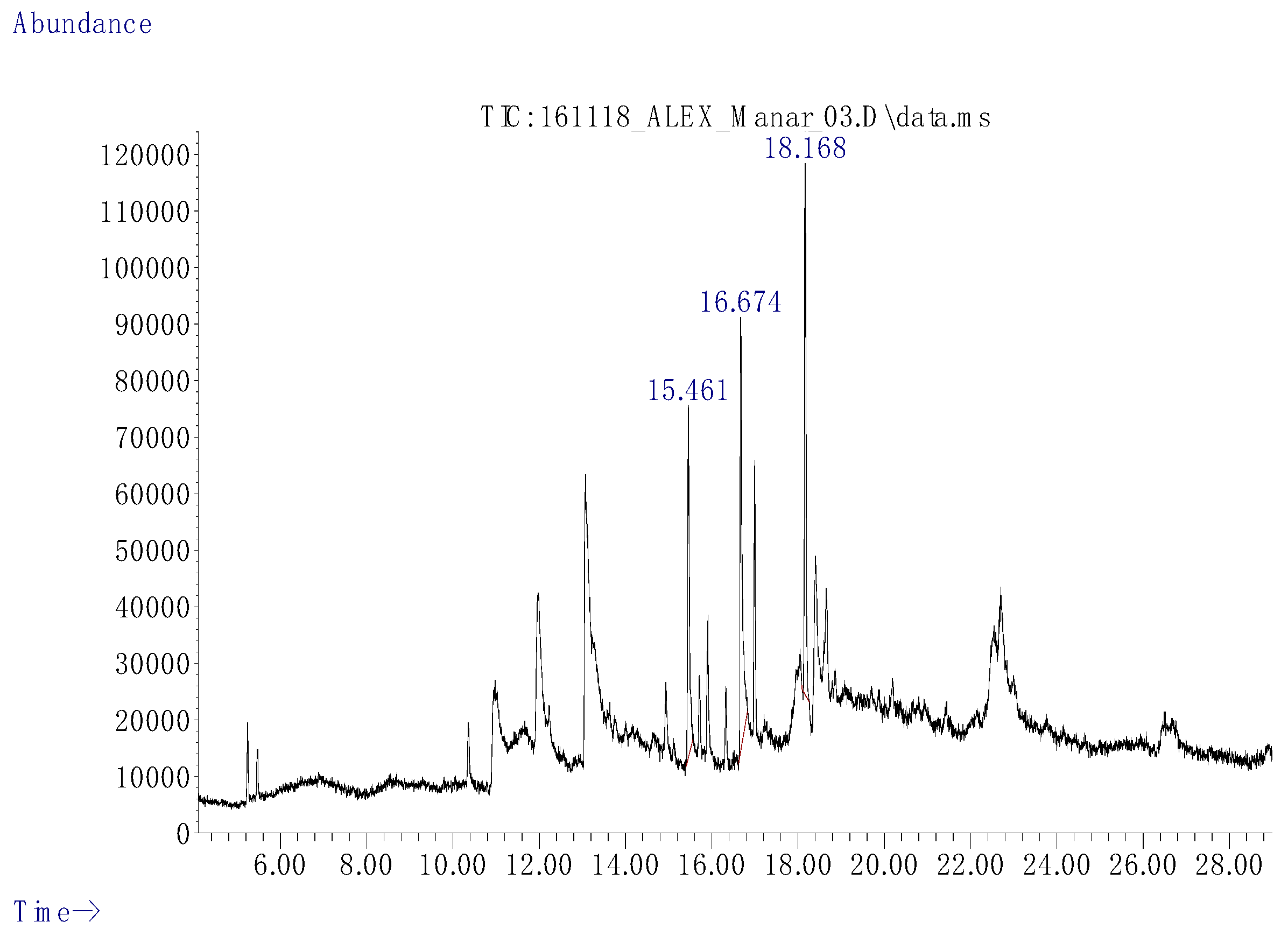
3.2. GC-MS
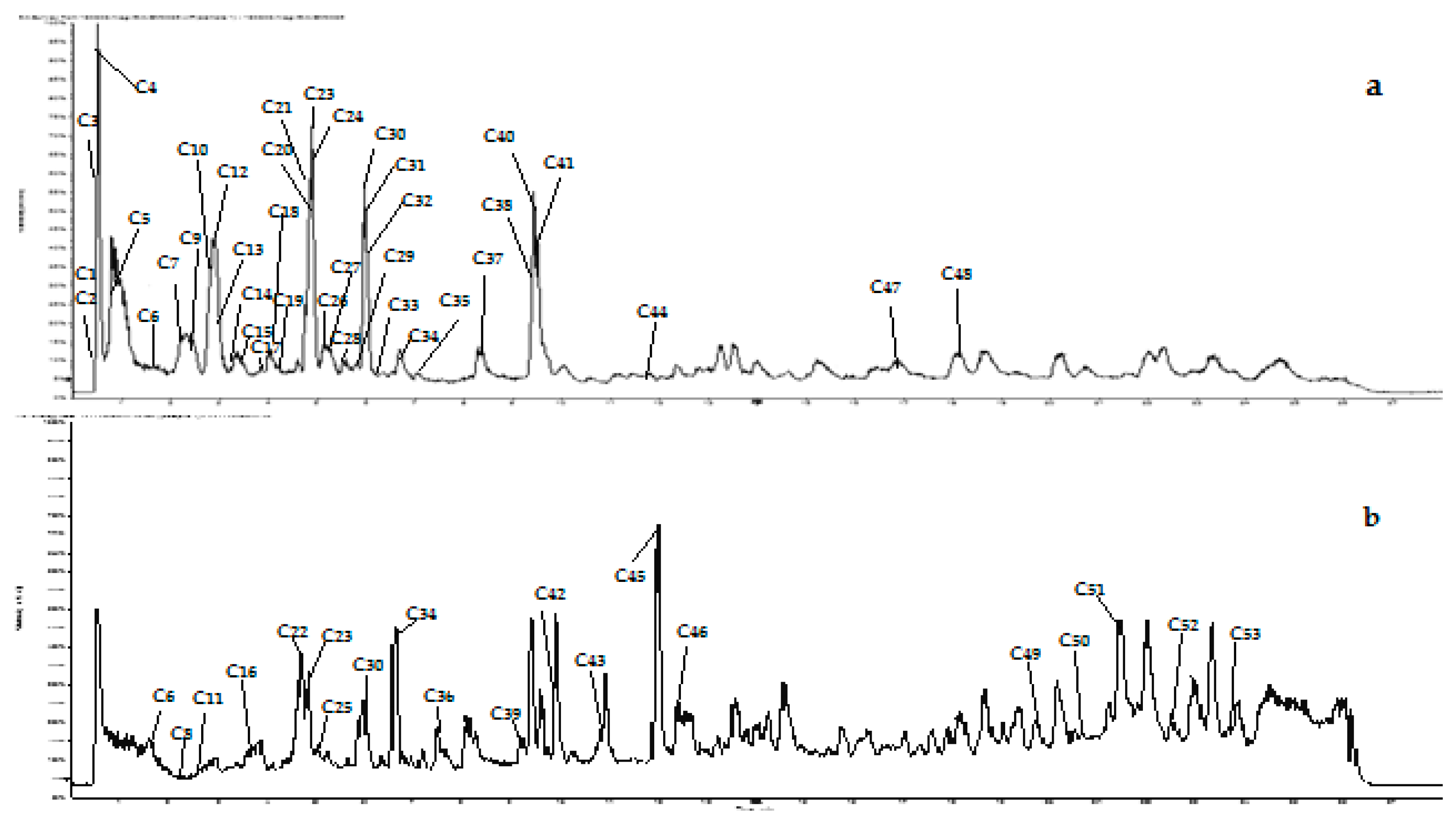
3.3. LC/Q-TOF-MS
3.3.1. Phenolic Acids
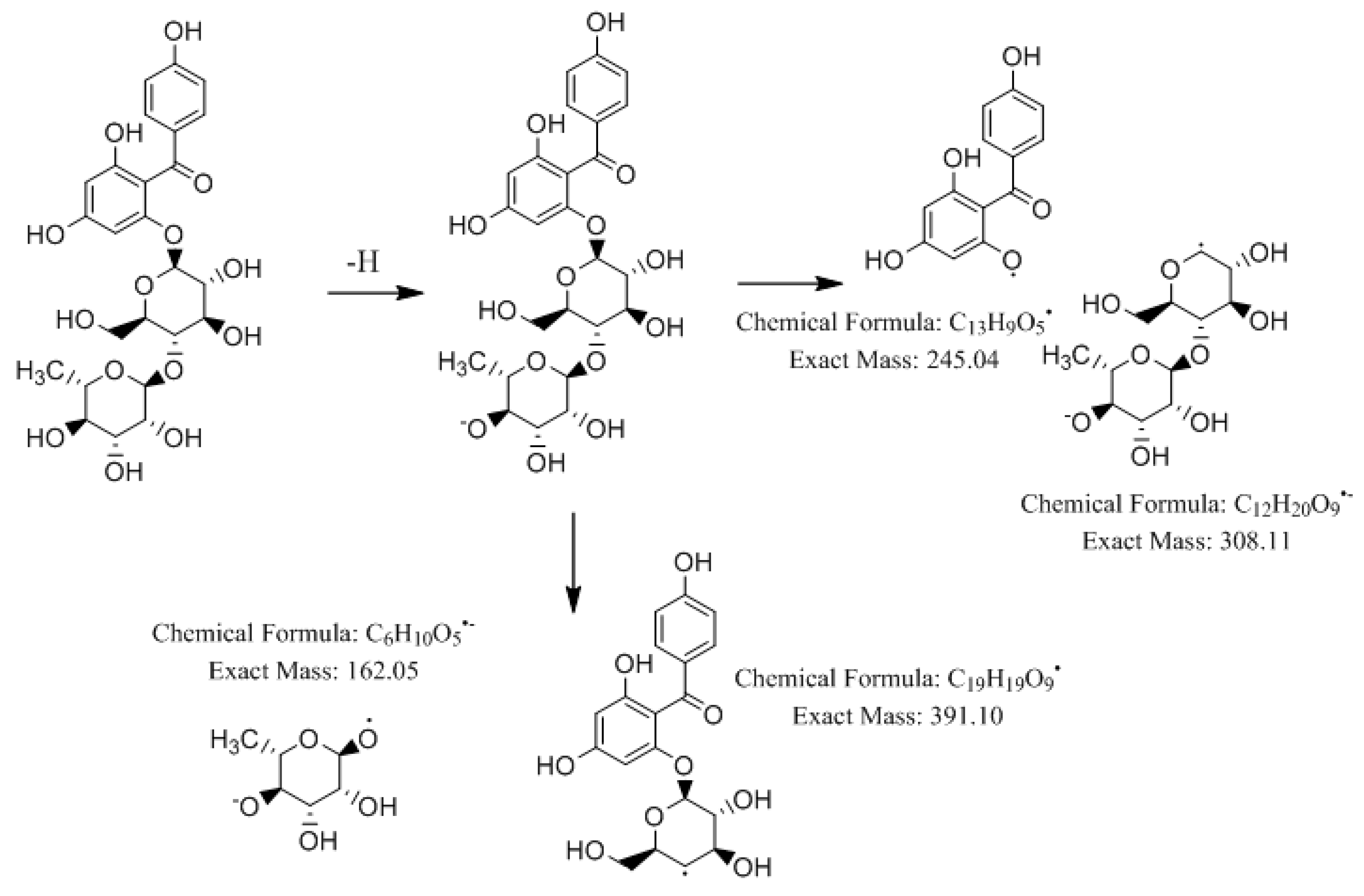
3.3.2. Benzophenones
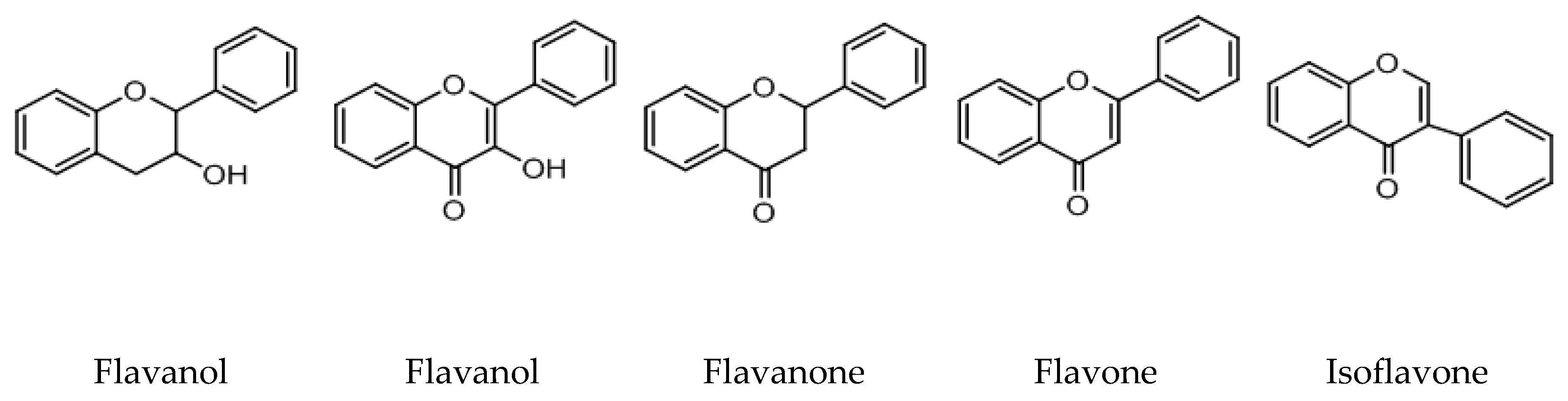
3.3.3. Flavonoids
3.3.4. Xanthones
3.3.5. Sterols
3.3.6. Terpenoids
3.3.7. Coumarins
3.3.8. Lignans
3.3.9. Fatty Acids
3.3.10. Tocopherols
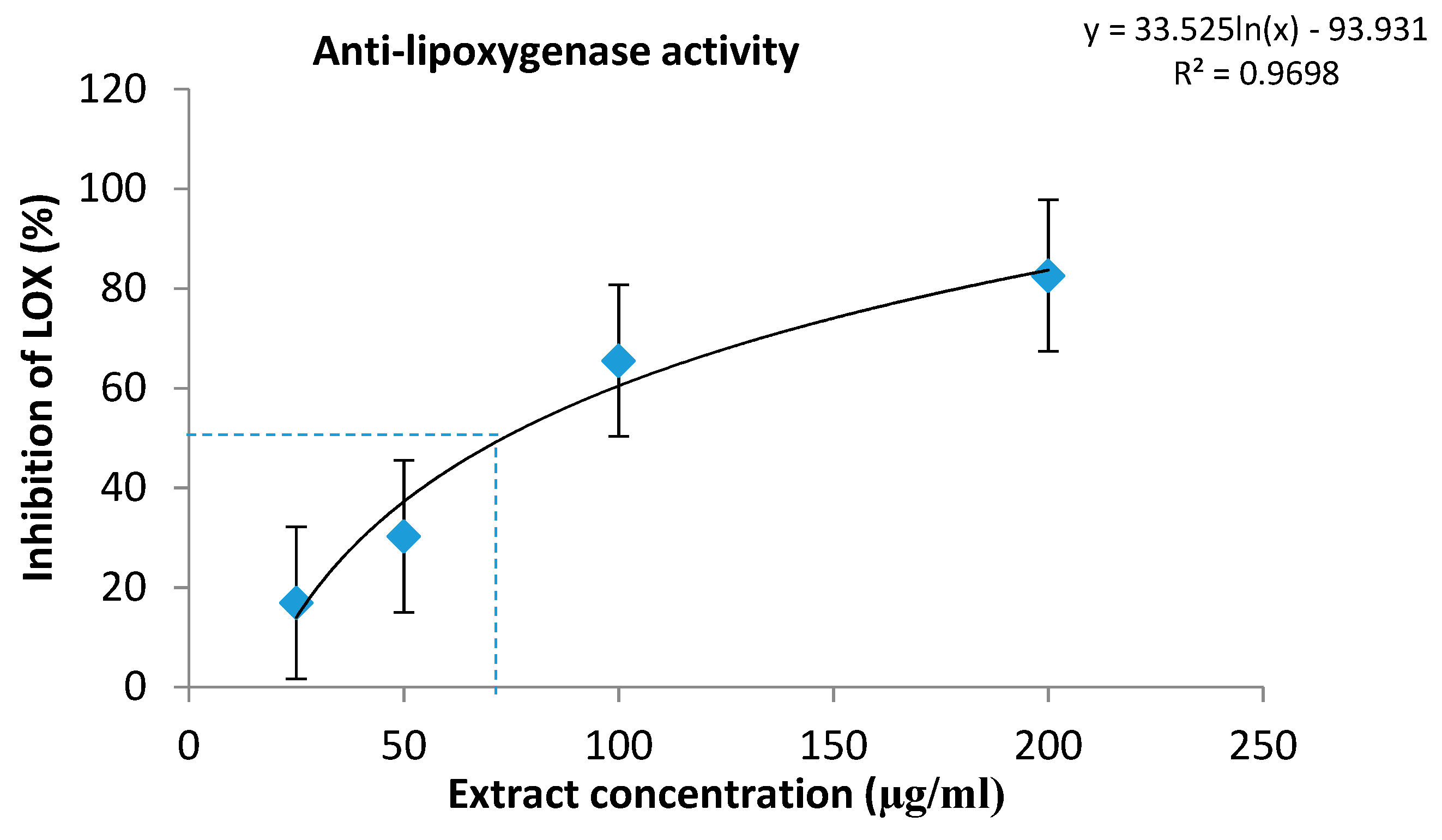
3.4. The Antilipoxygenase Assay
3.5. Toxicity Screening Assay
4. Conclusions
Supplementary Materials
Author Contributions
Acknowledgments
Conflicts of Interest
References
- Lee, S.Y.; Mohamed, R. The Origin and Domestication of Aquilaria, an Important Agarwood-Producing Genus. In Agarwood, 1st ed.; Mohamed, R., Ed.; Springer: Singapore, 2016; pp. 1–20. [Google Scholar]
- Hashim, Y.Z.H.-Y.; Kerr, P.G.; Abbas, P.; Salleh, H.M. Aquilaria Spp. (Agarwood) as Source of Health Beneficial Compounds: A Review of Traditional Use, Phytochemistry and Pharmacology. J. Ethnopharmacol. 2016, 189, 331–360. [Google Scholar] [CrossRef]
- Hendra, H.; Moeljopawiro, S.; Nuringtyas, T.R. Antioxidant and Antibacterial Activities of Agarwood (Aquilaria malaccensis Lamk.) Leaves. AIP Conf. Proc. 2016, 1755. [Google Scholar]
- Adam, A.Z.; Lee, S.Y.; Mohamed, R. Pharmacological Properties of Agarwood Tea Derived from Aquilaria (Thymelaeaceae) Leaves: An Emerging Contemporary Herbal Drink. J. Herb. Med. 2017, 10, 37–44. [Google Scholar] [CrossRef]
- Valentine, N.E.; Apridamayanti, P.; Sari, R. Fici Value of Aquilaria malaccensis Leaves Extract and Amoxicillin against Proteus Mirabilis and Pseudomonas Aeruginosa. Kartika J. Ilm. Farm. 2019, 6, 86–90. [Google Scholar] [CrossRef]
- Nadilah, W.A.; Ali, A.M.; Nur, W.A.; Hasima, N. Evaluation of DPPH Free Radical Scavenging, α-Glucosidase Inhibitory, and Antimicrobial Activities of Aquilaria malaccensis Leaf Extracts. J. Agrobiotech. 2019, 10, 36–45. [Google Scholar]
- Dash, M.; Patra, J.K.; Panda, P.P. Phytochemical and antimicrobial screening of extracts of Aquilaria agallocha Roxb. Afr. J. Biotechnol. 2008, 7, 3531–3534. [Google Scholar]
- Batubara, R.; Hanum, T.I.; Pulungan, W. Phytochemical and Antioxidant Activity of Gaharu Leaf Tea (Aquilaria malaccensis Lamk) as Raw Material of Tea from Middle Tapanuli Regency, North Sumatera Province. IOP Conf. Ser. Earth Environ. Sci. 2019, 260, 012101. [Google Scholar]
- Rahman, H.; Eswaraiah, M.C.; Dutta, A.M. Anti-arthritic activity of leaves and oil of Aquilaria agallocha. Saudi J. Life Sci. 2016, 1, 34–43. [Google Scholar]
- Alam, J.; Mujahid, M.; Jahan, Y.; Bagga, P.; Rahman, M.A. Hepatoprotective Potential of Ethanolic Extract of Aquilaria agallocha Leaves against Paracetamol Induced Hepatotoxicity in SD Rats. J. Tradit. Complement. Med. 2017, 7, 9–13. [Google Scholar] [CrossRef] [Green Version]
- Dyary, H.O.; Arifah, A.K.; Sharma, R.S.; Rasedee, A.; Mohd-Aspollah, M.S.; Zakaria, Z.A.; Zuraini, A.; Somchit, M.N. Antitrypanosomal screening and cytotoxic effects of selected medicinal plants. Trop. Biomed. 2014, 31, 89–96. [Google Scholar]
- Eissa, M.; Hashim, Y.Z.H.Y.; Zainurin, N.A.A. Aquilaria malaccensis Leaf as an Alternative Source of Anti-Inflammatory Compounds. Int. J. Adv. Sci. Eng. Inf. Technol. 2018, 8, 1625–1632. [Google Scholar] [CrossRef]
- Ismail, F.; Wahab, A.Y.A.; Isa, M.L.M.; Muhammad, H.; Ismail, R.A.S.R.; Razak, R.N.H.A. The Effects of Aquilaria malaccensis Leaves Aqueous Extract on Sperm of Sprague Dawley Rats towards Early Embryogenesis. Int. Med. J. Malays. 2019, 18, 59–68. [Google Scholar]
- Musa, N.H.C.; Zain, H.H.M.; Ibrahim, H.; Jamil, N.N.M. Evaluation of Acute and Sub-Acute Oral Toxicity Effect of Aquilaria malaccensis Leaves Aqueous Extract in Male ICR Mice. Nat. Prod. Sci. 2019, 25, 157–164. [Google Scholar] [CrossRef] [Green Version]
- Hegde, K.; Shree, D.P.M.; Sajjan, P.C. Evaluation of Immunomodulatory Potentials of the Leaves of Aquilaria malaccensis. Res. J. Pharmacol. Pharmacodyn. 2019, 11, 32–36. [Google Scholar] [CrossRef]
- Afiffudden, S.K.N.; Alwi, H.; Hamid, K.H.K. Determination of 4′-Hydroxyacetanilide in Leaves Extract of Aquilaria malaccencis by High Pressure Liquid Chromatograph. Procedia-Soc. Behav. Sci. 2015, 195, 2726–2733. [Google Scholar] [CrossRef] [Green Version]
- Pesic, M.; Greten, F.R. Inflammation and Cancer: Tissue Regeneration Gone Awry. Curr. Opin. Cell Biol. 2016, 43, 55–61. [Google Scholar] [CrossRef]
- Rao, U.M.; Ahmad, B.A.; Mohd, K.S. In Vitro Nitric Oxide Scavenging and AntiInflammatory Activities of Different Solvent Extracts of Various Parts of Musa paradisiaca. Malays. J. Anal. Sci. 2016, 20, 1191–1202. [Google Scholar] [CrossRef]
- Sultana, N.; Saify, Z.S. Naturally Occurring and Synthetic Agents as Potential Anti-Inflammatory and Immunomodulants. Anti-Inflamm. Anti-Allergy Agents Med. Chem. 2012, 11, 3–19. [Google Scholar] [CrossRef]
- Bijekar, S. Phytochemical profile of Codiaeum variegatum (L.). Int. J. Pharmacol. Pharm. Sci. 2015, 2, 22–31. [Google Scholar]
- Hashim, Y.Z.H.-Y.; Ismail, N.I.; Abbas, P. Analysis Of Chemical Compounds of Agarwood Oil From Different Species By Gas Chromatography Mass Spectrometry (Gcms). IIUM Eng. J. 2014, 15, 55–60. [Google Scholar] [CrossRef] [Green Version]
- Azhar-Ul-Haq, A.-U.-H.; Malik, A.; Anis, I.; Khan, S.B.; Ahmed, E.; Ahmed, Z.; Nawaz, S.A.; Choudhary, M.I. Enzymes Inhibiting Lignans from Vitex negundo. Chem. Pharm. Bull. 2005, 52, 1269–1272. [Google Scholar] [CrossRef] [PubMed] [Green Version]
- Sharma, A.; Marceau, C.; Hamaguchi, R.; Burridge, P.W.; Rajarajan, K.; Churko, J.M.; Wu, H.; Sallam, K.I.; Matsa, E.; Sturzu, A.C.; et al. Human Induced Pluripotent Stem Cell–Derived Cardiomyocytes as an In Vitro Model for Coxsackievirus B3–Induced Myocarditis and Antiviral Drug Screening Platform. Circ. Res. 2014, 115, 556–566. [Google Scholar] [CrossRef] [PubMed]
- Alaufi, O.M.; Noorwali, A.; Zahran, F.; Al-Abd, A.M.; Al-Attas, S. Cytotoxicity of Thymoquinone Alone or in Combination with Cisplatin (CDDP) against Oral Squamous Cell Carcinoma in Vitro. Sci. Rep. 2017, 7, 1–12. [Google Scholar] [CrossRef] [PubMed] [Green Version]
- Batubara, R.; Hanum, T.I.; Surjanto. Phytochemical and Tannin Content in Two Species of Agarwood Leaves from Mandailing Natal Regency North Sumatera Province. In AIP Conference Proceedings; AIP Publishing: New York, NY, USA, 2018; Volume 2049, p. 030009. [Google Scholar]
- Maharani, R.; Fernandes, A.; Turjaman, M.; Lukmandaru, G.; Kuspradini, H. The characterization of phytochemical and gc-ms analysis on borneo agarwood (Aquilaria malaccensis lamk) leaves and its utilization as an anti- browning in apple juice. Int. J. Pharmacogn. Phytochem. Res. 2016, 8, 1576–1582. [Google Scholar]
- Khalil, A.S.; Rahim, A.A.; Taha, K.K.; Abdallah, K.B. Characterization of Methanolic Extracts of Agarwood Leaves. J. Appl. Ind. Sci. 2013, 1, 78–88. [Google Scholar]
- Aparna, V.; Dileep, K.V.; Mandal, P.K.; Karthe, P.; Sadasivan, C.; Haridas, M. Anti-Inflammatory Property of n-Hexadecanoic Acid: Structural Evidence and Kinetic Assessment. Chem. Biol. Drug Des. 2012, 80, 434–439. [Google Scholar] [CrossRef]
- Prakasia, P.; Nair, A. Chemical fingerprint of essential oil components from fresh leaves of Glycosmis pentaphylla (Retz.) Correa. Pharma. Innov. J. 2015, 3, 50–56. [Google Scholar]
- Jong, P.L.; Tsan, P.; Mohamed, R. Gas Chromatography-Mass Spectrometry Analysis of Agarwood Extracts from Mature and Juvenile Aquilaria malaccensis. Int. J. Agric. Biol. 2017, 16, 644–648. [Google Scholar]
- Sz, S.; Sárváry, A.; Cain, T.; Ádány, R. Method Validation for the Simultaneous Determination of Fecal Sterols in Surface Waters by Gas Chromatography—Mass Spectrometry. J. Chromatogr. Sci. 2006, 44, 70–76. [Google Scholar]
- Adam, A.Z.; Tajuddin, S.N.; Sudmoon, R.; Chaveerach, A.; Abdullah, U.H.; Mahat, M.N.; Mohamed, R. Chemical Constituents and Toxicity Effects of Leaves from Several Agarwood Tree Species (Aquilaria). J. Trop. For. Sci. 2018, 30, 342–353. [Google Scholar]
- Kachlicki, P.; Piasecka, A.; Stobiecki, M.; Marczak, Ł. Structural Characterization of Flavonoid Glycoconjugates and Their Derivatives with Mass Spectrometric Techniques. Molecules 2016, 21, 1494. [Google Scholar] [CrossRef] [PubMed] [Green Version]
- Zou, D.; Wang, J.; Zhang, B.; Xie, S.; Wang, Q.; Xu, K.; Lin, R. Analysis of Chemical Constituents in Wuzi-Yanzong-Wan by UPLC-ESI-LTQ-Orbitrap-MS. Molecules 2015, 20, 21373–21404. [Google Scholar] [CrossRef] [PubMed]
- Son, S.-W.; Kim, H.-G.; Han, J.-M.; Lee, J.-S.; Choi, M.-K.; Lee, J.-S.; Son, C.-G. Anti-Melanoma Activity of Cynanchi atrati Radix Is Mediated by Regulation of NF-Kappa B Activity and pro-Apoptotic Proteins. J. Ethnopharmacol. 2014, 153, 250–257. [Google Scholar] [CrossRef] [PubMed]
- Aromdee, C.; Pranakhon, R.; Pannangpetch, P. Effects of Iriflophenone 3-C-β-Glucoside on Fasting Blood Glucose Level and Glucose Uptake. Pharmacogn. Mag. 2015, 11, 82–89. [Google Scholar] [CrossRef] [Green Version]
- Shan, L.; Wu, Y.; Yuan, L.; Zhang, Y.; Xu, Y.; Li, Y. Rapid Screening of Chemical Constituents in Rhizoma anemarrhenae by UPLC-Q-TOF/MS Combined with Data Postprocessing Techniques. Evid.-Based Complement. Altern. Med. 2017, 2017. [Google Scholar] [CrossRef] [Green Version]
- Yu, Q.; Qi, J.; Yu, H.-X.; Chen, L.-L.; Kou, J.-P.; Liu, S.-J.; Yu, B.-Y. Qualitative and Quantitative Analysis of Phenolic Compounds in the Leaves of Aquilaria sinensis Using Liquid Chromatography-Mass Spectrometry. Phytochem. Anal. 2013, 24, 349–356. [Google Scholar] [CrossRef]
- Qi, J.; Lu, J.-J.; Liu, J.-H.; Yu, B.-Y. Flavonoid and a Rare Benzophenone Glycoside from the Leaves of Aquilaria sinensis. Chem. Pharm. Bull. 2009, 57, 134–137. [Google Scholar] [CrossRef] [Green Version]
- Feng, J.; Yang, X.-W.; Wang, R.-F. Bio-Assay Guided Isolation and Identification of α-Glucosidase Inhibitors from the Leaves of Aquilaria sinensis. Phytochemistry 2011, 72, 242–247. [Google Scholar] [CrossRef]
- Fabre, N.; Rustan, I.; Hoffmann, E.; Quetin-Leclercq, J. Determination of Flavone, Flavonol, and Flavanone Aglycones by Negative Ion Liquid Chromatography Electrospray Ion Trap Mass Spectrometry. Am. Soc. Mass Spectrom. 2001, 12, 707–715. [Google Scholar] [CrossRef] [Green Version]
- Said, R.B.; Hamed, A.I.; Mahalel, U.A.; Al-Ayed, A.S.; Kowalczyk, M.; Moldoch, J.; Oleszek, W.; Stochmal, A. Tentative Characterization of Polyphenolic Compounds in the Male Flowers of Phoenix dactylifera by Liquid Chromatography Coupled with Mass Spectrometry and DFT. Int. J. Mol. Sci. 2017, 18, 512. [Google Scholar] [CrossRef]
- Flamini, R. Recent Applications of Mass Spectrometry in the Study of Grape and Wine Polyphenols. ISRN Spectrosc. 2013, 2013. [Google Scholar] [CrossRef]
- Tay, P.; Tan, C.; Abas, F.; Yim, H.; Ho, C. Assessment of Extraction Parameters on Antioxidant Capacity, Polyphenol Content, Epigallocatechin Gallate (EGCG), Epicatechin Gallate (ECG) and Iriflophenone 3-C-β-Glucoside of Agarwood (Aquilaria Crassna) Young Leaves. Molecules 2014, 19, 12304–12319. [Google Scholar] [CrossRef] [Green Version]
- Li, T.; Li, F.; Liu, X.; Liu, J.; Li, D. Synergistic Anti-Inflammatory Effects of Quercetin and Catechin via Inhibiting Activation of TLR4-MyD88-Mediated NF-ΚB and MAPK Signaling Pathways. Phytother. Res. 2019, 33, 756–767. [Google Scholar] [CrossRef]
- Cheng, A.-W.; Tan, X.; Sun, J.-Y.; Gu, C.-M.; Liu, C.; Guo, X. Catechin Attenuates TNF-α Induced Inflammatory Response via AMPK-SIRT1 Pathway in 3T3-L1 Adipocytes. PLoS ONE 2019, 14. [Google Scholar] [CrossRef] [Green Version]
- Mena, P.; Sánchez-Salcedo, E.M.; Tassotti, M.; Martínez, J.J.; Hernández, F.; Rio, D.D. Phytochemical Evaluation of Eight White (Morus alba L.) and Black (Morus nigra L.) Mulberry Clones Grown in Spain Based on UHPLC-ESI-MSn Metabolomic Profiles. FRIN 2016, 89, 1116–1122. [Google Scholar] [CrossRef]
- Álvarez-Fernández, M.A.; Cerezo, A.B.; Cañete-Rodríguez, A.M.; Troncoso, A.M.; García-Parrilla, M.C. Composition of Nonanthocyanin Polyphenols in Alcoholic-Fermented Strawberry Products Using LC–MS (QTRAP), High-Resolution MS (UHPLC-Orbitrap-MS), LC-DAD, and Antioxidant Activity. J. Agric. Food Chem. 2015, 63, 2041–2051. [Google Scholar] [CrossRef]
- Vuković, N.; Vukić, M.; Đelić, G.; Kacaniova, M.; Cvijović, M. The Investigation of Bioactive Secondary Metabolites of the Methanol Extract of Eryngium amethystinum. Kragujev. J. Sci. 2018, 40, 113–129. [Google Scholar] [CrossRef]
- Zhao, H.; Wang, J.; Xie, C.; Song, S.; Bai, G.; Luo, G. Study on Fingerprints of Different Organs of Arabidopsis thaliana by Using UPLC/ESI-Q-TOF MS. Asian J. Chem. 2013, 25, 3023–3029. [Google Scholar] [CrossRef]
- Gu, D.; Yang, Y.; Abdulla, R.; Aisa, H.A. Characterization and Identification of Chemical Compositions in the Extract of Artemisia rupestris L. by Liquid Chromatography Coupled to Quadrupole Time-of-Flight Tandem Mass Spectrometry. Rapid Commun. Mass Spectrom. 2011, 26, 83–100. [Google Scholar] [CrossRef]
- Rosso, M.D.; Panighel, A.; Vedova, A.; Gardiman, M.; Flamini, R. Characterization of Non-Anthocyanic Flavonoids in Some Hybrid Red Grape Extracts Potentially Interesting for Industrial Uses. Molecules 2015, 20, 18095–18106. [Google Scholar] [CrossRef] [Green Version]
- Mcnab, H.; Ferreira, E.S.; Hulme, A.N.; Quye, A. Negative Ion ESI–MS Analysis of Natural Yellow Dye Flavonoids—An Isotopic Labelling Study. Int. J. Mass Spectrom. 2009, 284, 57–65. [Google Scholar] [CrossRef] [Green Version]
- Kang, J.; Price, W.E.; Ashton, J.; Tapsell, L.C.; Johnson, S. Identification and Characterization of Phenolic Compounds in Hydromethanolic Extracts of Sorghum Wholegrains by LC-ESI-MSn. Food Chem. 2016, 211, 215–226. [Google Scholar] [CrossRef] [Green Version]
- Bhat, G.; Shawl, A.S.; Shah, Z.; Tantry, M. HPLC-DAD-ESI-MS/MS Identification and Characterization of Major Constituents of Iris crocea, Iris germanica and Iris spuria Growing in Kashmir Himalayas, India. J. Anal. Bioanal. Tech. 2014, 5. [Google Scholar] [CrossRef] [Green Version]
- Justesen, U. Negative Atmospheric Pressure Chemical Ionisation Low-Energy Collision Activation Mass Spectrometry for the Characterisation of Flavonoids in Extracts of Fresh Herbs. J. Chromatogr. A 2000, 902, 369–379. [Google Scholar] [CrossRef]
- Chen, G.; Li, X.; Saleri, F.; Guo, M. Analysis of Flavonoids in Rhamnus davurica and Its Antiproliferative Activities. Molecules 2016, 21, 1275. [Google Scholar] [CrossRef]
- Wang, S.-L.; Hwang, T.-L.; Chung, M.-I.; Sung, P.-J.; Shu, C.-W.; Cheng, M.-J.; Chen, J.-J. New Flavones, a 2-(2-Phenylethyl)-4H-Chromen-4-One Derivative, and Anti-Inflammatory Constituents from the Stem Barks of Aquilaria sinensis. Molecules 2015, 20, 20912–20925. [Google Scholar] [CrossRef] [Green Version]
- Chu, C.-W.; Li, W.-J.; Li, H.-T.; Huang, J.-C.; Chung, M.-I.; Chen, C.-Y. Flavonoids from the Flowers of Aquilaria sinensis. Chem. Nat. Compd. 2016, 52, 497–498. [Google Scholar] [CrossRef]
- Grabsk, A.; Avincola, A.; Claus, T.; Porto, C.; Visentainer, J.; Pilau, E. Direct Incorporation of Ginger and Oregano Antioxidants in Canola Oil. J. Braz. Chem. Soc. 2016, 28, 995–1002. [Google Scholar] [CrossRef]
- Liu, S.; Che, Y.; Wang, F.; Shang, Z.; Lu, J.; Dai, S.; Zhang, J.; Cai, W. Identification of Metabolites of 6′-Hydroxy-3,4,5,2′,4′-Pentamethoxychalcone in Rats by a Combination of Ultra-High-Performance Liquid Chromatography with Linear Ion Trap-Orbitrap Mass Spectrometry Based on Multiple Data Processing Techniques. Molecules 2016, 21, 1266. [Google Scholar] [CrossRef] [Green Version]
- Yang, X.B.; Feng, J.; Yang, X.W.; Zhao, B.; Liu, J.X. Aquisiflavoside, a New Nitric Oxide Production Inhibitor from the Leaves of Aquilaria sinensis. J. Asian Nat. Prod. Res. 2012, 14, 867–872. [Google Scholar] [CrossRef]
- Ito, T.; Kakino, M.; Tazawa, S.; Watarai, T.; Oyama, M.; Maruyama, H.; Araki, Y.; Hara, H.; Iinuma, M. Quantification of Polyphenols and Pharmacological Analysis of Water and Ethanol-Based Extracts of Cultivated Agarwood Leaves. J. Nutr. Sci. Vitaminol. 2012, 58, 136–142. [Google Scholar] [CrossRef] [Green Version]
- Ito, T.; Kakino, M.; Tazawa, S.; Oyama, M.; Maruyama, H.; Araki, Y.; Hara, H.; Iinuma, M. Identification of Phenolic Compounds in Aquilaria crassna Leaves Via Liquid Chromatography-Electrospray Ionization Mass Spectroscopy. Food Sci. Technol. Res. 2012, 18, 259–262. [Google Scholar] [CrossRef]
- Davis, B.D.; Needs, P.W.; Kroon, P.A.; Brodbelt, J.S. Identification of Isomeric Flavonoid Glucuronides in Urine and Plasma by Metal Complexation and LC-ESI-MS/MS. J. Mass Spectrom. 2006, 41, 911–920. [Google Scholar] [CrossRef]
- MUN, Y.M. Chemical Constituents and Bioactivity of Selected Malaysian Plants. Ph.D. Thesis, Monash University, Melbourne, Malaysia, 2017. [Google Scholar]
- Chen, D.; Bi, D.; Song, Y.-L.; Tu, P.-F. Flavanoids from the Stems of Aquilaria sinensis. Chin. J. Nat. Med. 2012, 10, 287–291. [Google Scholar] [CrossRef]
- Xu, X.; Li, X.; Liang, X. Application of Ultra-Performance Liquid Chromatography Coupled with Quadrupole Time-of-Flight Mass Spectrometry in Identification of Three Isoflavone Glycosides and Their Corresponding Metabolites. Rapid Commun. Mass Spectrom. 2018, 32, 262–268. [Google Scholar] [CrossRef]
- Gampe, N.; Darcsi, A.; Lohner, S.; Béni, S.; Kursinszki, L. Characterization and Identification of Isoflavonoid Glycosides in the Root of Spiny Restharrow (Ononis spinosa L.) by HPLC-QTOF-MS, HPLC–MS/MS and NMR. J. Pharm. Biomed. Anal. 2016, 123, 74–81. [Google Scholar] [CrossRef]
- Münger, L.H.; Boulos, S.; Nyström, L. UPLC-MS/MS Based Identification of Dietary Steryl Glucosides by Investigation of Corresponding Free Sterols. Front. Chem. 2018, 6, 342. [Google Scholar] [CrossRef]
- Naumoska, K.; Vovk, I. Analysis of Triterpenoids and Phytosterols in Vegetables by Thin-Layer Chromatography Coupled to Tandem Mass Spectrometry. J. Chromatogr. A 2015, 1381, 229–238. [Google Scholar] [CrossRef]
- Perveen, S.; Al-Taweel, A. Introductory Chapter: Terpenes and Terpenoids. In Terpenes and Terpenoids; IntechOpen: London, UK, 2018; pp. 1–12. [Google Scholar]
- Pandey, R.; Chandra, P.; Srivastva, M.; Arya, K.R.; Shukla, P.K.; Kumar, B. A Rapid Analytical Method for Characterization and Simultaneous Quantitative Determination of Phytoconstituents in Piper betle landraces Using UPLC-ESI-MS/MS. Anal. Methods 2014, 6, 7349–7360. [Google Scholar] [CrossRef]
- Heras, B.L.; Rodriguez, B.; Bosca, L.; Villar, A. Terpenoids: Sources, Structure Elucidation and Therapeutic Potential in Inflammation. Curr. Top. Med. Chem. 2003, 3, 171–185. [Google Scholar] [CrossRef]
- Kim, J.-S.; Ha, T.-Y.; Ahn, J.; Kim, S. Analysis and Distribution of Esculetin in Plasma and Tissues of Rats after Oral Administration. Prev. Nutr. Food Sci. 2014, 19, 321–326. [Google Scholar] [CrossRef] [Green Version]
- Eklund, P.C.; Backman, M.J.; Kronberg, L.Å.; Smeds, A.I.; Sjöholm, R.E. Identification of lignans by liquid chromatography-electrospray ionization ion-trap mass spectrometry. J. Mass Spectrom. 2007, 43, 97–107. [Google Scholar] [CrossRef]
- Bartosińska, E.; Buszewska-Forajta, M.; Siluk, D. GC–MS and LC–MS Approaches for Determination of Tocopherols and Tocotrienols in Biological and Food Matrices. J. Pharm. Biomed. Anal. 2016, 127, 156–169. [Google Scholar] [CrossRef]
- Lang, L.; Dong, N.; Wu, D.; Yao, X.; Lu, W.; Zhang, C.; Ouyang, P.; Zhu, J.; Tang, Y.; Wang, W.; et al. 2-Arylbenzo [b] Furan Derivatives as Potent Human Lipoxygenase Inhibitors. J. Enzym. Inhib. Med. Chem. 2016, 31 (Suppl. 4), 98–105. [Google Scholar] [CrossRef] [Green Version]
- Sadeghian, H.; Jabbari, A. 15-Lipoxygenase Inhibitors: A Patent Review. Expert Opin. Ther. Pat. 2015, 26, 65–88. [Google Scholar] [CrossRef]
- Chung, L.Y.; Soo, W.K.; Chan, K.Y.; Mustafa, M.R.; Goh, S.H.; Imiyabir, Z. Lipoxygenase inhibiting activity of some Malaysian plants Lipoxygenase inhibiting activity of some Malaysian plants. Pharm. Biol. 2009, 47, 1142–1148. [Google Scholar] [CrossRef]
- Ngamwongsatit, P.; Banada, P.P.; Panbangred, W.; Bhunia, A.K. WST-1-Based Cell Cytotoxicity Assay as a Substitute for MTT-Based Assay for Rapid Detection of Toxigenic Bacillus Species Using CHO Cell Line. J. Microbiol. Methods 2008, 73, 211–215. [Google Scholar] [CrossRef]


| Phytochemical | Test | Result | Observation |
|---|---|---|---|
| Flavonoids | NaOH test | + | Yellow color |
| Shinoda test | + | Orange red color | |
| Pb acetate test | + | Formation of yellow ppt | |
| Saponins | Froth Test | + | Formation of froth |
| Steroids | Liebermann-Burchard test | + | Change in color |
| Terpenoids | Salkowski test | + | Reddish brown color |
| Tannins | Ferric chloride test | + | Dark green color |
| Alkaloids | Dragendroff’s test | − | No reddish brown ppt |
| Mayer’s test | − | No creamy ppt | |
| Phenols | Ferric chloride test | + | Dark blue color |
| PK | RT | Area Pct | Library/ID | Ref | Qual |
|---|---|---|---|---|---|
| 1 | 5.2411 | 1.7647 | 3,4-dihydroxy-3,4-dimethylhexane-2,5-dione | 41,472 | 40 |
| 2 | 5.4741 | 1.1581 | N-Hydroxycarbamic acid,2-(isopropoxycarbonylamino)ethyl ester | 66,362 | 25 |
| 3 | 6.8827 | 0.3698 | Trimethylurea | 4204 | 27 |
| 4 | 8.699 | 0.1852 | 2R,3S-9-[1,3,4-Trihydroxy-2-butoxymethyl]guanine | 131,542 | 17 |
| 5 | 9.991 | 0.1905 | 1H-Imidazole-4-acrylamide | 16,605 | 38 |
| 6 | 10.3564 | 1.7308 | 1,1,3,3-Tetramethyl-1,3-disiletane | 20,576 | 38 |
| 7 | 10.6 | 0.2157 | 3,4,5,6,7-pentahydroxy-1-phenoxyheptan-2-one | 132,320 | 50 |
| 8 | 10.7853 | 0.2232 | methyl N-amino-N-methylcarbamodithioate | 15,931 | 35 |
| 9 | 10.9601 | 3.438 | N-Butylacetamide | 7803 | 37 |
| 10 | 11.9556 | 3.5325 | 4-Cyclopropylmethylbenzonitrile | 29,225 | 43 |
| 11 | 13.0729 | 17.2609 | 4-Methyldecane | 28,433 | 22 |
| 12 | 14.9368 | 1.9729 | 5-Methyl-2-(1-methylethylidene)cyclohexan-1-one oxime | 35,916 | 27 |
| 13 | 15.4611 | 10.8317 | Pinane | 17,016 | 50 |
| 14 | 15.7152 | 2.0621 | 1,2-Dipentylcyclopropene | 45,727 | 50 |
| 15 | 15.9112 | 3.0504 | 1-Ethynyl-1-cyclohexanol | 10,508 | 38 |
| 16 | 16.3242 | 2.0009 | Methyl hexadecanoate | 119,407 | 93 |
| 17 | 16.6684 | 17.9539 | n-Hexadecanoic acid | 107,547 | 90 |
| 18 | 16.9967 | 6.1465 | Ethyl hexadecanoate | 131,288 | 98 |
| 19 | 18.167 | 15.9946 | Phytol | 141,393 | 91 |
| 20 | 18.4 | 9.9175 | 9-octadecenoic acid | 129,341 | 94 |
| Peak | Mode | RT (min) | M+/− | Formula | Error | Fragmentation Pattern | Identification | Class | Literature Data |
|---|---|---|---|---|---|---|---|---|---|
| C1 | N | 0.452 | 133.0152 | C4H5O5− | −2.2 | 115, 89, 71 | Malic acid | Phenolic acid | [34], MoNA |
| C2 | N | 0.452 | 117.0182 | C4H5O4− | 2.9 | 99, 73 | Succinic acid | Phenolic acid | [34], HMDB |
| C3 | N | 0.499 | 153.0199 | C7H5O4− | −0.7 | 109, 91, 81 | Protocatechuic acid | Phenolic acid | [34], MoNA |
| C4 | N | 0.515 | 421.0770 | C19H17O11− | 1.2 | 403, 331, 301,259 | Mangiferin/Isomangiferin | Xanthone | [38], MoNA |
| C5 | N | 0.659 | 163.0390 | C9H7O3− | 1.7 | 147, 119 | Coumaric acid | Phenolic acid | [34], HMDB |
| C6 | P | 1.673 | 291.0896 | C15H15O6+ | 7.8 | 165, 147, 139, 123 | Catechin | Flavanol | [42], MoNA |
| N | 1.709 | 289.0739 | C15H13O6− | 1.1 | 271, 245, 179, 151, 137, 109 | [43], MoNA | |||
| C7 | N | 2.202 | 385.1896 | C17H21O10− | −0.6 | 223, 205, 179 | Glucopyranosyl sinapate | Phenolic acid | [35] |
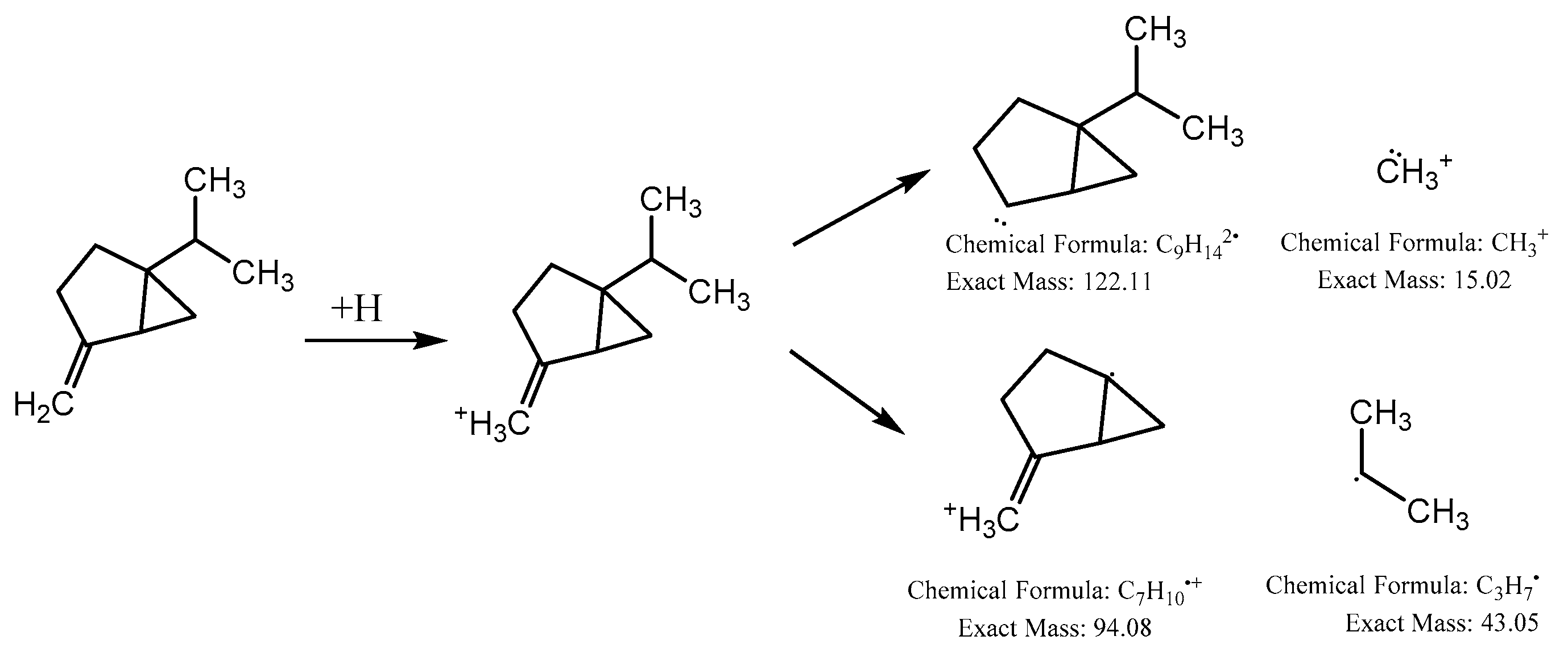
| C8 | P | 2.408 | 137.0596 | C10H17+ | −0.1 | 122, 94, 81, 77 | Sabinene | Monoterpene | --- |
| C9 | N | 2.484 | 435.0979 | C20H19O11− | 0.7 | 417, 345, 315, 272 | Homomangiferin | Xanthone | [38] |
| C10 | N | 2.627 | 461.0711 | C21H17O12− | 0.8 | 285, 175 | Kaempferol-O-Glucuronide | Flavonol | [48], MoNA |
| C11 | P | 2.728 | 593.1263 | C28H33O14+ | 0.8 | 477, 285 | Acacetin-O-rutinoside | Flavone | [56], NIST |
| C12 | N | 2.864 | 391.1029 | C19H19O9− | 0.4 | 245, 151 | Iriflophenone-2-O-α-L-rhamnopyranoside | Benzophenone | [38] |
| C13 | N | 3.067 | 245.0472 | C13H9O5− | 3.9 | 151, 107 | Iriflophenone | Benzophenone | [38] |
| C14 | N | 3.362 | 549.1127 | --- | 1.1 | 531, 115 | Unknown | --- | --- |
| C15 | N | 3.424 | 463.0889 | C21H18O12− | 3.6 | 301, 271 | Quercetin-O-hexoside | Flavonol | [47], HMDB |
| C16 | P | 3.681 | 179.1070 | C9H7O4+ | 0.5 | 161, 146, 133 | Dihydroxycoumarin (Esculetin) | Coumarin | [75], HMDB |
| C17 | N | 3.807 | 447.0946 | C21H19O11− | 1.6 | 285 | Luteolin-O-hexoside | Flavone | [56], HMDB |
| C18 | N | 4.242 | 477.1034 | C22H21O12− | −0.9 | 315, 271 | Isorhamnetin-O-hexoside | Flavonol | [51], MoNA |
| C19 | N | 4.282 | 507.1551 | C23H23O13− | −0.5 | 344 | Syringetin-O-hexoside | Flavonol | [52], MoNA |
| C20 | N | 4.512 | 317.0315 | C15H9O8− | 0.7 | 179, 151 | Myricetin | Flavonol | [53], HMDB |
| C21 | N | 4.550 | 431.0995 | C21H19O10− | −0.7 | 311, 269 | Apigenin-C-hexoside (Vitexin) | Flavone | [57], MoNA |
| C22 | P | 4.664 | 579.1703 | C27H31O14+ | −1.7 | 285 | Kaempferol-O-dirhamnoside | Flavonol | [50] |
| C23 | P | 4.820 | 435.1296 | C21H23O10+ | −1.7 | 247 | Iriflophenone, [2-(2-O-actyΑ-L-Rhamnopyranosyl) oxy] | Benzophenone | --- |
| N | 4.833 | 433.1136 | C21H21O10− | 0.3 | 245, 151 | [38] | |||
| C24 | N | 4.954 | 607.1660 | C28H31O15− | 3.4 | 313, 298 | Aquisiflavoside | Flavone | --- |
| C25 | P | 5.022 | 447.1280 | C22H23O10+ | 0.6 | 285, 270 | Sissotrin | Isoflavone | [68], NIST |
| C26 | N | 5.097 | 445.1136 | C21H17O11− | 0.2 | 268 | Baicalein-O-glucuronide | Flavone | [65], MoNA |
| C27 | N | 5.214 | 287.0579 | C15H11O6− | −0.1 | 151, 135, 107 | tetrahydroxyflavanone | Flavanone | [54] |
| C28 | N | 5.565 | 285.0409 | C15H9O6− | 1.1 | 133 | Luteolin | Flavone | [57], HMDB |
| C29 | N | 5.728 | 301.0360 | C15H9O7− | −0.9 | 273, 179, 151 | Quercetin | Flavonol | [47], HMDB |
| C30 | N | 5.895 | 553.1350 | C25H29O14− | −0.9 | 391, 245, 151 | Aquilarisinin | Benzophenone | --- |
| P | 5.939 | 555.1508 | C25H31O14+ | 0.7 | 291, 247, 121, 83 | --- | |||
| C31 | N | 6.094 | 417.1582 | C22H25O8− | −2.6 | 402, 181, 166, 151 | Syringaresinol | Lignan | [76], MoNA |
| C32 | N | 6.167 | 623.1359 | C28H31O16− | 3 | 315 | Isorhamnetin-O-rutinoside (narcissoside) | Flavonol | [51], MoNA |
| C33 | N | 6.254 | 593.1518 | C27H29O15− | 0.8 | 299, 285 | Kaempferol-O-neohesperidoside | Flavonol | [49], MoNA |
| C34 | N | 6.499 | 269.0473 | C15H9O5− | 0.1 | 151, 149, 117 | Apigenin | Flavone | [51], MoNA |
| P | 6.638 | 271.0601 | C15H11O5+ | 3.8 | 229, 153 | [60], MoNA | |||
| C35 | N | 6.931 | 301.0588 | C16H13O6− | 0.2 | 286, 255 | Hesperetin | Flavanone | [55,56], MoNA |
| C36 | P | 8.019 | 297.1127 | C20H41O+ | 1.9 | 281, 265, 248 | Phytol | Diterpene | MoNA |
| C37 | N | 8.352 | 299.0561 | C16H11O6− | −4.3 | 284, 256 | Hydroxygenkwanin | Flavone | [38], HMDB |
| C38 | N | 9.302 | 285.0774 | C16H13O5− | 4.6 | 165, 119, 93 | Sakuranetin | Flavanone | [57], HMDB |
| C39 | P | 9.310 | 285.0748 | C16H13O5+ | 1.1 | 270, 242, 176, 124 | Acacetin | Flavone | [56] MoNA |
| C40 | N | 9.445 | 283.0617 | C16H11O5− | 2.5 | 268 | Genkwanin | Flavone | [38], MoNA |
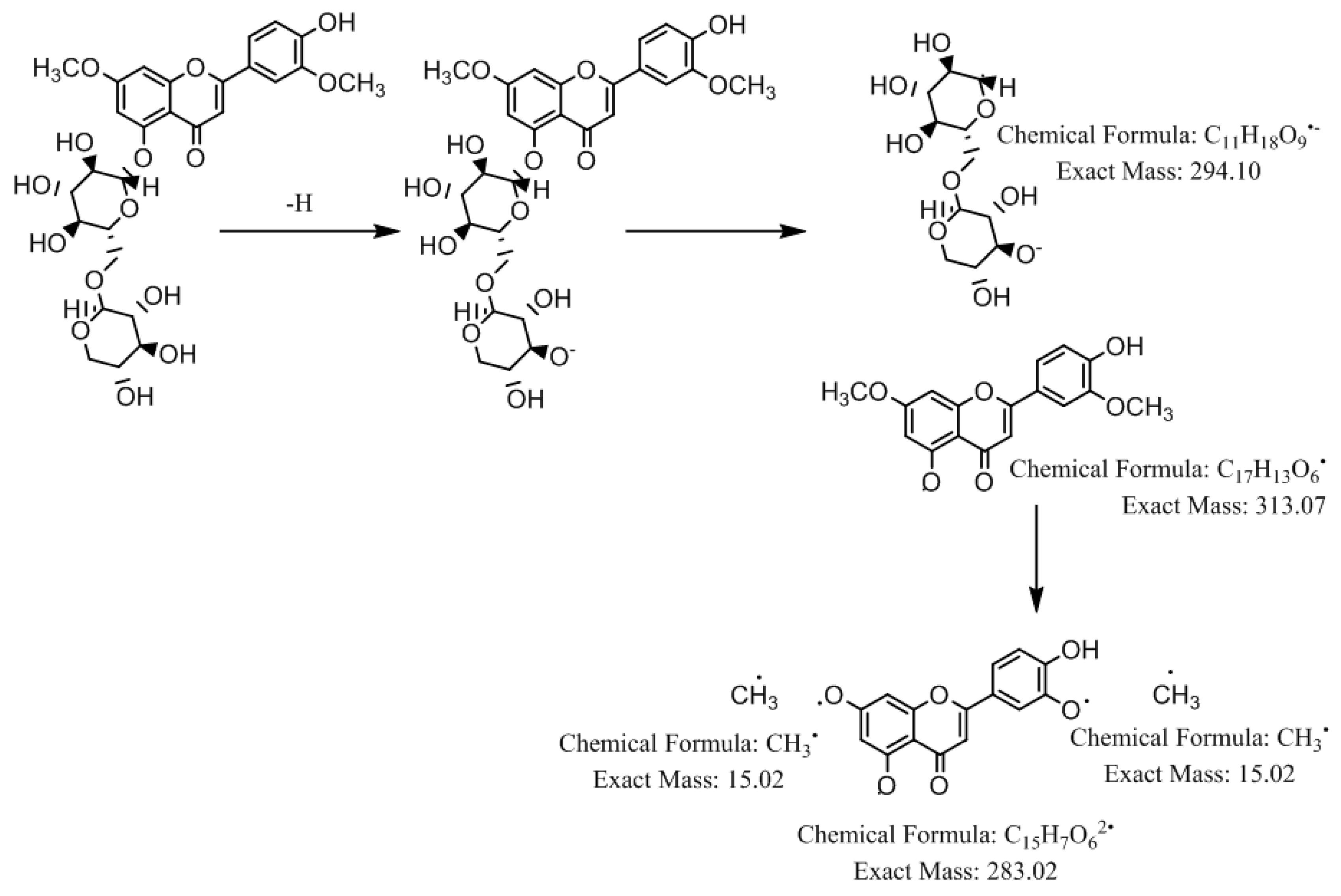
| C41 | N | 9.683 | 313.0726 | C17H13O6− | 1.8 | 298, 283, 270, 255 | Velutin | Flavone | [61], MoNA |
| C42 | P | 9.891 | 307.2488 | --- | 2.3 | 289, 271, 220 | Unknown | --- | --- |
| C43 | P | 10.856 | 329.1022 | --- | −0.4 | 313, 285 | Unknown | --- | --- |
| C44 | N | 11.757 | 577.2710 | C27H29O14− | −0.3 | 268 | Violanthin | Flavone | MoNA |
| C45 | P | 11.878 | 269.0810 | C16H13O4+ | 1.3 | 254, 197 | Formononetin | Isoflavone | [69] |
| C46 | P | 12.302 | 223.1681 | C15H27O+ | −0.6 | 207, 191, 177, 149 | Nerolidol | Sesquiterpenes | [73], MoNA |
| C47 | N | 16.856 | 279.2330 | C18H31O2− | 4 | 261, 234 | Linoleic acid | Fatty acid | MoNA |
| C48 | N | 17.976 | 255.2334 | C16H31O2− | −0.9 | 237 | Hexadecanoic acid | Fatty acid | MoNA |
| C49 | P | 19.775 | 427.3582 | C30H51O+ | −3 | 409, 367, 217, 191 | α-amyrin | Sesquiterpenes | [71] |
| C50 | P | 20.870 | 415.3518 | C29H51O+ | 1 | 397, 189, 161, 119 | β-sitosterol | Sterols | [71], HMDB |
| C51 | P | 21.572 | 413.3785 | C29H49O+ | −1.7 | 395, 255, 159 | Stigmasterol | Sterols | [71], HMDB |
| C52 | P | 22.407 | 427.3911 | C30H51O+ | −1.6 | 409, 185 | Lupeol | Triterpenoid | [71], NIST |
| C53 | P | 23.782 | 431.3790 | C29H51O2+ | 0.7 | 165 | α-Tocopherol | Tocopherols | [77], NIST |
© 2020 by the authors. Licensee MDPI, Basel, Switzerland. This article is an open access article distributed under the terms and conditions of the Creative Commons Attribution (CC BY) license (http://creativecommons.org/licenses/by/4.0/).
Share and Cite
Eissa, M.A.; Hashim, Y.Z.H.-Y.; El-Kersh, D.M.; Abd-Azziz, S.S.S.; Salleh, H.M.; Isa, M.L.M.; Abd Warif, N.M. Metabolite Profiling of Aquilaria malaccensis Leaf Extract Using Liquid Chromatography-Q-TOF-Mass Spectrometry and Investigation of Its Potential Antilipoxygenase Activity In-Vitro. Processes 2020, 8, 202. https://0-doi-org.brum.beds.ac.uk/10.3390/pr8020202
Eissa MA, Hashim YZH-Y, El-Kersh DM, Abd-Azziz SSS, Salleh HM, Isa MLM, Abd Warif NM. Metabolite Profiling of Aquilaria malaccensis Leaf Extract Using Liquid Chromatography-Q-TOF-Mass Spectrometry and Investigation of Its Potential Antilipoxygenase Activity In-Vitro. Processes. 2020; 8(2):202. https://0-doi-org.brum.beds.ac.uk/10.3390/pr8020202
Chicago/Turabian StyleEissa, Manar A., Yumi Z. H-Y. Hashim, Dina M. El-Kersh, Saripah S. S. Abd-Azziz, Hamzah Mohd. Salleh, Muhammad Lokman Md. Isa, and Nor Malia Abd Warif. 2020. "Metabolite Profiling of Aquilaria malaccensis Leaf Extract Using Liquid Chromatography-Q-TOF-Mass Spectrometry and Investigation of Its Potential Antilipoxygenase Activity In-Vitro" Processes 8, no. 2: 202. https://0-doi-org.brum.beds.ac.uk/10.3390/pr8020202