Article

Infrared Irradiation Drying Impact on Bee Pollen: Case Study on the Phenolic Composition of Eucalyptus globulus Labill and Salix atrocinerea Brot. Pollens

1
Laboratory of Pharmacognosy, Faculty of Pharmacy, Health Sciences Campus, University of Coimbra, Azinhaga de Santa Comba, 3000-548 Coimbra, Portugal
2
CQ-Centre of Chemistry—Coimbra, Department of Chemistry, Faculty of Sciences and Technology, University of Coimbra, Rua Larga, 3004-535 Coimbra, Portugal
3
Life Science Institute, University of Agricultural Sciences and Veterinary Medicine Cluj-Napoca, 3-5 Mănăştur Street, 400372 Cluj-Napoca, Romania
4
Department of Microbiology and Immunology, Faculty of Animal Science and Biotechnologies, University of Agricultural Sciences and Veterinary Medicine, 3-5 Mănăştur Street, 400372 Cluj-Napoca, Romania
5
REQUIMTE/LAQV, Laboratório de Farmacognosia, Departamento de Química, Faculdade de Farmácia, Universidade do Porto, R. Jorge Viterbo Ferreira, nº 228, 4050-313 Porto, Portugal
*
Author to whom correspondence should be addressed.
Submission received: 2 April 2021 / Revised: 25 April 2021 / Accepted: 26 April 2021 / Published: 18 May 2021

Abstract

:
Bee pollen is commonly reputed as a rich source of nutrients, both for bees and humans. Its composition is well balanced and can be taken as a stand-alone food or as supplement, including for the elderly owing its low caloric value. However, storage conditions frequently lead to product degradation, namely due to the high moisture content that enable the proliferation of molds and bacteria. Herein, an infrared (IR)-based technology is proposed as a mean to determine moisture content, setting also a new scalable approach for the development of a drying technology to be used for bee pollen processing, which can be carried out in a short time, without impacting the phenolic and flavonoid content and associated bioactive effects. Proof-of-concept was attained with an IR moisture analyzer, bee pollen samples from Eucalyptus globulus Labill and Salix atrocinerea Brot. being selected as models. Impact of the IR radiation towards the phenolic and flavonoid profiles was screened by HPLC/DAD profiling and radical scavenging ability by the DPPH assay. The IR-based approach shows good reproducibility while simultaneously reducing drying time and energy consumption, thus implying a low environmental impact and being suitable for industrial scale-up once no degradation has been found to occur during the radiation process.

Graphical Abstract

1. Introduction

Bee pollen is long known and classified as a food product [1], with a series of health benefits mainly attributed to a high content in phenolic constituents. While widely consumed, its preservation and quality control remain critical and call for further studies in this matter. Bee pollen is produced by the agglutination of selected flower pollens made by the worker bees, with nectar (and/or honey) and salivary substances, being collected at the entrance of the hive as small bolls, often named as pellets [2].
While scarce, currently available legislation defines bee pollen as a food, but some gaps dealing with its quality control remain to be filled, additional guidelines being currently proposed in the framework of an International Standard ISO normalization. The International Organization for Standardization includes the Working Group “TC 34/SC 19 Bee products, subgroup W3-Bee pollen”, that intends to implement guidelines and procedures for the standardization of the processing and circulation of bee products. One of the main challenges is to ensure a convenient preservation of the nutrients along with the bioactive compounds, which prompted us to conceptualize the current study.
Bee pollen is mostly commercialized in a dehydrated form, major chemical modifications of the components being assured through an appropriate drying process, which is crucial, for example, to avoid mold contamination [2]. However, most of the currently available drying methods are characterized by high energy consumption and a concomitant environmental impact.
Indeed, several studies delivered experimental evidence on the nutritional value of bee pollen, being portrayed as a fine food supplement [3,4] due to its high content in macronutrients, with well-balanced proportions of proteins, lipids, and carbohydrates, along with micronutrients and bioactive compounds, namely simple phenolic constituents and a series of polyphenols, flavonoids being particularly reputed in this matter. Nowadays, there is a high demand for food sources with low caloric impact but with a high value in nutrients, and bee pollen is certainly one of the most popular food products amongst the elderly [5]. According to Peris [6], 15 g of bee pollen supplies the required daily dose of amino acids. The caloric value of bee pollen was estimated by others at 381.70 kcal (1595.51 Kj) for 100 g [7] which gives a significant additional value to hypocaloric diets. In addition to its nutritional value, a myriad of potential therapeutic properties of bee pollen have been also suggested based on an increasing number of studies being carried out in the last two decades (among many others [8,9,10,11]). However, it is worth to mention that only a few address the allergenicity of bee pollen and the storage of fresh and dry samples [12,13].
Besides the requirements dealing with the nutritional content and the possible presence of additives, such as pesticides or antibiotics, unequivocal identification of the floral origin of bee-pollen and the preservation process are of utmost importance.
The floral composition is affected by phytogeographical, genetic modifications and seasonal factors [14,15,16]. A consistent work being carried out by us delivering data on hand-collected pollen, herbarium specimens and bee pollen samples, gathered in different locations and years, to perform the identification of bee pollen floral origins [17,18,19,20,21,22,23]. The method previously described by Campos et al., [17,18] enables the identification of pollen taxon by the HPLC-DAD-based phenolic/polyphenolic profiling of hydroalcoholic extracts. Generation of a chemical fingerprint that is species-specific (specific for each species of pollen origin), was found to be more sensitive and precise than the microscopic analysis, as it allows the identification of each taxon, to genera and species. Nevertheless, pollen shells, obtained after centrifugation of the hydroalcoholic extracts should be further analyzed on their morphological features. Relevantly, cumulative evidence suggests that the profile of each taxon is independent of geographical or climatic factors, which makes this method universal. Considering the above, the current study aims (i) to unequivocally identify the floral source of the crude materials used in this “Case Study”, and (ii) to investigate the impact of infrared (IR) radiation on the phenolic fingerprint in selected pollen samples. Radical scavenging ability was also assayed to further detail on the potential influence of IR on the extracts and the impact on the bioactive properties known to rely on phenolic compounds. The main end-point of the current work is to mimic a drying process with IR irradiation, with a low environmental impact due to the rapid, cheap, and accurate drying approach, thus setting a new perspective for further industrial drying applications.

2. Materials and Methods

2.1. Chemicals

Reference compounds were purchased from various suppliers: 2,2-diphenyl-1-picrylhydrazyl radical (DPPH), L-ascorbic acid and rutin were purchased from Sigma-Aldrich (St. Louis, MO, USA), ethanol 99% from Panreac (Castellar del Vallès, Spain), o-phosphoric acid and acetonitrile from Merck (Darmstradt, Germany). Water was treated in a Milli-Q (Millipore, Bedford, MA, USA) water purification system.

2.2. Bee Pollen Samples
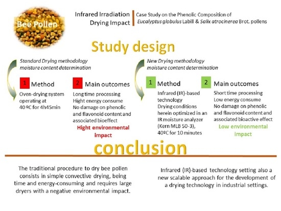
Samples of bee pollen were collected in Lavos (Figueira da Foz, Portugal), immediately frozen and stored at −21 °C until analysis. Pollen pellets were separated by hand, according to their morphological features. Identification of each taxon was carried out by HPLC/DAD analysis of their hydroalcoholic extracts (details in the extraction section) according to [17,18]. Briefly, HPLC/DAD profiling delivers different phenolic fingerprints, their comparison with an internal database of floral pollen sources allowing the identification of the species of each taxon under analysis. The sediments, obtained after centrifugation of the hydroalcoholic extracts, were further analyzed on their morphological characters in a Leitz Laborlux microscope to further confirm the taxon of the separated taxa from the entire mixture [17]. After confirmation of the two main floral sources in a mixed sample, a representative amount of bee pollen pellets was separated, and identified as follows: Eucalyptus globulus Labill (sample 1) and Salix atrocinerea Brot. (sample 2).

2.3. Drying Process

The IR drying process has been performed with a moisture analyzer (Kern MLB 50-3) and optimized to reach a residual humidity of ca. 4% as recommended by others [2]. To determine the reproducibility of the IR method, five random samples of bee pollen with different water content were used. A standard drying process was also performed in an oven-drying system, operating at 40 °C, until constant weight was recorded. This procedure is done with a mean time of approximately 4 h and 45 min in a heater, until the relative humidity reached the required values.

2.4. Moisture Determination

Water content determination was also performed by two different methods. For the IR-based method the moisture analyzer (Kern MLB 50-3) was used. Five aliquots of 1 g of sample were dried at 50 °C until constant weight for three steps of 45 s.
The methods described on the Codex Alimentarius, European Pharmacopoeia and Portuguese Pharmacopoeia, are very similar, and were used as references [24].

2.5. Extracts Preparation

Fresh and dry samples of the two different taxa (E. globulus and S. atrocinerea) were extracted with a mixture of ethanol:water (1:1, v/v) at the concentration of 20 mg/mL and 10 mg/mL, respectively, using ultrasonication (30 min). Insoluble material was separated by centrifugation (6000 rpm/5 min) and used for microscopic analysis confirmation. The supernatant was immediately analyzed by HPLC/DAD for phenolic fingerprint and spectrophotometric analysis of free radical scavenging activity using the DPPH method, as below described.

2.6. Chromatographic Analysis

Chromatographic analyses were carried out by HPLC/DAD [18]. Briefly, 20 μL of fresh pollen extract and 10 μL of dried bee pollen extract were analyzed in a Gilson 170, separation being attained with a Waters Spherisorb ODS2 (5 mm) column (4.6 × 250 mm) by an acidified water-acetonitrile gradient with a flow rate of 0.8 mL/min, and a column temperature of 24 °C. Standard chromatograms were plotted at λmax 260 and 340 nm. Spectral data for all peaks were accumulated in the range 220–400 nm using DAD (Gilson 170) and further analyzed with the software Unipoint. The suitability of the method was previously evaluated by Campos [17].
All the extracts were submitted to a qualitative and quantitative analysis of the main phenolic constituents. Structural determination of the phenolic compounds was performed according to the theoretical rules presented in Campos and Markham [25] and by comparison with our internal spectral database [25]. Concentrations were determined using the following standard curve equation obtained with rutin (1) (A = HPLC peak area):
y = 4.2159 × 10⁻⁹A + 0.0062 R2 = 0.9996

2.7. Free Radical Scavenging Activity

To evaluate the impact of IR radiation in the bioactivity of the extracts and the potential interference with the phenolic content, as phenolic constituents act as reference compounds in the preservation of the integrity of the samples, both pollen extracts were also used to determine the free radical scavenging activity upon the 2,2 diphenyl-picrylhydrazyl (DPPH) radical. The method was performed according to Campos et al., [19]. Measurements were carried out on a UV/VIS spectrophotometer Hitachi U-2000. Briefly, 2.5 mL of DPPH solution (5.96 mg in 250 mL of 1:1 ethanol/water solution) were mixed with an appropriate amount of extract (10, 20, 40, 60, 80, 100 µL), followed immediately by homogenization. After 10 min, quantification of the remaining DPPH radicals was recorded from the absorption at 517 nm. The reference standard was ascorbic acid with an EC50 value of 2.41 μg/mL.

2.8. Statistical Analysis

All determinations were performed in quintuplicate, for the moisture determination, or in triplicate, for the DPPH assay and chromatographic analysis. Graphics were created with STATISTICA 7 software. Classical statistical analysis was performed. DPPH test’s results for the different drying methods were compared by t-test for variance (p ≤ 0.05) with Microsoft Excel software. Validation of the moisture determination by Kern MLB 50-3 was done by Z-score test versus a standard method.

3. Results

3.1. Water Content Determination

The IR method reproducibility was studied using five bee pollen samples with a different water content (Table 1). The calculated relative standard deviation (RSD) ranged from 58.9%, determined for the fresh pollen (RH% = 27.77%), to 7.494% recorded with the dried samples (RH% = 2.25%). The method was validated by comparison with the standard methods described in the Portuguese Pharmacopoeia [24] using Z-score test. Admitting an error of the average of ±0.2% (the measurement uncertainty indicated by the Kern MLB 50-3 specifications) the moisture analyzer did not show any relevant differences comparing with the standard drying method.
Briefly, as evaluated with samples (1 g) of bee pollen, optimal drying performance was observed at 40 °C for 10 min. These parameters were optimized after a previous determination of the better conditions to be used in this IR-based drying process. No differences on the water content were observed between the two species under study, determined as 24.73 ± 0.21% for E. globulus and 24.77 ± 0.35% for S. atrocinerea.

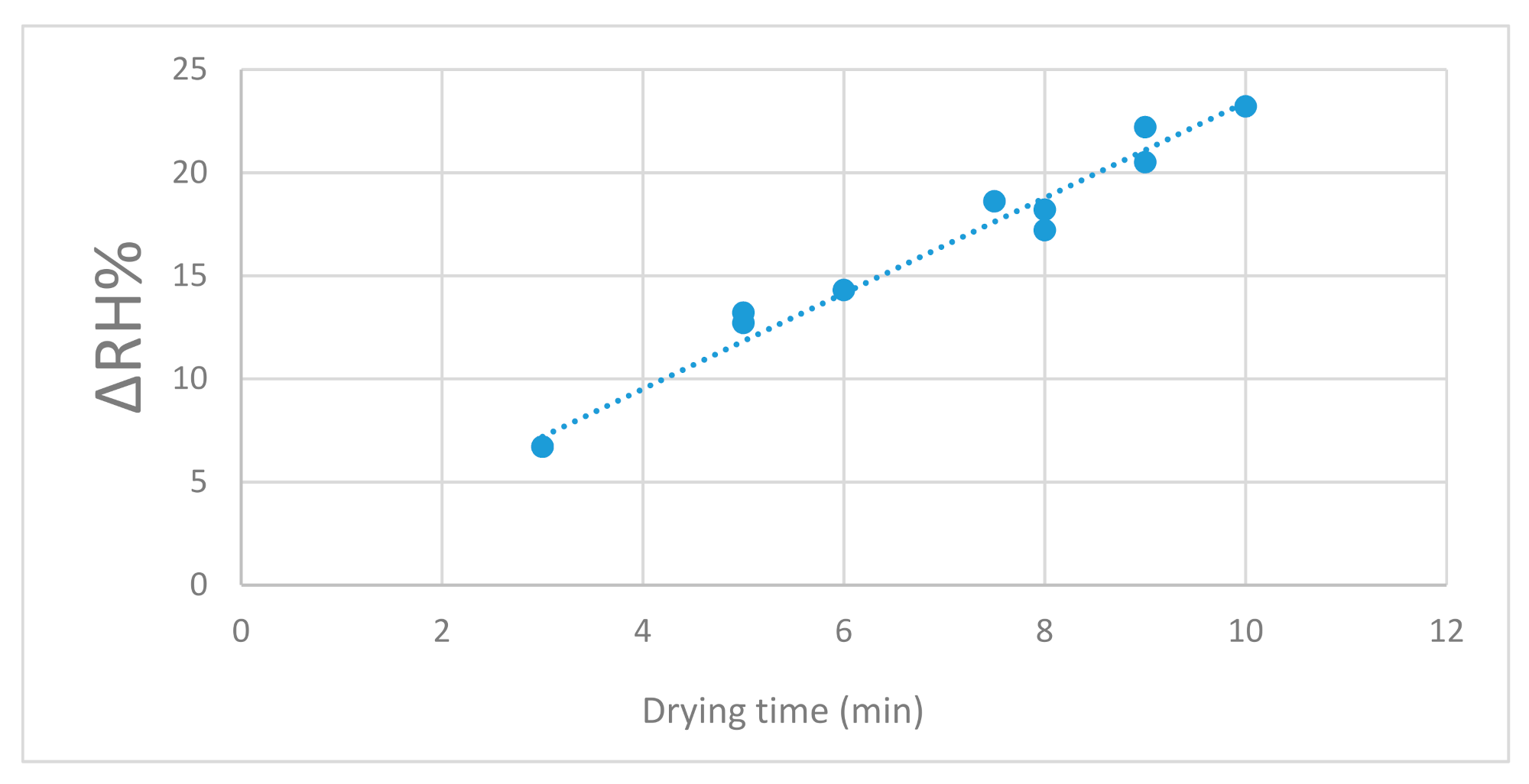
3.2. Infrared Drying Process

The IR drying process was optimized considering the drying rate of different samples (1 g each), i.e., the time (in minutes) required to achieve a final moisture of 4%. As showed in Figure 1, a linear relationship was found between the IR exposure time and the relative humidity loss (ΔRH%) at 40 °C. This relationship is described by the equation:
Dt = 2.3172 × ΔRH% + 0.2445; R2 = 0.9776, where Dt is the drying time (min)

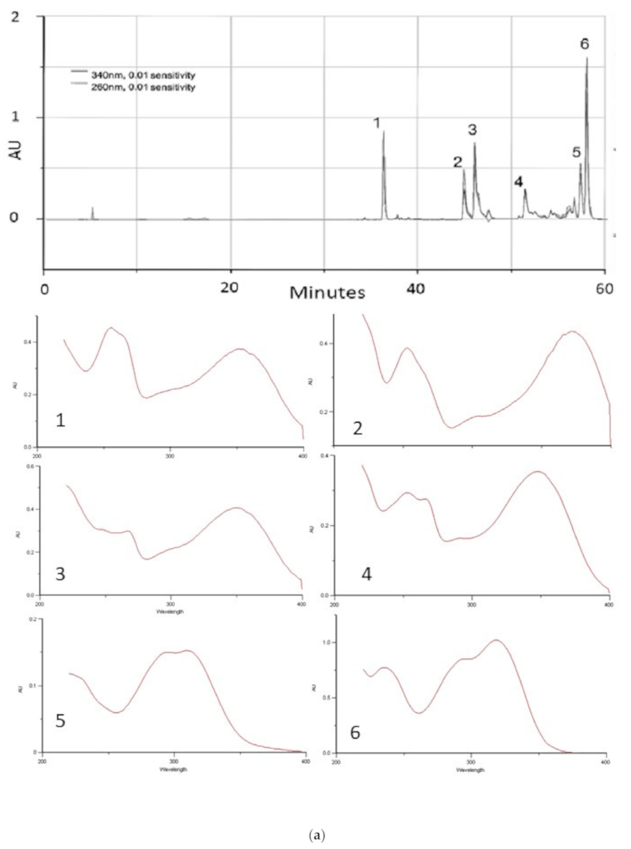
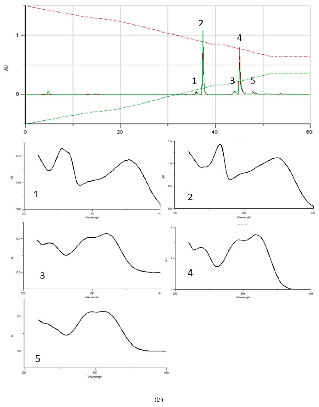
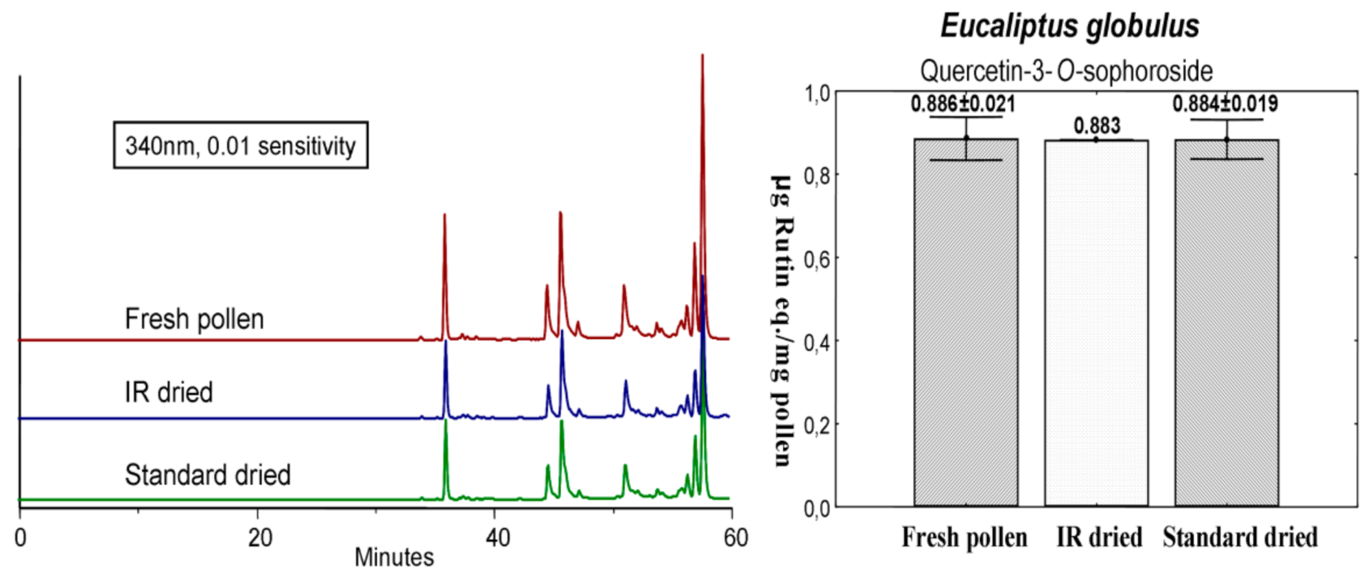
3.3. Pollen Phenolic/Flavonoid Profile

HPLC/DAD chromatographic analysis of the hydroethanolic pollen extracts was performed aiming to determine the pollen floral origin [18,26] and also to detect a possible interference with the qualitative and/or quantitative phenolic profiles (used as biomarkers) due to exposure to the IR radiation during the drying process. Phenolic profiles of the two samples, and the corresponding UV spectral data, are presented in Figure 2. The hydroethanolic extract obtained from E. globulus bee pollen (Figure 2a) was characterized by the occurrence quercetin-3-O-(β-D-glucopyranosyl-2-β-D-glucopyranoside) (namely quercetin-3-O-sophoroside) (RT 35.63), myricetin (RT 44.31), tricetin (RT 45.28), luteolin (RT 50.58) and 3-O-methylquercetin (RT 51.62), in addition to two cinnamic acid derivatives (RTs 57.11 and 57.67). These compounds were previously isolated from samples of E. globulus pollen, their structures being elucidated by NMR experiments (1H and 13C-NMR; 1H,1H-COSY; 1H,13C-COSY) [17,18]. Kaempferol-3-O-(β-D-rhamnopyranosyl-2-β-D-glucopyranoside) (namely kaempferol-3-O-neohesperoside) (RT 38.62) was detected as the main phenolic constituent on the hydroethanolic extract obtained from S. atrocinerea bee pollen samples, lower amounts of 3-O-glycosylated derivatives of quercetin (RT 37.04) and kaempferol (RT 39.26) being determined. Two caffeic acid derivatives (RT 45.31 and RT 46.52) were also identified. The profile (Figure 2b) was found to be identical to a previously described for S. atrocinerea in Campos et al. [18].
The identification of each taxon was in agreement and further corroborated with the results obtained by microscopic analysis.

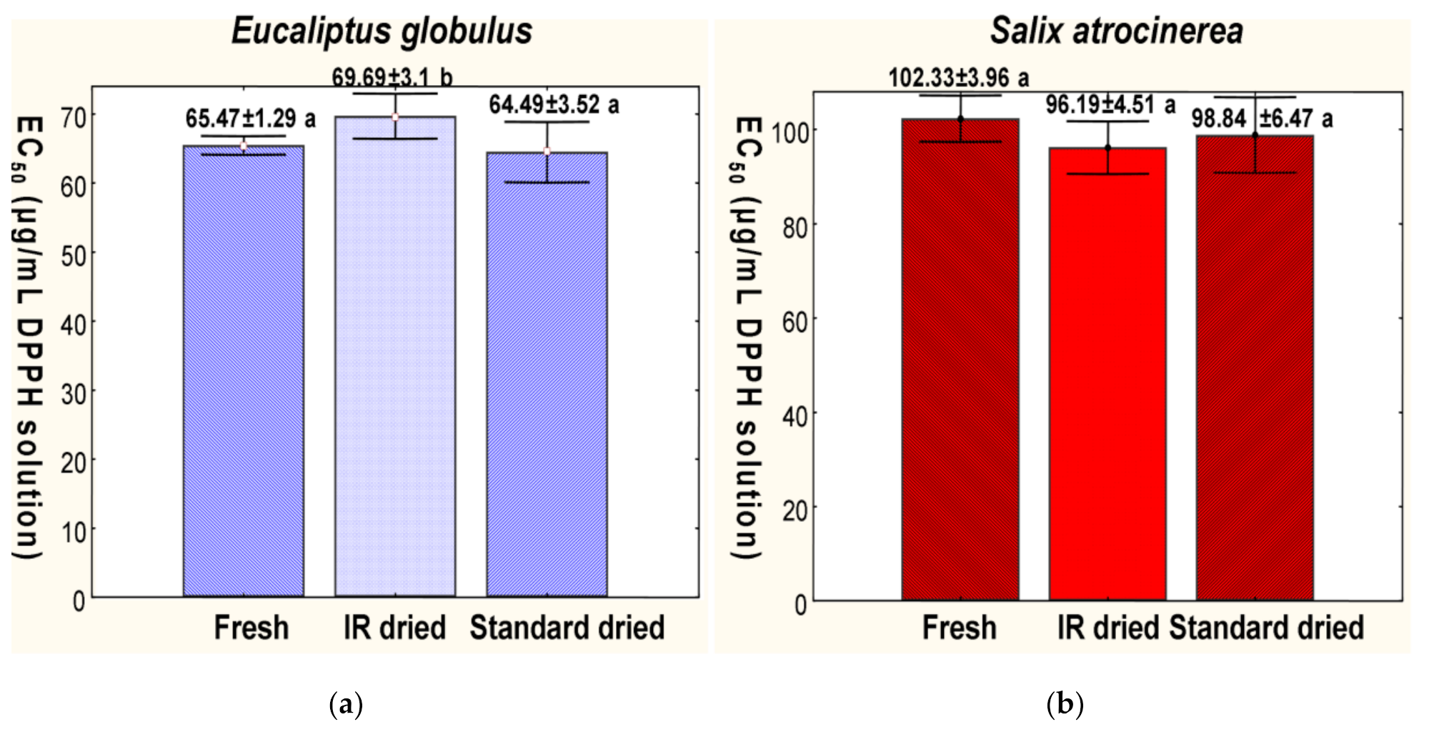
3.4. IR Radiation Effect on Phenolic Composition and on the Radical Scavenger Bioactivity

It is well known that bee pollen has a significant free radical scavenging activity [19,27,28,29], which is normally species-specific, dependent on similar phenolic/flavonoid profiles [19], and independent from exogenous parameters (geographical origin, climate, etc.). As such, these two parameters can be used as biomarkers of quality for bee pollen. As expected, the free radical scavenger activity of bee pollen is time-dependent and decreases with prolonged storage time [17,19].
Currently, there is no data dealing with the influence of post harvesting processing, namely with the drying processes by irradiation with IR, on the radical scavenging properties and/or in the HPLC/DAD profile of the phenolic/flavonoid compounds from bee pollen. The DPPH radical scavenging activity, expressed in EC50, was assayed to evaluate the impact of the IR radiation.
All samples exhibited antiradical activity (Figure 3) with a linear concentration-dependency. Samples of E. globulus bee pollen showed EC50 values slightly above from those of previously reported data, suggesting a slight decrease in the antiradical ability [17,19]. Comparison of the EC50 values determined for the fresh pollen samples and the dried ones (by T-test for variance; p ≤ 0.05) denotes a small decrease in the scavenging activity of the IR dried samples. No statistically relevant variation was detected in comparison with the traditional (standard) drying method. Fresh samples of S. atrocinera bee pollen showed high scavenging activity (around 100 μg/mL DPPH solution). As show in Figure 3, the activity remained unchanged with both drying processes.
Subsequently, the profile obtained by HPLC-DAD was used to study the influence of the drying process on the qualitative and quantitative profiles of the major phenolic constituents. Pollen samples were submitted to two distinct drying procedures, the standard drying method in oven-drying at 40 °C until the RH% reached 4%, and the optimized IR drying process. Then, hydroethanolic extracts obtained with the dried pollen samples were analyzed by HPLC-DAD and assessed on their radical scavenging activity (DPPH-method), to further investigate the impact of the IR radiation in the drying processing.
Chromatographic profiles of extracts obtained from the dried samples of E. globulus (IR and standard drying methods) matched perfectly with the phenolic profile of the extract obtained from fresh bee pollen samples (Figure 4), attesting the absence of any qualitative modification on the phenolic/flavonoidic profile. To assess the possible influence on the quantitative profile, quercetin-3-O-sophoroside (used as biomarker) was quantitated. As observed in Figure 4, no differences on the amounts of the glycosylated quercetin derivative were recorded between the samples.
Likewise, the chromatographic profile of S. atrocinera bee pollen dried samples did not reveal any difference comparing to fresh pollen chromatographic profile (Figure 5). As seen in Figure 5, no difference in the concentration of the major flavonoid kaempferol-3-O-neohesperidoside (biomarker), was noted within the three samples.
To further corroborate the absence of degradation, extracts (fresh and dried samples) were assayed on their free radical scavenging activity.
The extract obtained from E. globulus (Figure 4) fresh bee pollen samples was found to exhibit significant antiradical activity, an EC50 value of 65.47 ± 1.29 μg/mg, being recorded. No significant differences have been recorded in comparison with the extract obtained from samples dried under the standard approach (EC50 = 64.49 ± 3.52 μg/mL). The EC50 value estimated for the IR-based dried samples (EC50 of 69.69 ± 3.10 μg/mL) suggested a statistically significant, but slight, decrease on the antiradical activity. As no qualitative or quantitative differences have been observed in regard to the phenolic profiles of E. globulus pollen samples, the recorded decrease on the radical scavenging activity might be related with the occurrence of other, non-phenolic, constituents such has carotenoids. In contrast, the radical scavenging ability of the extracts obtained from fresh and dried samples of S. atrocinera bee pollen remained similar, as shown in Figure 5. Antiradical activity of the extracts obtained from samples of fresh pollen and dried samples (IR-based and standard method) was estimated at EC50 values of 102.33 ± 3.96, 96.19 ± 4.51) and 98.84 ± 6.47 μg/mL, respectively.
Considering the above, it is evident that neither of the drying methods elicited changes on the phenolic profiles (biomarkers) as well as on the derived free radical scavenging properties.

4. Discussion

Bee pollen is widely popular as a food supplement due to its equilibrate composition of proteins, lipids and sugars [4,23], high content in vitamins, as well all essential amino acids and unsaturated fatty acids (ω-3, ω-6, ω-9) [30,31,32,33].
Despite its widespread use, both due to the nutritional value and biological properties, water content determination and management are items of capital significance for the quality control and a safe consumption. Due to its richness in several nutrients, bee pollen is an ideal substrate for the development of microorganisms, especially mycotoxins-producing fungi, such as Aspergillus ochraceus Wilh, a frequent producer of ochratoxin A [34,35]. Various preservation methods have been proposed to avoid microbial contamination and growth, but drying processes remain the most convenient and appropriate if a RH% of ca. 4% is obtained. Recalling in mind the absence of a specific legislation in various countries, the routinely used methods for relative humidity determination (among others, AOAC or Pharmacopoeia methods) are generally time-consuming and susceptible to operator errors. In addition, the amount of energy being consumed has an environmental impact that must be considered.
To overcome such limitations, we propose a new IR-based drying method. Based on the obtained results (Table 1), it is clear that this method revealed to be reproducible in all the range of RH% assessed (±2–28%). Through Z-score test, the IR drying procedure was also compared with the official method [24] (which is similar to other Pharmacopoeias and Codex Alimentarius) and did not show any relevant performance difference. As such, while the methods are equivalent on their performance, the IR method greatly reduced the drying time (ca. 15 min for the moisture determination in samples) in comparison with standard methods requiring nearly 5 h to ensure the convenient reduction in RH%.
Drying conditions herein optimized in an IR moisture analyzer (Kern MLB 50-3), are scalable and guarantee the preservation of the matrix under these conditions. IR radiation did not affect phenolic biomarkers, but other structural classes such as proteins, lipids and vitamins, should be screened.
The HPLC/DAD profiles of the extracts, the quantification of the selected biomarkers and the reference bioactivity, as it is the radical free scavenging activity, were the preliminary models used in this study, and successfully applied to the different botanical origin samples. As showed in Figure 1, the IR dryer performance revealed an evident linear relationship between the amount of removed water (ΔRH%) and the drying time, and as seen in Figure 4 and Figure 5, the phenolic/flavonoid profiles of fresh and dried pollen samples, show a stable correlation with the biomarker used for these taxon. These results reveal the high potential of this method for further development of a dry methodology in industrial settings.
Despite previous approaches dealing with water content determination [36,37,38,39,40,41], the effects of IR radiation in the bioactivity and chemical stability of the bee pollen samples are herein reported for the first time. The current methodology might well be used in other edible products of plant origin, thus calling for further studies in this matter.
Recently, experimental data came to light associating the evidence that Far IR (FIR) is efficient in drying processes due to its energy-saving performance, mainly because energy is directly supplied to the object to be heated without dispersing energy to unnecessary objects, and in the case of certain phenolic compounds, an increase of the total amount and even in the free radical scavenging effect was observed [42].
Once, bee pollen is mainly rich in phenolic compounds, that frequently underlie its biological properties, in particular the free radical scavenging and antioxidant activities [8,9,17,19,43,44,45], it is relevant to understand the influence of IR radiation in the processed product if used for these further purposes.
Such data will be crucial for the development of further studies with foods and medicinal plants, that are frequently required to be dry for preservation.

5. Conclusions

The traditional procedure to dry bee pollen samples consists in simple convective drying, being time and energy-consuming and requires large dryers with a negative environmental impact. The prolonged exposure to heat can also promote chemical modifications, namely on the content of metabolites that are responsible for the biological activity of this matrix. In the present study, an IR-based drying method is optimized, discussed and provided the basis for an accurate drying process. The results from this IR-based method are of utmost importance, especially due to its low time- and energy-consumption when compared with the standard drying method. No impact being caused by IR has been observed with the samples under study, namely on their phenolic profiles and selected biomarkers. Complementary methodologies (morphological analysis and antiradical activity) further corroborated that this technology as the potential to be developed in a large scale for industrial applications.

Author Contributions

Conceptualization, M.G.C.; methodology, M.G.C., C.F. and N.G.M.G., software, M.G.C., C.F. and N.G.M.G.; validation, C.F. and N.G.M.G.; formal analysis, C.F. and N.G.M.G.; investigation, M.G.C., C.F. and N.G.M.G.; resources, M.G.C.; data curation, M.G.C.; writing—original draft preparation, M.G.C., C.F., N.G.M.G., O.B. and A.C.U.; writing—review and editing, O.B. and A.C.U.; supervision, M.G.C.; project administration, M.G.C.; funding acquisition, M.G.C. All authors have read and agreed to the published version of the manuscript.

Funding

The authors wish to thank to “Projeto Estratégico—(UI0204): UIDB/00313/2020 (Portugal).

Institutional Review Board Statement

Not applicable.

Informed Consent Statement

Not applicable.

Conflicts of Interest

The authors declare that they have no conflict of interest.

References

  1. EFSA Supporting Pub. Technical Report. EN-1234. 2017; Volume 14. Available online: https://0-efsa-onlinelibrary-wiley-com.brum.beds.ac.uk/doi/abs/10.2903/sp.efsa.2017.EN-1234 (accessed on 1 April 2021). [CrossRef]
  2. Campos, M.G.; Bogdanov, S.; Almeida-Muradian, L.B.; Szczesna, T.; Mancebo, Y.; Frigerio, C.; Ferreira, F. Pollen composition and standardisation of analytical methods. J. Apic. Res. 2008, 47, 154–161. [Google Scholar] [CrossRef]
  3. Kroyer, G.; Hegedus, N. Evaluation of bioactive properties of pollen extracts as functional dietary food supplement. Innov. Food Sci. Emerg. Technol. 2001, 2, 171–174. [Google Scholar] [CrossRef]
  4. Mărgăoan, R.; Mărghitas, L.A.; Dezmirean, D.S.; Dulf, F.V.; Bunea, A.; Socaci, S.A.; Bobiş, O. Predominant and secondary pollen botanical origins influence the carotenoid and fatty acid profile in fresh honeybee-collected pollen. J. Agric. Food Chem. 2014, 62, 6306–6316. [Google Scholar] [CrossRef] [PubMed]
  5. Figueiredo, F.; Encarnação, T.; Campos, M.G. Algae as functional foods for the elderly. Food Nut. Sci. 2016, 7, 1122–1148. [Google Scholar] [CrossRef] [Green Version]
  6. Peris, M.J. Producción y comércio de los produtos apícolas en España. 1º Congreso Nacional de Apicultura (Madrid). El Campo del Banco de Bilbao. Apicultura 1984, 93, 40–68. [Google Scholar]
  7. Orzaez-Villanueva, M.T.; Diaz-Marquina, A.; Bravo-Serrano, R.; Blazquez-Abellan, G. The importance of bee-collected pollen in the diet: A study of its composition. Int. J. Food Sci. Nut. 2002, 53, 217–224. [Google Scholar] [CrossRef] [PubMed]
  8. Leja, M.A.; Mareczek, G.; Wyżgolik, J.; Klepacz-Baniak, K.C. Antioxidative properties of bee pollen in selected plant species. Food Chem. 2007, 100, 237–240. [Google Scholar] [CrossRef]
  9. Pascoal, A.; Rodrigues, S.; Teixeira, A.; Feásc, X.; Estevinho, L.M. Biological activities of commercial bee pollens: Antimicrobial, antimutagenic, antioxidant and anti-inflammatory. Food Chem. Toxic. 2014, 63, 233–239. [Google Scholar] [CrossRef]
  10. Silva, D.N.A.; João, J.S.S.; Campos, M.G. Biological and Functional Properties of Bee Products for Medicinal Purposes. In Traditional and Folk Herbal Medicine: Recent Researches, 1st ed.; Gupta, V.K., Ed.; Daya Publishing House: New Delhi, India, 2014; Chapter 18; pp. 541–562, ISBN-10: 8170358744. [Google Scholar]
  11. Mărgăoan, R.; Stranț, M.; Varadi, A.; Topal, E.; Yücel, B.; Cornea-Cipcigan, M.; Campos, M.G.; Vodnar, D.C. Bee pollen and bee bread: Bioactive constituents and health benefits. Antioxidants 2019, 8, 568. [Google Scholar] [CrossRef] [Green Version]
  12. Anjos, O.; Paula, V.; Delgado, T.; Estevinho, L. Influence of the storage conditions on the quality of bee pollen. Zemdirb. Agric. 2019, 106, 87–94. [Google Scholar] [CrossRef]
  13. Jagdis, A.; Sussman, G. Anaphylaxis from bee pollen supplement. Canadian Med. Assoc. J. 2012, 184, 1167–1169. [Google Scholar] [CrossRef] [Green Version]
  14. EFSA Panel on Genetically Modified Organisms (GMO). Statement on the safety of MON810 maize pollen occurring in or as food. panel on genetically modified organisms (GMO). EFSA J. 2011, 11, 2434. [Google Scholar]
  15. EFSA. Scientific opinion on an application (EFSA-GMO-NL-2012-107) for the placing on the market of maize MON 810 pollen under Regulation (EC) No 1829/2003 from Monsanto. EFSA J. 2012, 10, 3022. [Google Scholar] [CrossRef] [Green Version]
  16. Szczesna, T.; Rybak-Chimielewska, H.; Chmielewsky, W. Sugar composition of pollen loads harvested at different periods of the beekeeping season. J. Apic. Sci. 2002, 46, 107–115. [Google Scholar]
  17. Campos, M.G. Caracterização do Pólen Apícola Pelo Seu Perfil em Compostos Fenólicos e Pesquisa de Algumas Actividades Biologicas. Ph.D. Thesis, Faculty of Pharmacy, University of Coimbra, Coimbra, Portugal, 1997. [Google Scholar]
  18. Campos, M.G.; Mitchel, K.; Cunha, A.; Markham, K.R. A systematic approach to the characterisation of bee pollens via their flavonoid/phenolic profiles. Phytochem. Anal. 1997, 8, 181–185. [Google Scholar] [CrossRef]
  19. Campos, M.G.; Webby, R.F.; Markham, K.R.; Mitchell, K.A.; Cunha, A.P. Age-induced diminution of free radical scavenging capacity in bee pollens and the contribution of constituent flavonoids. J. Agric. Food Chem. 2003, 51, 742–745. [Google Scholar] [CrossRef] [Green Version]
  20. Bakour, M.; Campos, M.G.; Imtara, H.; Lyoussi, B. Antioxidant content and identification of phenolic/flavonoid compounds in the pollen of fourteen plants using HPLC-DAD. J. Apic. Res. 2020, 59, 35–41. [Google Scholar] [CrossRef]
  21. Bovi, T.S.; Caeiro, A.; Santos, S.A.; Zaluski, R.; Shinohara, A.J.; Lima, G.P.P.; Campos, M.G.; Junior, A.J.; Orsi, R.O. Flavonoid content in bee bread suffers seasonal variation and affects hypopharyngeal gland development in Apis mellifera honey bees. J. Apic. Res. 2019, 20, 1–8. [Google Scholar]
  22. Negri, G.; Barreto, L.M.R.C.; Sper, F.L.; Carvalho, C.D.; Campos, M.G. Phytochemical analysis and botanical origin of Apis mellifera bee pollen from the municipality of Canavieiras, Bahia State, Brazil. Braz. J. Food Techn. 2018, 21, 1–16. [Google Scholar] [CrossRef] [Green Version]
  23. Urcan, A.C.; Criste, A.D.; Dezmirean, D.S.; Mărgăoan, R.; Caeiro, A.; Campos, M.G. Similarity of data from bee bread with the same taxa collected in India and Romania. Molecules 2018, 23, 2491. [Google Scholar] [CrossRef] [Green Version]
  24. Infarmed-National Authority of Medicines and Health Products. I.P. Perda Por Secagem. In Portuguese Pharmacopoeia, 7th ed; Infarmed, Ed.; Imprensa Nacional: Lisboa, Portugal, 2005; Chapter 2.2.32.d; pp. 49–50. [Google Scholar]
  25. Campos, M.G.; Markham, K.R. Structure Information from HPLC and on-Line Measured Absorption Spectra: Flavones, Flavonols and Phenolic Acids, 1st ed.; Imprensa da Universidade de Coimbra: Coimbra, Portugal, 2007; pp. 14, 26–29, 91, 104. ISBN 978-989-8074-05-8. [Google Scholar]
  26. Almaraz-Abarca, N.; Campos, M.G.; Delgado-Alvarado, E.A.; Ávila-Reyes, J.A.; Herrera-Corral, J.; González-Valdez, L.S.; Naranjo-Jiménez, N.; Frigerio, C.; Tomatas, A.F.; Almeida, A.J.; et al. Pollen flavonoid/phenolic acid composition of four species of cactaceae and its taxonomic significance. Am. J. Agric. Biol. Sci. 2008, 3, 534–543. [Google Scholar]
  27. LeBlanc, B.W.; Davis, O.K.; Boue, S.; DeLucca, A.; Deeby, T. Antioxidant activity of Sonoran Desert bee pollen. Food Chem. 2009, 115, 1299–1305. [Google Scholar] [CrossRef]
  28. Rebiai, A.; Lanez, T. Chemical composition and antioxidant activity of Apis mellifera bee pollen from northwest Algeria. J. Fund. Appl. Sci. 2012, 4, 155–163. [Google Scholar] [CrossRef] [Green Version]
  29. Šaric, A.; Balog, T.; Sobočanec, S.; Kušić, B.; Šverko, V.; Rusak, G.; Likić, S.; Bubalo, D.; Pinto, B.; Reali, D.; et al. Antioxidant effects of flavonoid from Croatian Cystus incanus L. rich bee pollen. Food Chem. Toxic. 2009, 47, 547–554. [Google Scholar] [CrossRef] [PubMed]
  30. Almeida-Muradian, L.B.; Pamplona, L.C.; Coimbra, S.; Barth, O.M. Chemical composition and botanical evaluation of dried bee pollen pellets. J. Food Comp. Anal. 2005, 18, 106–111. [Google Scholar] [CrossRef]
  31. Conte, G.; Bednelli, G.; Serra, A.; Signorini, F.; Bientinesi, M.; Nicolella, C.; Mele, M.; Canale, M. Lipid characterization of chestnut and willow honeybee-collectged pollen: Impact of freeze-drying and microwave-assisted drying. J. Food Comp. Anal. 2016. [Google Scholar] [CrossRef]
  32. Gardana, C.; Del Bo’, C.; Quicazán, M.C.; Correa, A.R.; Simonetti, P. Nutrients, phytochemicals and botanical origin of commercial bee pollen from different geographical areas. J. Food Comp. Anal. 2018, 73, 29–38. [Google Scholar] [CrossRef]
  33. Bedlina-Aldemita, D.; Opper, C.; Schreiner, M.; D’Amico, S. Nutritional composition of pot-pollen produced by stingless bees (Tetragona biroi Friese) from the Philippines. J. Food Comp. Anal. 2019, 82, 103215. [Google Scholar] [CrossRef]
  34. Medina, A.; Gonzalez, G.; Saez, J.M.; Mateo, R.; Jimenez, M. Bee pollen, a substrate that stimulates Ochratoxin A production by Aspergillus ochraceus Wilh. Syst. Appl. Microb. 2004, 27, 261–267. [Google Scholar] [CrossRef]
  35. Rodríguez-Carrasco, Y.; Font, G.; Mañes, J.; Berrada, H. Determination of mycotoxins in bee pollen by gas chromatography–tandem mass spectrometry. J. Agric. Food Chem. 2013, 61, 1999–2005. [Google Scholar] [CrossRef]
  36. Melo, I.; Almeida-Muradian, L. Comparison of methodologies for moisture determination on dried bee pollen samples. Food Sci. Techn. 2011, 31, 194–197. [Google Scholar] [CrossRef] [Green Version]
  37. Arruda, V.A.S.; Pereira, A.A.S.; de Freitas, A.S.; Barth, O.M.; Almeida-Muradian, L.B. Dried bee pollen: B complex vitamins, physicochemical and botanical composition. J. Food Comp. Anal. 2013, 29, 100–105. [Google Scholar] [CrossRef]
  38. Isik, A.; Ozdemir, M.; Doymaz, I. Infrared drying of bee pollen: Effects and impacts on food components. Czech J. Food Sci. 2019, 37, 69–74. [Google Scholar] [CrossRef] [Green Version]
  39. Isik, A.; Ozdemir, M.; Doymaz, I. Effect of hot air drying on quality characteristics and physicochemical properties of bee pollen. Food Sci. Tech. 2019, 39, 224–231. [Google Scholar] [CrossRef] [Green Version]
  40. Kanar, Y.; Mazi, B.G. Effect of different drying methods on antioxidant characteristics of bee-pollen. J. Food Measur. Charact. 2019, 13, 3376–3386. [Google Scholar] [CrossRef]
  41. Keskin, M.; Özkök, A. Effects of drying techniques on chemical composition and volatile constituents of bee pollen. Czech J. Food Sci. 2020, 38, 203–208. [Google Scholar] [CrossRef]
  42. Azad, M.O.K.; Piao, J.P.; Park, C.H.; Cho, H.D. Far infrared irradiation enhances nutraceutical compounds and antioxidant properties in Angelica gigas nakai powder. Antioxidants 2018, 7, 189. [Google Scholar] [CrossRef] [Green Version]
  43. Mărghitaş, L.A.; Stanciu, O.G.; Dezmirean, D.S.; Bobiş, O.; Popescu, O.; Bogdanov, S.; Campos, M.G. In-vitro antioxidant capacity of honeybee-collected pollen of selected floral origin harvested from Romania. Food Chem 2009, 115, 878–883. [Google Scholar] [CrossRef]
  44. Rzepecka-Stojko, A.; Pilawa, B.; Ramos, P.; Stojko, J. Antioxidative properties of bee pollen extracts examined by EPR spectroscopy. J. Apic. Sci. 2012, 56, 23–30. [Google Scholar] [CrossRef] [Green Version]
  45. Silva, T.M.S.; Câmara, C.A.; Silva Lins, A.C.; Barbosa-Filho, J.M.; Freitas da Silva, E.M.S.; Santos, F.B.M. Chemical composition and free radical scavenging activity of pollen loads from stingless bee Melipona subnitida Ducke. J. Food Comp. Anal. 2006, 19, 507–511. [Google Scholar] [CrossRef]
Figure 1. Relationship between the IR drying time and the relative humidity loss (ΔRH%) at 40 °C.
Processes 09 00890 g001
Figure 2. Phenolic/Flavonoidic profile of the two samples. (a) E. globulus; (b) S. atrocinerea obtained with HPLC/DAD analysis and plotted at 260 and 340 nm; Right side of the figure show the respective UV-spectra for each flavonoid and phenolic acid derivatives. (a) Compounds: 1. Quercetin-3-O-sophoroside; 2. myricitin; 3. tricetin; 4. luteolin; 5 and 6. derivatives of cinnamic acid-1 and 2 (spermidine); (b) Compounds: 1. Quercetin-3-O-sophoroside; 2. kaempferol-3-O-neohesperidoside; 3. 4 and 5. derivatives of caffeic acid.
Processes 09 00890 g002aProcesses 09 00890 g002b
Figure 3. DPPH assay results for different drying methods expressed as EC50 (μg/mL DPPH solution). Results represents means ± SD (n = 3) (a,b) groups concordance by t-test analysis for variance (p ≤ 0.05): (a) Data from Eucalyptus globulus bee pollen samples; (b) Data from Salix atrocinera bee pollen samples.
Processes 09 00890 g003
Figure 4. Chromatogram of E. globulus fresh and dried pollen extract and quercetin-3-O-sophoroside quantification, expressed as rutin equivalent mg/g of dry pollen. Results represents means ± SD (n = 3).
Processes 09 00890 g004
Figure 5. Chromatogram of S. atrocinerea fresh and dried pollen extract and kaempferol-3-O-neohesperidoside quantification, expressed as rutin equivalent mg/g dry pollen. Results are expressed as means ± SD (n = 3).
Processes 09 00890 g005
Table 1. IR method reproducibility with different bee pollen samples containing different amounts of water.
Sample 1Sample 2Sample 3Sample 4Sample 5
11.7927.8226.12.2622.34
11.4827.7926.11.323.94
11.0827.8825.882.1922.05
11.4927.4825.092.4822.69
11.6127.8625.862.0822.79
Std deviation0.260.160.420.170.34
Average11.4927.7725.812.2522.47
RSD%2.270.591.617.491.51
Publisher’s Note: MDPI stays neutral with regard to jurisdictional claims in published maps and institutional affiliations.

