Article

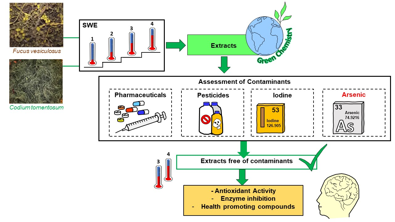
Multi-Step Subcritical Water Extracts of Fucus vesiculosus L. and Codium tomentosum Stackhouse: Composition, Health-Benefits and Safety

1
LAQV, REQUIMTE, Instituto Superior de Engenharia do Instituto Politécnico do Porto, Rua Dr. António Bernardino de Almeida 431, 4249-015 Porto, Portugal
2
LAQV, REQUIMTE, Departamento de Química, Faculdade de Ciências e Tecnologia, Universidade NOVA de Lisboa, Quinta da Torre, 2829-516 Caparica, Portugal
3
CBQF—Centro de Biotecnologia e Química Fina—Laboratório Associado, Escola Superior de Biotecnologia, Universidade Católica Portuguesa, Rua Diogo Botelho 1327, 4169-005 Porto, Portugal
4
iBET, Instituto de Biologia Experimental e Tecnológica, Apartado 12, 2781-901 Oeiras, Portugal
5
LAQV, REQUIMTE, Departamento de Ciências Químicas, Laboratório de Química Aplicada, Faculdade de Farmácia, Universidade do Porto, Rua de Jorge Viterbo Ferreira 228, 4050-313 Porto, Portugal
6
Departamento de Saúde Ambiental, Escola Superior de Saúde, Instituto Politécnico do Porto, R. Dr. António Bernardino de Almeida 400, 4200-072 Porto, Portugal
7
Instituto de Tecnologia Química e Biológica António Xavier, Universidade Nova de Lisboa, Av. da República, 2780-157 Oeiras, Portugal
8
FFULisboa, Faculty of Pharmacy, University of Lisbon, Avenida Professor Gama Pinto, 1649-003 Lisboa, Portugal
*
Author to whom correspondence should be addressed.
Submission received: 4 May 2021 / Revised: 14 May 2021 / Accepted: 15 May 2021 / Published: 19 May 2021
(This article belongs to the Section Biological Processes and Systems)

Abstract

:
Mental health and active aging are two of the main concerns in the 21st century. To search for new neuroprotective compounds, extracts of Codium tomentosum Stackhouse and Fucus vesiculosus L. were obtained through multi-step (four step) subcritical water extraction using a temperature gradient. The safety assessment of the extracts was performed by screening pharmaceutical compounds and pesticides by UHPLC-MS/MS, and iodine and arsenic levels by ICP-MS. Although the extracts were free of pharmaceutical compounds and pesticides, the presence of arsenic and high iodine contents were found in the first two extraction steps. Thus, the health-benefits were only evaluated for the fractions obtained in steps 3 and 4 from the extraction process. These fractions were tested against five brain enzymes implicated in Alzheimer’s, Parkinson’s, and major depression etiology as well as against reactive oxygen and nitrogen species, having been observed a strong enzyme inhibition and radical scavenging activities for the step 4 fractions from both seaweed species. Regarding the variation of the chemical composition during the extraction, step 1 fractions were the richest in phenolic compounds. With the increase in temperature, Maillard reaction, caramelization and thermo-oxidation occurred, and the resulting products positively affected the antioxidant capacity and the neuroprotective effects.

Graphical Abstract

1. Introduction

Seaweeds are a heterogeneous group of aquatic photosynthetic organisms taxonomically classified as green (Chlorophyta), red (Rhodophyta), and brown (Ochrophyta) algae, depending on the nature of their pigments. They are a rich source of several essential nutrients, such as dietary fiber, protein, vitamins (A, B1, B2, B3, B5, B9, B12, C, D, and E), and minerals (e.g., Na, Ca, Mg, P, Cl, Mn, K, I, Fe, Zn, Cu, Se, Mb, B, Ni, Co), and they are also a good source of omega-3 fatty acids, such as eicosapentaenoic acid (EPA) and docosahexaenoic acid (DHA) [1,2,3]. In addition to their nutritional value, seaweeds also contain a myriad of bioactive compounds, namely, phenolic compounds, terpenoids, meroterpenoids, phytosterols, glycolipids, polysaccharides, and pigments (carotenoids and phycobilins), which are responsible for health-promoting effects, such as anti-diabetic, anti-hypertensive, anti-inflammatory, antimicrobial, anti-tumor, antiviral, fat-reducing and neuroprotective effects, among others [1,2].
Being mentally healthy and active during aging is one of the biggest concerns in the 21st century, which has led to the urgent search for new neuroprotective compounds. Various seaweed crude extracts and compounds (such as phlorotannins, phytosterols, carotenoid, sulfated polysaccharides, triterpenoid-saponins) have shown potent cholinesterase, β-secretase, and tyrosinase inhibitory activity and a protective effect against Aβ-induced neurotoxicity, highlighting their usefulness in combating neurodegenerative diseases, such as Alzheimer’s and Parkinson’s disease [4,5]. In addition, although cognitive impairments are the most notable features of Alzheimer’s disease, neuropsychiatric disorders, such as depression and anxiety, profoundly impact the quality of life of both patients and their caregivers [6].
Since there is evidence that brown, red, and green seaweeds affect the monoaminergic system, exhibiting anti-depressant-like activities [7,8,9], the study of seaweeds as potential sources of bioactive compounds may pave the way for new strategies to fight those diseases, either through the development of new pharmaceutical products or through the direct use of algae in food.
Although the consumption of seaweed is widespread in the Asian diet and a low incidence of chronic diseases has been associated with it, regular consumption of seaweed has also raised safety concerns due, in particular, to the potential intake of contaminants, including pesticides and pharmaceutical compounds and, above all, the presence of arsenic and excessively high levels of iodine [10,11]. Due to their intensive use, pharmaceuticals and pesticides were detected in the soil of urban, suburban, and agricultural areas, untreated sewages, effluents from wastewater treatment plants (WWTP), landfills and agricultural and urban runoff, in surface waters and riverbed sediments [12,13,14]. However, their detection in streams, stream sediments and groundwater not affected by WWTP discharges but which may be affected by local wastewater disposal systems or other local sources of contamination is also documented [13,15]. There is a growing concern about risks to public health and the environment due to excessive and continuous use of pesticides because they can migrate from treated fields to air, other lands, and water bodies [16]. Pharmaceuticals have emerged as a major group of environmental contaminants in the 1990s [17,18,19,20]. Over the past 30 years, increasing attention has been paid to understanding the presence and impacts of pharmaceutical compounds entering and detected in freshwater ecosystems [21]. In contrast, much less attention has been paid to understanding the release of pharmaceutical compounds into coastal environments and their potential marine impacts [22,23]. Some seaweeds exhibit a high affinity for heavy metals and metalloids, whose levels in algae are strongly dependent on environmental parameters and the stage of the specimen’s life cycle. Seaweed surfaces can absorb a variety of toxic elements through electrostatic attraction to negatives sites or through slower active absorption, in which metal ions are transported across the cell membrane to the cytoplasm [24]. Arsenic (As) can be incorporated as arsenate in seaweeds due to its similarity to their main nutrient, phosphate from seawater [25]. The total content of As in species of marine macroalgae has been reported to range from 0.1 to 250 mg/kg dry weight (dw) [26]. Chronic exposure to As, especially to inorganic forms, is associated with the occurrence of several types of cancers. However, there is also evidence of other health problems, such as diabetes mellitus, cardiovascular disorders, skin lesions, reproductive and developmental toxicity, nervous system dysfunction, respiratory system and gastrointestinal tract irritation, and anemia [25]. On the other hand, care should also be taken with excessive iodine intake. Iodine is an essential micronutrient necessary for the synthesis of thyroid hormones. However, both deficiency and excess iodine can increase the risk of developing thyroid disorders. It is known that excessive iodine intake can result in both hypothyroidism and hyperthyroidism, with or without a goiter [27]. Therefore, it is important to monitor these different types of contaminants in seaweeds and seaweed extracts before consumption or use as ingredients in food, health, and cosmetic products.
In addition to the contaminants naturally present in seaweed samples, the extraction process can also leave solvent residues. Therefore, efficient and solvent-free methods, using mild processing conditions to avoid thermal degradation of bioactive compounds, are welcome. Among these green extraction methods, subcritical water extraction (SWE) offers the unique advantage of using tunable solvent properties of water to maximize extraction efficiency without the use of hazardous organic solvents. SWE is faster than conventional methods, and high extraction yields are achieved by optimizing the combination of the physico-chemical properties of water as a function of both temperature and pressure [28,29]. Moreover, by increasing the temperature up to 250 °C, the ionic product of water increases by three orders of magnitude compared to its value at room temperature. This makes water reactive and capable of promoting the generation of new bioactive compounds through Maillard reactions, caramelization, and thermoxidation [30].
Taking into account all the issues mentioned, the objectives of this study were to evaluate the neuroprotective effect of the extracts obtained by SWE from two seaweeds widely present on the Portuguese coast and on the Portuguese market: the green Codium tomentosum Stackhouse and the brown Fucus vesiculosus L. In our first approach, 82 pharmaceutical compounds and 33 pesticides were screened in all fractions, as well as their levels of arsenic and iodine determined; the contaminant-free fractions were subsequently evaluated from the biological point of view against five brain enzymes implicated in Alzheimer’s, Parkinson’s, and major depression etiology (acetylcholinesterase, butyrylcholinesterase, monoamine oxidase A, monoamine oxidase B and tyrosinase) as well as against reactive oxygen and nitrogen species (superoxide radical anion and nitric oxide), and some classes of bioactive compounds were quantified by spectrophotometric methods. Since the SWE was carried out with temperatures ranging from room temperature to 250 °C, several parameters were monitored, including pH and the content of reducing sugars, free amino groups, and intermediate and final Maillard reaction products.

2. Materials and Methods

2.1. Samples

F. vesiculosus and C. tomentosum were produced in an Integrated multi-trophic aquaculture system and supplied in dried form by ALGAplus (Ílhavo, Portugal). The samples were hydrated for 5 min in salted water (35 g NaCl/L) and then washed in ultrapure water to eliminate NaCl. Then, they were dehydrated at 41 °C (Excalibur, model 4926T, Dublin, Ireland) for 18 h and ground to obtain particles in the 1–2 µm range.

2.2. Reagents, Solvents, and Materials

Monoamine oxidase A (MAO-A, human recombinant), monoamine oxidase B (MAO-B, human recombinant), kynuramine, clorgyline, tris(hydroxymethyl)aminemethane (Tris), 5,5′-dithiobis(2-nitrobenzoic acid) (DTNB), acetylthiocholine iodide (ATCI), S-butyrylthiocholine iodide (BTCI), galantamine, acetylcholinesterase (AChE) from Electrophorus electricus, butyrylcholinesterase (BuChE) from equine serum, bovine serum albumin (BSA), potassium phosphate monobasic (KH2PO4), potassium phosphate dibasic trihydrate (K2HPO4.3H2O), 2′,2′-azobis (2-amidinopropane) dihydrochloride (AAPH), β-nicotinamide adenine dinucleotide (NADH) disodium salt hydrate, phenazine methosulphate (PMS), nitrotetrazolium blue chloride (NBT), sodium nitroprusside dihydrate (SNP), sulphanilamide, naphthylethylenediamine dihydrochloride, ortho-phosphoric acid 85%, sodium carbonate, Folin–Ciocalteau reagent, gallic acid, sodium nitrite, 2,4-dimethoxybenzaldehyde, phloroglucinol, 2-mercaptoethanol, L-leucine, o-phthaldialdehyde (OPA), neocuproine, perchloric acid 70%, propanol LC-MS and sodium hydroxide were purchased from Sigma-Aldrich (St. Louis, MO, USA, and Steinheim, Germany). Magnesium chloride hexahydrate and o-boric acid were obtained from VWR (Leuven, Belgium), and sodium chloride from Fisher Scientific (Fair Lawn, NJ, USA). Aluminum chloride was purchased from Honeywell Fluka (Seelze, Germany), and catechin from Extrasynthèse (Genay, France). D(+)-glucose anhydrous, acetonitrile and methanol LC-MS grade were obtained from Scharlau (Barcelona, Spain), ethanol absolute and sodium carbonate anhydrous from Panreac (Barcelona, Spain) and formic acid (PA-ACS), hydrochloric acid, acetic acid glacial and copper (II) sulfate pentahydrate from Carlo Erba (Rodano, Italy). Glycine was obtained from Merck (Darmstad, Germany). Ultrapure water (resistivity of 18.2 MΩ.cm at 25 °C) was produced using a Simplicity 185 system (Millipore, Molsheim, France). The eluents used in UHPLC-MS/MS analysis were filtered through a 0.22 µm nylon membrane filter (Fioroni Filters, Ingré, France) using a vacuum pump (Dinko D-95, Barcelona, Spain) and degassed for 15 min in an ultrasonic bath (Sonorex Digital 10P, Bandelin DK 255P, Germany). Iodide (TraceCERT®, 1000 mg/L iodide in water) was obtained from Supelco (Bellefonte, PA). Calibration standards for ICP-MS analysis were prepared from a 10 mg/L multi-element standard solution (Periodic table mix 1, Sigma-Aldrich, Buchs, Switzerland). A total of 115 compounds embracing 82 pharmaceuticals, metabolites, and degradation product and 33 pesticides were tested. CAS number, molecular weight and formula, the solvent used for the preparation of the stock solution and supplier company are presented in Table S1 for pharmaceutical and Table S2 for pesticides of the Supplementary Material.

2.3. Seaweed Proximate Composition

The water content of seaweeds was determined using a thermogravimetric moisture analyzer (Kern DAB 100-3, Balingen, Germany) at 105 °C. The analysis was performed in triplicate. The nitrogen content was determined by elementary analysis and the protein content was calculated from those values using a nitrogen-to-protein conversion factor of 6.25 [31]. The analysis was performed in triplicate. For the determination of ash content, ca. 0.8 g of seaweed was weighed in a porcelain crucible and placed in a muffle at 550 °C for 6 h. The crucible was then removed and placed in a desiccator to cool down, before weighing. The ash content was determined by the weight difference [32]. The analysis was performed in duplicate. Total lipid content was measured by performing a soxhlet extraction with n-hexane. Ca. 2 g of each seaweed was extracted with 70 mL of n-hexane for 3 h. At the end of the extraction, the solvent was evaporated. The residue was dried overnight at 40 °C, to remove traces of solvent, and weighed. The analysis was performed in triplicate.
To quantify soluble carbohydrates, 0.8 g of the defatted seaweed fraction was extracted with 40 mL of ethanol:water solution (80:20, v/v) in an ultrasonic bath for 15 min at room temperature. After this extraction, the mixture was centrifuged at 10,000 rpm, for 10 min at 4 °C. This process was repeated three times, then the supernatants were combined, and the ethanol evaporated at 50 °C, under vacuum, in a rotary evaporator. The remaining solution was diluted with 80 mL of water and used for carbohydrate analysis. The residue was left to dry overnight at 40 °C.
To hydrolyze the insoluble structural carbohydrates, 3 mL of 72% (w/w) H2SO4 were added to 0.3 g of the dry residue (above). This mixture was incubated in a water bath at 30 °C, under stirring, for 1 h, and then diluted to 4% H2SO4 (w/w) by adding 84 mL of water and incubated at 121 °C in a silicone bath, under stirring, for another 1 h. The mixture was then filtered with a silica filter, under vacuum, and the supernatant was used for the quantification of carbohydrates [32]. The solid remaining after acid hydrolysis was washed with water, dried at 105 °C overnight and weighed. Its ash and nitrogen content was determined as mentioned above and subtracted from the amount of dry solid to obtain the amount of Klason lignin [32].
The total amount of carbohydrates in the solutions obtained during biomass characterization was determined using the phenol-sulphuric acid colorimetric method [33]. A calibration curve was obtained with solutions of D(+)-glucose monohydrate. To 500 µL of a standard solution or carbohydrate-rich liquor, 1.5 mL of H2SO4 (96%) and 300 µL of a 5% (w/v) aqueous solution of phenol were added. The resulting mixtures were well stirred. After incubation for 5 min at 90 °C in a dry block bath, the mixtures were well stirred and cooled to room temperature by immersion in a water bath. The absorbance was measured at 490 nm and the results obtained were expressed in g/L glucose equivalent. All procedures were performed in triplicate.

2.4. Subcritical Water Extraction (SWE) of Seaweeds

The SWE of seaweeds was carried out in an apparatus described elsewhere [34]. In brief, a high-pressure liquid pump (Knauer 40 Preparative pump 1800, Berlin, Germany) injects distilled water into a stainless-steel packed bed reactor (51 cm long, 2.6 cm i.d.) that is placed inside an electric oven with temperature control (Nabertherm model 30–3000 C N641, Lilienthal, Germany). The water flow is heated before entering the reactor by a heating wire connected to a temperature controller. The water temperature in the reactor is continuously monitored by a thermocouple, and the system pressure is controlled by a back-pressure regulator, BPR (Tescom, model 26–1000, Selmsdorf, Germany). The aqueous liquor that flows from the reactor passes through a 15 µm filter and is continuously collected after the BPR valve.
In each experiment, the reactor was filled with ca. 20 g of seaweed, placed between porous discs. The pressure was kept constant at 100 bar during the experiment. A constant water flow of 10 mL/min was used for the tests carried out in this work. Once it took a certain time to reach the desired temperature in the reactor, the liquor leaving the reactor was collected in a series of sampling tubes, each corresponding to a temperature interval, as follows: E1 fraction—the liquor flowing from the reactor when the temperature increased from room temperature to 90 °C (corresponding to a time period of ca. 100 min); E2 fraction—increase in temperature from 90 to 140 °C (ca. 90 min); E3 fraction—increase in temperature from 140 to 190 °C (ca. 90 min); and E4 fraction—increase in temperature from 190 to 250 °C (ca. 100 min).
All the liquors collected were lyophilized, and the amount of dried fractions obtained was used to calculate the extraction yield of the assay. The dried extracts were stored at 4 °C until further analysis. All experiments were replicated.

2.5. Chemical Characterization of SWE Extracts

The chemical composition of E1–E4 fractions was evaluated considering the presence of potentially hazardous compounds and bioactive substances, as described in the following sections.

2.5.1. UHPLC-MS/MS Analysis of Drugs and Pesticides

A Shimadzu Nexera UHPLC-MS/MS-ESI (LCMS-8030) was used for chromatographic analysis, and was equipped with a CBM-20A controller, a LC-30 AD two solvent delivery pumps, a CTO-20 AC column oven, a SIL-30 AC auto-sampler, a DGU-20A 5R degasser, and a CBM-20A system controller (Kyoto, Japan). The equipment was coupled to a triple-quadrupole mass spectrometer and operated in electrospray ionization (ESI) mode. Lab Solutions software (Shimadzu Corporation, Kyoto, Japan) was used for system control and data processing. Argon at a pressure of 230 kPa was used as collision-induced dissociation gas (CID), and nitrogen was used as drying and nebulizing gas.
Individual stock standard solutions were prepared at a concentration of 1 g/L on a weight basis and stored at −20 °C. Mixed working standard solutions containing all compounds were prepared in acetonitrile:ultrapure water (30:70, v/v). A mixture of isotopically labeled internal standards (ILIS) was prepared to be used for internal standard. The concentration of each ILIS is shown in Table S3 (Supplementary Material). The sample extracts were filtered through 0.22 µm PTFE syringe filters (Sicherheitssiegel, China) before chromatographic analysis.
Based on previous work by the authors [35,36,37], chromatographic conditions (columns, mobile phases, isocratic or gradient elution, flow rate, oven temperature) were tested and optimized for the set of pharmaceutical compounds studied. The MS conditions are summarized in Table S4 (Supplementary Material).
For the analysis of pesticides, the precursor ion and the ionization mode for each analyte were selected by obtaining individual standard chromatograms (100 mg/L) in full scan mode in the Q1 and Q3 quadrupoles. Then, direct injection of individual standard solutions (10 mg/L) was performed and the mass spectrometer operated in the multiple reaction monitoring (MRM) mode. The optimization of the MRM was conducted to determine the product ions (quantification and qualifier ions) and collision energies for each pesticide. The two most intense MRM transitions were monitored for each analyte, the first for quantification and the second as a qualifier. The individual MS/MS parameters are shown in Table S5 (Supplementary Material).
Source-dependent parameters were also optimized by direct injection, using a mixed standard solution of pesticides (10 mg/L of each compound). The optimized parameters were: 3.0 L/min for nebulizing gas (nitrogen), 16 L/min for drying gas (nitrogen), 5.0 kV for interface voltage, 200 °C for desolvation temperature, and 250 °C for heat block temperature.
Chromatographic conditions were optimized to obtain good resolution, peak shape and reproducibility. Different mobile phases comprising different combinations of 0.1% formic acid in ultrapure water (A) and acetonitrile (B) were tested using isocratic or gradient elution. Two chromatographic columns were tested: Luna Omega 1.6 µm, 150 × 2.1 mm (Phenomenex, CA, USA) and Cortecs™ UPLC® C18+ 1.6 µm, 100 × 2.1 mm (Waters, Milford, MA, USA). The best results were found using the Luna Omega column in gradient elution mode. The optimized program starts with 5% (B), increasing for 3 min to 100% (B), holding for 2 min and returning to the initial conditions in 1 min. The total time was set at 10 min. A dwell time of 2.5 ms was used for all compounds. A summary of the chromatographic programs used for the analysis of pharmaceutical compounds and pesticides is presented in Table S6 (Supplementary material).

2.5.2. Iodine and Arsenic Determination

The determination of iodine was performed as previously described by Costa Leite et al. [38] and the determination of arsenic was performed according to Cabrita et al. [39], using an iCAP™ Q ICP-MS (Thermo Fisher Scientific, Germany) instrument. Analyses were performed in triplicate.

2.5.3. Total Phenolic, Total Flavonoid and Total Phlorotannin Content

E1–E4 fractions of each seaweed were dissolved in water at concentrations ranging from 2.0 mg/mL to 0.4 mg/mL, depending on the extracts’ solubility, and diluted, when necessary, before analysis. Seaweed raw material (c.a. 1 g) was extracted with 10 mL of water at 70 °C for 30 min. After a 5 min centrifugation at 4500 rpm, the supernatant was separated. The solid residue was extracted again with an additional 10 mL of water, repeating the previous step. Supernatants were mixed and diluted as needed before further analysis.
The total phenolic content of seaweeds and fractions was quantified through a colorimetric assay using the Folin–Ciocalteau reagent [40], using gallic acid (GA) as the standard. Measurements were made using 96-well plates in a Synergy HT W/TRF multimode microplate reader (BioTek Instruments, Winooski, VT, USA) using Gen5 2.0 software (BioTek Instruments). Three assays were performed, each one in quadruplicate and the results were expressed as mg of gallic acid equivalents (GAE) per gram extract (dry weight, dw).
The total flavonoid content (TFC) of the fractions was determined as described before [41]. Briefly, 1 mL of extract solution (2 mg/mL), 4 mL of d-H2O and 0.3 mL of 5% NaNO2 were added to a 10 mL volumetric flask. After 5 min, 0.3 mL of 10% AlCl3 solution was added to the mixture and after 6 min, 2 mL of 1 M NaOH was added. Immediately, the total volume of the flask was completed with d-H2O and the content well homogenized. The absorbance of the colored mixture was measured at 510 nm using an Ultrospec 3000, UV/Visible spectrophotometer (Pharmacia Biotech, Uppsala, Sweden) against a blank solution prepared with d-H2O. Six catechin solutions of different concentrations (20–100 mg/L) were used to obtain a calibration curve. The results were expressed as mg of catechin equivalents (CE) per g of extract (dw).
The quantification of total phlorotannins (1,3,5-substituted phenols) was based on the reaction with 2,4-dimethoxybenzaldehyde (DMBA) to form a colored product, as previously described [42]. Measurements were made using 96-well plates in a Synergy HT (BioTek Instruments) microplate reader, as described by Lopes et al. [43], using phloroglucinol (Phl) as the standard. Three assays were performed, each one in quadruplicate and the results were expressed as mg of Phl equivalents per g of extract (dw).

2.5.4. Determination of pH, Available Free Amino Acids and Reducing Sugars

The pH (at 20 °C) of the aqueous solutions of fractions and seaweeds was determined with a Crison (Barcelona, Spain) combined glass electrode, and the measurements were performed using a Crison micro pH 2002 at 20 °C.
Available free amino groups were determined in seaweeds and fractions by the OPA assay as described by Plaza et al. [30]. To simultaneously extract molecules with free amino groups, 1 g of seaweed or 10 mg of E1-E4 fractions were mixed with 5 or 0.5 mL of a 0.2 M perchloric acid solution. The mixture was well homogenized in a vortex for 2 min and kept in an ultrasonic bath at 30 °C for 30 min, followed by centrifugation for 20 min at 4000 rpm [44]. Prior to their analysis, the samples were appropriately diluted with borate buffer. Fluorescence was measured at λexc = 340 nm and λem = 455 nm using a Synergy HT (BioTek Instruments) microplate reader. Sample blanks at the same concentration as in the assay were also analyzed to correct for possible interferences due to the sample’s natural fluorescence. Leucine (Leu) was used as the standard, and the data were expressed as mg of Leu equivalents per g of sample (dw).
Reducing sugars were quantified using the neocuproine method [45] with minor modifications. Briefly, to extract the reducing sugars, 1 g of seaweeds (or 100 mg of fractions) were mixed with 10 mL of 80% ethanol solution (or 5 mL for the fraction solution). After vortexing for 1 min, the solutions were heated in a water bath at 80 °C for 15 min with constant stirring. After centrifugation at 9000 rpm for 20 min, 1 mL of the supernatant was mixed with 0.2 mL of a 5% HCl and heated to 90 °C for 30 min to hydrolyze the polysaccharides. After cooling to room temperature, the solution was neutralized with a few drops of 25% NaOH solution. Then, 250 µL of sample solution were mixed with 750 µL of a Cu(II) solution (4.0 g anhydrous sodium carbonate, 1.6 g glycine and 0.045 g copper sulfate solution in 100 mL water) and 750 µL of neocuproine solution (0.120 g of neocuproine in 100 mL of a 1:4 ethanol:water solution). After vortexing, the final solution was heated in a boiling water bath for 15 min. An orange color was produced, and the samples were diluted if necessary, before analysis. D(+)-glucose was used to prepare standard solutions (range 20 to 100 mg/L), and the 80% ethanol solution was used as the blank. 250 µL of samples, standards and blanks were dispensed in a 96-well plate, and the orange color was measured at 455 nm in a Synergy HT (BioTek Instruments) microplate reader. All procedures were performed in triplicate.

2.5.5. Maillard Reaction Products and Browning Index

Melanoidins were estimated through the browning intensity of the solutions of fractions and seaweeds. The browning intensity was measured at 294, 360, and 420 nm, using a Synergy HT (BioTek Instruments) microplate reader. When necessary, the samples were diluted to obtain an absorbance value below 1.5 [30,46]. The data were expressed as absorbance units (AU). The analysis was performed in quadruplicate.
The formation of fluorescent advanced glycation end-products (AGEs) was estimated by measuring the fluorescence at an excitation wavelength of 360 ± 40 nm and an emission wavelength of 460 ± 40 nm in a Synergy™ HT microplate reader, after appropriate dilution of samples [30]. The data were expressed as fluorescence units (FU). The analysis was performed in quadruplicate.
The color parameters of yellow to yellow-brown were evaluated as reported by Matiacevich and Buera [47]. These parameters were used to obtain the tristimulus values X, Y and Z according to the following equations:
X = T625 × 0.42 + T550 × 0.35 + T445 × 0.21
Y = T625 × 0.2 + T550 × 0.63 + T495 × 0.17
Z = T495 × 0.24 + T445 × 0.94
where T625, T495, T445, and T550 are the transmittance values at 625, 495, 445 and 550 nm, respectively.
The transmittance values at 445, 495, 550 and 625 nm were measured on a Synergy™ HT microplate reader. The browning index (Br), proposed as an adequate measure of visual browning, was calculated according to Equation (4).
Br = 100 × (x − 0.31)/0.172
where
x = X/(X + Y + Z)
The analysis was performed in quadruplicate.

2.6. Bioactivities

2.6.1. ORAC

The oxygen radical absorbance capacity (ORAC) test was based on the method developed by Huang et al. [48] which assesses the antioxidant capacity of the tested samples toward the peroxyl radicals (ROO) generated during the thermal decomposition of the 2′,2′-azobis (2-amidinopropane) dihydrochloride (AAPH). Briefly, disodium fluorescein (FL) was added to sample dilutions and the resulting mixture was equilibrated at 37 °C. Then, the reaction was initiated by the addition of AAPH, and the fluorescence was recorded for 40 min with a FLX800 microplate fluorescence reader (BioTek Instruments), under the control of Gen5 software. The final concentrations in the reaction mixture were 3 × 10⁻⁴ mM for FL and 0.0414 g/mL for AAPH, and all solutions were prepared in 75 mM phosphate-buffered saline (PBS), pH 7.4.
Five different concentrations of 6-hydroxy-2,5,7,8-tetramethylchroman-2-carboxylic acid (Trolox) solutions (5–40 µM/mL) were used to obtain a calibration curve. Three assays were performed, and the results were expressed as micromoles of Trolox equivalents (TE) per milligram of extract (dw).

2.6.2. Antiradical Activity

Superoxide anion radical (O2) and nitric oxide (NO) scavenging activities were evaluated according to Oliveira et al. [49]. The fractions were dissolved in 19 mM KH2PO4/K2HPO4 buffer (pH 7.4) for O2 scavenging activity or 0.1 M KH2PO4/K2HPO4 (pH 7.4) for NO scavenging activity. The radical O2 was generated by the NADH/PMS system. Phosphate buffer was used as negative control. The blanks contained all reagents except PMS, and kinetic curves were obtained at 560 nm in a Synergy HT (BioTek Instruments) microplate reader. IC50 values were compared with those of ascorbic acid (positive control). To evaluate the NO scavenging activity, sodium nitroprusside was incubated with the sample for 60 min, at room temperature, under light. After incubation, Griess reagent (1% sulphanilamide and 0.1% naphthylethylenediamine in 2% phosphoric acid) was added to each well. The mixture was incubated at room temperature for 10 min, and the absorbance (due to the colored compound formed by diazotization reaction) was read at 560 nm (Biotek Synergy HT microplate reader). Phosphate buffer was used as negative control and 2% phosphoric acid, instead of the Griess reagent, was added to the blanks.
The results were compared with those of ascorbic acid (positive control, IC50 = 160.04 µg/mL against O2 and 446.43 µg/mL against NO). All assays were done three times, each time in triplicate.

2.6.3. Enzyme Inhibition

The method for evaluating the inhibitory activity of the fractions against monoamine oxidase A (MAO-A) and B (MAO-B) was based on the production of 4-hydroxyquinoline from the deamination of kynuramine [50], adopting a procedure previously described [51,52], with minor modifications. 215 μL of sample dissolved in 0.1 M KH2PO4/K2HPO4 (pH 7.4) were mixed with 10 μL of 3.75 mM of knuramine. The reaction was started by adding 75 µL of 17 U/mL MAO-A or MAO-B solution. Negative controls and blanks were prepared with buffer instead of extracts or enzymes, respectively. The production of 4-hydroxyquinoline was determined by measuring the absorbance at 314 nm with a Synergy HT (Biotek Instruments) microplate reader during incubation at 37 °C for 70 min. Clorgyline was used as a positive control (IC50 = 25.0 ng/mL against MAO-A).
The inhibition of AChE and BuChE activity was measured in a 96-well microplate, according to the Ellman method [53,54], with minor modifications. Briefly, the fractions were dissolved in Tris-HCl buffer (50 mM, pH = 8) and 25 µL were added to the wells, along with 125 µL of DTNB reagent, 50 µL of buffer B (Tris-HCl buffer + 0.1% albumin), 25 µL of ATCI/BTCI and 25 µL of 0.44 U/mL of AChE or 0.40 U/mL of BuChE solutions. Slopes were calculated from the kinetic curve obtained from six absorbance measurements at 405 nm in a total reaction time of 1 min 44 s, after subtracting the blanks (wells containing all reagents except the enzyme). Galantamine was used as a positive control (IC50 = 0.92 µg/mL against AChE and IC50 = 4.92 µg/mL against BuChE) and Tris-HCl buffer as a negative control.
Tyrosinase inhibition was measured according to Masuda et al. [55]. The fractions were dissolved in 1/15 M of Na2HPO4/KH2PO4 (pH = 6.8) and 40 µL of the solution were dispensed into 96-well microplates (40 µL of buffer for a negative control) followed by 80 µL of buffer and 40 µL of 46 U/mL tyrosinase solution. After 10 min of incubation at room temperature, 40 µL of L-DOPA were added. After another 10 min, the absorbance was measured at 475 nm. L-DOPA was replaced by buffer solution in blanks and kojic acid was used as a positive control (IC50 = 1.82 µg/mL). All assays were performed three times, in triplicate each time.

2.7. Statistical Analysis

All data were recorded as mean ± SD or mean ± SEM. The IC50 values were calculated using the Graph Pad Prism Software, version 6. For the comparison of IC50 values and compounds content, the samples were grouped according to one-way analysis of variance (ANOVA), using Tukey as post-test (comparison of more than three samples) or by t-test (comparison of two samples) (Graph Pad Prism Software, version 6). Differences were considered statistically significant for p < 0.05.

3. Results and Discussion

3.1. Chemical Characterization of Seaweeds

The chemical composition of the seaweeds studied is shown in Table 1. The moisture of the seaweeds differed considerably between both species. Rodrigues et al. [56] reported a moisture content of 9.0 ± 0.2 g/100 g for C. tomentosum, and Lorenzo et al. [57] a moisture content of 11.23 ± 0.08 g/100 g for F. vesiculosus.
The lipid and ash content of F. vesiculosus and C. tomentosum agree generally with that described in the literature [56,58,59,60,61]. The lipid content found in this work was 3.4 ± 0.5 and 2.2 ± 0.6 g/100 g (dw) for F. vesiculosus and C. tomentosum, respectively. Lorenzo et al. [57] reported a total lipid content of 3.75 ± 0.20 g/100 g for F. vesiculosus, while for C. tomentosum, Rodrigues et al. [56] reported a total lipid content of 3.6 ± 0.2 g/100 g (dw). The relatively high ash content found is an important feature of seaweeds and is related to a high concentration of minerals [58]. The ash content for F. vesiculosus and C. tomentosum was 17.3 ± 0.1 and 24.0 ± 0.1 g/100 g (dw), respectively. These values are comparable to those reported in the literature, 20.71 ± 0.04 g/100 g for F. vesiculosus [57] and 35.99 ± 0.48 for C. tomentosum [56].
The protein content is known to be higher in green and red seaweeds (10–47% dw) than in brown seaweeds (5–24% dw) [2]. The protein content found for F. vesiculosus is within the range observed by other authors [57,60,61]. On the other hand, C. tomentosum showed a relatively higher protein content than that reported in some studies [62,63], but similar to that obtained by Rodrigues et al. [56]. It is known that the protein content of seaweeds varies throughout the year and depends on the geographic area and environmental conditions [64].
The main components that give Fucus sp. its claimed health effects are non-digestible polysaccharides (dietary fiber) and polyphenols [65]. These polysaccharides and polyphenols are present in a higher proportion than in other edible seaweeds [66,67]. The content of soluble and insoluble polysaccharides found for F. vesiculosus are below those obtained by Rupérez and Saura-Calixto [68], and in both cases the seaweeds showed a higher proportion of insoluble than soluble polysaccharides.
In this study, a total sugar content of 28.5 g/100 g (dw) was obtained for C. tomentosum (Table 1). The total sugar content of C. tomentosum was reported by Rodrigues et al. [56] as 32.8 g/100 g (dw). In other Codium species, the content ranged from 8.8 g/100 to 66.8 g/100 g (dw) [69,70,71,72]. Conversely to their high carbohydrate content, seaweeds are known for their low lignin content, usually less than 10% [73]. Although the content of carbohydrates and lignin was similar for F. vesiculosus, lignin content was 10 times lower than that of carbohydrates for C. tomentosum.

3.2. SWE of Seaweeds

F. vesiculosus and C. tomentosum were extracted by SWE at a fixed pressure of 100 bar and temperatures varying up to 250 °C. The objective was to evaluate the possibility of obtaining fractions with different biological properties and chemical compositions starting from the same matrix.
The overall extraction yield obtained for the two seaweeds as a function of the temperature is shown in Table 2. The increase in the temperature led to an increase in the cumulative yield of extraction for both seaweeds, slightly higher for C. tomentosum. This was expected since the increase in the temperature positively affects the solubility of the compounds and, at the same time, reduces the viscosity of the water, allowing a better penetration of the solvent in the matrix. In addition, the water dielectric constant decreases markedly with increasing temperature, from nearly 80 at 25 °C to ca. 30 at 250 °C, close to the dielectric constant of methanol at room temperature, thus promoting the solubilization of less polar compounds. Additionally, the ionic product of water increases with increasing temperature, making water a more reactive medium for hydrolysis reactions [30,74]. The combination of all these factors contributed to the increase in extraction yield that was observed with the increase of temperature.

3.3. UHPLC-MS/MS Analysis of Drugs and Pesticides in Seaweed Extracts

The linearity of the method was established through linear regression analysis for calibration curves obtained with twelve concentration levels in the range of 0.5 to 1000 μg/L. The limit of detection (LOD) and the limit of quantification (LOQ) were calculated as the analyte concentration corresponding to a signal-to-noise ratio of 3 and 10, respectively. The ion ratio for each compound was calculated as the ratio between the quantifying and the qualifying ions. The results obtained for the SWE extracts are presented in Tables S7 and S8 (Supplementary Material). No pollutants were detected in any of the analyzed fractions.

3.4. Iodine and Arsenic Levels in Seaweed Extracts

The levels of iodine and arsenic are presented in Table 3. F. vesiculosus showed a higher content of iodine in E1 fraction (90 °C), with 14.6 mg/g dw, decreasing throughout the extraction steps and consequently with the increasing of temperature (from 90 in E1 to 250 °C in E4), with the lowest values found in E4 fraction, with 1.25 mg/g at 250 °C. Ferreira et al. [75] also evaluated the effect of different extraction temperatures (25, 50, 75, 100, and 120 °C) and extraction times (5 min, 1 h, 2 h, and 4 h) on the recovery of bioactive compounds, including iodine from F. vesiculosus. These authors reported that for temperatures between 50 and 120 °C, the amount of iodine extracted from F. vesiculosus increased with the time, with the highest value (0.4903 mg/g dw) obtained at 120 °C/4 h. In the present study, the E1 fraction was the one with the highest iodine content.
A similar trend was obtained for C. tomentosum, with the highest iodine content obtained for E1 fraction (42.0 µg/g) and the lowest for E4 fraction (0.511 µg/g). These results suggest that iodine appears to be present mainly as inorganic iodine, readily extractable with water [75], so that the first extraction step at lower temperature (90 °C) is the one with the highest yield. The high values for F. vesiculosus when compared to C. tomentosum are related to the ability of Fucus species to accumulate iodine, with Fucus species showing iodine levels as high as 500 mg/g dw [76] and Codium species values between 75.4 to 475.0 µg/g dw [39], which is in agreement with the values found in this study.
The highest As values were obtained for E1 fraction in both species (144 µg/g for F. vesiculosus and 23.1 µg/g for C. tomentosum). For C. tomentosum, the lowest value was for E4 fraction (0.511 µg/g). A different behavior was found for F. vesiculosus. For this species, the lowest content was obtained in E2 fraction (8.75 µg/g), with an increase in E3, followed by E4 (32.3 and 11.9 µg/g, respectively). These results are probably related to the fact that arsenic species are closely bound to metallothioneins (small cysteine-rich proteins) in the seaweed F. vesiculosus [77]. During SWE, protein cleavage occurs at temperatures above 160 °C [78]. The temperatures for E3 and E4 fractions were 140–190 and 190–250 °C, respectively, suggesting that protein-bound As is released into the aqueous phase at these temperatures while at lower temperatures were extracted soluble forms of As. A total As content of 327 µg/g in F. vesiculosus has been reported in the literature [79]. No reports were found on the As content in C. tomentosum, but for Codium lucasii Setchell a total As content of 16.6 µg/g and 13.7 µg/g after microwave-assisted methanol/water (78% v/v) extraction was reported [80].

3.5. Chemical Composition of Seaweed Extracts

The phenolic and phlorotannin contents of the F. vesiculosus and C. tomentosum SWE fractions are shown in Table 4.
F. vesiculosus fractions had a relatively higher total phenolic content (TPC) than C. tomentosum ones, as expected since brown seaweed species have higher levels of phenols compared to green seaweeds [59,65,66,67]. For F. vesiculosus, the fractions richest in phenolics were obtained at the lowest temperatures, while for C. tomentosum the opposite was found. For both seaweeds, and considering the mass of the fractions collected at each operating temperature as well as respective TPC, the total mass of phenolics extracted by subcritical water was significant and higher than the initial mass of phenolics in the original seaweed, as determined by conventional extraction with water at 70 °C (see Table 4). This is even more evident for C. tomentosum, where the respective SWE extracts showed measurable TPC values, although the aqueous extract obtained at 70 °C has shown a residual value. This behavior has been observed before [30,34] due to the removal of phenolics trapped in the seaweed structure with the SWE treatment. On the one hand, the effect of temperature on the water dielectric constant will cause a higher solubility of less polar polyphenols. On the other hand, the increase in the ionic product of water with temperature up to 250 °C makes the water reactive. This can promote the formation of other compounds from Maillard and caramelization reactions, such as reducing sugars, amino acids and melanoidins, which are known to reduce the Folin–Ciocalteu reagent and thus contribute to the overestimation of the determined phenolics content [30,81,82]. Plaza et al. [30] reported that when using SWE at 100 and 200 °C, the increase in the extraction temperature produced an enhancement of the total phenols for the brown seaweed Undaria pinnatifida of more than a ten-fold increase. In addition, the total flavonoid content was consistently higher for E4 fraction (2.0 ± 0.1 g CE/100 g dw for F. vesiculosus and 0.7 ± 0.1 g CE/100 g dw for C. tomentosum) than for E3 (1.5 ± 0.1 g CE/100 g dw for F. vesiculosus and 0.2 ± 0.1 g CE/100 g dw for C. tomentosum). Again, these results may be an overestimate, since other non-flavonoid compounds with catechol groups can also absorb at 510 nm [82].
Phlorotannins were also totally extracted from F. vesiculosus by subcritical water in the first two fractions (E1 and E2), collected at temperatures up to 140 ºC, similar to what was observed by Ferreira et al. [75].
The total protein content of the SWE extracts is shown in Table 5. Subcritical water extracted and hydrolyzed the protein fraction of the seaweeds, and the temperature was beneficial to obtain extracts richer in protein, confirming the behavior reported by other authors [30,83]. C. tomentosum extracts had higher protein contents than F. vesiculosus extracts, which was expected, given the much higher initial protein content of C. tomentosum (see Table 1). Considering the mass of the fractions collected at each operating temperature and the respective protein content, subcritical water extracted ca. 85% of the original protein content of F. vesiculosus and ca. 50% of C. tomentosum. Two consecutive processes occur during SWE extraction/hydrolysis of seaweeds: the direct extraction of the most soluble proteins, especially at lower temperatures, and the hydrolysis of the proteins into small peptide units and/or free amino acids at the higher operating temperatures [83].
Amino acids and reducing sugars can react at high temperatures producing non-enzymatic browning reactions or Maillard reactions and generating different flavors and brown color [84,85]. Since the SWE was carried out at temperatures ranging from room temperature and 250 °C, several chemical parameters were measured, including reducing sugars, free amino groups, intermediate and final Maillard reaction products and pH (Table 5). In general, the free amino groups content increased from fraction E1 to fraction E4 in both seaweeds due to the hydrolysis of protein and peptide with increasing temperature [30,86].
Reducing sugars did not show a marked increase with temperature, possibly indicating that subcritical water did not significantly hydrolyze the seaweed carbohydrate fraction to monosaccharides or small oligosaccharides [30].
Maillard reaction influences the pH of the systems more than the thermal degradation. Lan et al. [86] observed a decrease in pH from 6.5 to ca. 4.5 in a Maillard reaction of the xylose–soybean peptide system, probably due to the formation of organic acids. These authors [86] also investigated whether the phenomenon occurred during thermal degradation of the soybean peptides and observed that the pH changed only slightly, meaning that the release of amino acids from the peptide degradation had little effect on the pH of the system without sugar. Similarly, Liu et al. [87] reported a trend toward a decrease in pH in galactose/glycine model systems due to the consumption of amino groups and the production of acids.
In this study, a decrease in the pH of the C. tomentosum fractions was clearly observed (Table 5), whereas for F. vesiculosus fractions, only a relatively small change was noticed, indicating a stronger Maillard reaction rate for C. tomentosum than for F. vesiculosus. In fact, and as discussed below, higher production of intermediate and final Maillard reaction products was obtained for fraction E4 of C. tomentosum than for fraction E4 of F. vesiculosus.
UV absorbance at 294 nm is often used to detect the colorless intermediate Maillard reaction products, while the final stage is monitored at 360 and 420 nm. These intermediate compounds could be the precursors of the browning products in the Maillard reaction or caramelization [88]. According to Table 5, the absorbance at 294 nm increased from fraction E1 to fraction E4 for C. tomentosum, while the values varied randomly for F. vesiculosus. The absorbance at 360 and 420 nm is commonly used to monitor the formation of brown products from the Maillard reaction. Browning is also associated with caramelization processes [30,86]. The temperature favored the formation of these products, since an increase in absorbance at 360 and 420 nm was observed for both C. tomentosum and F. vesiculosus. These results are in agreement with the browning index obtained (Table 5).
Another class of Maillard reaction products, the fluorescent advanced glycation end-products (AGEs) or melanoidins, have intense light emission between 400 and 500 nm when excited at a wavelength of 360 or 370 nm [30]. Although some fluctuations were noticed for F. vesiculosus, in general, a pattern of increasing the fluorescent AGEs content with temperature was observed, with the fraction E4 for C. tomentosum showing the highest value of all the extracts collected. This seems to indicate that a higher production of intermediate and final Maillard reaction products was obtained at the highest temperature studied for C. tomentosum seaweed.

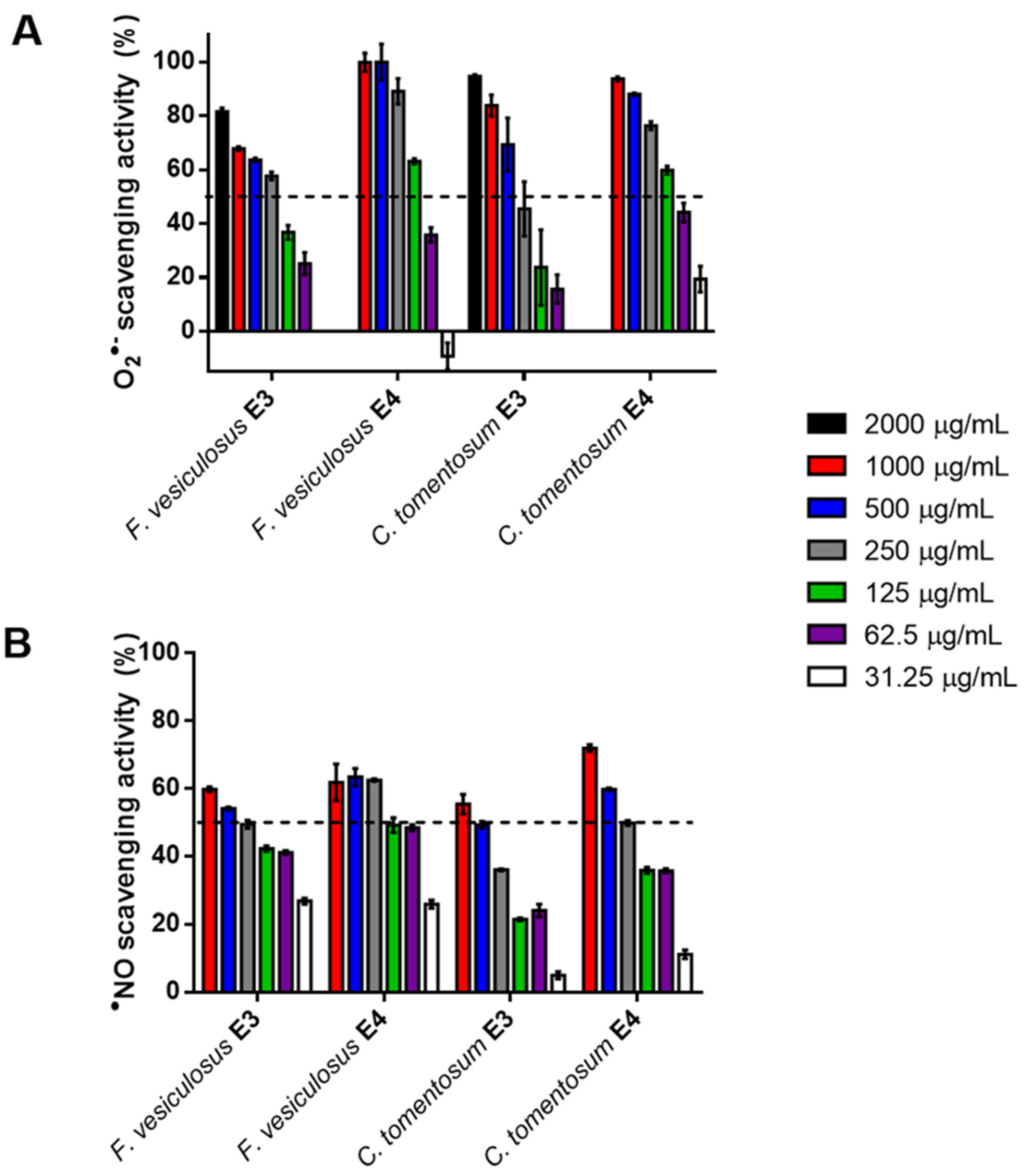
3.6. Antioxidant Activity

Having found high levels of arsenic and iodine in the first two fractions (E1 and E2) for both seaweeds, the antioxidant capacity and antiradical activities were evaluated only for E3 and E4 fractions. The antioxidant capacity measured by the ORAC assay showed that, for both species, E4 was more active than E3, with the order of potencies being as follows: C. tomentosum E4 (2.6 ± 0.5 µM TEAC/mg dw) > F. vesiculosus E4 (1.6 ± 0.2 µM TEAC/mg dw) > F. vesiculosus E3 (1.2 ± 0.1 µM TEAC/mg dw) > C. tomentosum E3 (0.8 ± 0.3 µM TEAC/mg dw). These results are in agreement with those obtained for O2●– scavenging activity (Figure 1A), with C. tomentosum E4 fraction showing the lowest IC50 values (85.7 µg/mL). On the other hand, regarding the NO scavenging activity, F. vesiculosus E4 fraction was more active than C. tomentosum E4 (IC50 = 132.4 µg/mL vs. 254.2 µg/mL) (Figure 1B). A previous study with these seaweeds not subjected to the pre-treatment described in Section 2.1 (washing step with salt water (35 g NaCl/L) followed by ultrapure water) also showed that C. tomentosum E4 fraction was the most antioxidant fraction with 104.8 mg AAE/g dw for ABTS●+ assay and 687.2 µg TE/g dw for ORAC assay [89].
Literature data point out the greater antioxidant potential of brown seaweeds over red and green seaweeds due to the higher amounts of phenolic compounds and phlorotannins [90]. In fact, Corsetto et al. [91] not only demonstrated the antioxidant capacity of F. vesiculosus water extracts through ORAC, DPPH radical scavenging and ferrous ion-chelating ability assays, but also reported that the extracts were able to prevent the production of radicals in HepG2 cells exposed to oxidative stress. Conversely, for C. tomentosum, Silva et al. [92] used three complementary methods (DPPH, FRAP, ORAC) to test the antioxidant activity of MEOH and dichloromethane crude extracts of this species. Crude extracts were also fractionated, and the corresponding fractions were tested. None of the tested fractions showed significant antioxidant potential, which was correlated with their low levels of TPC. In addition, an aqueous extract of C. tomentosum has already been tested for O2●– and NO scavenging activity, showing less activity than fraction E4 in the present study [93,94]. Therefore, considering the low TPC values observed for the E3 and E4 fractions of C. tomentosum, other compounds may be influencing the overall antioxidant activity.
In fact, Plaza et al. [30] reported the formation of neoantioxidants in natural complex matrices during SWE, which correspond to Maillard reaction products. SWE extracts from plants, macroalgae and microalgae were able to scavenge ABTS●+, ROO and O2●– radicals. In all cases, samples obtained at 200 °C showed higher antioxidant capacity than the corresponding ones extracted at 100 °C.
Therefore, the overall antioxidant activity of F. vesiculosus and C. tomentosum E3 and E4 fractions is a combination of phenolic compounds and Maillard reaction products.

3.7. CNS Enzymes Inhibition

The inhibitory potential of the SWE E3 and E4 fractions of F. vesiculosus and C. tomentosum was tested against AChE, BuChE, MAO-A, MAO-B and tyrosinase (Figure 2).
AChE and BuChE are enzymes related to cognitive function, since they regulate the neurotransmitter acetylcholine in the synaptic cleft of neurons. The rapid breakdown of this neurotransmitter leads to cholinergic dysfunction and, ultimately, memory impairment. Thus, cholinesterase inhibitors have been developed to alleviate the cholinergic deficit in patients with Alzheimer’s disease [5]. Regarding cholinesterase inhibition, only C. tomentosum E4 fraction was able to suppress enzymes activity over 50%, with IC50 [AChE] = 239.8 µg/mL and IC50 [BuChE] = 430.1 µg/mL (Figure 2A,B). These results are in agreement with our previous results with these samples not submitted to the pre-treatment described in Section 2.1, showing the higher inhibitory potential of C. tomentosum compared to F. vesiculosus for AChE [89]. Andrade et al. [94] reported the absence of cholinesterase inhibition for the aqueous extract of C. tomentosum. However, aqueous extracts from three different samples of F. vesiculous exhibited anti-AChE activity with IC50 values in the range of 15 to 841 µg/mL [95]. MAO-A and MAO-B inhibitors are in clinical use to treat psychiatric disorders, such as depression, and neurological disorders, such as Alzheimer’s and Parkinson’s diseases [96]. Both E4 fractions were active against MAO isoforms A and B, with C. tomentosum and F. vesiculosus presenting IC50 = 181.2 (MAO-A)/422.5 (MAO-B) µg/mL and IC50 = 102.2 (MAO-A)/481.5 (MAO-B) µg/mL, respectively (Figure 2C,D). These results are better than those found in the literature. An ethanolic extract of F. vesiculosus exhibited moderate MAO-B inhibitory activity of IC50 > 4000 < 7000 µg/mL [97], while a phlorotannin-rich extract of F. vesiculosus exhibited low activity against AChE, BuChE, MAO-A, MAO-B and tyrosinase [98].
The neuromelanin production mechanism includes a rate-limiting step of oxidation of L-DOPA to the melanin precursor DOPAquinone, catalyzed by tyrosinase. Neuromelanin accumulates with age and, in Parkinson’s disease, there is a selective degeneration of neuromelanin-containing neurons. Therefore, tyrosinase may also be a potential target for the development of drugs to treat Parkinson’s disease [99,100]. F. vesiculosus E4 fraction was highly potent against tyrosinase (IC50 = 15.3 µg/mL) (Figure 2E).
It is well known that polyphenols have strong activity against the tested enzymes. However, other compounds may also contribute to the greater enzyme inhibition potential showed by E4 compared to E3 fractions. Zhao et al. [101] and Xu et al. [102] reported that the Maillard reaction could increase the bioactivity of hydrolysates, including antioxidant, anti-cholinesterase and anti-tyrosinase activity.

4. Conclusions

Although the consumption of seaweeds could become an interesting strategy to increase the consumption of anti-aging compounds, concerns about their potential chemical contamination may hinder widespread use. SWE is a green process that has proven to be a very efficient technique for eliminating heavy metals and high amounts of iodine from seaweeds while, at the same time, it has enabled obtaining highly active antioxidant and neuroprotective fractions, recognized for their anti-aging properties. In this study on C. tomentosum and F. vesiculosus, the levels of iodine and arsenic in the fractions proved to be dependent on the extraction temperature and varied between the different fractions extracted, with the third and fourth steps of the extraction showing the lowest concentrations of these potentially hazardous substances. A wide panel of environmental pollutants was analyzed in the fractions, including widely used pharmaceutical compounds and pesticides, but these were not detected. These two fractions (E3 and E4) were also the ones with the highest biological activities, which can be tentatively explained due to the combined presence of phenolic compounds and Maillard reaction products generated at very high temperatures (250 °C).

Supplementary Materials

The following are available online at https://0-www-mdpi-com.brum.beds.ac.uk/article/10.3390/pr9050893/s1, Table S1: Pharmaceuticals, metabolites, transformation products, isotopically labelled internal standards (ILIS), chemical abstracts service (CAS), formula, molecular weight, supplier company, and solvent used for the preparation of each stock solution, Table S2: Pesticides, transformation products, isotopically labelled internal standards (ILIS), chemical abstracts service (CAS), formula, molecular weight, supplier company, and solvent used for the preparation of each stock solution, Table S3: Concentration in the standards and in the analyzed samples of the fifteen isotopically labelled internal standards (ILIS), Table S4: Therapeutic class, pharmaceuticals, ionization mode, precursor and product ions, mass spectrometry conditions, ion ratio, and isotopically labelled internal standards (ILIS) for each pharmaceutical in study. Legend: P- Chromatographic program, Table S5: Chemical family, pesticides type, name of each pesticides, ionization mode, precursor and product ions, mass spectrometry conditions, ion ratio, and isotopically labelled internal standards (ILIS). Legend: P- Chromatographic program; Table S6: Chromatographic programs and mass spectrometry conditions used in the negative and positive ionization modes for pharmaceutical and pesticides analysis, Table S7: Retention time, regression, detection and quantitation limits for each transition and ion ratio for each pharmaceutical, Table S8: Retention time, regression, detection and quantitation limits for each transition and ion ratio for each pesticide.

Author Contributions

Conceptualization, P.P., A.P.C., A.P., P.S., C.D.-M. and C.G.; methodology, C.S, P.P., M.M., T.N., A.P.C., A.P., L.C., A.B., E.P. and C.G.; validation, P.S., N.F., A.A., M.R.B., C.D.-M. and C.G.; formal analysis, C.S., P.P., P.S., N.F. and C.G.; investigation, C.S, P.P., M.M., T.N., A.P.C., A.P., L.C., A.B., E.P. and C.G.; resources, P.P., A.P.C., A.P., P.S., C.D.-M. and C.G.; writing—original draft preparation, C.S., P.P., P.S., N.F. and C.G.; writing—review and editing, C.S., P.P., A.P.C., A.P., P.S., N.F., E.P., A.A., M.R.B., C.D.-M. and C.G.; supervision, C.G..; project administration, C.D.-M. and C.G.; funding acquisition, P.P., A.P.C., A.P., P.S., C.D.-M. and C.G. All authors have read and agreed to the published version of the manuscript.

Funding

This work received financial support from EU and Fundação para a Ciência e a Tecnologia (FCT) through the project PTDC/OCE-ETA/30240/2017-SilverBrain-From sea to brain: Green neuroprotective extracts for nanoencapsulation and functional food production (PO-CI-01-0145- FEDER-030240).

Institutional Review Board Statement

Not applicable.

Informed Consent Statement

Not applicable.

Data Availability Statement

Not applicable.

Acknowledgments

This work was supported by projects REQUIMTE/LAQV—UIDB/50006/2020 and UIDP/50006/2020, and iNOVA4Health—UIDB/04462/2020 and UIDP/04462/2020, financed by FCT/Ministério da Ciência, Tecnologia e Ensino Superior (MCTES), through national funds. AP also acknowledge FCT for the project IF/01146/2015. We acknowledge the help of Nuno Costa and Carla Rodrigues from Laboratório de Análises (LAQV, REQUIMTE).

Conflicts of Interest

No conflict of interest to declare.

References

  1. Cao, L.; Lee, S.G.; Lim, K.T.; Kim, H.-R. Potential anti-aging substances derived from seaweeds. Mar. Drugs 2020, 18, 564. [Google Scholar] [CrossRef]
  2. Peñalver, R.; Lorenzo, J.M.; Ros, G.; Amarowicz, R.; Pateiro, M.; Nieto, G. Seaweeds as a functional ingredient for a healthy diet. Mar. Drugs 2020, 18, 301. [Google Scholar] [CrossRef]
  3. Gullón, P.; Astray, G.; Gullón, B.; Franco, D.; Campagnol, P.C.B.; Lorenzo, J.M. Inclusion of seaweeds as healthy approach to formulate new low-salt meat products. Curr. Opin. Food Sci. 2021, 40, 20–25. [Google Scholar] [CrossRef]
  4. Manandhar, B.; Wagle, A.; Seong, S.H.; Paudel, P.; Kim, H.-R.; Jung, H.A.; Choi, J.S. Phlorotannins with potential anti-tyrosinase and antioxidant activity isolated from the marine seaweed Ecklonia stolonifera. Antioxidants 2019, 8, 240. [Google Scholar] [CrossRef] [PubMed] [Green Version]
  5. Olasehinde, T.A.; Olaniran, A.O.; Okoh, A.I. Macroalgae as a valuable source of naturally occurring bioactive compounds for the treatment of Alzheimer’s disease. Mar. Drugs 2019, 17, 609. [Google Scholar] [CrossRef] [PubMed] [Green Version]
  6. Burke, A.D.; Goldfarb, D.; Bollam, P.; Khokher, S. Diagnosing and treating depression in patients with Alzheimer’s disease. Neurol. Ther. 2019, 8, 325–350. [Google Scholar] [CrossRef] [PubMed] [Green Version]
  7. Siddiqui, P.J.A.; Khan, A.; Uddin, N.; Khaliq, S.; Rasheed, M.; Nawaz, S.; Hanif, M.; Dar, A. Antidepressant-like deliverables from the sea: Evidence on the efficacy of three different brown seaweeds via involvement of monoaminergic system. Biosci. Biotechnol. Biochem. 2017, 81, 1369–1378. [Google Scholar] [CrossRef] [PubMed] [Green Version]
  8. Abreu, T.M.; Monteiro, V.S.; Martins, A.B.S.; Teles, F.B.; da Conceição Rivanor, R.L.; Mota, É.F.; Macedo, D.S.; de Vasconcelos, S.M.M.; Júnior, J.E.R.H.; Benevides, N.M.B. Involvement of the dopaminergic system in the antidepressant-like effect of the lectin isolated from the red marine alga Solieria filiformis in mice. Int. J. Biol. Macromol. 2018, 111, 534–541. [Google Scholar] [CrossRef]
  9. Violle, N.; Rozan, P.; Demais, H.; Nyvall Collen, P.; Bisson, J.-F. Evaluation of the antidepressant- and anxiolytic-like effects of a hydrophilic extract from the green seaweed Ulva sp. in rats. Nutr. Neurosci. 2018, 21, 248–256. [Google Scholar] [CrossRef]
  10. Bouga, M.; Combet, E. Emergence of seaweed and seaweed-containing foods in the UK: Focus on labeling, iodine content, toxicity and nutrition. Foods 2015, 4, 240–253. [Google Scholar] [CrossRef]
  11. Murai, U.; Yamagishi, K.; Kishida, R.; Iso, H. Impact of seaweed intake on health. Eur. J. Clin. Nutr. 2020, 1–13. [Google Scholar] [CrossRef]
  12. Battaglin, W.A.; Bradley, P.M.; Iwanowicz, L.; Journey, C.A.; Walsh, H.L.; Blazer, V.S. Pharmaceuticals, hormones, pesticides, and other bioactive contaminants in water, sediment, and tissue from Rocky Mountain National Park, 2012–2013. Sci. Total Environ. 2018, 643, 651–673. [Google Scholar] [CrossRef]
  13. Focazio, M.J.; Kolpin, D.W.; Barnes, K.K.; Furlong, E.T.; Meyer, M.T.; Zaugg, S.D.; Barber, L.B.; Thurman, M.E. A national reconnaissance for pharmaceuticals and other organic wastewater contaminants in the United States—II) Untreated drinking water sources. Sci. Total Environ. 2008, 402, 201–216. [Google Scholar] [CrossRef]
  14. Kolpin, D.W.; Furlong, E.T.; Meyer, M.T.; Thurman, E.M.; Zaugg, S.D.; Barber, L.B.; Buxton, H.T. Pharmaceuticals, hormones, and other organic wastewater contaminants in U.S. streams, 1999−2000:  A National Reconnaissance. Environ. Sci. Technol. 2002, 36, 1202–1211. [Google Scholar] [CrossRef] [Green Version]
  15. Barnes, K.K.; Kolpin, D.W.; Furlong, E.T.; Zaugg, S.D.; Meyer, M.T.; Barber, L.B. A national reconnaissance of pharmaceuticals and other organic wastewater contaminants in the United States—I) Groundwater. Sci. Total Environ. 2008, 402, 192–200. [Google Scholar] [CrossRef] [PubMed]
  16. Özkara, A.; Akyıl, D.; Konuk, M. Pesticides, environmental pollution, and health. In Environmental Health Risk—Hazardous Factors to Living Species; Larramendy, M., Soloneski, S., Eds.; Interchopen: London, UK, 2016. [Google Scholar]
  17. Mezzelani, M.; Gorbi, S.; Regoli, F. Pharmaceuticals in the aquatic environments: Evidence of emerged threat and future challenges for marine organisms. Mar. Environ. Res. 2018, 140, 41–60. [Google Scholar] [CrossRef] [PubMed]
  18. Christensen, F.M. Pharmaceuticals in the environment—A human risk? Regul. Toxicol. Pharmacol. 1998, 28, 212–221. [Google Scholar] [CrossRef]
  19. Daughton, C.G.; Ternes, T.A. Pharmaceuticals and personal care products in the environment: Agents of subtle change? Environ. Health Perspect. 1999, 107 (Suppl. S6), 907–938. [Google Scholar] [CrossRef] [PubMed]
  20. Ternes, T.A. Occurrence of drugs in German sewage treatment plants and rivers. Water Res. 1998, 32, 3245–3260. [Google Scholar] [CrossRef]
  21. Hughes, S.R.; Kay, P.; Brown, L.E. Global synthesis and critical evaluation of pharmaceutical data sets collected from river systems. Environ. Sci. Technol. 2013, 47, 661–677. [Google Scholar] [CrossRef]
  22. Daughton, C.G. Cradle-to-cradle stewardship of drugs for minimizing their environmental disposition while promoting human health. I. Rationale for and avenues toward a green pharmacy. Environ. Health Perspect. 2003, 111, 757–774. [Google Scholar] [CrossRef] [PubMed] [Green Version]
  23. Vieno, N.; Hallgren, P.; Wallberg, P.; Pyhälä, M.; Zandaryaa, S.; Baltic Marine Environment Protection Commission. Pharmaceuticals in the Aquatic Environment of the Baltic Sea Region: A Status Report (Vol. 1); UNESCO: Paris, France, 2017. [Google Scholar]
  24. Besada, V.; Andrade, J.M.; Schultze, F.; González, J.J. Heavy metals in edible seaweeds commercialised for human consumption. J. Mar. Syst. 2009, 75, 305–313. [Google Scholar] [CrossRef]
  25. Camurati, J.R.; Salomone, V.N. Arsenic in edible macroalgae: An integrated approach. J. Toxicol. Environ. Health B 2020, 23, 1–12. [Google Scholar] [CrossRef]
  26. Ma, Z.; Lin, L.; Wu, M.; Yu, H.; Shang, T.; Zhang, T.; Zhao, M. Total and inorganic arsenic contents in seaweeds: Absorption, accumulation, transformation and toxicity. Aquaculture 2018, 497, 49–55. [Google Scholar] [CrossRef]
  27. Aakre, I.; Tveito Evensen, L.; Kjellevold, M.; Dahl, L.; Henjum, S.; Alexander, J.; Madsen, L.; Markhus, M.W. Iodine status and thyroid function in a group of seaweed consumers in Norway. Nutrients 2020, 12, 3483. [Google Scholar] [CrossRef]
  28. Bordoloi, A.; Goosen, N.J. A greener alternative using subcritical water extraction to valorize the brown macroalgae Ecklonia maxima for bioactive compounds. J. Appl. Phycol. 2020, 32, 2307–2319. [Google Scholar] [CrossRef]
  29. Soares, C.; Švarc-Gajić, J.; Oliva-Teles, M.T.; Pinto, E.; Nastić, N.; Savić, S.; Almeida, A.; Delerue-Matos, C. Mineral composition of subcritical water extracts of Saccorhiza Polyschides, a brown seaweed used as fertilizer in the north of portugal. J. Mar. Sci. Eng 2020, 8, 244. [Google Scholar] [CrossRef] [Green Version]
  30. Plaza, M.; Amigo-Benavent, M.; del Castillo, M.D.; Ibáñez, E.; Herrero, M. Facts about the formation of new antioxidants in natural samples after subcritical water extraction. Food Res. Int. 2010, 43, 2341–2348. [Google Scholar] [CrossRef] [Green Version]
  31. Hames, B.; Scarlata, C.; Sluiter, A. Determination of Protein Content in Biomass; Laboratory Analytical Procedure (LAP), Technical Report NREL/TP-510-42625; National Renewable Energy Laboratory: Golden, CO, USA, 2008. [Google Scholar]
  32. Sluiter, A.; Hames, B.; Ruiz, R.; Scarlata, C.; Sluiter, J.; Templeton, D. Determination of Ash in Biomass; Laboratory Analytical Procedure (LAP), Technical Report NREL/TP-510-42618; National Renewable Energy Laboratory: Golden, CO, USA, 2012. [Google Scholar]
  33. Masuko, T.; Minami, A.; Iwasaki, N.; Majima, T.; Nishimura, S.-I.; Lee, Y.C. Carbohydrate analysis by a phenol–sulfuric acid method in microplate format. Anal. Biochem. 2005, 339, 69–72. [Google Scholar] [CrossRef]
  34. Pedras, B.; Salema-Oom, M.; Sá-Nogueira, I.; Simões, P.; Paiva, A.; Barreiros, S. Valorization of white wine grape pomace through application of subcritical water: Analysis of extraction, hydrolysis, and biological activity of the extracts obtained. J. Supercrit. Fluids 2017, 128, 138–144. [Google Scholar] [CrossRef]
  35. Paíga, P.; Lolić, A.; Hellebuyck, F.; Santos, L.H.M.L.M.; Correia, M.; Delerue-Matos, C. Development of a SPE–UHPLC–MS/MS methodology for the determination of non-steroidal anti-inflammatory and analgesic pharmaceuticals in seawater. J. Pharm. Biomed. Anal. 2015, 106, 61–70. [Google Scholar] [CrossRef] [PubMed] [Green Version]
  36. Paíga, P.; Correia, M.; Fernandes, M.J.; Silva, A.; Carvalho, M.; Vieira, J.; Jorge, S.; Silva, J.G.; Freire, C.; Delerue-Matos, C. Assessment of 83 pharmaceuticals in WWTP influent and effluent samples by UHPLC-MS/MS: Hourly variation. Sci. Total Environ. 2019, 648, 582–600. [Google Scholar] [CrossRef]
  37. Lolić, A.; Paíga, P.; Santos, L.H.M.L.M.; Ramos, S.; Correia, M.; Delerue-Matos, C. Assessment of non-steroidal anti-inflammatory and analgesic pharmaceuticals in seawaters of North of Portugal: Occurrence and environmental risk. Sci. Total Environ. 2015, 508, 240–250. [Google Scholar] [CrossRef] [Green Version]
  38. Costa Leite, J.; Keating, E.; Pestana, D.; Cruz Fernandes, V.; Maia, M.L.; Norberto, S.; Pinto, E.; Moreira-Rosário, A.; Sintra, D.; Moreira, B.; et al. Iodine status and iodised salt consumption in portuguese school-aged children: The iogeneration study. Nutrients 2017, 9, 458. [Google Scholar] [CrossRef] [PubMed] [Green Version]
  39. Cabrita, A.R.J.; Maia, M.R.G.; Oliveira, H.M.; Sousa-Pinto, I.; Almeida, A.A.; Pinto, E.; Fonseca, A.J.M. Tracing seaweeds as mineral sources for farm-animals. J. Appl. Phycol. 2016, 28, 3135–3150. [Google Scholar] [CrossRef]
  40. Barroso, M.F.; Ramalhosa, M.J.; Alves, R.C.; Dias, A.; Soares, C.M.D.; Oliva-Teles, M.T.; Delerue-Matos, C. Total antioxidant capacity of plant infusions: Assessment using electrochemical DNA-based biosensor and spectrophotometric methods. Food Control 2016, 68, 153–161. [Google Scholar] [CrossRef]
  41. Çam, M.; Hışıl, Y. Pressurised water extraction of polyphenols from pomegranate peels. Food Chem. 2010, 123, 878–885. [Google Scholar] [CrossRef]
  42. Stern, J.L.; Hagerman, A.E.; Steinberg, P.D.; Winter, F.C.; Estes, J.A. A new assay for quantifying brown algal phlorotannins and comparisons to previous methods. J. Chem. Ecol. 1996, 22, 1273–1293. [Google Scholar] [CrossRef]
  43. Lopes, G.; Sousa, C.; Silva, L.R.; Pinto, E.; Andrade, P.B.; Bernardo, J.; Mouga, T.; Valentão, P. Can phlorotannins purified extracts constitute a novel pharmacological alternative for microbial infections with associated inflammatory conditions? PLoS ONE 2012, 7, e31145. [Google Scholar] [CrossRef] [PubMed]
  44. Pinho, O.v.; Ferreira, I.M.P.L.V.O.; Mendes, E.; Oliveira, B.M.; Ferreira, M. Effect of temperature on evolution of free amino acid and biogenic amine contents during storage of Azeitão cheese. Food Chem. 2001, 75, 287–291. [Google Scholar] [CrossRef]
  45. Dygert, S.; Li, L.H.; Florida, D.; Thoma, J.A. Determination of reducing sugar with improved precision. Anal. Biochem. 1965, 13, 367–374. [Google Scholar] [CrossRef]
  46. Benjakul, S.; Lertittikul, W.; Bauer, F. Antioxidant activity of Maillard reaction products from a porcine plasma protein–sugar model system. Food Chem. 2005, 93, 189–196. [Google Scholar] [CrossRef]
  47. Matiacevich, S.B.; Pilar Buera, M. A critical evaluation of fluorescence as a potential marker for the Maillard reaction. Food Chem. 2006, 95, 423–430. [Google Scholar] [CrossRef]
  48. Huang, D.; Ou, B.; Hampsch-Woodill, M.; Flanagan, J.A.; Prior, R.L. High-throughput assay of oxygen radical absorbance capacity (ORAC) using a multichannel liquid handling system coupled with a microplate fluorescence reader in 96-well format. J. Agric. Food Chem. 2002, 50, 4437–4444. [Google Scholar] [CrossRef] [PubMed]
  49. Oliveira, A.P.; Valentão, P.; Pereira, J.A.; Silva, B.M.; Tavares, F.; Andrade, P.B. Ficus carica L.: Metabolic and biological screening. Food Chem. Toxicol. 2009, 47, 2841–2846. [Google Scholar] [CrossRef] [PubMed]
  50. Weissbach, H.; Smith, T.E.; Daly, J.W.; Witkop, B.; Udenfriend, S. A rapid spectrophotometric assay of monoamine oxidase based on the rate of disappearance of kynuramine. J. Biol. Chem. 1960, 235, 1160–1163. [Google Scholar] [CrossRef]
  51. Dittmann, K.; Riese, U.; Hamburger, M. HPLC-based bioactivity profiling of plant extracts: A kinetic assay for the identification of monoamine oxidase-A inhibitors using human recombinant monoamine oxidase-A. Phytochemistry 2004, 65, 2885–2891. [Google Scholar] [CrossRef] [PubMed]
  52. Novaroli, L.; Reist, M.; Favre, E.; Carotti, A.; Catto, M.; Carrupt, P.-A. Human recombinant monoamine oxidase B as reliable and efficient enzyme source for inhibitor screening. Bioorg. Med. Chem. 2005, 13, 6212–6217. [Google Scholar] [CrossRef]
  53. Ingkaninan, K.; de Best, C.M.; van der Heijden, R.; Hofte, A.J.P.; Karabatak, B.; Irth, H.; Tjaden, U.R.; van der Greef, J.; Verpoorte, R. High-performance liquid chromatography with on-line coupled UV, mass spectrometric and biochemical detection for identification of acetylcholinesterase inhibitors from natural products. J. Chromatogr. A 2000, 872, 61–73. [Google Scholar] [CrossRef]
  54. Vinholes, J.; Grosso, C.; Andrade, P.B.; Gil-Izquierdo, A.; Valentão, P.; Pinho, P.G.d.; Ferreres, F. In vitro studies to assess the antidiabetic, anti-cholinesterase and antioxidant potential of Spergularia rubra. Food Chem. 2011, 129, 454–462. [Google Scholar] [CrossRef] [PubMed]
  55. Masuda, T.; Yamashita, D.; Takeda, Y.; Yonemori, S. Screening for tyrosinase inhibitors among extracts of seashore plants and identification of potent inhibitors from Garcinia subelliptica. BioSci. BioTechnol. Biochem. 2005, 69, 197–201. [Google Scholar] [CrossRef] [PubMed] [Green Version]
  56. Rodrigues, D.; Freitas, A.C.; Pereira, L.; Rocha-Santos, T.A.P.; Vasconcelos, M.W.; Roriz, M.; Rodríguez-Alcalá, L.M.; Gomes, A.M.P.; Duarte, A.C. Chemical composition of red, brown and green macroalgae from Buarcos bay in Central West Coast of Portugal. Food Chem. 2015, 183, 197–207. [Google Scholar] [CrossRef] [PubMed]
  57. Lorenzo, J.M.; Agregán, R.; Munekata, P.E.S.; Franco, D.; Carballo, J.; Şahin, S.; Lacomba, R.; Barba, F.J. Proximate composition and nutritional value of three macroalgae: Ascophyllum nodosum, Fucus vesiculosus and Bifurcaria bifurcata. Mar. Drugs 2017, 15, 360. [Google Scholar] [CrossRef] [Green Version]
  58. Pereira, L. A review of the nutrient composition of selected edible seaweeds. In Seaweed: Ecology, Nutrient Composition and Medicinal Uses; Pomin, V.H., Ed.; Nova Science Publishers, Inc.: New York, NY, USA, 2011; pp. 15–47. [Google Scholar]
  59. Holdt, S.L.; Kraan, S. Bioactive compounds in seaweed: Functional food applications and legislation. J. Appl. Phycol. 2011, 23, 543–597. [Google Scholar] [CrossRef]
  60. Catarino, M.D.; Silva, A.M.S.; Cardoso, S.M. Phycochemical constituents and biological activities of Fucus spp. Mar. Drugs 2018, 16, 249. [Google Scholar] [CrossRef] [Green Version]
  61. Olsson, J.; Toth, G.B.; Albers, E. Biochemical composition of red, green and brown seaweeds on the Swedish west coast. J. Appl. Phycol. 2020, 32, 3305–3317. [Google Scholar] [CrossRef]
  62. Manivannan, K.; Thirumaran, G.; Karthikai Devi, G.; Hemalatha, A.; Anantharaman, P. Biochemical composition of seaweeds from Mandapam coastal regions along southeast coast of India. Am. Eurasian J. Bot 2008, 1, 32–37. [Google Scholar]
  63. Sudhakar, M.P.; Kumar, B.R.; Mathimani, T.; Arunkumar, K. A review on bioenergy and bioactive compounds from microalgae and macroalgae-sustainable energy perspective. J. Clean Prod. 2019, 228, 1320–1333. [Google Scholar] [CrossRef]
  64. Ito, K.; Hori, K. Seaweed: Chemical composition and potential food uses. Food Rev. Int. 1989, 5, 101–144. [Google Scholar] [CrossRef]
  65. Díaz-Rubio, M.E.; Pérez-Jiménez, J.; Saura-Calixto, F. Dietary fiber and antioxidant capacity in Fucus vesiculosus products. Int. J. Food Sci. Nutr. 2008, 60 (Suppl. S2), 23–34. [Google Scholar] [CrossRef] [PubMed]
  66. Jiménez-Escrig, A.; Jiménez-Jiménez, I.; Pulido, R.; Saura-Calixto, F. Antioxidant activity of fresh and processed edible seaweeds. J. Sci. Food Agric. 2001, 81, 530–534. [Google Scholar] [CrossRef]
  67. Rupérez, P.; Toledano, G. Indigestible fraction of edible marine seaweeds. J. Sci. Food Agric. 2003, 83, 1267–1272. [Google Scholar] [CrossRef] [Green Version]
  68. Rupérez, P.; Saura-Calixto, F. Dietary fibre and physicochemical properties of edible Spanish seaweeds. Eur. Food Res. Technol. 2001, 212, 349–354. [Google Scholar]
  69. Ortiz, J.; Uquiche, E.; Robert, P.; Romero, N.; Quitral, V.; Llantén, C. Functional and nutritional value of the Chilean seaweeds Codium fragile, Gracilaria chilensis and Macrocystis pyrifera. Eur. J. Lipid Sci. Technol. 2009, 111, 320–327. [Google Scholar] [CrossRef] [Green Version]
  70. Turan, F.; Ozgun, S.; Sayın, S.; Ozyılmaz, G. Biochemical composition of some red and green seaweeds from Iskenderun Bay, the northeastern Mediterranean coast of Turkey. J. Black Sea/Medit. Environ. 2015, 21, 239–249. [Google Scholar]
  71. Arguelles, E.D. Evaluation of nutritional composition and in vitro antioxidant and antibacterial activities of Codium intricatum Okamura from Ilocos Norte (Philippines). Jordan J. Biol. Sci. 2020, 13, 375–382. [Google Scholar]
  72. Garcia, J.; Palacios, V.; Roldán, A. Nutritional potential of four seaweed species collected in the barbate estuary (Gulf of Cadiz, Spain). J. Nutr. Food Sci. 2016, 6, 1000505. [Google Scholar]
  73. Bayu, A.; Handayani, T. High-Value Chemicals from Marine Macroalgae: Opportunities and Challenges for Marine-Based Bioenergy Development; IOP Conference Series: Earth and Environmental Science; IOP Publishing: Bristol, UK, 2018; Volume 209, p. 012046. [Google Scholar] [CrossRef]
  74. Brunner, G. Near critical and supercritical water. Part I. Hydrolytic and hydrothermal processes. J. Supercrit. Fluids 2009, 47, 373–381. [Google Scholar] [CrossRef]
  75. Ferreira, R.M.; Ramalho Ribeiro, A.; Patinha, C.; Silva, A.M.S.; Cardoso, S.M.; Costa, R. Water extraction kinetics of bioactive compounds of Fucus vesiculosus. Molecules 2019, 24, 3408. [Google Scholar] [CrossRef] [Green Version]
  76. Nitschke, U.; Walsh, P.; McDaid, J.; Stengel, D.B. Variability in iodine in temperate seaweeds and iodine accumulation kinetics of Fucus vesiculosus and Laminaria digitata (Phaeophyceae, Ochrophyta). J. Phycol. 2018, 54, 114–125. [Google Scholar] [CrossRef] [PubMed]
  77. Merrifield, M.E.; Ngu, T.; Stillman, M.J. Arsenic binding to Fucus vesiculosus metallothionein. Biochem. Biophys. Res. Commun. 2004, 324, 127–132. [Google Scholar] [CrossRef]
  78. Powell, T.; Bowra, S.; Cooper, H.J. Subcritical water processing of proteins: An alternative to enzymatic digestion? Anal. Chem. 2016, 88, 6425–6432. [Google Scholar] [CrossRef] [PubMed] [Green Version]
  79. Truus, K.; Vaher, M.; Taure, I. Algal biomass from Fucus vesiculosus (Phaeophyta): Investigation of the mineral and alginate components. Proc. Estonian Acad. Sci. Chem. 2001, 50, 95–103. [Google Scholar]
  80. Tukai, R.; Maher, W.A.; McNaught, I.J.; Ellwood, M.J.; Coleman, M. Occurrence and chemical form of arsenic in marine macroalgae from the east coast of Australia. Mar. Freshw Res. 2002, 53, 971–980. [Google Scholar] [CrossRef]
  81. Pérez-Martínez, M.; Caemmerer, B.; De Peña, M.P.; Cid, C.; Kroh, L.W. Influence of brewing method and acidity regulators on the antioxidant capacity of coffee brews. J. Agric. Food Chem. 2010, 58, 2958–2965. [Google Scholar] [CrossRef]
  82. Silva, K.D.R.R.; Sirasa, M.S.F. Antioxidant properties of selected fruit cultivars grown in Sri Lanka. Food Chem. 2018, 238, 203–208. [Google Scholar] [CrossRef]
  83. Melgosa, R.; Trigueros, E.; Sanz, M.T.; Cardeira, M.; Rodrigues, L.; Fernández, N.; Matias, A.A.; Bronze, M.R.; Marques, M.; Paiva, A.; et al. Supercritical CO2 and subcritical water technologies for the production of bioactive extracts from sardine (Sardina pilchardus) waste. J. Supercrit. Fluids 2020, 164, 104943. [Google Scholar] [CrossRef]
  84. Tamanna, N.; Mahmood, N. Food processing and Maillard reaction products: Effect on human health and nutrition. Int. J. Food Sci. 2015, 2015, 526762. [Google Scholar] [CrossRef] [PubMed]
  85. Aalaei, K.; Rayner, M.; Sjöholm, I. Chemical methods and techniques to monitor early Maillard reaction in milk products; A review. Crit. Rev. Food Sci. Nutr. 2019, 59, 1829–1839. [Google Scholar] [CrossRef] [PubMed]
  86. Lan, X.; Liu, P.; Xia, S.; Jia, C.; Mukunzi, D.; Zhang, X.; Xia, W.; Tian, H.; Xiao, Z. Temperature effect on the non-volatile compounds of Maillard reaction products derived from xylose–soybean peptide system: Further insights into thermal degradation and cross-linking. Food Chem. 2010, 120, 967–972. [Google Scholar] [CrossRef]
  87. Liu, S.-C.; Yang, D.-J.; Jin, S.-Y.; Hsu, C.-H.; Chen, S.-L. Kinetics of color development, pH decreasing, and anti-oxidative activity reduction of Maillard reaction in galactose/glycine model systems. Food Chem. 2008, 108, 533–541. [Google Scholar] [CrossRef]
  88. Zhou, Y.-Y.; Li, Y.; Yu, A.-N. The effects of reactants ratios, reaction temperatures and times on Maillard reaction products of the L-ascorbic acid/L-glutamic acid system. Food Sci. Technol. 2016, 36, 268–274. [Google Scholar] [CrossRef] [Green Version]
  89. Carvalho, A.P.; Paíga, P.; Ribeiro, I.; Pedras, B.; Craveiro, R.; Mano, F.; Simões, P.; Delerue-Matos, C. Assessment of functional properties and determination of pharmaceuticals in subcritical water extracts from two seaweeds. In Proceedings of the 14° Encontro de Química dos Alimentos, Viana do Castelo, Portugal, 6–9 November 2018. [Google Scholar]
  90. Pinteus, S.; Silva, J.; Alves, C.; Horta, A.; Fino, N.; Rodrigues, A.I.; Mendes, S.; Pedrosa, R. Cytoprotective effect of seaweeds with high antioxidant activity from the Peniche coast (Portugal). Food Chem. 2017, 218, 591–599. [Google Scholar] [CrossRef] [PubMed]
  91. Corsetto, P.A.; Montorfano, G.; Zava, S.; Colombo, I.; Ingadottir, B.; Jonsdottir, R.; Sveinsdottir, K.; Rizzo, A.M. Characterization of antioxidant potential of seaweed extracts for enrichment of convenience food. Antioxidants 2020, 9, 249. [Google Scholar] [CrossRef] [Green Version]
  92. Silva, J.; Martins, A.; Alves, C.; Pinteus, S.; Gaspar, H.; Alfonso, A.; Pedrosa, R. Natural approaches for neurological disorders—The neuroprotective potential of Codium tomentosum. Molecules 2020, 25, 5478. [Google Scholar] [CrossRef] [PubMed]
  93. Valentão, P.; Trindade, P.; Gomes, D.; Guedes de Pinho, P.; Mouga, T.; Andrade, P.B. Codium tomentosum and Plocamium cartilagineum: Chemistry and antioxidant potential. Food Chem. 2010, 119, 1359–1368. [Google Scholar] [CrossRef]
  94. Andrade, P.B.; Barbosa, M.; Matos, R.P.; Lopes, G.; Vinholes, J.; Mouga, T.; Valentão, P. Valuable compounds in macroalgae extracts. Food Chem. 2013, 138, 1819–1828. [Google Scholar] [CrossRef]
  95. André, R.; Guedes, L.; Melo, R.; Ascensão, L.; Pacheco, R.; Vaz, P.D.; Serralheiro, M.L. Effect of food preparations on in vitro bioactivities and chemical components of Fucus vesiculosus. Foods 2020, 9, 955. [Google Scholar] [CrossRef] [PubMed]
  96. Finberg, J.P.M.; Rabey, J.M. Inhibitors of MAO-A and MAO-B in psychiatry and neurology. Front. Med. 2016, 7, 340. [Google Scholar] [CrossRef] [Green Version]
  97. Mazzio, E.; Deiab, S.; Park, K.; Soliman, K.F.A. High throughput screening to identify natural human monoamine oxidase B inhibitors. Phytother. Res. 2013, 27, 818–828. [Google Scholar] [CrossRef]
  98. Barbosa, M.; Valentão, P.; Ferreres, F.; Gil-Izquierdo, Á.; Andrade, P.B. In vitro multifunctionality of phlorotannin extracts from edible Fucus species on targets underpinning neurodegeneration. Food Chem. 2020, 333, 127456. [Google Scholar] [CrossRef] [PubMed]
  99. Alves, A.; Sousa, E.; Kijjoa, A.; Pinto, M. Marine-derived compounds with potential use as cosmeceuticals and nutricosmetics. Molecules 2020, 25, 2536. [Google Scholar] [CrossRef] [PubMed]
  100. Carballo-Carbajal, I.; Laguna, A.; Romero-Giménez, J.; Cuadros, T.; Bové, J.; Martinez-Vicente, M.; Parent, A.; Gonzalez-Sepulveda, M.; Peñuelas, N.; Torra, A.; et al. Brain tyrosinase overexpression implicates age-dependent neuromelanin production in Parkinson’s disease pathogenesis. Nat. Commun. 2019, 10, 973. [Google Scholar] [CrossRef] [Green Version]
  101. Zhao, T.; Zhang, Q.; Wang, S.; Qiu, C.; Liu, Y.; Su, G.; Zhao, M. Effects of Maillard reaction on bioactivities promotion of anchovy protein hydrolysate: The key role of MRPs and newly formed peptides with basic and aromatic amino acids. LWT Food Sci. Technol. 2018, 97, 245–253. [Google Scholar] [CrossRef]
  102. Xu, H.; Zhang, X.; Karangwa, E. Inhibition effects of Maillard reaction products derived from l-cysteine and glucose on enzymatic browning catalyzed by mushroom tyrosinase and characterization of active compounds by partial least squares regression analysis. RSC Adv. 2016, 6, 65825–65836. [Google Scholar] [CrossRef]
Figure 1. Superoxide anion radical (O2●–) (A) and nitric oxide (NO) (B) scavenging activity of F. vesiculosus and C. tomentosum E3 and E4 fractions. Results are expressed as mean ± SEM of three assays performed each one in triplicate.
Processes 09 00893 g001
Figure 2. Enzyme inhibition activity of F. vesiculosus and C. tomentosum E3 and E4 fractions: (A) AChE inhibition, (B) BuChE inhibition, (C) MAO-A inhibition, (D) MAO-B inhibition, (E) Tyrosinase inhibition. Results are expressed as mean ± SEM of three assays performed each one in triplicate.
Processes 09 00893 g002
Table 1. Proximate composition of the two seaweeds studied (mean ± SD).
ComponentF. vesiculosusC. tomentosum
Moisture (g/100 g seaweed)4.5 ± 0.4 a8.0 ± 0.2 b
Ash (g/100 g dw)17.3 ± 0.1 a24.0 ± 0.1 b
Lipids (g/100 g dw)3.4 ± 0.5 a2.2 ± 0.6 a
Protein (g/100 g dw)3.1 ± 0.6 a25.3 ± 0.9 b
Total carbohydrate content (g GE/100 g dw)17.7 ± 0.12 a28.5 ± 2.5 b
Carbohydrates, soluble (g/100 g dw)0.5 ± 0.02 a0.7 ± 0.1 b
Carbohydrates, insoluble (g/100 g dw)17.2 ± 0.1 a27.8 ± 2.4 b
Lignin (g/100 g dw)21.9 ± 1.1 a2.1 ± 0.1 b
dw: dry weight. Different letters in the same row mean statistically significant differences (p < 0.05).
Table 2. Extraction yields of the SWE extracts.
Temperature Range (°C)Cumulative Extraction Yield
(g/100 g dw)
Fractions F. vesiculosusC. tomentosum
E120–9020.54.1
E290–14056.07.0
E3140–19065.933.6
E4190–25073.751.4
Table 3. Iodine and arsenic content of SWE extracts (mean ± SD).
F. vesiculosusC. tomentosum
FractionI * (µg/g dw)As ** (µg/g dw)I * (µg/g dw)As ** (µg/g dw)
E114600 ± 500 a144 ± 4 a42.0 ± 1.2 a23.1 ± 1.7 a
E25000 ± 70 b8.75 ± 0.30 b14.5 ± 0.2 b14.8 ± 0.4 b
E32850 ± 100 c32.3 ± 2.7 c8.08 ± 0.37 c8.51 ± 0.11 c
E41250 ± 10 d11.9 ± 1.0 b1.16 ± 0.13 d0.511 0.033 d
Different letters in the same column mean statistically significant differences (p < 0.05). * I has a recommended daily intake (RDI) of 150 µg and a tolerable upper intake level (UL) of 600 µg/day (www.who.int accessed on 14 May 2021). ** According to the Joint FAO/WHO Expert Committee on Food Additive (JECFA) based on data from an epidemiology study conducted on a highly-exposed population, the inorganic arsenic lower limit on the benchmark dose for a 0.5% increased incidence of lung cancer was calculated to be 3 μg/kg body weight (bw) per day (range: 2–7 μg/kg bw per day). Assuming that all the As present in the extracts is inorganic and considering an average weight of 70 kg for an adult, more than 1 g of each extract can be consumed safely per day.
Table 4. Phenolic and phlorotannin contents of SWE extracts (mean ± SEM).
TPC
(g GAE/100 g dw)
Phlorotannin Content
(g PhloE/100 g dw)
TFC
(g CE/100 g dw)
FractionsF. vesiculosusC. tomentosumF. vesiculosusC. tomentosumF. vesiculosusC. tomentosum
E116.4 ±0.7 a0.5 ± 0.02 a0.8 ± 0.1 a-n.t.n.t.
E26.4 ± 0.6 b1.0 ± 0.1 a0.1 ± 0.01 b-n.t.n.t.
E36.4 ± 0.3 b1.7 ± 0.1 b--1.5 ± 0.1 a0.2 ± 0.1 a
E46.0 ± 0.3 b4.7 ± 0.5 c--2.0 ± 0.1 b0.7 ± 0.1 b
Different letters in the same column mean statistically significant differences (p < 0.05).
Table 5. Reducing sugar, total protein, free amino groups and Maillard reaction products of seaweeds and SWE extracts (mean ± SD).
Reducing Sugar Content (g GE/100 g dw)Protein Content (g/100 g dw)Free Amino Groups (mg LE/100 g dw)Browning
Index %
Maillard Reaction Intermediate Products
(Abs = 294 nm)
Browning
(Abs = 360 nm)
Browning
(Abs = 420 nm)
Fluorescent AGEs
exc 360/40;
λem 460/40)
pH
F. vesiculosus
Raw material0.3 ± 0.03 a 1.9 ± 0.1 a
E10.5 ± 0.02 b2.9 ± 0.4 a130.1 ± 7.3 b42.2 ± 1.0 a4.9 ±0.02 a1.7 ±0.02 a0.7 ±0.01 a435.0 ± 23.8 a6.0 ± 0.1 a,c
E21.0 ± 0.1 c1.6 ± 0.5 b9.1 ± 0.7 a29.7 ± 0.5 b10.0 ±0.1 b0.9 ±0.01 b0.5 ± 0.01 a155.0 ± 7.1 b5.8 ± 0.1 a
E31.1 ± 0.1 c7.2 ± 0.6 c144.5 ± 5.0 b36.3 ± 0.4 a,b15.9 ± 0.3 c1.9 ± 0.02 c0.8 ± 0.01 a2052.5 ± 77.6 c5.2 ± 0.1 b
E41.0 ± 0.02 c9.8 ± 0.5 d1428.6 ± 63.8 c73.2 ± 7.8 c6.2 ± 0.1 d3.0 ±0.04 d1.9 ±0.03 b1632.5 ± 22.2 d6.2 ± 0.1 c
C. tomentosum
Raw material0.1 ± 0.01 a 10.9 ± 1.0 a
E11.3 ± 0.1 b10.3 ± 0.3 a81.3 ± 3.5 b8.4 ± 0.5 a0.8 ± 0.03 a0.4 ± 0.02 a0.3 ± 0.02 a133.0 ± 4.8 a6.8 ±0.01 a
E21.8 ± 0.1 c21.5 ± 0.3 b99.8 ± 4.7 b13.2 ± 0.4 b1.0 ± 0.01 b0.7 ± 0.01 b0.4 ± <0.01 b364.0 ± 8.5 a6.5 ±0.04 b
E31.2 ± 0.1 b22.9 ± 0.4 c341.8 ±23.8 c19.0 ± 0.3 c4.7 ± 0.1 c0.9 ±0.02 c0.5 ±0.01 c903.8 ± 21.7 b5.0 ±0.1 c
E41.4 ± 0.1 b31.4 ± 0.4 d1196.8 ± 41.8 c60.6 ± 0.9 d7.0 ± 0.1 d3.4 ±0.1 d1.7 ± 0.03 d6915.0 ± 196.7 c4.9 ± 0.1 c
Values were compared within each seaweed extracts, and different letters in the same column mean statistically significant differences (p < 0.05).
Publisher’s Note: MDPI stays neutral with regard to jurisdictional claims in published maps and institutional affiliations.

