Article

High-Performance Thin-Layer Chromatography for Rutin, Chlorogenic Acid, Caffeic Acid, Ursolic Acid, and Stigmasterol Analysis in Periploca aphylla Extracts

1
Department of Pharmacognosy, College of Pharmacy, King Saud University, P.O. Box 22452, Riyadh 11495, Saudi Arabia
2
Laboratorio de Bioquímica Aplicada, Facultad de Ciencias de la Salud, Universidad Arturo Prat, Iquique 1110939, Chile
3
Department of Chemistry, Abbottabad Campus, COMSATS University Islamabad, Abbottabad 22060, Pakistan
4
Division of Science and Technology, Department of Chemistry, University of Education, Township, Lahore 54770, Pakistan
5
National Center for Natural Products Research, School of Pharmacy, University of Mississippi, Oxford, MS 38677, USA
*
Author to whom correspondence should be addressed.
Submission received: 14 February 2021 / Revised: 16 March 2021 / Accepted: 24 March 2021 / Published: 2 April 2021

Abstract

:
Periploca aphylla (PA), an interesting Saudi medicinal plant, is used in folk medicine to treat urticaria, cerebral fever, tumors, and swelling. To prove its use in folk medicine, two different extracts from the aerial parts of the plant: chloroform P-1, and n-butanol P-2 were subjected to biological assays to screen peroxisome proliferator-activated receptors (PPARα and PPARγ) agnostic, anti-inflammatory, antioxidant, cytotoxic, and estrogenic activities. In addition, five bioactive secondary metabolites were isolated from the aerial parts of the plant: rutin, chlorogenic acid, caffeic acid, ursolic acid, and stigmasterol. P-1 and P-2 decreased cellular oxidative stress by 47.0% and 62.0%, respectively, compared to the standard drug quercetin, while one of the compounds rutin PA-1 isolated from P-1 extract and significantly decreased cellular oxidative stress by 67.0% compared to quercetin (75.0%). P-1 and P-2 also significantly activated PPARγ agnostic. P-1 and P-2 did not inhibit nuclear factor kappa B and inducible nitric oxide synthase activity and showed no cytotoxic or estergenic effects on four human cancer cell lines. In this study, both extracts were standardized using high-performance thin-layer chromatography (HPTLC). RP-HPTLC showed sharp and compact bands of rutin (Rf = 0.09), caffeic acid (Rf = 0.25), and chlorogenic acid (Rf = 0.39) scanned at λmax = 340 nm using the water: methanol (60:40 v/v) mobile phase. At λmax = 539 nm ursolic acid (Rf = 0.20) and stigmasterol (Rf = 0.48) were scanned using the chloroform: methanol (98:2 v/v) as NP-HPTLC mobile phase. Therefore, the developed RP- and NP-HPTLC systems are a precise, sensitive, and specific analytical tool for the quantification of compounds isolated from PA, which can be used as phytomarkers for taxonomical identification and assessment of PA.

1. Introduction

Herbal medicine is used by ~80% of people in both developed and developing countries for the treatment of different diseases [1,2]. The genus Periploca (Apocynaceae) comprises ten species distributed in different temperate regions over the world [3]. These species have long served as traditional medicine, especially P. sepium and P. forrestii [3]. In Saudi Arabia, four species, P. aphylla (PA), P. somaliensis, P. visciformis, and P. brevicoronata, are found. PA is widely distributed in the South Hijaz and Najd regions of Saudi Arabia and locally known as suwwas [4]. In Saudi traditional medicine, practitioners use PA for stomach ache, to treat edema, urticaria, cerebral fever, and hyperthermia, and to control constipation. In addition, its milky juice is applied topically for tumors and swelling [4,5,6]. Phytochemical studies on Periploca have shown that the plant contains metabolites such as phenylpropanoids, carbohydrates, steroids, flavonoids, terpenoids, aromatics, and quinones. Various constituents isolated from PA include steroids and their glucosides, terpenoids, flavonoids, and their glucosides, phenylpropanoids, quinines, and phenolics compounds [3].
One of the barriers to the acceptance of Ayurvedic or herbal medicine is a lack of standard quality control because of the complex nature of plants’ chemical constituents [7]. High-performance thin-layer chromatography (HPTLC) is a powerful analytical tool that provides chromatographic information about complex mixtures, such as natural products, because of the low mobile phase requirement, low operational cost, and high sample throughput [8]. However, there are few HPTLC studies on the qualitative and quantitative determination of ursolic acid, stigmasterol, rutin, chlorogenic acid, and caffeic acid in the plant family [9,10].
This study performed pharmacological experiments on two PA extracts from the aerial parts of the plant with one compound, rutin (isolated from butanol fraction) to discover new biological activities. In addition, rutin, chlorogenic acid, caffeic acid, ursolic acid, and stigmasterol proceeded for HPTLC studies to determine whether they could be considered phytomarkers in PA.

2. Materials and Methods

2.1. Plant Materials

The aerial parts of PA were collected from Al-Baha, Saudi Arabia, which were identified by Dr. Mohammed Yousef, a plant taxonomist at the College of Pharmacy, King Saud University, Saudi Arabia. A voucher specimen (No. Pa-3-2008) is kept in the herbarium of the College of Pharmacy.

2.2. PA Extracts and Phytochemical Isolation

The shade-dried aerial parts (500 g) of PA were crushed into small pieces and successfully extracted with ethanol (3 × 2.0 L) at room temperature (25 °C ± 2 °C). The ethanol extract was concentrated using a rotary evaporator (Büchi Rotavapor RII, Flawil, Switzerland) under reduced pressure. Next, 55 g of the lyophilized ethanolic extract were suspended in 500 mL of water and partitioned successively with chloroform (3 × 1.0 L) and n-butanol (3 × 1.0 L). A part (10 g) of the chloroform soluble fraction was subjected to vacuum liquid chromatography using an n-hexane/ethyl acetate gradient (9.0:1.0→2.0:8.0) to obtain two major subfractions (C1–C2). C1 fraction were chromatographed over an open glass silica gel column using an n-hexane/ethyl acetate gradient (8.0:2.0→6.0:4.0) to obtain 25 mg of stigmasterol (white amorphous powder), while C2 was chromatographed using an n-hexane/ethyl acetate gradient (8.0:2.0→6.0:4.0) to obtain 40 mg of ursolic acid (white amorphous powder). In addition, 25 g of the n-butanol soluble fraction was subjected to a Sephadex LH-20 column and eluted with a water: methanol gradient (100% water→50% water: 50% methanol) to obtain five major subfractions (B1–B5) based on their high-performance liquid chromatography (HPLC) and thin-layer chromatography (TLC) images.
B1 was subjected to an RP18 silica gel open glass column using a water: methanol gradient (70% water: methanol→30% water: methanol) to obtain 55 mg of rutin (yellow amorphous powder). B2 was subjected to C-18 silica gel column chromatography and eluted under medium pressure with a water: methanol gradient (8.5: 1.5→6.0: 4.0) to obtain 45 mg of chlorogenic acid (white amorphous powder). B3 was loaded on a C-18 silica gel column and eluted with a water: methanol gradient mixture (8.0: 2.0→7.0: 3.0) to obtain 25 mg of caffeic acid (white amorphous powder).

2.3. Standards, Solvents, and Stock Preparation

The commercial biomarkers rutin, caffeic acid, chlorogenic acid, ursolic acid, and stigmasterol were purchased from Sigma-Aldrich (St. Louis, MO, USA) and p-anisaldehyde and the organic solvents butanol, chloroform, and methanol from BDH (London, UK). HPLC-grade methanol (Merck, Darmstadt, Germany) was used to prepare standard stock solutions (1000 μg/mL) and their serial dilutions (10–80 μg/mL). Ultrapure water was used from the Milli-Q Direct water purification system (Merck Millipore, Burlington, MA, USA).

2.4. Apparatus

Silica gel 60F254 RP-HPTLC and NP-HPTLC plates (Merck) were used for the application of different track standards (P-1 and P-2). In addition, CAMAG automatic TLC sampler-4 was used for the application of PABuF and PAChF band-wise to the plates developed in an automatic development chamber (ADC2) (CAMAG, Muttenz, Switzerland). CAMAG TLC Reprostar3 and the CATS 4 scanner (CAMAG) was used for documenting the developed HPTLC plates. Columns used for column chromatography were open glass columns and purchased from Sigma Aldrich company.

2.5. HPTLCconditions

HPTLC analyses of rutin, caffeic acid, chlorogenic acid (method I) in P-2 and ursolic acid and stigmasterol (method II) in P-1 were performed on 10 × 10 cm2 RP-HPTLC and NP-HPTLC plates, respectively; each track was 6 mm wide and 7.3 mm apart. Next, 10 μL of each solvent dilution was used to furnish a linearity range of 100–800 ng/band of rutin, caffeic acid, chlorogenic acid, ursolic acid, and stigmasterol at a rate of 160 nL/s. The plates were developed in a presaturated 20 × 10 cm2 twin-trough glass chamber at room temperature (25 ± 2 °C) and 60 ± 5% humidity. The solvents used were water and methanol (60:40 v/v) for method I and chloroform and methanol (98:2 v/v) for method II. The developed plate for method II was dried, derivatized with p-anisaldehyde, and then redried and quantified at a wavelength of 539 nm while the plate for method I was quantified at UV λmax of 340 nm. The proposed HPTLC was validated (for the limit of detection [LOD], the limit of quantification [LOQ], precision, and recovery as accuracy and robustness of the proposed method), as previously described [5].

2.6. Biological Studies

2.6.1. PPARα and PPARγ Agonistic Activity

A reporter gene assay was performed, as previously described method [11]. Briefly, upon confluence, different concentrations of the test samples were exposed to transfected cells for 24 h. Vehicle control was considered when the fold induction in luciferase activity was calculated. Rosiglitazone and ciprofibrate were used as control drugs for PPARα and PPARγ, respectively.

2.6.2. Assay for Inducible Nitric Oxide Synthase Inhibition

Mouse macrophage (RAW264.7) cells were seeded in 96-well plates and kept for 24 h for confluence, as previously described [11,12]. Next, the test samples were treated with 5 μg/mL of lipopolysaccharide (LPS), followed by 24 h incubation. The nitrite level in the cell supernatant was measured using Griess reagent, and the dose–response curves indicated the half-maximal inhibitory concentration (IC50). Parthenolide was used as a positive control.

2.6.3. Reporter Gene Assay for NF-κB Activity Inhibition

Human chondrosarcoma cells (SW1353) transfected with a nuclear factor kappa B (NF-κB) luciferase plasmid construct were seeded in 96-well plates at a density of 1.25 × 10⁵ cells/well, as previously described [11,13]. After incubation for 24 h, the cells were treated with the test samples for 30 min, followed by 70 ng/mL of phorbol 12-myristate 13-acetate (PMA) for 8 h. The luciferase assay kit (Promega, Madison, WI, USA) was used to determine luciferase activity, and parthenolide was used as a positive control.

2.6.4. Cytotoxicity Assay

Four human cancer cell lines (SK-OV-3, SK-MEL, BT-549, and KB) and two noncancerous kidney cell lines (VERO and LLC-PK1) were used for an in vitro cytotoxicity test. Different concentrations of the test samples were added to the cell lines and incubated them for 48 h. The cell viability was determined at the end of incubation using the Borenfreund method [14], with doxorubicin as a positive control.

2.7. Statistical Analysis

Statistical analysis was used to determine the total variation in a data set using Dunnet’s test and one-way analysis of variance. Results were indicated as the mean ± standard deviation (SD). p < 0.05 was considered statistically significant.

3. Results

3.1. Method Development

Suitable mobile phases for quantitative analysis of rutin, caffeic acid, and chlorogenic acid in method I (RP-HPTLC) and for ursolic acid and stigmasterol in method II (NP-HPTLC) were performed using various compositions of different solvents. Water and methanol (60:40 v/v) were found to be the most suitable mobile phase for method I, and chloroform and methanol (98:2 v/v) for method II. The NP-HPTLC developed plates were derivatized by spraying p-anisaldehyde and then heated to obtain clear spots of standards as well as the different phytoconstituents present in PAChF.
In method I, densitometric analysis was performed at 340 nm in absorbance mode, obtaining compact, sharp, symmetrical, and high-resolution bands of rutin, caffeic acid, and chlorogenic acid at Rf = 0.09 ± 0.001, 0.25 ± 0.004, and 0.39 ± 0.003, respectively (Figure 1A). In method II, quantification was performed at 539 nm, obtaining compact, sharp, and high-resolution bands of ursolic acid and stigmasterol at Rf = 0.20 ± 0.003 and 0.48 ± 0.004, respectively (Figure 1B). Thus, the RP- and NP-HPTLC methods developed were found to be quite selective with good baseline resolution.

3.2. Method Validation

The linearity of rutin, caffeic acid, chlorogenic acid, ursolic acid, and stigmasterol was validated by using the linear regression equation (Y) and correlation coefficient (r2). The six-point calibration curve for rutin, caffeic acid, chlorogenic acid, ursolic acid, and stigmasterol was linear in the range 100–800 ng/band. The values of Y and r2 for the phytomarkers were as follows: rutin, Y = 4.27X + 320.01 and r2 = 0.9945 ± 0.002; caffeic acid, Y = 21.13X + 6538.26 and r2 = 0.9927 ± 0.0013; chlorogenic acid, Y = 26.51X + 854.12 and r2 = 0.9939 ± 0.0012; ursolic Y = 3.272X + 241.02 and r2 = 0.9944 ± 0.0001; and stigmasterol Y = 5.153X + 285.72 and r2 = 0.9926 ± 0.0021. Further analysis revealed a good linearity response for HPTLC methods I and II (Table 1).
The mean ± SD of the slope and intercept were 4.27 ± 0.018 and 320.01 ± 10.89, respectively, for rutin; 21.13 ± 1.58 and 6538.26 ± 50.19, respectively, for caffeic acid; 26.51 ± 1.399 and 854.12 ± 38.07, respectively, for chlorogenic acid; 3.272 ± 0.023 and 241.02 ± 12.76, respectively, for ursolic acid; and 5.153 ± 0.045 and 285.72 ± 10.447, respectively, for stigmasterol. Table 2 shows the recovery as the accuracy of the proposed method for HPTLC methods I and II for rutin, caffeic acid, and chlorogenic acid, while Table 3 shows the recovery as the accuracy of the proposed method for ursolic acid and stigmasterol. The recovery was 96.66–99.96% for rutin, 96.33–98.18% for caffeic acid, 95.81–97.33% for chlorogenic acid, 97.63–99.07 for ursolic acid, and 96.57–97.81% for stigmasterol. The %RSD was 3.10–4.16% for rutin, 2.77–3.91% for caffeic acid, 3.12–3.68% for chlorogenic acid, 2.98–3.74% for ursolic acid, and 2.96–4.85% for stigmasterol. Table 4 shows the intra-/interday precision (n = 6) recorded for rutin, caffeic acid, and chlorogenic acid in method I, and Table 5 shows that recorded for ursolic acid and stigmasterol in method II. In method I, the intra-/interday %RSD for rutin, caffeic acid, and chlorogenic acid was 2.41–3.73%/2.46–3.88%, 3.67–4.22%/3.61–4.16%, and 3.23–3.71%/3.18–3.69%, respectively, indicating good precision of this method. Similarly, in method II, the intra-/interday %RSD for ursolic acid and stigmasterol was 3.25–3.80%/3.28–3.83% and 3.39–3.87%/3.33–3.81%, respectively, again indicating good precision.
In addition, data for the robustness of the proposed method for rutin, caffeic acid, and chlorogenic acid (Table 6), and ursolic acid and stigmasterol were documented (Table 7). The low SD and %RSD obtained after introducing small deliberate changes indicated the robustness of HPTLC methods I and II. The LOD/LOQ for rutin, caffeic acid, chlorogenic acid, ursolic acid, and stigmasterol was 14.39/43.62, 32.56/98.67, 17.41/52.78, 22.94/69.52, and 28.51/86.41 ng/band, respectively (Table 1).

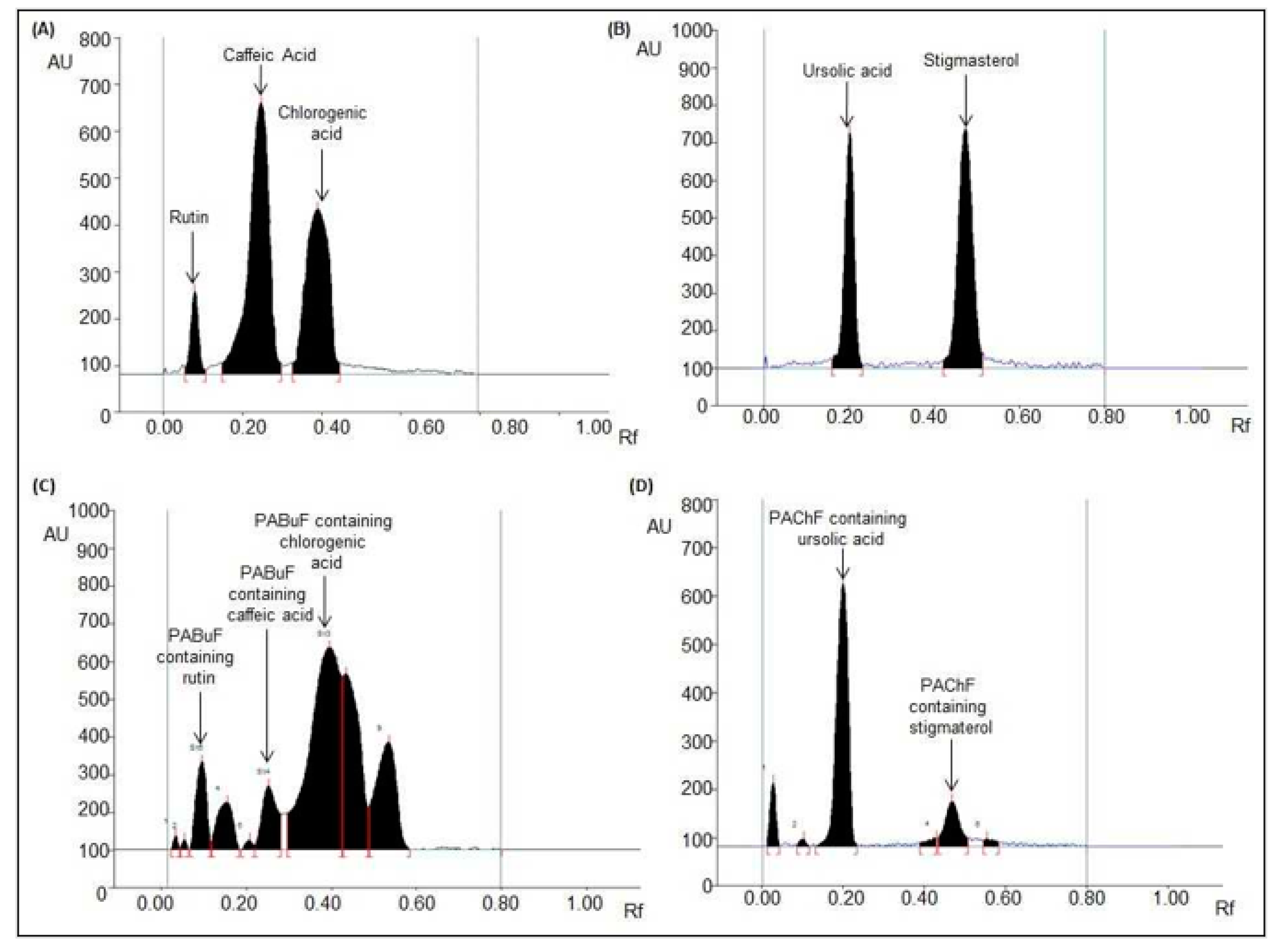
3.3. Application of Validated HPTLC Methods for Quantitative Analysis of Rutin, Chlorogenic Acid, Caffeic Acid, Ursolic Acid, and Stigmasterol in Butanol and Chloroform PA Extracts

Further applicability of the developed and validated HPTLC methods I and II were tested for quantitative analysis of rutin, caffeic acid, and chlorogenic acid (Figure 1C) in PABuF and ursolic acid and stigmasterol (Figure 1D) in PAChF. HPTLC methods I and II estimated the contents of rutin, caffeic acid, and chlorogenic acid to be 14.4, 1.04, and 17.10 μg/mg, respectively, of the dry weight of PABuF, and the contents of ursolic acid and stigmasterol to be 62.17 and 7.31 μg/mg, respectively, of the dry weight of PAChF.

3.4. Biological Evaluation

The fold induction was detected in PPAR activity in response to P-1 and P-2 fractions and the rutin (PA-1) compared to untreated controls. P-1 and P-2 showed a specific activation effect (fold induction ≥1.5) (Table 8).
From the results shown in Table 9, it could be seen that the P-1, P-2, and PA-1 decreased cellular oxidative stress. PA-1 significantly decreased cellular oxidative stress (67.0% decrease at 250 µg/mL), while P-1 and P-2 showed a 47.0% and 62.0% decrease in cellular oxidative stress, respectively, at a dose of 500 µg/mL compared to the standard drug quercetin. However, neither of the extracts showed NF-κB and inducible nitric oxide synthase (iNOS) inhibitory activity or cytotoxic and estrogenic effects on the four human cancer cell lines.

4. Discussion

Four species of the genus Periploca are found in Saudi Arabia: P. aphylla (PA), P. somaliensis, P. visciformis, and P. brevicoronata. PA has many benefits for human health in preventing and treating diseases because it has antimicrobial, antioxidant, insecticidal, analgesic, and sedative properties [1,3]. In this study, we isolated ursolic acid and stigmasterol from P-1 and three polyphenolic compounds rutin, chlorogenic acid, and caffeic acid from P-2 after subjecting P-1 and P-2 to a series of silica gel and Sephadex LH-20 column chromatography for separation and purification. Ursolic acid shows a variety of pharmaceutical properties, such as anticancer, anti-inflammatory, and antimicrobial properties. It also has a protective effect on many organs such as the liver, lungs, and brain [15]. In addition, stigmasterol has been investigated for its pharmacological properties, such as antihypercholesterolemic, hypoglycemic, antitumor, antiosteoarthritic, antimutagenic, anti-inflammatory, and antioxidant properties [16]. Flavonoids and other phenolic compounds are used as interesting alternative sources and are isolated from several medicinal plants. They have potential applications in pharmaceutical and medical fields because of their immune system–promoting, anti-inflammatory, antibacterial, antioxidant, anticancer, cardioprotective, and skin-protective (against UV radiation) effects [17]. We isolated P-1, P-2, and PA-1 (pure rutin) in good quantity and performed various biological studies to explore new, interesting biological activities not reported previously.
Several studies have been performed to explore the potential of selective PPARγ modulators. In this study, PPAR activity by fold induction of P-1, P-2, and PA-1 was detected against untreated controls. A fold induction of 1.5 means a 50% increase in PPAR activity. Our results indicated that P-1 shows PPARγ-specific antagonistic activity, followed by P-2, and both have a moderate effect compared to rosiglitazone (10, 5, 2.5 µM).
Historically, natural remedies have provided a favorable pool of structures for drug discovery. Recently, many studies have explored the PPARγ antagonistic potential of many natural herbs. PPARγ agonists are used to treat hyperglycemia accompanied by type II diabetes and metabolic syndrome [18]. In addition, PPAR activators, especially PPARγ, can be good neuroprotective drugs against inflammatory responses in cerebral ischemia and reperfusion injury. Generally, PPARγ antagonistic activity is involved in the regulation of insulin sensitivity, inflammation, fatty acid storage, and glucose metabolism; therefore, because of its role in decreasing insulin resistance and inflammation, it is implicated in the pathology of several diseases, including cancer, obesity, atherosclerosis, and diabetes [19].
Ursolic acid is a PPARγ antagonistic and an anti-inflammatory agent [18]. In addition, stigmasterol affects PPARγ activity [20]. In contrast, rutin is a better PPARγ agonist than thiazolidinediones, confirming its ability to bind at the active site of PPARγ [19]. Previous studies on caffeic acid and chlorogenic acid have shown their highest affinity for PPARγ [21,22]. Perhaps, the interesting PPARγ antagonistic effect observed in P-1 and P-2 is due to the synergistic effect of active secondary metabolites, although it was slightly high in P-1. This is the first study on the PPARγ antagonistic activity of PA extracts.
P-2 also showed antioxidant potential due to a higher decrease in cellular oxidative stress (62.0% decrease at a dose of 500 µg/mL). P-1 also possesses potent antioxidant activity but, in less intensity, compared to P-2. P-1 decreased cellular oxidative stress by 47.0% at a dose of 500 µg/mL. PA-1 (rutin) showed a significant decrease in cellular oxidative stress (67.0%) compared to quercetin (75.0%) at a low dose of 250 µg/mL. The antioxidant effect of P-2 could be due to the presence of rutin in good enough quantity. It is reported that rutin is a favorable neuroprotective compound for the treatment of various neurodegenerative diseases, such as Parkinson’s disease, Alzheimer’s disease, prion diseases, and Huntington’s disease, because of its beneficial effects as a powerful antioxidant [23]. Natural remedies and herbs have antioxidant properties that are believed to help reverse the effect of aging, support a healthy brain, maintain capillary integrity, and also maintain a healthy cardiovascular system. However, many studies have reported that caffeic acid, ursolic acid, stigmasterol, and chlorogenic acid show free-radical-scavenging activity and are recommended to be used as natural antioxidants against oxidative deprivation to prevent many diseases [16,24,25].
Because of its action on PPARγ as well as its effect against radical oxidative stress that may damage cells, our results highlight a new role of PA in treating many cardiovascular diseases. However, further in vivo studies are required.
The anti-inflammatory properties of PA extracts are described in terms of the inhibition of NF-κB’s transcriptional activity and iNOS. However, P-1 and P-2 as well as PA-1 do not show NF-κB and iNOS inhibitory activity. The Saudi PA does not show any cytotoxic, anti-inflammatory, and estrogenic activity, indicating the antagonistic action of various constituents present in it, which has not been reported earlier in the literature nor mentioned in Saudi traditional medicine with regard to the use of PA or its extracts in treating cancer, treating inflammation, or even showing estrogenic activity.
The LOD/LOQ for rutin, caffeic acid, chlorogenic acid, ursolic acid, and stigmasterol of 14.39/43.62, 32.56/98.67, 17.41/52.78, 22.94/69.52, and 28.51/86.41 ng/band, respectively, indicate that the two HPTLC methods I and II have good sensitivity for the quantification of the five compounds in PA extracts. To the best of our knowledge, this is the first report on accurate, simple, and rapid HPTLC methods developed and validated for the simultaneous quantification of rutin, caffeic acid, chlorogenic acid, ursolic acid, and stigmasterol in PA extracts [9,26]. Nowadays, HPTLC has become a routine analytical technique due to its advantages of reliability in quantitation of analytes at micro and even in nanogram levels and cost-effectiveness. It has proved a very useful technique because of its low operating cost, high sample throughput, and need for minimum sample clean-up. The major advantage of HPTLC is in reducing analysis time and cost per analysis TLC has been known as the fast tool for the detection of compounds [27,28,29]. Different HPTLC studies had been reported for the quantitative analysis of ursolic acid in many plant extracts, such as in the aerial parts of Nepeta deflersiana [9] and Wattakaka volubilis [26]. HPTLC densitometric quantification of stigmasterol from Ficus religiosa, Rauvolfia serpentina, and Bryophyllum pinnatum has been done previously. Traditional uses of these plants in ethnomedicine might be attributed due to the presence of stigmasterol as an important class of compound. Many phenolic constituents have been identified and their presence was confirmed by an HPTLC analysis. It has been proved to be a good analytical method for the rapid identification of secondary metabolites from medicinal plants. HPTLC quantification of chlorogenic acid has been carried out in Pluchea indica leaves found in Thailand [30,31]. To our knowledge, this is the first report of concurrent estimation of these compounds in PA. This HPTLC quantitative study has reported that a good quantity of rutin, caffeic acid, chlorogenic acid, ursolic acid, and stigmasterol are present in PA extracts, which is consistent with our previously reported results and could be responsible for their antioxidant and PPARα antagonistic activity [14,15,16,17,18].

5. Conclusions

Densitometric HPTLC is the most suitable method of assessing the secondary metabolites present in plant products. It can be used for the simultaneous quantitative determination of rutin, chlorogenic acid, caffeic acid, ursolic acid, and stigmasterol in PA because of its simplicity, accuracy, and selectivity. The results of HPTLC analysis for secondary metabolites could be considered phytomarkers in PA. The chloroformic and butanolic extracts of PA with active pure principle possess moderate free-radical-scavenging potency and selective PPARγ antagonistic activity, which are evidenced by the presence of active secondary metabolites confirmed by HPTLC fingerprinting analysis. In the future, these bioactive compounds may lead to the development of novel drugs for the treatment of many diseases.

Author Contributions

Conceptualization, R.O.; S.P. and H.Y.A.; P.A.; methodology, S.P.; software, O.M.N., J.P.; validation, B.S.S.A.-K. and A.M.A.-T.; formal analysis, A.K. and R.M. investigation, biological activities S.I.K. All authors have read and agreed to the published version of the manuscript.

Funding

The authors would like to extend their sincere appreciation to the Deanship of Scientific Research at King Saud University for its funding of this research through the Research Group Project no. RGP-221.

Institutional Review Board Statement

Not applicable.

Informed Consent Statement

Not applicable.

Data Availability Statement

The data used to support this study are available from the corresponding author.

Acknowledgments

The authors would like to extend their sincere appreciation to the Deanship of Scientific Research at King Saud University for its funding of this research through the Research Group Project no. RGP-221.

Conflicts of Interest

The authors have declared that no conflict of interest exist.

References

  1. Fabio, F.; Luigi, G. Herbal Medicine Today: Clinical and Research Issues. Evid. Based Complementary Altern. Med. 2007, 4, 37–40. [Google Scholar]
  2. Mohammed, M. Herbal Medicine in Healthcare-An Overview. Nat. Prod. Commun. 2012, 7, 807–812. [Google Scholar]
  3. Huang, M.; Shen, S.; Luo, C.; Ren, Y. Genus Periploca (Apocynaceae): A review of its classification, phytochemistry, biological activities and toxicology. Molecules 2019, 24, 2749. [Google Scholar] [CrossRef] [PubMed] [Green Version]
  4. Rehman, N.; Khan, A.; Fatima, U.; Akram, M.N.; Al-Musayeib, N.; Al-Massarani, S.; El-Gamal, A.; Gilani, A.H. Presence of laxative and antidiarrheal activities in Periploca aphylla: A Saudi medicinal plant. Int. J. Pharmacol. 2013, 9, 190–196. [Google Scholar] [CrossRef] [Green Version]
  5. Mustafa, G.; Anis, E.; Ahmed, S. Lupene-type triterpenes from Periploca Aphylla. J. Nat. Prod. 2000, 63, 881–883. [Google Scholar] [CrossRef]
  6. Tundis, R.; Loizzo, M.R.; Menichini, F.; Statti, G.A. Biological and pharmacological activities of iridoids: Recent developments. Mini-Rev. Med. Chem. 2008, 8, 399–420. [Google Scholar] [CrossRef] [PubMed]
  7. Ashish, K.S.; Rajesh, K.; Anurag, M.; Rajiv, G. Problems associated with clinical trials of Ayurvedic medicines. Braz. J. Pharmacogn. 2010, 20, 276–281. [Google Scholar]
  8. Parvez, M.K.; Alam, P.; Arbab, A.H.; Al-Dosari, M.S.; Alhowiriny, T.A.; Alqasoumi, S.I. Analysis of antioxidative and antiviral biomarkers β-amyrin, β-sitosterol, lupeol, ursolic acid in Guiera senegalensis leaves extract by validated HPTLC methods. Saudi Pharm. J. 2018, 26, 685–693. [Google Scholar] [CrossRef]
  9. Raha, O.; Nasir, S.; Perwez, A.; Tawfeq, A.; Areej, A.; Sami, A.; Najwa, M.; Rsshad, M.; Shabana, I.K.; Shagufta, P. Quantitative and biological analysis of iridoid glycoside & ursolic acid in aerial parts of Nepeta deflersiana by validated HPTLC method. Evid. Based Complementary Altern. Med. 2018, 2018, 1–11. [Google Scholar]
  10. Mariia, S.; Izabela, J.; Ewa, M.; Natalia, S.; Volodymyr, S.; Piotr, P.W. Development of high-performance thin layer chromatography method for identification of phenolic compounds and quantification of rosmarinic acid content in some species of the Lamiaceae family. J. Pharm. Bioallied Sci. 2020, 12, 139–145. [Google Scholar]
  11. Fawzy, G.A.; Al-Taweel, A.M.; Perveen, S.; Khan, S.I.; Al-Omary, F.A. Bioactivity and chemical characterization of Acalypha fruticosa growing in Saudi Arabia. Saudi Pharm. J. 2017, 25, 104–109. [Google Scholar] [CrossRef] [Green Version]
  12. Taweel, A.M.A.; Shafae, A.M.E.; Perveen, S.; Fawzy, G.A.; Khan, S.I. Anti-Inflammatory and cytotoxic constituents of Bauhina retusa. Int. J. Pharmacol. 2015, 11, 372–376. [Google Scholar] [CrossRef] [Green Version]
  13. Alam, P.; Siddiqui, N.; Al-Rehaily, A.; Alajmi, A.; Basudan, O.; Khan, T. Stability indicating densitometric HPTLC method for quantitive analysis of biomarker naringin in the leaves and stems of Rumex vesicarius L. J. Planar Chromatogr. 2014, 20, 204–209. [Google Scholar] [CrossRef]
  14. Borenfreund, E.; Babich, H.; Martin-Alguacil, N. Rapid chemosensitivity assay with human normal and tumor cells in vitro. Vitr. Cell. Dev. Biol. 1990, 26, 1030–1034. [Google Scholar] [CrossRef] [PubMed]
  15. Wozniak, L.; Skąpska, S.; Marszałek, K. Ursolic acid: A pen-tacyclic triterpenoid with a wide spectrum of pharmacological activities. Molecules 2019, 20, 9721. [Google Scholar] [CrossRef] [PubMed] [Green Version]
  16. Kaur, N.; Chaudhary, J.; Jain, A.; Kishore, L. Stigmasterol: A comprehensive review. IJPSR 2011, 2, 2259. [Google Scholar]
  17. Ghasemzadeh, A.; Ghasemzadeh, N. Flavonoids and phenolic acids: Role and biochemical activity in plants and human. J. Med. Plants Res. 2011, 5, 6697–6703. [Google Scholar] [CrossRef]
  18. Wang, Y.; He, Z.; Deng, S. Ursolic acid reduces the metalloprotease/anti-metalloprotease imbalance in cerebral ischemia and reperfusion injury. Drug Des. Dev. Ther. 2016, 10, 1663–1674. [Google Scholar] [CrossRef] [Green Version]
  19. Elekofehinti, O.O. In silico Studies on Plant Derived Rutin as Potent Agonist of Peroxisome Proliferator-activated Receptor Gamma (PPARγ). Br. J. Med. Med Res. 2016, 14, 1–8. [Google Scholar] [CrossRef]
  20. Wang, L.; Waltenberger, B.; Pferschy-Wenzig, E.; Blunder, M.; Liu, X.; Malainer, C.; Blazevic, T.; Schwaiger, S.; Rollinger, J.M.; Heiss, H.E.; et al. Natural product agonists of peroxisome proliferator-activated receptor gamma (PPARγ): A review. Biochem. Pharmacol. 2014, 92, 73–89. [Google Scholar] [CrossRef] [Green Version]
  21. Grzelczyk, J.; Budryn, G.; Pérez‑Sánchez, H. Evaluation of affinity of bioactive isolates from various coffee extracts through binding with PPAR‑γ with the use of isothermal titration calorimetry and docking simulation to prevent antidiabetic effects. J. Therm. Anal. Calorim. 2020, 142, 877–887. [Google Scholar] [CrossRef]
  22. Peng, S.G.; Pang, Y.L.; Zhu, Q.; Kang, J.H.; Liu, M.X.; Wang, Z. Chlorogenic acid functions as a novel agonist of PPARγ2 during the differentiation of mouse 3T3-L1 Preadipocytes. Biomed Res. Int. 2018, 2018, 8594767. [Google Scholar] [CrossRef] [PubMed]
  23. Enogieru, A.B.; Haylett, W.; Hiss, D.C.; Bardien, S.; Ekpo, O.E. Rutin as a potent antioxidant: Implications for neurodegenerative disorders. Oxidative Med. Cell. Longev. 2018, 2018, 6241017. [Google Scholar] [CrossRef] [PubMed]
  24. Tomac, I.; Šeruga, M.; Labuda, J. Evaluation of antioxidant activity of chlorogenic acids and coffee extracts by an electrochemical DNA-based biosensor. Food Chem. 2020, 325, 126787. [Google Scholar] [CrossRef]
  25. Chang, M.; Liu, C.; Shieh, D.; Chen, C. Evaluation and analysis of phytochemical antioxidant capacity. Res. Artic. Biomed. Res. 2017, 28, 6431–6434. [Google Scholar]
  26. Velmani, G.; Vivekananda, M.; Subhash, C.M. HPTLC evaluation of oleanolic acid and ursolic acid from the methanol extract of Wattakaka volubilis. J. Acute Dis. 2014, 3, 59–61. [Google Scholar]
  27. Deepti, R.; Sushila, R.; Permender, R.; Aakash, D.; Sheetal, A.; Dharmender, R. HPTLC densitometric quantification of stigmasterol and lupeol from Ficus religiosa. Arab. J. Chem. 2015, 8, 366–371. [Google Scholar]
  28. Anjoo, K.; Ajay, K.S. Development of validated HPTLC method for quantification of stigmasterol from leaf and stem of Bryophyllum pinnatum. Arab. J. Chem. 2017, 10, S2644–S2650. [Google Scholar]
  29. Abhijit, D.; Devendra, K.P. HPTLC Method for Quantitative Evaluation of Seasonal Variation of stigmasterol in Rauvolfia serpentina (L). Benth. ex Kurz. J. Biol. Act. Prod. Nat. 2014, 4, 254–261. [Google Scholar]
  30. Savita, C.; Boonyadist, V. Simultaneous HPTLC quantification of three caffeoylquinic acids in Pluchea indica leaves and their commercial products in Thailand. Rev. Bras. Farmacogn. 2017, 2017, 177–181. [Google Scholar]
  31. Beata, P.; Adam, T.; Marta, K.; Małgorzata, K. Comparison of Phenolic Compound Separations by HPTLC and PPEC with SDS as the Mobile Phase Component. J. Anal. Methods Chem. 2019, 2019, 1–15. [Google Scholar]
Figure 1. Chromatograms of phytomarker analysis in PABuF and PAChF. (A) Chromatogram of rutin (Rf = 0.09), caffeic acid (Rf = 0.25), and chlorogenic acid (Rf = 0.39) scanned at λmax = 340 nm; mobile phase (method I; RP-HPTLC) water: methanol (60: 40 v/v). (B) Chromatogram of ursolic acid (Rf = 0.20) and stigmasterol (Rf = 0.48) scanned at λmax = 539 nm; mobile phase (method II; NP-HPTLC) chloroform: methanol (98: 2, v/v). (C) Chromatogram of PABuF (rutin, spot 3, Rf = 0.09; caffeic acid, spot 6, Rf = 0.25; and chlorogenic acid, spot 7, Rf = 0.39) scanned at λmax = 340 nm (method I; RP-HPTLC). (D) Chromatogram of PAChF (ursolic acid, spot 3, Rf = 0.20 and stigmasterol, spot 5, Rf = 0.48) scanned at λmax = 539 nm (method II; NP-HPTLC). PABuF, P. aphylla butanol fraction; and PAChF, P. aphylla chloroform fraction; HPTLC, high-performance thin-layer chromatography).
Separations 08 00044 g001
Table 1. Rf and linear regression data for calibration curves of rutin, caffeic acid, chlorogenic acid, ursolic acid, and stigmasterol (n = 6).
ParametersMethod IMethod II
RutinCaffeic AcidChlorogenic AcidUrsolic AcidStigmasterol
Linearity range (ng/spot)100–800100–800100–800100–800100–800
Regression equationY = 4.27X + 320.01Y = 21.13X + 6538.26Y = 26.51X + 854.12Y = 3.272X + 241.02Y = 5.153X + 285.72
Correlation (r2) coefficient0.9945 ± 0.0020.9927 ± 0.00130.9939 ± 0.00120.9944 ± 0.00010.9926 ± 0.0021
Slope ± SD4.27 ± 0.01821.13 ± 1.5826.51 ± 1.3993.272 ± 0.0235.153 ± 0.045
Intercept ± SD320.01 ± 10.896538.26 ± 50.19854.12 ± 38.07241.02 ± 12.76285.72 ± 10.447
Standard error of the slope0.0070.0850.0570.0090.018
Standard error of intercept4.44920.4815.5415.2064.264
Rf0.09 ± 0.0010.25 ± 0.0040.39 ± 0.0030.20 ± 0.0030.48 ± 0.004
LOD (ng)14.3932.5617.4122.9428.51
LOQ (ng)43.6298.6752.7869.5286.41
SD, standard deviation; LOD, limit of detection; LOQ, limit of quantification, ng, nanogram; Rf, relative flow.
Table 2. Recovery as the accuracy of the proposed method for rutin, caffeic acid, and chlorogenic acid (n = 6). SD, standard deviation; RSD, relative standard deviation.
Method I
Standard Added to
Analyte (%)
Theoretical
Concentration of Standard (ng/spot)
RutinCaffeic AcidChlorogenic Acid
Concentration Found
(ng/spot) ± SD
%RSD%RecoveryConcentration Found
(ng/spot) ± SD
%RSD%RecoveryConcentration Found
(ng/spot) ± SD
%RSD%Recovery
0200199.93 ± 7.294.1699.96196.17 ± 7.613.8898.08194.52 ± 6.573.3897.26
50300295.61 ± 9.193.1098.53289.01 ± 11.293.9196.33287.41 ± 8.973.1295.81
100400386.67 ± 14.233.6896.66392.32 ± 14.733.7598.18389.33 ± 13.413.4497.33
150500486.96 ± 16.273.3497.39482.77 ± 13.372.7796.55484.58 ± 17.813.6896.91
SD (standard deviation); RSD (relative standard deviation); ng, nanogram.
Table 3. Recovery as the accuracy of the proposed method for ursolic acid and stigmasterol (n = 6).
Method II
Standard Added to Analyte (%)Theoretical Concentration of Standard (ng/spot)Ursolic AcidStigmasterol
Concentration Found (ng/spot) ± SD%RSD%RecoveryConcentration Found (ng/spot) ± SD%RSD%Recovery
0200197.11 ± 7.083.5698.56195.63 ± 7.252.9697.81
50300293.26 ± 10.973.7497.75289.72 ± 9.673.9596.57
100400390.52 ± 11.632.9897.63388.06 ± 11.894.8597.01
150500495.36 ± 14.923.0199.07487.41 ± 10.514.2897.48
Table 4. Precision of the proposed HPTLC method I (n = 6).
Method I
Concentration of Standard Added (ng/spot)RutinCaffeic AcidChlorogenic Acid
Intraday PrecisionInterday PrecisionIntraday PrecisionInterday PrecisionIntraday PrecisionInterday Precision
Average Concentration Found ± SD%RSDAverage Concentration Found ± SD%RSDAverage Concentration Found ± SD%RSDAverage Concentration Found ± SD%RSDAverage Concentration Found ± SD%RSDAverage Concentration Found ± SD%RSD
200195.20 ± 7.293.73192.86 ± 7.493.88195.52 ± 8.264.22192.30 ± 8.014.16194.43 ± 7.213.71190.66 ± 7.053.69
300293.42 ± 8.192.79288.96 ± 8.012.77291.68 ± 11.583.96288.84 ± 11.133.85291.66 ± 9.813.36287.89 ± 9.893.43
400387.24 ± 9.372.41382.55 ± 9.432.46389.01 ± 14.293.67385.70 ± 13.913.61394.20 ± 17.743.23386.59 ± 12.313.18
HPTLC, high-performance thin-layer chromatography; SD, standard deviation; RSD, relative standard deviation; ng, nanogram.
Table 5. Precision of the proposed HPTLC method II (n = 6).
Method II
Concentration of Standard Added (ng/spot)Ursolic AcidStigmasterol
Intraday PrecisionInterday PrecisionIntraday PrecisionInterday Precision
Average Concentration Found ± SD%RSDAverage Concentration Found ± SD%RSDAverage Concentration Found ± SD%RSDAverage Concentration Found ± SD%RSD
200196.63 ± 6.583.34192.65 ± 6.793.52197.30 ± 6.773.43193.42 ± 6.893.56
300294.17 ± 11.193.80288.06 ± 11.033.83294.22 ± 11.383.87289.17 ± 11.033.81
400391.29 ± 12.733.25385.18 ± 12.653.28392.32 ± 13.313.39387.28 ± 12.913.33
HPTLC, high-performance thin-layer chromatography; SD, standard deviation; RSD, relative standard deviation; ng, nanogram.
Table 6. Robustness of the proposed HPTLC method I at 400 ng/band (n = 6).
Method I
Optimization ConditionRutinCaffeic AcidChlorogenic Acid
SD%RSDSD%RSDSD%RSD
Mobile phase composition; (water: methanol)
a (6:4)9.452.3611.682.9814.133.63
(5.8:4.2)9.212.3211.933.0414.593.70
(6.2:3.8)9.872.5112.173.0914.873.76
Mobile phase volume (for saturation)
(18 mL)9.712.4410.542.6914.033.64
(20 mL)9.542.4110.892.7714.093.65
(22 mL)9.912.5110.232.6114.073.64
Duration of saturation
(10 min)8.922.2413.093.3414.413.68
(20 min)8.572.1612.183.1114.473.69
(30 min)8.732.2012.013.0614.583.71
HPTLC (high-performance thin-layer chromatography); SD (standard deviation); RSD (relative standard deviation). a Different ratio of water: methanol used.
Table 7. Robustness of the proposed HPTLC method II at 400 ng/band (n = 6).
Method II
Optimization ConditionUrsolic AcidStigmasterol
SD%RSDSD%RSD
Mobile phase composition; (chloroform: methanol)
a (98:2)10.972.8210.192.61
(97.8:2.2)10.512.6910.272.63
(98.2:1.8)10.372.6710.542.68
Mobile phase volume (for saturation)
(18 mL)10.892.829.972.56
(20 mL)10.932.829.812.53
(22 mL)10.792.789.912.54
Duration of saturation
(10 min)10.672.7310.772.77
(20 min)10.512.7010.832.78
(30 min)10.432.6810.692.73
HPTLC, high-performance thin-layer chromatography; SD, standard deviation; RSD, relative standard deviation. a Different ratio of water: methanol used.
Table 8. PPAR agonistic activity of PA extracts and the pure compound.
Sample NamePPARα Fold Induction (µg/mL)PPARγ Fold Induction (µg/mL)Ciprofibrate (µM)Rosiglitazone (µM)
502512.5502512.51052.51052.5
P-1NANANA2.12.51.72.31.61.33.83.53.0
P-2NANANA1.91.71.3
PA-1NANANANANANA
PPAR, peroxisome proliferator–activated receptor; PA, Periploca aphylla; NA, not active; P-1, chloroform extract; P-2, n-butanol extract; PA-1, pure compound (rutin).
Table 9. Anti-inflammatory activity of PA extracts and the pure compound.
a Sample% Decrease in Cellular Oxidative StressNF-κB InhibitionSp-1 Inhibition IC50 (µg/mL)iNOS Inhibition IC50 (µg/mL)
P-147NANANA
P-262NANANA
PA-167NANANA
b Quercetin 50 µM75---
b Parthenolide-0.86.50.29
a At 500 µg/mL for the extracts and 250 µg/mL for rutin; b Positive control; PA, Periploca aphylla; NA, no activity up to 100 µg/mL (NF-κB, iNOS) and 500 µg/mL (cellular oxidative stress); P-1, chloroformic extract; P-2, butanolic extract; PA-1, pure compound (rutin); NF-κB, nuclear factor kappa B; iNOS, inducible nitric oxide synthase; IC50, half-maximal inhibitory concentration. NF-κB, Nuclear factor kappa B; Sp-1, serine protease 1; iNOS, Inducible nitric oxide synthase.
Publisher’s Note: MDPI stays neutral with regard to jurisdictional claims in published maps and institutional affiliations.

