Article

Valorisation of Prunus avium L. By-Products: Phenolic Composition and Effect on Caco-2 Cells Viability

1
CICS-UBI—Health Sciences Research Center, University of Beira Interior, 6200-506 Covilhã, Portugal
2
CNC—Centre for Neuroscience and Cell Biology, University of Coimbra, 3004-504 Coimbra, Portugal
3
Laboratory of Pharmacology, Faculty of Pharmacy, University of Coimbra, 3000-548 Coimbra, Portugal
4
CIBIT—Coimbra Institute for Biomedical Imaging and Translational Research, University of Coimbra, 3000-548 Coimbra, Portugal
5
Phytochemistry and Healthy Foods Laboratory, Food Science and Technology Department, CEBAS-CSIC, 30100 Murcia, Spain
*
Authors to whom correspondence should be addressed.
Submission received: 9 April 2021 / Revised: 16 May 2021 / Accepted: 20 May 2021 / Published: 25 May 2021
(This article belongs to the Topic Fruit and Vegetable Juice Processing and Nutrition)

Abstract

:
Prunus avium L. by-products, such as stems, leaves, and flowers, are used in folk medicine to prevent and treat some diseases. However, their phenolic composition and in vitro bioactivities against tumor cells are poorly known. In this work, we compared the phenolic profile and the biological potential of aqueous infusions and hydroethanolic extracts of P. avium leaves, stems, and flowers from Saco cultivar, collected from the Fundão region (Portugal). Among the fifty-two phenolic compounds tentatively identified by HPLC-DAD-ESI/MSn, the hydroxycinnamic acids were the most abundant. Both extracts of stems revealed a higher activity against DPPH. Meanwhile, hydroethanolic extracts from stems and flowers and aqueous infusions of flowers were the most effective in inhibiting the growth of the human epithelial colorectal adenocarcinoma (Caco-2) cells at concentrations above 200 μg/mL. More detailed knowledge about the phenolic composition and health-promoting properties of Portuguese P. avium by-products allows for increasing the biological and commercial value of these bio-wastes, which may have a positive impact on food and pharmaceutical industries, as on the valorization of the local economy.

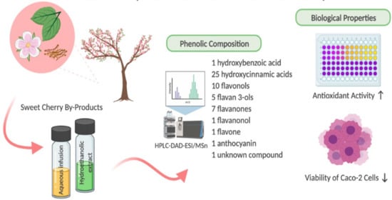
Graphical Abstract

1. Introduction

Prunus avium L. is a deciduous tree belonging to the Rosaceae family, Prunoideae subfamily, Prunus genus, and subspecies avium, which produces one of the most popularly appreciated red fruits, the sweet cherry [1]. This fruit possesses significant amounts of phenolic compounds, namely chlorogenic acids and anthocyanins, whose powerful antioxidant, anti-inflammatory, and chemopreventive properties are well-described [2,3,4,5,6,7].
Sweet cherry has been the focus of many studies due to its capacity to scavenge and neutralize free radicals, and consequently, attenuate or even mitigate the occurrence of oxidative stress-related diseases [4,7,8]. Nevertheless, tons of cherries are produced annually, resulting in large quantities of by-products as crop residues (e.g., leaves, stems, flowers, non-marketable fruits, and pits). The incorporation of these by-products in dietary supplements, functional foods, food fortification, food preservation, nutraceuticals, and pharmaceuticals has been encouraged as the cherry fruit and its by-products present significant bioactive compounds [9,10]. Few studies have already reported the richness of cherry leaves, stems, and flowers in hydroxycinnamic acids (e.g., 3- and 5-O-caffeoylquinic acids), flavan-3-ols (e.g., epigallocatechin and catechin), flavonols (e.g., quercetin 3-O-rutinoside, quercetin 3-O-glucoside, and kaempferol 3-O-rutinoside), and flavanones (naringenin and sakuranetin and their derivatives) [2,9,11] Moreover, their use can also be considered part of a strategy for sustainable fruit production, bringing numerous benefits for the regional circular economy and reducing agro-waste.
P. avium leaves and stems have been used since ancient times to boost the immune system due to their sedative, diuretic, anti-inflammatory, and draining properties [12,13]. On the other hand, their flowers are a symbol of renewal and vitality in Japanese culture [14].
In the present work, the identification of phenolic compounds from P. avium leaves, stems, and flowers of Saco cultivar using high-performance liquid chromatography (HPLC) coupled with mass spectrometer electrospray ionization mass (ESI-MS/MS) detection was performed. HPLC–DAD was used for the phenolic quantification of all samples. We also assessed their antioxidant capacity against 2,2-diphenyl-1-picrylhydrazil radical (DPPH) and their effects on human epithelial colorectal adenocarcinoma (Caco-2) cells, herein reported for the first time.

2. Materials and Methods

2.1. Plant Material

P. avium leaves, stems, and flowers of the Saco cultivar (collected between April and June 2018) were provided by a local producer in Cimo da Aldeia—Pêro Viseu (40°12′36″ N 7°26′21″ W), Fundão region (Beira Interior, Portugal). After collection, the samples were transported to the CICS-UBI laboratory. The samples were frozen using liquid nitrogen, freeze-dried (ScanVac CoolSafe, LagoGene APS, Allerød, Denmark), and converted to a dried powder with a mean particle size lower than 910 μm. Posteriorly, they were stored at −20 °C and protected from light until analysis.

2.2. Aqueous Infusions and Hydroethanolic Extracts Preparation

Aqueous infusions of cherry by-products were prepared following a previously reported procedure [9]. Briefly, the dried powdered samples were subjected to infusion (1 g/100 mL) at 100 °C for 3 min. After that, they were filtered with a membrane filter (0.45 μm, Millipore, Bedford, MA, USA), frozen, freeze-dried, and kept at −20 °C in the dark until analysis. This protocol is similar to that involved in the infusions prepared commercially for human consumption. The obtained yields were 26.07 ± 1.22%, 28.42 ± 0.86%, and 18.40 ± 1.18% for leaves, stems, and flowers, respectively. On the other hand, and regarding the hydroethanolic extracts, 1 g of each sample was extracted with ethanol/water (50:50, v/v). The process involved 30 min of sonication, 2 h of maceration (at a temperature of 25 °C with agitation at 200 r/min), and another 30 min of sonication. The obtained extract was filtered (membrane with 0.45 μm, Millipore, Bedford, MA, USA), evaporated, and kept at −20 °C until further [9]. The extraction yields from dry material were 25.20 ± 1.90%, 28.89 ± 0.14%, and 22.31 ± 1.23% for leaves, stems, and flowers, respectively.

2.3. Total Phenolic Compounds Determination

The total phenolic compounds in P. avium leaves, stems, and flowers were estimated via a colorimetric assay, based on procedures described in [15] with some modifications. Briefly, 50 µL of the methanolic samples (2.5 mg/mL) was mixed with 450 µL of distilled water and 2.5 mL of 0.2 N Folin-Ciocalteu reagent. After 5 min of incubation at room temperature, 2 mL of a saturated sodium carbonate solution was added to the mixture, and the volume was adjusted to 5 mL with distilled water. Then, the reaction was kept in a water-bath at 30 °C for 90 min while protected from light. After that, the absorbance was read at 765 nm. Gallic acid was used to calculate the standard curve (50–500 mg/mL; y = 0.0011x + 0.0057; R2 = 0.9958). The experiments were performed in triplicate. The results were mean values ± standard deviations and expressed as molar concentration of gallic acid.

2.4. Total Flanonoids Determination

Flavonoid contents were assessed by the aluminum chloride colorimetric method [16]. Methanolic solutions of quercetin (purity ≥ 95%, 12.5–200 mg/L) were used to calculate the calibration curve (y = 0.0068x − 0.0135; R2 = 0.9969). The results were expressed in mg quercetin equivalents (QE) per gram of extract.

2.5. Phenolic Compounds Composition

2.5.1. HPLC-DAD-ESI/MSn

Phenolics of leaves, stems, and flowers were analyzed by HPLC-DAD-ESI/MSn following the methodology previously described [5,17], with slight modifications. We used an Agilent HPLC 1100 series coupled to a mass detector ion trap spectrometer (model G2445A) equipped with an electrospray ionization interface and controlled by LC/MS software (Esquire Control Ver. 6.1. Build No. 534.1., Bruker Daltoniks GmbH, Bremen, Germany). The compounds were identified using their elution order, retention time, ultraviolet–visible spectra, fragmentation patterns, and diagnostic fragments in MS2 and MS3, with those obtained from standard compounds analyzed under the same conditions, and comparing the results obtained with the data reported in the literature [2,5,17,18].

2.5.2. HPLC-DAD

Aqueous infusions and hydroethanolic extracts of leaves, stems, and flowers were dissolved in methanol (2 mg for leaves and flowers, and 10 mg for stems) and analyzed by HPLC-DAD (Agilent, Santa Clara, CA, USA), according to the method previously reported [3]. The quantification was performed by injecting 20 µL of each extract sample onto an LC model Agilent 1260 system (Agilent, Santa Clara, CA, USA), composed of an auto-sampler, coupled with a photodiode array detector, and the column used was a Nucleosil® 100-5 C18 (Macherey-Nagel, Düren, Germany) [3]. The identification of the phenolics was completed through the absorbance recorded in the chromatograms relative to external standards at 280 nm for hydroxybenzoic acids, flavan-3-ols, flavanones, and flavones; 320 nm for hydroxycinnamic acids; 350 nm for flavanonols and flavonols; 500 nm for anthocyanins [5]. The phenolic compounds found in infusions and hydroethanolic extracts were identified by comparing their retention times and ultraviolet–visible absorption spectra in the 200–500 nm range with those of authentic standards. All the calibration curves of the external standards (concentration range of 1.5–100 µg/mL) used in the phenolics quantification are described in Table 1.

2.6. 2.2-Diphenyl-1-Picrylhydrazil Radical (DPPH•)-Scavenging Activity

A DPPH-scavenging assay was evaluated following a reported procedure [9]. Briefly, seven different dilutions of infusion and hydroethanolic extracts of P. avium leaves, stems, and flowers were prepared in methanol, placed in a 96-well plate, and read at 515 nm, using a microplate reader Bio-Rad Xmark spectrophotometer. Three experiments were performed in triplicate. Ascorbic acid was used as a positive control.

2.7. Caco-2 Cell Culture and Treatment with By-products Extracts

Caco-2 cells were purchased from the American Type Culture Collection (ATCC; Manassas, VA, USA) and cultured as a monolayer in DMEM supplemented with 10% FBS and 1% penicillin/streptomycin (all from Sigma-Aldrich, MI, USA) at 37 °C in a humidified atmosphere of 95% air and 5% CO2. To determine the antiproliferative effects of infusions and hydroethanolic extracts of cherry by-products, cells were incubated for 24 h with different concentrations (50, 100, 200, 400, and 800 µg/mL) of these extracts, accordingly to the method previously described [4], with slight modifications. After 24 h of extract incubation, the 3-(4,5-dimethylthiazol-2-yl)-2,5-diphenyltetrazolium bromide (MTT) reduction and lactate dehydrogenase (LDH) release assays were performed.

2.7.1. MTT Assay

Cell viability was assessing using the MTT assay following the protocol previously described [4]. The absorbance was measured at 570 nm using a microplate reader Bio-Rad Xmark spectrophotometer. Six independent experiments were performed in triplicate. Cell viability was measured as the percentage of absorbance compared to control.

2.7.2. LDH Assay

LDH activity was spectrophotometrically evaluated at 340 nm, based on the conversion of pyruvate to lactate, using NADH as a cofactor, as previously described [4]. Six independent experiments were performed in triplicate.

2.8. Statistical Analysis

The results are expressed as mean values ± standard deviation (SD) and the experiments were carried out in triplicate. The results were analyzed using one-way ANOVA followed by Tukey’s test at a 95% level of significance. In the case of DPPH assay, the statistical comparison of all infusions and hydroethanolic extracts was performed using the Kruskal–Wallis test. Concerning the cellular-based assays, data from different extracts were compared by one-way ANOVA followed by Dunnett’s test as a post hoc test. Differences were considered statistically significant for p < 0.05 using Graphpad Prism Version 7.05 Software (San Diego, CA, USA).

3. Results and Discussion

3.1. Total Phenolics Content

Phenolic compounds constitute one of the major groups of phytochemicals, with a relevant antioxidant activity such as free radical-scavenging, hydrogen donation, singlet oxygen quenching, metal ion chelation, and also acting as a substrate for radicals such as hydroxyl and superoxide [19]. In order to make a general screening of the phenolics of P. avium leaves, stems, and flowers, the total phenols content was evaluated. The obtained results showed that P. avium stems presented a higher content of phenols (301.38 ± 5.9 mg GAE/g extract), followed by leaves and flowers (100.71 ± 8.30 and 81.20 ± 2.75 mg GAE/g extract, respectively (Table 2)). The obtained data are in line with those reported by [20], which found similar values in sweet cherry fruit. However, Serra et al. [21] reported higher values (1309 ± 12 mg GAE/100 g dw) in cherries from the Saco cultivar. In a study conducted by Prvulovic et al. [11], the total phenolics content in P. avium stems ranged from 12.96 to 31.85 mg GAE/g dry. These differences may be associated with some factors, such as the type of extraction used, the harvest region, and the climate, among others [22,23].

3.2. Total Flavonoids Content

Flavonoids are the main class of phenolics found in plants, and they are common components in the human diet. According to their chemical structure, they can be divided into flavonols, flavones, flavanones, isoflavones, flavan-3-ols, and anthocyanins [1]. These compounds have relevant biological activities, such as the ability to neutralize free radicals, and for that reason, their quantification in vegetal extracts is important [1]. The total flavonoids content was evaluated with the aluminum chloride colorimetric method (Table 3). As expected, the hydroethanolic extracts of P. avium by-products present higher concentrations of flavonoids than the respective infusions. The results showed that leaf hydroethanolic extract presented a higher content of flavonoids (35.17± 5.9 mg QE/g extract), followed by hydroethanolic extracts of flowers and stems (24.62 ± 0.39 and 15.25 ± 1.18 mg QE/g extract, respectively) (Table 3). Based on the obtained values for the total flavonoids content, it is possible to verify that the highest concentration of these compounds is in the extracts obtained using a solvent of moderate polarity (ethanol). Recently, similar results were found in P. avium by-products, such as stalk, pulp, seed, and leaf [24].

3.3. Analysis of Phenolic Compounds.

The individual phenolics present in P. avium leaves, stems, and flowers were tentatively identified based on a comparison of their retention times, ultraviolet–visible spectra, and the interpretation of their fragmentation patterns as obtained from MSn spectra and by their relationship with other data reported in the literature. This study identified a total of fifty-two phenolic compounds, some of which were previously described in P. avium fruit [4,5,7] and its by-products [2,9]. The analysis allowed the identification of one hydroxybenzoic acid, twenty-five hydroxycinnamic acids, ten flavonols, five flavan-3-ols, seven flavanones, one flavanonol, one flavone, one anthocyanin, and one unknown compound (Table 4 and Table 5). As far as we know, this is the first study that characterizes the phenolic composition of leaves and flowers of sweet cherry, and the first concerned with the characterization of Saco variety stems from the Fundão region (Portugal), as evaluated by HPLC-DAD-ESI/MSn.

3.3.1. Hydroxybenzoic Acids

In this work, one hydroxybenzoic acid has been described for the first time in the leaves and stems of P. avium. One signal (peak 5) at m/z 153 was detected and eluted at 15.7 min (Table 4). This compound was identified as a protocatechuic acid aglycone that corresponds to a loss of 162 Da (hexose moiety). This phenolic acid had already been reported in cherry fruits [5,6].

3.3.2. Hydroxycinnamic Acids

A total of twenty-five hydroxycinnamic acids (peaks 1–4, 6–10, 12, 14–17, 20–21, 28, 33, 35, 38–39, 42, 44, 46–47) were identified in P. avium by-products (Table 4 and Table 5) using previously developed studies [2,6,9,25,26].
Peaks 1, 3, and 20 were identified as ferulic acids since they showed a fragmentation pattern at m/z 193 [ferulic acid-H]- after the loss of a hexosyl moiety (−162 mu), and the presence of a feruloyl fragment [25]. Bastos et al. [2] also detected their presence in P. avium stem extracts, infusions, and decoctions. On the other hand, these compounds have also been described in sweet cherry fruits [5,6]. Feruloyl di-hexose (peak 1) was not quantified in all infusions and hydroethanolic extracts (Table 5). Feruloyl hexose (peak 3) and feruloylquinic acid (peak 20) were not detected in leaves and stems, and it was not possible to quantify them in the flower infusion and hydroethanolic extract (Table 5).
Twelve signals (peaks 2, 4, 6–7, 9–10, 15, 21, 33, 35, 38, and 39) were detected that eluted between 13.7 and 31.3 min (Table 4). These components were identified as caffeoylquinic acids, and they have a fragmentation pattern at m/z 191 and/or at 179 [27]. Caffeoylquinic acid-glycoside (peak 7), 3-caffeoylquinic acid cis (peak 9), 3,5-dicaffeoylquinic acid 1 and 2 (peaks 10 and 35, respectively), 3,4-dicaffeoylquinic acid (peak 33), 5-caffeoylquinic acid trans (peak 21), and 4,5-dicaffeoyquinic acid (peak 39) had already been reported by Martini et al. [6] in sweet cherry cultivars. Moreover, peaks 4 and 7 were also identified as caffeoylquinic acid glycosides (m/z 515) (Table 4 and Table 5), and both possess a caffeoyl glucoside moiety (m/z 341) [28]. Peak 17 corresponds to 4-caffeyolquinic acid (molecular ion m/z z 353) with a fragment pattern at m/z 173 [25]. As far as we know, this is the first report concerning the presence of 4-caffeyolquinic acid in P. avium leaf infusion (ca. 908.93 ± 91.95 µg/g of dw) and hydroethanolic extract (ca. 466.37 ± 19.7 µg/g of dw), as shown in Table 5. However, its presence has already been reported in sweet cherries [5,6,27].
Peaks 14 and 16 were identified as p-coumaroylquinic acid derivative and p-coumaric acid derivative, respectively. They exhibited a molecular ion at m/z 337 and fragmentation ions at m/z 191 (Table 4 and Table 5) [2,25,28]. P-Coumaroylquinic acid has already been described in P. avium stems [2]. Moreover, peak 16 was described as a p-coumaroylquinic acid derivative, showing fragmentation ions at m/z 337.
The phenolic compounds corresponding to the peaks 46 and 47 were classified as coumaroyl-caffeoylquinic acids (molecular ion at m/z 499) (Table 4 and Table 5). Nevertheless, they possess distinct fragment patterns due to dehydration and the loss of caffeoyl and p-coumaroyl residues [29]. 3-Coumaroyl-5-caffeoylquinic acid (peak 46) was identified only in sweet cherry fruits [6].
Peak 12 was identified as a caffeoyl hexose (m/z 341) with a fragmentation at m/z 179 (Table 4 and Table 5) [6]. Similarly, in another study, Bastos et al. [2] tentatively identified these compounds as caffeic acid and trans-caffeic acid hexoside, based on the same pseudo-molecular ion, ultraviolet spectra, and fragmentation pattern. Additionally, two caffeoyl hexose derivatives (m/z 341) were tentatively identified (peaks 38 and 42), based on similar fragmentation (Table 4 and Table 5).
In sum, regarding hydroxycinnamic acids, P. avium leaves proved to be the by-products richest in these types of compounds. Totals of 51345.69 and 57605.22 µg/g of dw were obtained for the infusion and hydroethanolic extract, respectively (Table 5). Trans-5-caffeoylquinic acid (peak 21) was the major hydroxycinnamic acid found in both extracts, followed by 3-caffeoylquinic acid cis (peak 9). Our results were similar to those obtained by Jesus et al. [9], who also reported that hydroxycinnamics were the main compounds in leaves, representing 75.3% and 63.7% of total phenolic compounds in the infusion and hydroethanolic extract, respectively.

3.3.3. Flavonols

Ten flavonols were found in P. avium leaves, stems, and flowers (Table 4 and Table 5), according to previous studies [2,5,30]. In this study, four quercetins (peaks 25, 26, 32, and 49) were detected (molecular ion at m/z 789, 771, 625, 609, and 463), as was a quercetin ion at m/z 301 (Table 4 and Table 5). Quercetin 3-O-rutinoside (peak 49) was the only flavonol quantified in all infusions and hydroethanolic extracts of P. avium leaves, stems, and flowers (Table 5). The leaf infusion was the richest one in this flavonol (ca. 6175.93 ± 148.22 µg/g of dw, comprising about 17.08% of total phenolic compounds), followed by flower hydroethanolic extract (1823.94 ± 38.9 µg/g of dw, containing about 7.71% of total phenolic compounds) (Table 5). These results agree with other recent work [9] showing that quercetin 3-O-rutinoside was the major flavonol found in leaf extracts. According to the literature, flavonols are the main phenolics found in leaves, and quercetin 3-O-rutinoside and quercetin 3-O-glucoside were also found in P. avium stems [2]. On the other hand, peak 26 was tentatively identified as quercetin O-rutinoside-O-hexoside. This compound exhibits a molecular ion at m/z 771, and typical fragmentations at m/z 609, 463, and 301 (Table 4). The presence of this flavonol in P. avium by-products agrees with other previous work (Bastos et al., 2015). Additionally, peak 26 was identified as quercetin di-hexoside and was only quantified in the flower hydroethanolic extract (Table 4 and Table 5). Quercetin 3-O-hexoside (peak 32) was detected at some levels in the stem and flower infusions and hydroethanolic extracts (Table 4 and Table 5), comprising less than 6% of total phenolic compounds.
On the other hand, six kaempferols (peaks 23, 27, 30, 34, 36, and 40) were found, all of which displayed a kaempferol fragment at m/z 285 [28]. Kaempferol O-rutinoside-O-hexoside (peak 23) and kaempferol 3-glucoside (peak 36) were found for the first time by Bastos et al. [2] in P. avium stems. Our study, as we far as known, detected them for the first time in flowers and leaves (Table 4 and Table 5). Kaempferol 3-O-rutinoside (peak 34) was identified in leaf infusions (1298.58 ± 44.2 µg/g) (Table 4 and Table 5), its presence being identified in previous works [9].

3.3.4. Flavan 3-ols

Five flavan 3-ols (peaks 11, 13, 18–19, and 22) (Table 4 and Table 5) already described in cherries and their by-products were identified [2,5,6,9]. Catechin hexoside (peak 11) was tentatively identified, exhibiting a molecular ion at m/z 451 and fragmentation ions at m/z 289 and 245 (Table 4). This catechin was found in leaves and stems of P. avium (Table 5).
Furthermore, four procyanidins were identified in our samples (peaks 13, 18, 19, and 22). Two of these compounds were tentatively classified as type-B procyanidin dimers at m/z 577 (peaks 13 and 22) (Table 4 and Table 5). The presence of a procyanidin is supported by the fragment patterns at m/z 425 as well as water elimination at m/z 407 [28]. Our team reported similar findings for sweet cherry composition, which comprised about 18.47% total non-colored phenolics [5]. Procyanidin dimer B type 1 was found in different sweet cherries [6], while procyanidin dimer B type 2 was quantified in infusion and hydroethanolic extracts of P. avium stems (7149.5 ± 510.5 and 8810.67 ± 592.2 µg/g of dw, respectively) (Table 4 and Table 5). Procyanidin tetramer (peak 18; m/z 1153 molecular ion) and procyanidin trimer (peak 19; m/z 865 molecular ion) were also identified in P. avium by-products. As far as we know, this is the first study assessing the presence of these compounds in the leaves and stems of sweet cherries.

3.3.5. Flavanones

A total of seven flavanones, eluted between 31.0 and 39.1 min, were identified in P. avium by-products, including four sakuranetins (peaks 37, 47, 51 and 52), two naringenins (peaks 41 and 48), and one pinocembrin (peak 44) (Table 4 and Table 5). Two sakuranetin 5-O-hexoside derivatives (peaks 37 and 52) were found for the first time in all cherry by-products studied in this work (Table 4 and Table 5). Both compounds have a fragmentation at m/z 285. Sakuranetin 5-O-hexoside (peak 51) was also identified in the same sample, presenting an ion at m/z 447 and releasing a fragment ion at m/z 285. This compound was quantified in the aqueous infusion and hydroethanolic extract (ca. 265.89 ± 9.8 and 214.66 ± 10.6 µg/g of dw, respectively) of P. avium leaves (Table 4), and these results are in accord with other previous works [2,9]. In addition, sakuranetin O-pentosyl-hexoside (peak 48) was tentatively identified based on the same fragmentation previously described (Table 4 and Table 5), and was found in stems. Previous data have already reported the presence of sakuranin in sweet cherries [21,31,32].
Naringenin-7-O-hexoside (peak 41) and naringenin hexoside (peak 48) presented a similar pseudo molecular ion at m/z 433 and 443 (Table 4 and Table 5), respectively. They release a fragmentation ion at m/z 271 (loss of hexose group) [2,9]. Naringenin 7-O-hexoside was quantified in stems infusion and hydroethanolic extracts (ca.1482.67 ± 15.94 and 1940.77 ± 51.2 µg/g of dw, respectively) (Table 5), comprising about 12% of the total phenolic compounds present in cherry stems. This compound was already described in P. avium stems and leaves, ranging between 51.9 and 4036.2 µg/g of dw, agreeing with other previous works [9]. Naringenin hexoside was also reported in sweet cherries [2,6]. The compound detected in peak 44 was tentatively identified as pinocembrin O-pentosyl-hexoside at a molecular ion m/z 549, releasing fragments at m/z 255 and 234. According to the literature, these fragments might be related to pinocembrin, a phenolic compound described in wood from different Prunus species [33]. A study conducted by Bastos et al. [2] reported this flavanone in P. avium stems extracts.

3.3.6. Flavanonols

One flavanonol was detected in P. avium stems, namely aromadendrin O-hexoside (peak 31), which was eluted to 26.5 min and presented a molecular ion m/z 449 (Table 4 and Table 5). This compound released a fragment ion from the loss of 162 mu (hexosyl moiety), and its existence has already been reported in P. avium woods [33]. In the stem hydroethanolic extract, the mean value found of aromadendrin O-hexoside was around 172.96 ± 12.8 µg/g of dw (Table 5). Similarly, Bastos et al. [2] also identified and quantified this compound in stem extracts, although at a lower amount.

3.3.7. Flavone

Chrysin 7-O-hexoside (peak 50) (Table 4 and Table 5) was the only flavone tentatively identified in cherry stems, exhibiting a molecular ion at m/z 415 and fragments at m/z 253 (Table 4). The identification of this flavone was achieved by considering previous reports focusing on P. avium and P. cerasus barks’ phenolic determinations [33,34,35]. This compound’s presence has already been reported in a study developed by Bastos et al. [2] in P. avium stems extracts.

3.3.8. Anthocyanins

Cyanidin 3-O-rutinoside was the only anthocyanin identified in P. avium leaves, and was identified for the first time in this study (Table 4 and Table 5). This compound presented a molecular ion at m/z 595 and fragments at m/z 449 and 287. This anthocyanin is the major one found in sweet cherries, comprising around 70 and 90% of their total phenolics and anthocyanins contents, respectively [5,6]. However, in this study, we were unable to quantify this compound by HPLC-DAD (Table 5).

3.4. 2,2-Diphenyl-1-Picrylhydrazil Radical (DPPH•)-Scavenging Activity

Th eDPPH assay is a colorimetric test generally used to evaluate the antioxidant potential of pure compounds or extracts. Thus, the general screening of the antioxidant potential of cherry leaf, stem, and flower aqueous infusions and hydroethanolic extracts was performed by evaluating the scavenging activity against DPPH. All extracts revealed a strong antioxidant activity (Table 6). The infusions and hydroethanolic extracts of stems were the most active (IC50 = 19.04 ± 0.31 and 28.41 ± 0.55 µg/mL, respectively) (Table 6). Even so, all extracts showed less activity than the ascorbic acid-positive control (IC50 = 11.06 ± 0.37 µg/mL). The obtained results agree with another study that reported an IC50 = 23.38 µg/mL for the stem infusion [36]. Comparatively to the literature’s data, our results showed that both extracts of cherry leaf had a higher activity than sweet cherry leaf crude methanol extracts [37]. Additionally, all samples’ infusions and hydroethanolic extracts proved to be less efficient than cherry fruit hydroethanolic extracts (IC50 ranged between 12.12 and 43.3 μg/mL of dried extract) [3]. The antioxidant capacity of vegetal plant parts is intimately linked to phenolic content. Particularly, positive correlations were already reported between this assay and total phenolic acids (r = 0.9767 and r = 0.9200, p < 0.05, respectively).

3.5. Antiproliferative Effect on Caco-2-Cells

As previously described, P. avium leaves, stems, and flowers are very rich in phenolic compounds, and several studies have already proved that these phytochemicals are the main ones responsible for their bioactive properties [3,4,6,9,21]. In a previous study [4], the authors characterized the phenolic profile of extracts of sweet cherry fruit and reported that the major concentrations (between 200 and 800 µg/mL) of the colored fraction extract had a significant cytotoxic effect on Caco-2 cells. In this context, the antiproliferative effects of cherry leaf, stem, and flower infusions and hydroethanolic extracts on Caco-2 cells were also studied.
Caco-2 cells were used, due to their relevance to the analysis of the action of food components in intestinal epithelium issue. Thus, cells were exposed to different concentrations of extracts (50, 100, 200, 400, and 800 µg/mL) for 24 h, and after which cell viability was assessed through MTT assay. As shown in Figure 1, when we compare the infusions and the hydroethanolic extracts of leaves, stems, and flowers, it is possible to verify that concentrations above 200 µg/mL reduced cell viability (Figure 1). The hydroethanolic extracts of stems and flowers (Figure 1D,F), and the infusions of flowers (Figure 1E), significantly reduced cell viability (IC50 = 328.74 ± 2.37, IC50 = 349.76 ± 0.60, and IC50 = 364.79 ± 1.83 µg/mL, respectively). Phenolic compounds may be involved in the loss of cellular viability in cancer cells. It is of note that flavonoids can act as antioxidants or pro-oxidants, interfering in the carcinogenesis process [38]. According to the literature, these compounds and other phenolics such as chlorogenic acids have cytotoxic effects on cancer cells, preventing their development [39].
The cell membrane damage of Caco-2 cells after the incubation of infusions and hydroethanolic extracts of P. avium by-products was measured by the release of LDH. The LDH leakage assay is based on a measurement of the activity of this cytoplasmic enzyme in the extracellular medium. When the cell membrane loses its integrity, the LDH is released, and this can be quantified in the supernatant. Our results showed a significant increase in this enzyme for concentrations above 50 µg/mL in the leaf infusion (Figure 1A), stem infusion (Figure 1C), and flower hydroethanolic extract (Figure 1F). Thus, according to previous studies [5,40], it is possible that the cell death in Caco-2 is triggered by apoptosis.

4. Conclusions

The present work is the first to report on the phenolic characterization of leaves, stems, and flowers of Saco sweet cherry from the Fundão region by HPLC-DAD-ESI/MSn, and the effects of these phenolics on Caco-2 cells viability.
The obtained results reveal that hydroxycinnamic acids and flavonol glycosides were the main phenolics found in the P. avium by-products studied. Moreover, these compounds may be considered the main contributors to the bioactive properties demonstrated. Concerning biological potential, stem hydroethanolic extract was the most effective against DPPH, while the aqueous infusion and hydroethanolic extract of flowers were the most cytotoxic for Caco-2 cells. In conclusion, this study may provide useful knowledge about cherry by-products’ phenolic compositions, contributing to their biological valorization, and thus offering an excellent source of bioactive compounds to be used by pharmaceutical and food industries. Furthermore, the recovery and valorization of cherry by-products may be beneficial for the regional circular economy and contribute to the reuse of agro-waste.

Author Contributions

Conceptualization: A.R.N., G.A. and L.R.S.; methodology: A.R.N., A.C.G., C.G-V., D.A.M. and L.R.S.; investigation: all authors; resources: G.A., A.F., C.G.-V., D.A.M. and L.R.S.; writing—original draft preparation: A.R.N., A.C.G., G.A. and L.R.S.; writing—review and editing: A.R.N., A.C.G., G.A., A.F., C.G.-V., D.A.M. and L.R.S. All authors have read and agreed to the published version of the manuscript.

Funding

This work was supported by the Portuguese Foundation for Science and Technology (SFRH/BD/139137/2018 to Ana R. Nunes and 2020.04947.BD to Ana C. Gonçalves) and FEDER funds through the POCI—COMPETE 2020—Operational Programme Competitiveness and Internationalization in Axis I—Strengthening research, technological development and innovation (Project POCI-01-0145-FEDER-007491), Operational Program of the Center (Project CENTRO-01-0247-FEDER-017547). Part of this work was also supported by the Grant for Research Group of Excellence —Fundación Seneca, Murcia Regional Agency for Science and Technology, Project 19900/GERM/15 at CEBAS-CSIC. Graphical abstract created with BioRender.com.

Institutional Review Board Statement

Not applicable.

Informed Consent Statement

Not applicable.

Data Availability Statement

Data are contained within this article.

Conflicts of Interest

The authors declare no conflict of interest.

References

  1. Gonçalves, A.C.; Bento, C.; Silva, B.; Simões, M.; Silva, L.R. Nutrients, Bioactive Compounds and Bioactivity: The Health Benefits of Sweet Cherries (Prunus avium L.). Curr. Nutr. Food Sci. 2018, 14, 1–20. [Google Scholar] [CrossRef]
  2. Bastos, C.; Barros, L.; Duenas, M.; Calhelha, R.C.; Queiroz, M.J.R.P.; Santos-Buelga, C.; Ferreira, C.F.R.F. Chemical characterisation and bioactive properties of Prunus avium L.: The widely studied fruits and the unexplored stems. Food. Chem. 2015, 173, 1045–1053. [Google Scholar] [CrossRef] [PubMed] [Green Version]
  3. Gonçalves, A.C.; Bento, C.; Silva, B.M.; Silva, L.R. Sweet cherries from Fundão possess antidiabetic potential and protect human erythrocytes against oxidative damage. Food Res. Int. 2017, 95, 91–100. [Google Scholar] [CrossRef] [PubMed] [Green Version]
  4. Gonçalves, A.; Rodrigues, M.; Santos, A.; Alves, G.; Silva, L.R. Antioxidant Status, Antidiabetic Properties and Effects on Caco-2 Cells of Colored and Non-Colored Enriched Extracts of Sweet Cherry Fruits. Nutrients 2018, 10, 1688. [Google Scholar] [CrossRef] [PubMed] [Green Version]
  5. Gonçalves, A.C.; Campos, G.; Alves, G.; García-Viguera, C.; Moreno, D.A.; Silva, L.R. Physical and phytochemical compositon of 23 Portuguese sweet cherries as conditioned by variety (or genotype). Food Chem. 2021, 335, 127637. [Google Scholar] [CrossRef]
  6. Martini, S.; Conte, A.; Tagliazucchi, D. Phenolic compounds profile and antioxidant properties of six sweet cherry (Prunus avium) cultivars. Food Res. Int. 2017, 97, 15–26. [Google Scholar] [CrossRef]
  7. Silva, G.R.; Vaz, C.V.; Catalão, B.; Ferreira, S.; Cardoso, H.J.; Duarte, A.P.; Socorro, S. Sweet Cherry Extract Targets the Hallmarks of Cancer in Prostate Cells: Diminished Viability, Increased Apoptosis and Suppressed Glycolytic Metabolism. Nutr. Cancer 2020, 72, 917–931. [Google Scholar] [CrossRef]
  8. Serra, A.T.; Matias, A.A.; Almeira, A.P.C.; Bronze, M.R.; Alves, P.M.; De Sousa, H.C.; Duarte, C.M.M. Processing cherries (Prunus avium) using supercritical fluid technology. Part 2. Evaluation of SCF extracts as promising natural chemotherapeutical agent. J. Supercrit. Fluids 2011, 55, 1007–1013. [Google Scholar] [CrossRef]
  9. Jesus, F.; Gonçalves, A.C.; Alves, G.; Silva, L.R. Exploring the phenolic profile, antioxidant, antidiabetic and anti-hemolytic potential of Prunus avium vegetal parts. Food. Res. Int. 2019, 116, 600–610. [Google Scholar] [CrossRef]
  10. Bento, C.; Gonçalves, A.C.; Silva, B.M.; Silva, L.R. Assessing the phenolic profile, antioxidant, antidiabetic and protective effects against oxidative damage in human erythrocytes of peaches from Fundão. J. Funct. Foods 2018, 43, 224–233. [Google Scholar] [CrossRef]
  11. Prvulovic, D.; Popovic, M.; Malenčić, Đ.; Ljubojevic, M.; Ognjanov, V. Phenolic compounds in sweet cherry (Prunus avium L.) petioles and their antioxidant properties. Res. J. Agric. Sci. 2011, 43, 198–202. [Google Scholar]
  12. Cagno, R.D.; Surico, R.F.; Minervini, G.; Rizzello, C.G.; Lovino, R.; Servili, M.; Taticchi, A.; Urbani, S.; Gobbetti, M. Exploitation of sweet cherry (Prunus avium L.) puree added of stem infusion through fermentation by selected autochthonous lactic acid bacteria. Food Microbiol. 2011, 28, 900–909. [Google Scholar] [CrossRef] [PubMed]
  13. Hooman, N.; Mojab, F.; Nickavar, B.; Pouryousefi-Kermani, P. Diuretic effect of powdered Cerasus avium (cherry) tails on healthy volunteers. Pak. J. Pharm. Sci. 2009, 22, 381–383. [Google Scholar]
  14. Yılmaz, F.M.; Görgüç, A.; Karaaslan, M.; Vardin, H.; Bilek, S.; Uygun, Ö.; Bircan, C. Sour Cherry By-products: Compositions, Functional Properties and Recovery Potentials—A Review. Crit. Rev. Food Sci. Nutr. 2019, 59, 1–49. [Google Scholar] [CrossRef] [PubMed]
  15. González-Molina, E.; Moreno, D.A.; García-Viguera, C. A new drink rich in healthy bioactives combining lemon and pomegranate juices. Food Chem. 2009, 115, 1364–1372. [Google Scholar] [CrossRef]
  16. Luís, A.; Neiva, D.; Pereira, H.; Gominho, J.; Domingues, F.; Duarte, A.P. Stumps of Eucalyptus globulus as a source of antioxindar and antimicrobial polyphenols. Molecules 2014, 19, 16428–16446. [Google Scholar] [CrossRef] [PubMed] [Green Version]
  17. Migues, I.; Baenas, N.; Gironés-Vilaplana, A.; Cesio, M.V.; Heinzen, H.; Moreno, D.A. Phenolic Profiling and Antioxidant Capacity of Eugenia uniflora L. (Pitanga) Samples Collected in Different Uruguayan Locations. Foods 2018, 7, 67. [Google Scholar] [CrossRef] [PubMed] [Green Version]
  18. Ferreres, F.; Grosso, C.; Gil-Izquierdo, A.; Valentão, P.; Andrade, P.B. Assessing Jasminum grandiflorum L. authenticity by HPLC-DAD-ESI/MSn and effects on physiological enzymes and oxidative species. J. Pharm. Biomed. Anal. 2014, 88, 157–161. [Google Scholar] [CrossRef]
  19. Barreira, J.C.M.; Ferreira, I.C.F.R.; Oliveira, M.B.P.P.; Pereira, J.A. Antioxidant activities of the extracts from chestnut flower, leaf, skins and fruit. Food Chem. 2008, 107, 1106–1113. [Google Scholar] [CrossRef]
  20. Souza, V.R.; Pereira, P.A.P.; Silva, T.L.T.; Lima, L.C.O.; Pio, R.; Queiroz, F. Determination of the bioactive compounds, antioxidant activity and chemical composition of Brazilian blackberry, red raspberry, strawberry, blueberry and sweet cherry fruits. Food Chem. 2014, 156, 362–836. [Google Scholar] [CrossRef] [Green Version]
  21. Serra, A.T.; Duarte, R.O.; Bronze, M.R.; Duarte, C.M.M. Identification of bioactive response in traditional cherries from Portugal. Food Chem. 2011, 125, 318–325. [Google Scholar] [CrossRef]
  22. Garcia-Salas, P.; Morales-Soto, A.; Segura-Carretero, A.; Fernández-Gutiérrez, A. Phenolic-compound-extraction systems for fruit and vegetable samples. Molecules 2010, 15, 8813–8826. [Google Scholar] [CrossRef]
  23. Wang, L.; Weller, C.L. Recent advances in extraction of nutraceuticals from plants. Trends Food Sci. Technol. 2006, 17, 300–312. [Google Scholar] [CrossRef]
  24. Yüksekkaya, Ş.; Başyiğit, B.; Sağlam, H.; Pekmez, H.; Cansu, Ü.; Karaaslan, A.; Karaaslan, M. Valorization of fruit processing by-products: Free, esterifed, and insoluble bound phytochemical extraction from cherry (Prunus avium) tissues and their biological activities. J. Food Meas Charact. 2021, 15, 1092–1107. [Google Scholar] [CrossRef]
  25. Clifford, M.N.; Johnston, K.L.; Knight, S.; Kuhnert, N. Hierarchical Scheme for LC-MSn Identification of Chlorogenic Acids. J. Agric. Food Chem. 2003, 51, 2900–2911. [Google Scholar] [CrossRef] [PubMed]
  26. Matteo, A.D.; Russo, R.; Graziani, G.; Ritieni, A.; Vaio, C.D. characterization of autochthonous sweet cherry cultivars (Prunus Avium L.) of Southern Italy for fruit quality, bioactive compounds and antioxidant activity. J. Sci. Food Agric. 2017, 97, 2782–2794. [Google Scholar] [CrossRef]
  27. Nawirska-Olszańska, A.; Kolniak-Ostek, J.; Oziembłowski, M.; Ticha, A.; Hyšpler, R.; Zadak, Z.; Zidova, P.; Paprstein, F. Comparison of Old Cherry Cultivars Grown in Czech Republic by Chemical Composition and Bioactive Compounds. Food Chem. 2017, 228, 136–142. [Google Scholar] [CrossRef] [PubMed]
  28. Ncube, E.N.; Mhlongo, M.I.; Piater, L.A.; Steenkamp, P.A.; Dubery, I.A.; Madala, N.E. analyses of chlorogenic acids and related cinnamic acid derivatives from nicotiana tabacum tissues with the Aid of UPLC-QTOF-MS/MS Based on the In-Source Collision-Induced Dissociation Method. Chem. Cent. J. 2014, 8, 66. [Google Scholar] [CrossRef] [Green Version]
  29. Pacifico, S.; Maro, A.D.; Petriccione, M.; Galasso, S.; Piccolella, S.; Giuseppe, A.M.A.D.; Scortichini, M.; Monaco, P. Chemical composition, nutritional value and antioxidant properties of autochthonous Prunus avium cultivars from Campania Region. Food Res. Int. 2014, 64, 188–199. [Google Scholar] [CrossRef]
  30. Zhao, X.; Zhang, W.; Yin, X.; Su, M.; Sun, C.; Li, X.; Chen, K. Phenolic composition and antioxidant properties of different peach [Prunus persica (L.) Batsch] Cultivars in China. Int. J. Mol. Sci. 2015, 16, 5762–5778. [Google Scholar] [CrossRef] [PubMed] [Green Version]
  31. Serra, A.; Seabra, I.; Braga, M.; Bronze, M.; Sousa, H.; Duarte, C. Processing cherries (Prunus avium) using supercritical fluid technology. Part 1: Recovery of extract fractions rich in bioactive compounds. J. Supercrit. Fluids. 2010, 55, 184–191. [Google Scholar] [CrossRef]
  32. Treutter, D.; Feucht, W.; Schmid, P.P.S. Ageing-dependent responses of phloem flavonoids of Prunus avium graftings: Flavanone-, flavone- and isoflavone-glucosides. Sci. Hortic. 1987, 32, 183–193. [Google Scholar] [CrossRef]
  33. Hasegawa, M. Flavonoids of Various Prunus Species. VI. The Flavonoids in the Wood of Prunus aequinoctialis, P. nipponica, P. Maximowiczii and P. avium. J. Am. Chem. Soc. 1957, 79, 1738–1740. [Google Scholar] [CrossRef]
  34. Geibel, M.; Geiger, H.; Treutter, D. Tectochrysin 5- and genistein 5-glucosides from the bark of Prunus cerasus. Phytochemistry 1990, 29, 1351–1353. [Google Scholar] [CrossRef]
  35. Wang, H.; Nair, M.G.; Strasburg, G.M.; Booren, A.M.; Gray, J.I. Antioxidant Polyphenols from Tart Cherries (Prunus cerasus). J. Agric. Food Chem. 1999, 47, 840–844. [Google Scholar] [CrossRef] [PubMed]
  36. Bursal, E.; Koksal, E.; Gulçin, I.; Bilsel, G.; Goren, A.C. Antioxidant activity and polyphenol content of cherry stem (Cerasus avium L.) determined by LC–MS/MS. Food Res. Int. 2013, 51, 66–74. [Google Scholar] [CrossRef]
  37. Kutlu, T.; Takim, K.; Çeken, B.; Kizil, M. DNA damage protecting activity and in vitro antioxidant potential of the methanol extract of Cherry (Prunus avium L). J. Med. Plants Res. 2014, 8, 715–726. [Google Scholar]
  38. Galati, G.; O’Brien, P. Potential toxicity of flavonoids and other dietary phenolics: Significance for their chemopreventive and anticancer properties. Free Radic. Biol. Med. 2004, 37, 287–303. [Google Scholar] [CrossRef]
  39. Yen, G.; Chen, C.; Chang, W.; Wu, M.; Cheng, F.; Shiau, D.; Hsu, C. Antioxidant activity and anticancer effect of ethanolic and aqueous extracts of the roots of Ficus beecheyana and their phenolic components. J. Food Drug. Anal. 2018, 26, 182–192. [Google Scholar] [CrossRef] [Green Version]
  40. Lin, W.; Huang, Y.-W.; Zhou, X.-D.; Ma, Y. In Vitro toxicity of silica nanoparticles in human lung cancer cells. Toxicol. Appl. Pharmacol. 2006, 217, 252–259. [Google Scholar] [CrossRef]
Figure 1. Effect of infusions and hydroethanolic extracts of P. avium leaves, stems, and flowers on Caco-2 cell line viability after 24 h of exposure, assessed by MTT reduction and LDH leakage assays. Values show mean ± SEM of at least six independent experiments performed in triplicate. (* p < 0.05, ** p < 0.01, and # p < 0.0001).
Foods 10 01185 g001
Table 1. Calibration curves of external standards (concentrations range of 1.5–100 µg/mL) used in the quantification of phenolic compounds of leaves, stems, and flowers in P. avium extracts from the Fundão region (Portugal).
Phenolic CompoundCalibration Curve EquationR2
Caffeic acidy = 233.49x + 185.440.999
Catechiny = 34.52x + 134.380.998
Chlorogenic acidy = 46.37x + 170.34 0.999
Chrysiny = 69.375x − 43.531 0.999
p-coumaric acidy = 260.73x -137.540.999
Ferulic acidy = 136.37x + 154.910.999
Gallic acidy = 54.13x + 131.110.997
p-hydroxybenzoic acidy = 36.18x + 121.900.995
Kaempferol 3-O-rutinosidey = 41.442x + 184.110.999
Naringenin 7-O-glucosidey = 41.039x + 183.360.999
Quercetiny = 48.56x − 11.2220.999
Feruloyl di-hexose (peak 1), feruloyl hexose (peak 3), and feruloylquinic acid (peak 20) were quantified as ferulic acid, while caffeoylquinic acid derivative 1 and 2 (peaks 2 and 6), caffeoylquinic acid glycoside-derivative (peak 4), caffeoylquinic acid-glycoside (peak 7), caffeoyl hexose derivative 1, 2 and 3 (peaks 8, 38 and 42), 3-caffeoylquinic acid cis and 5-caffeoylquinic acid trans (peak 9 and 21), 3,5-dicaffeyolquinic acid 1 and 2 (peaks 10 and 35), caffeoyl hexose (peak 12), dicaffeoylquinic acid 1 and 2 (peak 15 and 43), 4-caffeoylquinic acid (peak 17), 3,4-dicaffeyolquinic acid (peak 33), and 4,5-dicaffeoylquinic acid (peak 39) were quantified as chlorogenic acid. Protocatechuic acid-glycoside (peak 5) as p-hydroxybenzoic, while catechin hexoside (peak 11), procyanidin dimer B type 1 and 2 (peaks 13 and 22), procyanidin tetramer (peak 18), procyanidin trimer (peak 19), quercetin-3-O-rutinoside-O-hexoside (peak 25), quercetin di-hexoside (peak 26), quercetin-3-O-hexoside and quercetin-3-O-rutinoside (peaks 32 and 49) were quantified as quercetin. On the other hand, p-coumaroylquinic acid derivative (peak 14), p-coumaric acid derivative (peak 16), p-coumaroylquinic acid (peak 28), 3-coumaroyl-5-caffeoylquinic acid and 3-coumaroyl-4-caffeoylquinic acid (peaks 45 and 46) were quantified as p-coumaric acid. Kaempferol-O-rutinoside-O-hexoside and kaempferol-O-rutinoside-O-hexoside derivative (peaks 23 and 27), kaempferol-di-hexoside (peak 30), kaempferol-3-O-rutinoside (peak 34), kaempferol 3-hexoside (peak 36), and kaempferol-3-O-acteyl-hexoside (peak 40) were quantified as kaempferol 3-O-glucoside. Finally, sakuranetin-5-O-hexoside derivative 1 and 2 (peaks 37 and 52), sakuranetin-O-pentosylhexoside (peak 47), sakuranetin-5-O-hexoside (peak 51), pinocembrin-O-pentosylhexoside (peak 44), naringenin-7-O-hexoside, and naringenin hexoside (peaks 41 and 48) were quantified as naringenin-7-O-glucoside.
Table 2. Total phenolic content of leaves, stems, and flowers in P. avium from the Fundão region (Portugal).
LeavesStemsFlowers
Total Phenols Content (mg GAE per g dw)100.71 ± 8.30301.38 ± 5.91 a81.20 ± 2.75 a,b
Values are expressed as mean ± standard deviation of three experiments. a Significant results (p < 0.05) are indicated as vs. leaves, b as vs. stems.
Table 3. Total flavonoids content of leaves, stems, and flowers in P. avium infusion and hydroethanolic extracts from Fundão region (Portugal).
ExtractsTotal Flavonoids Content (mg QE per g dw)
LeavesInfusion31.63 ± 2.24
Hydroethanolic35.17 ± 2.62
StemsInfusion9.93 ± 1.19 a,b
Hydroethanolic15.25 ± 1.18 a,b,c
FlowersInfusion22.67 ± 0.73 a,b,c,d
Hydroethanolic24.62 ± 0.39 a,b,c,d
Values are expressed as mean ± standard deviation of three experiments. a Significant result (p < 0.05) is indicated as vs. leaves infusion, b as vs. leaves hydroethanolic, c s vs. stems infusion, d vs. stems hydroethanolic.
Table 4. Retention time (Rt), wavelengths of maximum absorption in the ultraviolet-visible region (λmax), mass spectral data and identification of phenolic compounds of leaves, stems, and flowers in P. avium extracts from Fundão region (Portugal).
PeakPhenolic CompoundsHPLC-DAD-ESI-MSn Characteristics
Rt (min)λmax (nm)Molecular Ion [M + H] (m/z)Fragments MS/MS (m/z)
1Feruloyl di-hexose13.1320518355/356, 337, 193, 176
2Caffeoylquinic acid derivative 113.7320371353, 191, 135
3Feruloyl hexose14.2320355193, 176
4Caffeoylquinic acid glycoside-derivative15.3320515341, 335, 179, 191
5Protocatechuic acid-glycoside15.7280315, dimer adduct 631153
6Caffeoylquinic acid derivative 216.1350503341, 179
7Caffeoylquinic acid-glycoside16.4320515341, 179
8Caffeoyl hexose derivative 117.2350683521, 529, 341, 315, 179
93-Caffeoylquinic acid cis17.6320353, dimer adduct 707191, 179, 135
103,5-diCaffeoylquinic acid 118.2320515353, 191, 179, 135
11Catechin hexoside18.4280451289, 245
12Caffeoyl hexose18.7320341179, 135
13Procyanidin dimer B type 119.2280577289, 407, 425, 451
14p-Coumaroylquinic acid derivative19.6320679337, 517, 337, 191, 162
15diCaffeoylquinic acid 119.8320515323, 191, 353
16p-Coumaric acid derivative20.1320337, dimer adduct 675162,191
174-Caffeoylquinic acid20.8320353, dimer adduct 707173
18Procyanidin tetramer21.02801153865, 577, 289, 245
19Procyanidin trimer21.2280865577, 289, 245
20Feruloylquinic acid 21.2320367193
215-Caffeoylquinic acid trans21.5320353191, 179
22Procyanidin dimer B type 222.0280577425, 289, 407, 451
23Kaempferol O-rutinoside-O-hexoside22.7350755593, 285, 695
24Cyanidin 3-O-rutinoside23.3280595449, 287
25Quercetin 3-O-rutinoside-O-hexoside24.4350771609, 463, 301
26Quercetin di-hexoside25.0350625453, 301
27Kaempferol O-rutinoside-O-hexoside derivative25.3350771593, 285, 327
28p-Coumaroylquinic acid25.5320337, dimer adduct 675337, 191
29Unknown 125.5360465285
30Kaempferol-di-hexoside25.6350609285, 447
31Aromandendrine O-hexoside26.5350447287
32Quercetin-3-O-hexoside28.0350463301, 271, 179
333,4-diCaffeoylquinic acid28.5320515353, 173
34Kaempferol 3-O-rutinoside29.2350593285, 256
353,5-diCaffeoylquinic acid 230.0320515353, 191
36Kaempferol 3-hexoside30.7350447285
37Sakuranetin 5-O-hexoside derivative 131.0280447285
38Caffeoyl hexose derivative 231.2320503341, 179
394,5-diCaffeoylquinic acid31.3320515353, 179, 173
40Kaempferol 3-O-acetyl-hexoside32.7350489285
41Naringenin 7-O-hexoside33.0280433271, 151, 313
42Caffeoyl hexose derivative 333.8320869451, 341
43diCaffeoylquinic acid 234.7320515353, 335, 191, 173
44Pinocembrin-O- pentosylhexoside35.0280549255, 234
453-Coumaroyl-5-caffeoylquinic acid35.0320499337,163, 173
463-Coumaroyl-4-caffeoylquinic acid35.3320499337, 353, 173
47Sakuranetin-O-pentosylhexoside35.5280579285, 270
48Naringenin hexoside35.9280443271
49Quercetin 3-O-rutinoside36.9350609301, 271
50Chrysin 7-O-hexoside37.0280415253, 208
51Sakuranetin 5-O-hexoside38.3280447285
52Sakuranetin 5-O-hexoside derivative 239.1280593447, 285
Table 5. Quantification of phenolic compounds (µg/g of dried samples) of leaves, stems, and flowers in P. avium infusion and hydroethanolic extracts from the Fundão region (Portugal).
PeakPhenolic CompoundsLeavesStemsFlowers
InfusionHydroethanolicInfusionHydroethanolicInfusionHydroethanolic
7Caffeoylquinic acid-glycosidenqnq1936.04 ± 18.92117.33 ± 25.4 cndnd
93-Caffeoylquinic acid cis18,667.85 ± 162.220,215.87 ± 917.3 a196.23 ± 14.1 a,b320.23 ± 46.7 a,b23,294.66 ± 653.4 a,b,c,d15,996.99 ± 335.4 a,b,c,d,e
103,5-diCaffeoylquinic acid 1nqnqnq158.85 ± 4.72948.25 ± 29.5 d1309.65 ± 47.9 d,e
15diCaffeoylquinic acid 12210.36 ± 99.33375.96 ± 98.8 andndnqnq
16p-Coumaric acid derivative1482.13 ± 14.91335.62 ± 51.9 andndndnd
174-Caffeoylquinic acid908.93 ± 81.95466.37 ± 19.7 andndndnd
215-Caffeoylquinic acid trans24,425.04 ± 897.327,210.54 ± 1415.7 a1095.56 ± 167.7 a,b1338.68 ± 40.2 a,b3841.41 ± 304.04 a,b,c,d640.77 ± 28.6 a,b,c,d,e
22Procyanidin dimer B type 2nqnq7149.5 ± 510.58810.67 ± 529.2 cndnd
23Kaempferol-O-rutinoside-O-hexosidenqnqndnd5313.35 ± 91.72676.57 ± 134.9 e
26Quercetin di-hexosidendndndndnq348.22 ± 6.3
28p-Coumaroylquinic acid473.68 ± 6.3 a450.79 ± 9.1ndndndnd
31Aromandrine O-hexosidendndnq172.96 ± 18.9ndnd
32Quercetin 3-O-hexosidenqnq665.76 ± 1.61025.78 ± 18.2 c702.74 ± 12.8 c,d555.16 ± 12.9 c,d
34Kaempferol 3-O-rutinoside1298.58 ± 44.2nqnqnqndnd
36Kaempferol 3-hexosidenq1542.19 ± 112.13ndndnqnq
41Naringenin 7-O-hexosidendnd1482.67 ± 15.941940.77 ± 51.2 cndnd
453-Coumaroyl-5-caffeoylquinic acid696.45 ± 10.9905.1 ± 11.9 andnd340.21 ± 0.95 a,b305.5 ± 36.2 a,b
463-Coumaroyl-4-caffeoylquinic acid2481.25 ± 51.83644.97 ± 64.00 andnd327.81 ± 21.1 a,bnq
48Naringenin hexoside326.60 ± 56.1689.26 ± 58.5 cndndndnd
49Quercetin 3-O-rutinoside6175.93 ± 148.223653.48 ± 22.7 a404.39 ± 9.7 a,b767.00 ± 19.4 a,b,c2512.03 ± 6.03 a,b,c,d1823.94 ± 38.9 a,b,c,d,e
51Sakuranetin 5-O-hexoside265.89 ± 9.8214.66 ± 10.6 anqnqnqnq
Σ36,142.4263,704.8112,932.1516,181.6539,280.4623,656.8
Values are expressed as mean ± standard deviation of three assays. Σ, sum of the determined phenolics; nd, not detected; nq, not quantified. a: Significant result (p < 0.05) is indicated as vs aqueous infusion leaves; b: vs hydroethanolic leaves; c: vs aqueous infusion flowers; d: vs hydroethanolic flowers; e: vs aqueous infusion stems.
Table 6. IC50 (µg/mL) values of DPPH radical-scavenging activity of P. avium leaf, stem, and flower infusions and hydroethanolic extracts from Saco cultivar.
ExtractsDPPH
LeavesInfusion55.12 ± 1.11
Hydroethanolic51.52 ± 0.84
StemsInfusion28.41 ± 0.55
Hydroethanolic19.04 ± 0.31 a
FlowersInfusion56.64 ± 0.91
Hydroethanolic194.1 ± 2.07 a
Values are expressed as mean ± standard deviation of three experiments. a Significant result (p < 0.05) as vs. infusion leaves.
Publisher’s Note: MDPI stays neutral with regard to jurisdictional claims in published maps and institutional affiliations.