Article

Integrated Phenotypic–Genotypic Analysis of Latilactobacillus sakei from Different Niches

1
State Key Laboratory of Food Science and Technology, School of Food Science and Technology, Jiangnan University, Wuxi 214122, China
2
State Key Laboratory of Dairy Biotechnology, Shanghai Engineering Research Center of Dairy Biotechnology, Dairy Research Institute, Bright Dairy & Food Co., Ltd., Shanghai 200436, China
3
Department of Agricultural, Food and Nutritional Science, University of Alberta, Edmonton, AB T6G 2R3, Canada
4
National Engineering Research Center for Functional Food, Jiangnan University, Wuxi 214122, China
*
Author to whom correspondence should be addressed.
Submission received: 2 July 2021 / Revised: 22 July 2021 / Accepted: 23 July 2021 / Published: 25 July 2021
(This article belongs to the Section Foodomics)

Abstract

:
Increasing attention has been paid to the potential probiotic effects of Latilactobacillus sakei. To explore the genetic diversity of L. sakei, 14 strains isolated from different niches (feces, fermented kimchi, and meat products) and 54 published strains were compared and analyzed. The results showed that the average genome size and GC content of L. sakei were 1.98 Mb and 41.22%, respectively. Its core genome mainly encodes translation and transcription, amino acid synthesis, glucose metabolism, and defense functions. L. sakei has open pan-genomic characteristics, and its pan-gene curve shows an upward trend. The genetic diversity of L. sakei is mainly reflected in carbohydrate utilization, antibiotic tolerance, and immune/competition-related factors, such as clustering regular interval short palindromic repeat sequence (CRISPR)–Cas. The CRISPR system is mainly IIA type, and a few are IIC types. This work provides a basis for the study of this species.

1. Introduction

Latilactobacillus sakei is a ubiquitous psychrophilic lactobacillus, which was first isolated from sake by KATAGIR et al. [1] in 1934. This species is a Gram-positive bacterium that exists in many niches, such as sour dough [2,3], fermented vegetables [4], fermented meat products [5], and human feces [6]. L. sakei has attracted the attention of researchers owing to its prominent bacteriocin-producing ability. The species can produce various class II bacteriocins such as Sakacin Q [7] and Sakacin P [8] and can inhibit a variety of food-borne pathogenic bacteria such as Listeria monocytogenes [9] and Staphylococcus aureus [10]. In addition, recent studies have shown that some antimicrobial proteins in L. sakei also act as inhibitors of foodborne pathogenic bacteria. Adriana Lopez-Arvizu et al. [11] found that the antimicrobial protein produced by L. sakei UAM-MG-3, had high homology with N-acetylmuramoyl-l-alanine amidase and was able to effectively inhibit the activity of Listeria innocua, promoting its application in food industry. Gao et al. [12] found that adding L. sakei C2 and its bacteriocin can inhibit pathogenic bacteria and prevent lipid oxidation of vacuum-packed cooked ham slices at cold storage temperatures. In addition, L. sakei is used as a starter for fermented meat products [13] and kimchi [14] in the food industry, ensuring product quality and accelerating the fermentation rate. It is worth noting that L. sakei also has a variety of probiotic functions, such as regulating immunity [15], improving metabolic syndrome [16], and relieving inflammatory diseases [17]. In recent years, some studies have shown that L. sakei can reduce the expression of pro-inflammatory cytokines related to psoriasis and improve the severity of psoriasis in mice [18]. Other research results show that L. sakei K040706 can enhance the phagocytosis of macrophages and increase the expression of immune regulators such as inducible nitric oxide synthase (iNOS) and cytokines, making it a candidate drug for immune stimulation [19].
In 2005, Chaillou et al. [20] sequenced the genome of L. sakei 23 K for the first time, which gives a foundation for the development of genome sequencing of L. sakei. With genome technology development, the genetic diversity and functional diversity of bacteria have been extensively studied. Chaillou et al. [21] revealed that L. sakei has three different phylogenetic lineages influenced by its different habitats. Nyquist et al. [22] analyzed the genome of L. sakei isolated from processed meat products, and found that the genes involved in nucleoside elimination, arginine catabolism, and coping with redox and oxygen level changes of these strains were all retained, which may be the key factors for their survival in meat products. Studies have shown that niches affect the genetic characteristics and evolution direction of specific species, and these strains show specific host adaptability in the evolution process. However, at present, the genome analysis of L. sakei is limited to a few isolated sources such as fermented meat products, and the gene analysis of L. sakei from feces and pickles is limited. To further explore the genetic information and evolution of L. sakei, more genomic analysis is necessary.
In this study, the genetic diversity of the core genome, pan-genome, carbohydrate utilization enzymes and CRISPR–Cas system of 54 publicly available genomes of L. sakei from the National Center for Biotechnology Information (NCBI) and 14 L. sakei isolated from human feces, fermented meat, and vegetables in this study, for a total of 68 L. sake strains, were analyzed using bioinformatics.

2. Materials and Methods

2.1. Isolation of Strains, Genome Sequencing, and Data Assembly

A total of 14 L. sakei strains were isolated from fecal samples, fermented meat products, and fermented vegetable products in different regions of China, as shown in Table 1. The strains were cultured in De Man, Rogosa, and Sharpe (MRS) medium [23] and incubated for 24 h at 37 °C. All the identified L. sakei strains using 16S rRNA sequencing were stored at −80 °C in 30% glycerol. Illumina Hiseq X Ten platform (Majorbio BioTech Co, Shanghai, China) was used to sequence the draft genomes of L. sakei, and 2 × 150 bp paired-end libraries and a paired-end library with an average read length of about 400 bp were constructed. SOAPdenovo was used to assemble the reads, and GapCloser software was used to fill the local inner gaps [24]. In addition, 54 publicly available genomes of L. sakei from NCBI (https://0-www-ncbi-nlm-nih-gov.brum.beds.ac.uk/, accessed on 30 March 2021) were used in this study.

2.2. Genome Features Prediction and Annotation

Glimmer 3.02 [39] (http://0-ccb-jhu-edu.brum.beds.ac.uk/software/glimmer/index.shtml, accessed on 5 April 2021) prediction software was used to predict the G+C content of each genome. The Swiss-Prot [40] and RefSeq non-redundant proteins (NR) databases [41] using Diamond software were used to annotate amino acid sequences, with an E-value of 1 × 10⁻⁵. Glimmer (http://0-ccb-jhu-edu.brum.beds.ac.uk/software/glimmer/index.shtml, accessed on 5 April 2021) can predict the coding sequence (CDS) of the genome.

2.3. Pan-Genome and Core-Genome Analysis

Using PGAP-1.2.1 software, genomes were examined based on protein sequences, annotation information, and nucleotide sequences, and then analyzed according to the Heap’s law pan-genome model [42], to calculate the pan-genome of L. sakei. The protein sequence alignment of 68 strains was completed using Orthomcl software, and a Venn diagram was constructed [43]. According to the clusters of orthologous groups (COG) (http://0-www-ncbi-nlm-nih-gov.brum.beds.ac.uk/COG/, accessed on 15 April 2021) assignments, the functions of the genome-encoded proteins were categorized.

2.4. Average Nucleotide Identity (ANI) Values and Phylogenetic Analyses

The method of calculating ANI was used to average the consistency of homologous genes of each pair for sequences [44]. The python script was used to calculate the ANI value between any two genomes (https://github.com/widdowquinn/pyani, accessed on 28 April 2021) [45], and the obtained matrix was clustered and visualized by R package heat map software.
Orthomcl v2.0.9 software was used to extract all orthologous protein sequences of 68 strains, and the homologous genes were clustered [46]. MEGA 7.0 software was used to construct the phylogenetic tree of the MAFFT-aligned sequence [47], which was modified by Evolgenius (https://evolgenius.info//evolview, accessed on 8 May 2021) [48].

2.5. Genotype/Phenotype Association Applied to Carbohydrate Metabolism

The annotation of carbohydrate utilization genes was performed using the Carbohydrate Active Enzyme Database (CAZy) [49]. Thereafter, the annotated results were analyzed by cluster analysis using HemI software [50].
The ability of 14 L. sakei strains to utilize 13 carbohydrates, D-cellose, soluble starch, glucose hydrochloride, saccharose, D-fructose, D-galactose, L-arabinose, D-mannose, D-xylose, D-maltose, L-sorbose, escalin, and D-trehalose, was determined. Carbohydrate stock solution at a concentration of 10 g/L was filtered through a 0.22 μm filter membrane, and then added to modified MRS medium (without carbohydrate). Thereafter, 0.5% (w/v) bromocresol purple solution was added as an indicator. L. sakei was cultured at 37 °C for 24 h with 1% inoculum in the culture medium, and the change in color was used to judge its utilization. The experiment was repeated three times.

2.6. Genotype/Phenotype Association Applied to Antibiotic Resistance

The predicted antibiotic resistance gene information in the genome was obtained by comparing the amino acid sequences of the strains with the comprehensive antibiotic research database (CARD, http://arpcard.mcmaster.ca, accessed on 24 May 2021) [51]. The strains were clustered using HemI software [50].
The microbroth dilution method was used to determine antibiotic resistance of L. sakei according to ISO 10932:2010 [52]. The following 11 antibiotics were detected: chloramphenicol, rifampicin, streptomycin, kanamycin, gentamycin, tetracycline, clindamycin, neomycin, erythromycin, ciprofloxacin, and vancomycin (all purchased from Sangon Biotech Co., Ltd., Shanghai, China). OD625 was determined using an enzyme-labeled instrument (Varioskan Lux, Thermo, Waltham, MA, USA) to determine the MIC of strain to antibiotics.

2.7. CRISPR Identification and Characterization of Isolated Strains

CRISPR loci, CRISPR repeats, and spacers in L. sakei were excavated and predicted using CRISPRFinder (https://crisprcas.i2bc.paris-saclay.fr/CrisprCasFinder/Index, accessed on 30 May 2021) [53]. The repeated secondary structure was predicted using RNAfold (http://rna.tbi.univie.ac.at/cgi-bin/RNAWebSuite/RNAfold.cgi, accessed on 30 May 2021) [54]. Phylogenetic analysis was performed based on amino acid sequence and CRISPR repeats of Cas1, Cas2, and Cas9 proteins. MEGA software (version 7.0; Sudhirkumar, PA, USA) was used to construct the phylogenetic tree.

3. Results

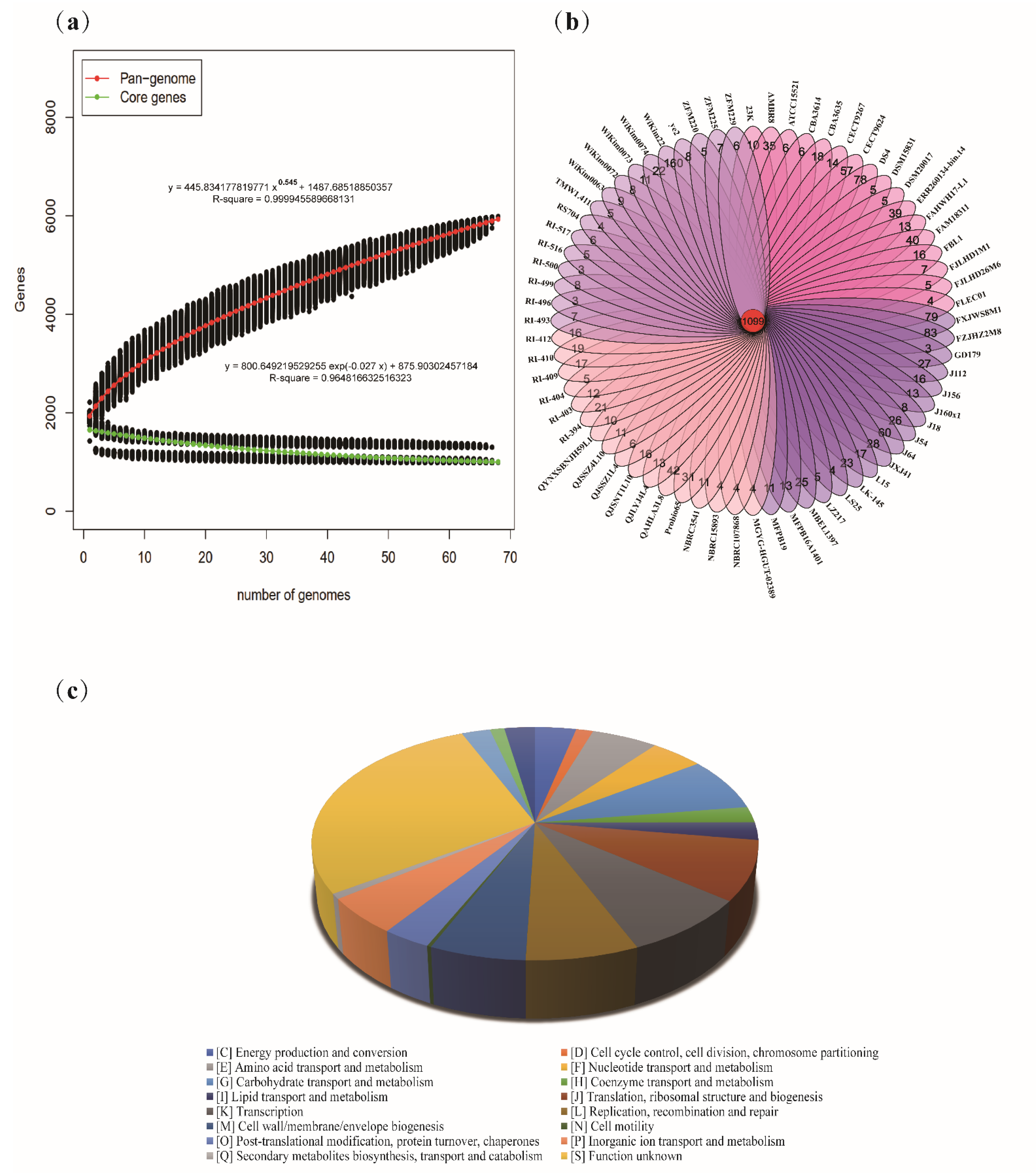
3.1. General Genome Characteristics of Latilactobacillus sakei

In our previous research, 14 L. sakei strains were isolated from different niches, such as human feces, fermented vegetable products, fermented meat products, and kimchi water (Table 1). Combined with the genome information of 54 L. sakei strains published in NCBI GenBank database, the genomes of 68 L. sakei strains were compared and analyzed (Table 1). The genome size of 68 L. sakei strains ranged from 1.54 Mb (L. sakei ERR260134-bin.14) to 2.19 Mb (L. sakei WiKim22) with an average size of 1.98 Mb. The average G+C content was 41.22%, ranging from 40.61% for L. sakei WiKim22 to 42.03% for L. sakei QAHLA3L8. The average predicted coding sequences (CDSs) of each genome numbered 1923, ranging from 1779 for L. sakei 23 K to 2142 for L. sakei FXJWS8M1.

3.2. Pan-Genome and Core Genes of Latilactobacillus sakei

To study the genetic diversity of L. sakei, the pan-genome and core genes were analyzed. The functional relationship between the number of core genes and pan-genes and the number of sequencing strains was plotted (Figure 1a). The results showed that with an increase in the number of L. sakei strains, the number of pan-genes increased continuously, and the number of core genes tended to be stable. When the 68th strain was added, the number of ubiquitin genes was stable at 5983, and the number of core genes reached 993. The pan-genome curve shows an asymptotic trend, which may indicate that L. sakei has an open pan-genome. The specific core genes and homologous core genes of L. sakei strains were analyzed, and Wayne diagram was drawn (Figure 1b). Sixty-eight L. sakei strains had 1099 common core genes, and each strain had 4–160 unique core genes. Functional analysis of the core genes of L. sakei revealed that the core genome includes replication, transcription, translation, nucleotide metabolism, carbohydrate metabolism, amino acid metabolism, lipid metabolism, and other related genes. Among them, genes related to carbohydrate metabolism accounted for approximately 7.53% of core functional genes, 5.68% of core functional genes were related to amino acid metabolism; however, 28.24% of core genome functions are unknown (Figure 1c).

3.3. ANI and Phylogenetic Analyses of Latilactobacillus sakei

Average nucleotide identity (ANI) is a classic method that can analyze unique species or potential subspecies within the same strain. The generally accepted ANI boundary value is 95–96% [55]. When the ANI value is less than this boundary value, the strain may be a potential subspecies. Among the 68 L. sakei strains, except for L. sakei DS4, the ANI values of the other strains were higher than 97% (Figure 2a). The ANI value between strain DS4 and other L. sakei strains was only 92–93%, indicating that L. sakei DS4 may be a potential subspecies. However, this conjecture has not been confirmed by research, and whether L. sakei DS4 is a potential subspecies needs further discussion.
The phylogenetic tree was constructed based on homologous genes of 68 L. sakei strains to explore the phylogenetic relationships (Figure 2b). The phylogenetic tree was divided into two large clades, among which L. sakei DS4 was divided into one, which may be related to its low ANI value. The remaining strains were mainly divided into three evolutionary clades (A–C), and the strains isolated in our laboratory exist in all three evolutionary clades. In clade A, three strains were from feces, and the other six strains were from fermented products. Clade B was isolated from feces, whereas clade C was isolated from fermented meat products.

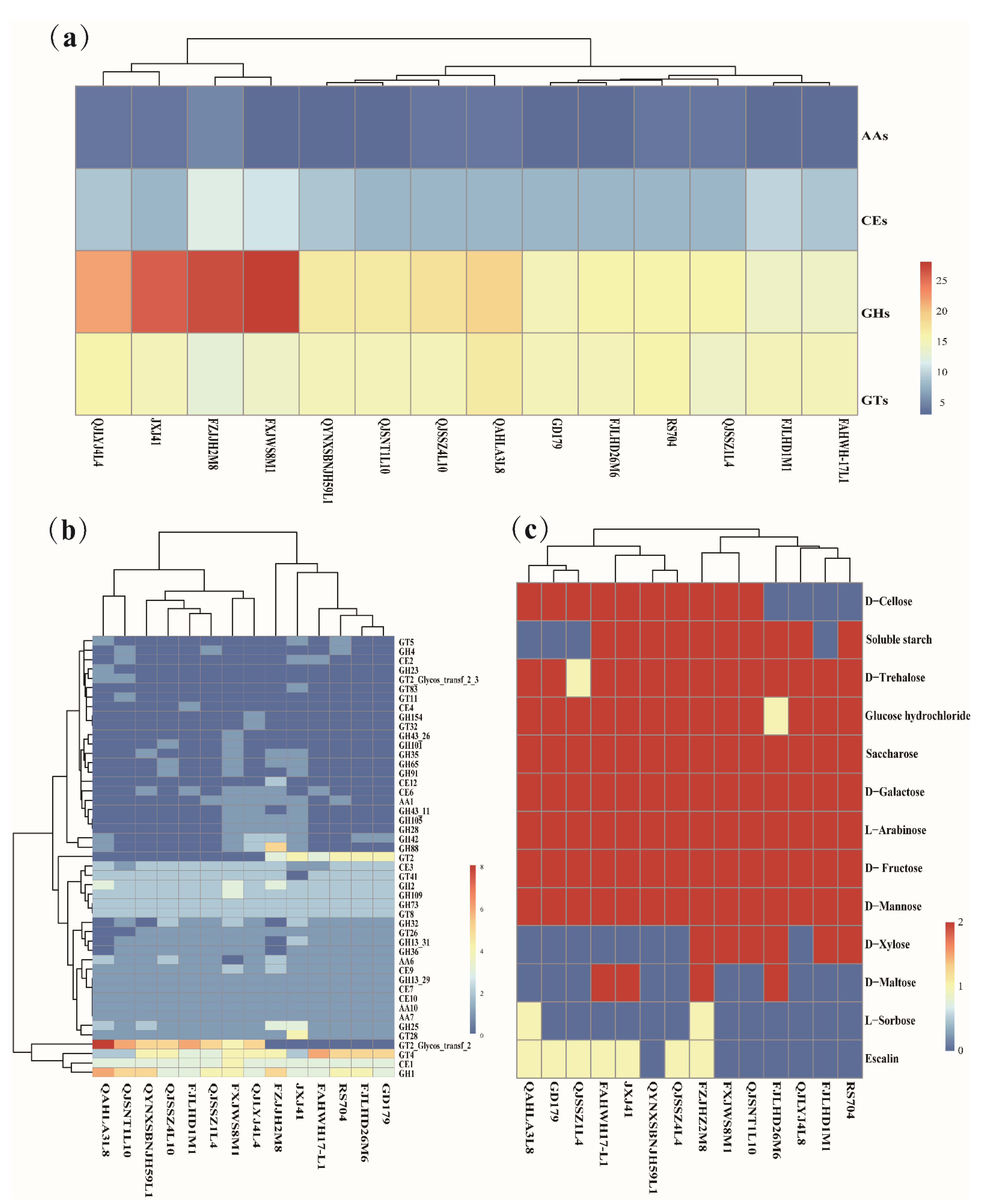
3.4. Genotype/Phenotype Association Analysis for Carbohydrates Utilization in Latilactobacillus sakei

To understand the carbohydrate utilization ability of L. sakei, the sequencing genomes of 14 L. sakei strains isolated in our laboratory were analyzed using the CAZy database. There are were carbohydrate-active enzyme families in 14 L. sakei strains, including auxiliary activity (AA) families, carbohydrate esterase (CE) families, glycoside hydrolase (GH) families, and glycosyltransferases (GTs) families. The GH family was the most abundant enzyme in L. sakei, followed by the GT family, and the number of carbohydrate-active enzyme genes encoding the CE and AA families decreased in turn (Figure 3a). Among these strains, AA10 (lytic cellulose monooxygenase (EC 1.14.99.56)), CE1 (feruloyl esterase (EC 3.1.1.73)), GH1 (β-glucosidase (EC 3.2.1.21)), GH2 (β-galactosidase (EC 3.2.1.23)), GH13_29 (α-amylase (EC 3.2.1.1)), GT4 (sucrose synthase (EC 2.4.1.13)), and another 16 active genes were found in all strains, whereas the distribution of the remaining genes was different in each strain (Figure 3b). For example, CE4 (acetyl xylan esterase (EC 3.1.1.72)) only existed in L. sakei FJLHD1M1, and AA6 (1,4-benzoquinone reductase (EC 1.6.5.6)) was absent in L. sakei FXJWS8M1. GH1, GH2, and GH109 accounted for a high proportion in the GH family, which are related to the catabolism of carbohydrates such as lactose and mannose. In addition, GT2 and GT4 accounted for a higher proportion in the GT family. Based on the clustering results of carbohydrate utilization active enzymes of L. sakei, it was found that there was no evident regularity between the clustering results of various strains and the host and geographical sources, indicating that there is diversity in the carbohydrate utilization ability of L. sakei.
To verify the genotype, the ability of L. sakei to utilize 13 carbohydrates was determined. All strains utilized glucose hydrochloride, D-mannose, D-fructose, L-arabinose, D-galactose, and sucrose, but had strain specificity for the utilization of D-maltose, D-xylose, soluble starch, L-sorbose, D-trehalose, D-cellobiose, and escin (Figure 3c). The metabolism of D-galactose is responsible for β-galactosidase, which belongs to GH2 and GH42 families. It is not difficult to find that all L. sakei strains containing GH2 or GH42 gene can utilize D-galactose. In addition, β-mannosidase in GH2 is responsible for mannose catabolism, promoting the utilization of mannose by 14 L. sakei strains. All strains contained GH13_29 family enzymes, which have α-amylase related to starch hydrolysis and trehalose-6-phosphate hydrolase related to trehalose hydrolysis. However, four strains did not show the ability to utilize soluble starch, and one strain showed the ability to utilize trehalose, which may be due to the lack of expression of GH13_29 in the four strains.

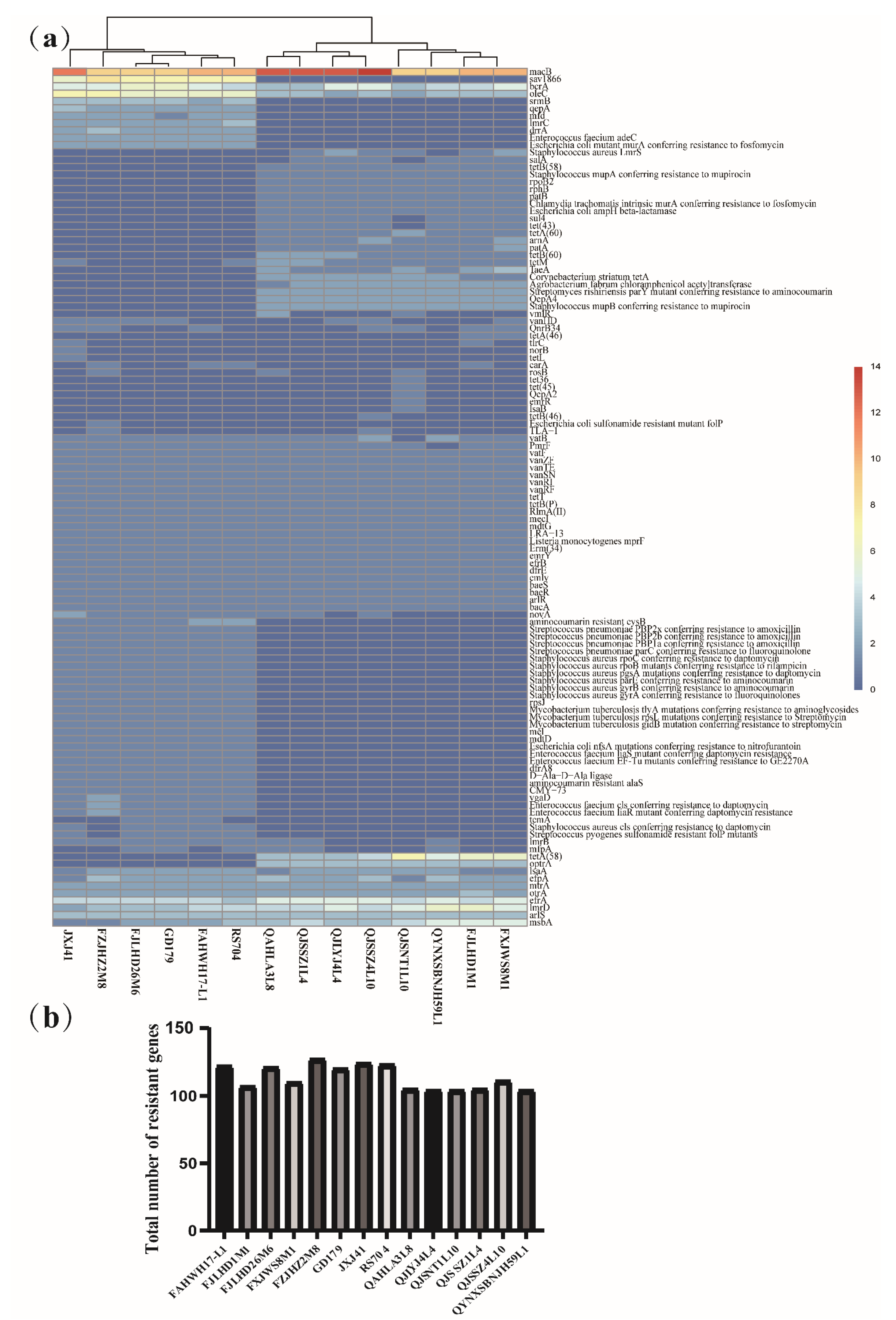
3.5. Genotype/Phenotype Association Analysis for Antibiotic Resistance in Latilactobacillus sakei

The genome of L. sakei was annotated using CRAD database. The annotation results showed that 117 resistance genes were predicted in 14 L. sakei strains (Figure 4a), including macrolide resistance genes (macB), glycopeptide resistance genes (vanRF and vanRI), aminoglycoside resistance genes (baeR and baeS), fluoroquinolone resistance genes (mfd and mfpA), lincoamide resistance genes (lmrB and lmrD), streptogramin resistance genes (vatB and vatF), tetracycline resistance genes (emrY, rpsJ, tetM, tetB(P), and tetT), and rifamycin resistance genes (rphB). By comparing the total number of resistance genes of 14 L. sakei isolates from different sources, it was found that the resistance genes of L. sakei isolated from feces and kimchi water were higher than those isolated from fermented products (Figure 4b). Among them, the number of resistance genes of L. sakei FZJHZ2M8 was the highest (127), whereas that of L. sakei QJLYJ4L4 and L. sakei QYNXSBNJH59L1 was the least (104). In addition, the distribution of some resistance genes appears to be related to the isolated sources. For example, rifamycin resistance gene rphB and tetracycline resistance gene tetB(60) existed in all strains isolated from fermented products and a few strains isolated from feces. However, the resistance genes dfrA8 and β-lactam CMY-73 were mostly found in the strains isolated from feces and kimchi water; however, none of the strains isolated from fermented products contained these genes (Figure 4a).
Furthermore, the genetic traits of 14 L. sakei strains were matched, and the tolerance of L. sakei to 10 antibiotics such as erythromycin, vancomycin, streptomycin, kanamycin, gentamycin, neomycin, tetracycline, rifampicin, ciprofloxacin, and clindamycin was analyzed (Table 2). There were glycopeptide resistance genes and aminoglycoside resistance genes in all strains; therefore, all strains have good tolerance to typical glycopeptide antibiotics such as vancomycin and aminoglycoside antibiotics such as streptomycin, kanamycin, and gentamycin. Macrolide resistance genes were abundant in 14 strains of L. sakei, and the strains had good tolerance to erythromycin, except for L. sakei QJSNT1L10. TetT and tetB(P) genes related to tetracycline resistance existed in all strains; however, six strains were sensitive to tetracycline, which may indicate that tetT and tetB(P) are not the key genes of tetracycline resistance.

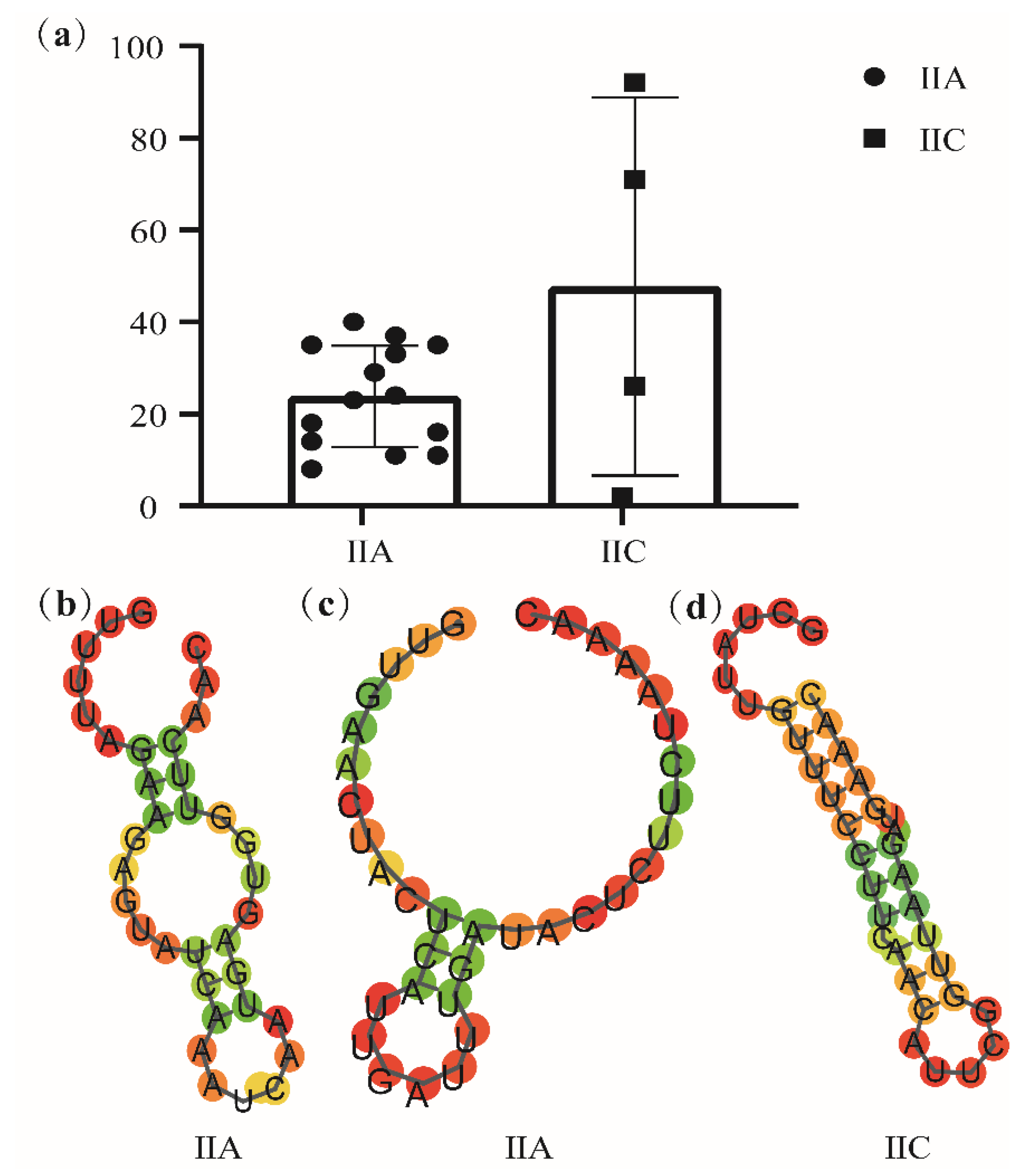
3.6. Prediction of CRISPR–Cas Systems in Latilactobacillus sakei

Fourteen L. sakei strains isolated from our laboratory were analyzed using the CRISPR–Cas system, and thirteen strains were identified as CRISPRs. However, only genomes with evidence levels above 1 were considered in this study due to differences in evidence level. Moreover, CRISPR, an orphan without Cas protein, was ignored because it could not silence foreign DNA. Two strains with complete CRISPR–Cas system were isolated from fermented products (QJSNT1L10 and QAHLA3L8), and one strain was isolated from baby feces (FZJHZ2M8), all of which belonged to class IIA or IIC, including Cas1, Cas2, Cas9, and Csn2 (Table 3). To further explore the distribution characteristics of the CRISPR–Cas system of L. sakei, the CRISPR–Cas system of 54 strains of L. sakei on NCBI was predicted. Among the 54 strains, 13 strains had complete CRISPR–Cas system, and these systems were all IIA or IIC. Most of these 14 strains were isolated from fermented products (Table 3).
By analyzing the number of spacer sequences of CRISPR loci with different subtypes of L. sakei (Figure 5a), it is clear that the number of interval sequences of type IIC loci is quite different, up to 92 and at least 2. Repeat sequences were explored by their secondary structure (Figure 5b–d). According to the repeated sequences, it is predicted that there are two typical secondary structures in subtype IIA (Figure 5b–c) and one typical secondary structure in subtype IIC (Figure 5d). Repeated sequence is a typical stem–loop stable structure, which contains a large loop and a small loop at both ends. The phylogenetic tree was constructed with Cas1, Cas2, and Cas9 protein genes, showing that Cas1, Cas2, and Cas9 protein genes of subtype IIA and IIC have strict single-line inheritance, but there were a few exceptions (Figure 6a–c).

4. Discussion

Latilactobacillus sakei is a potential candidate probiotic that exists in many niches. A study showed that L. sakei can not only be used as a starter [56] and biological protective agent [57] in the food industry, but also has a variety of probiotic functions, such as improving metabolic syndromes such as obesity [58], improving immunity and relieving atopic dermatitis [59], and alleviating inflammatory reactions in colitis mice [60]. The development of genomic tools provides strong support for diversity analysis of strains. However, there are few studies on the genome diversity of L. sakei, and the isolation sources of strains are relatively single. In this study, the genetic diversity and functional diversity of 68 L. sakei strains from different niches were analyzed using 14 L. sakei strains isolated in our laboratory and 54 strains with published genetic information on NCBI.
The average genome size of 68 strains of L. sakei was 1.98 Mb, and the average GC content was 41.22%, which is consistent with the study of Eisenbach et al. [61]. The average GC content is lower than that of Ligilactobacillus ruminis [62], Lacticaseibacillus casei [63], and other lactobacillus strains that belong to a free-living and nomadic lifestyle, although higher than that of Pediococcus pentosaceus [64] and Lactobacillus crispatus [65]. The pan-genome and core genome showed that L. sakei has an open pan-genome. In addition, the function and translation, defense mechanism, and general function prediction of L. sakei were revealed by annotating the core genes.
ANI is a classical index, used to distinguish whether a particular strain belongs to the same species, and commonly takes a 95% threshold as the species boundary [55]. Except for L. sakei DS4, the ANI value of other strains was more than 97%, whereas that of DS4 was only 92%, which reveals that DS4 may be a potential subspecies. Based on the phylogenetic tree of homologous gene sequence similarity, 68 L. sakei strains were divided into two major clades and three main sub-clades. There was no evident correlation between each branch and the isolated source.
To further understand the fermentation ability of L. sakei using carbohydrates, the genome of 14 strains of L. sakei isolated in our laboratory was analyzed and compared using CAZymes technology. GH family enzymes, including GH1, GH2, GH42, and GH73, are the relatively abundant carbohydrate enzymes in L. sakei. These enzymes are involved in the catabolism of various carbon compounds, such as soluble starch, mannose, xylose, glucose, trehalose, maltose, and galactose, consistent with the results of Eisenbach et al. [61]. Aside from the corresponding hydrolases, the phosphotransferase system will also affect the utilization of carbohydrates. Chaillou et al. [20] showed that there are phosphotransferase systems of glucose, mannose, fructose, sucrose, and trehalose in L. sakei, which provides a stronger basis for utilizing these carbohydrates by L. sakei. GH25, as lysozyme [66], participates in peptidoglycan and cell wall catabolism, which can promote cell division and defense. β-N-acetylglucosamine is encoded by GH73, and the β-1,4 glycosidic bond between N-acetylmuramic acid and N-acetylglucosamine of bacterial cell wall peptidoglycan is cleaved by it [67]. These results provide a competitive advantage for the survival of L. sakei. Microbes also produce a special enzyme called carbohydrate esterase (CE) to deacetylate hemicellulose and pectin units of plant polysaccharides [68]. CE is mainly divided into pectin deacetylating CE and hemicellulose deacetylation. L. sakei is rich in the CE1 family, which is an acetyl xylan esterase.
Owing to their intrinsic and nontransmissible characteristics, many LABs have high antibiotic susceptibility [69]. All 14 strains of L sakei isolated in our study have glycopeptide resistance genes; therefore, they have good tolerance to vancomycin, a typical antibiotic of glycopeptide. This is consistent with the results of Georgieva et al. [70], who found that heterofermentative lactic acid bacteria are resistant to vancomycin. In addition, 14 L. sakei strains have glycoside resistance genes, which are resistant to streptomycin, kanamycin, and gentamicin, which are inherent properties of lactobacillus [71]. There were 13 tetracycline resistance genes in the studied L. sakei, however, six strains were sensitive to tetracycline, among which four strains were isolated from fecal samples. The resistance of most bacteria to tetracycline seems to be obtained horizontally, and the tet gene encoding drug resistance is highly mobile because it is located on conjugated transposons [72]. Most tet genes encode tetracycline resistance efflux proteins, a part of the major facilitator (MFS) of transporters. These proteins are bound by membranes and exchange protons with tetracycline cation complexes under concentration gradient [73], which reduces the concentration of tetracycline in cells, thus protecting ribosomes in cells.
CRISPR and Cas combine to form the CRISPR–Cas system, providing adaptive immunity to bacterial invasive components [74]. Sixteen strains of 68 L. sakei strains were identified to have a complete CRISPR–Cas system, including 14 subtypes of IIA and 4 subtypes of IIC. This is consistent with the results of Ilkkan et al. [30] that subtype IIA is the main CRISPR–Cas subtype in L. sakei. In Latilactobacillus curvatus, Loigolactobacillus rennini, Pediococcus damnosus, and Secundilactobacillus paracollinoides, which exist in a similar niche to L. sakei, highly similar structures of type IIA CRISPR/Cas and similar Cas protein sequences were also found [33]. The activity of the CRISPR system is reflected by the number of spacer sequences, and continuous acquisition of spacer sequences has been proven in the active CRISPR–Cas system [75]. It can be inferred from the number of spacer sequences that the IIC subtype of L. sakei is more active and can better resist the insertion of foreign genes. The CRISPR–Cas structure can be used in genetic engineering, and its great potential has been proven, particularly Cas9 nuclease [76]. Cas protein is used as a programmable nuclease that is used for efficient and accurate genome editing in various fields of medicine, research, and biotechnology. The new possibility of Cas9-mediated genome editing is attributed to the identification of new PAMs(g/a) AAA (for type IIA) and (a/g) (c/t) AC (for type IIC CRISPR–Cas system), as well as localized tracrRNAs [32]. Therefore, the application potential of L. sakei Cas nuclease requires further study.

5. Conclusions

In this study, comparative genomics was used to analyze the genomes of 68 Latilactobacillus sakei strains, which provided a basis for analyzing the functional genes of this species. The results showed that niche affected the antibiotic resistance of L. sakei, and the strains from feces and pickle water had more abundant antibiotic resistance genes, and its genetic diversity is also reflected in carbohydrate utilization and some immune/competition related factors (CRISPR). Genome sequencing and genetic analysis in this study helped understand the biotechnology potential of L. sakei and promote its future development as a protective agent/starter and a therapeutic agent for microbial-related diseases.

Author Contributions

Writing—original draft, Y.C.; writing—reviewing and editing, N.L., S.Z., and C.Z.; data curation, formal analysis, methodology, L.Y., Q.Z., N.Q., and H.D.; supervision, Y.X., B.Y., and L.Y.; project administration, F.T.; funding acquisition, J.Z., Q.Z., L.Y., and W.C. All authors have read and agreed to the published version of the manuscript.

Funding

This work was supported by the National Natural Science Foundation of China (32001665), the Natural Science Foundation of Jiangsu Province (BK20180603), the National Natural Science Foundation of China (U1903205,31820103010,31772090), the Key Scientific and Technological Research Projects in the Key Areas of the Xinjiang Production and Construction Corps (2018AB010), the Postdoctoral Science Foundation of China (2018M642166), the General Financial Grant from the Jiangsu Postdoctoral Science Foundation (2018K016A), the Shanghai Engineering Research Center of Dairy Biotechnology(19DZ2281400), the National first-class discipline program of Food Science and Technology (JUFSTR20180102), and the Collaborative Innovation Center of Food Safety and Quality Control in Jiangsu Province.

Institutional Review Board Statement

Not applicable.

Informed Consent Statement

Not applicable.

Data Availability Statement

Not applicable.

Conflicts of Interest

The authors declare no conflict of interest.

References

  1. Katagiri, H.; Kitahara, K.; Fukami, K.; Sugase, M. The Characteristics of the Lactic Acid Bacteria Isolated from Moto, Yeast Mashes for Saké Manufacture. Bull. Agric. Chem. Soc. Jpn. 1934, 10, 153–157. [Google Scholar] [CrossRef]
  2. Moroni, A.V.; Arendt, E.K.; Bello, F.D. Biodiversity of lactic acid bacteria and yeasts in spontaneously-fermented buckwheat and teff sourdoughs. Food Microbiol. 2011, 28, 497–502. [Google Scholar] [CrossRef] [PubMed]
  3. Reale, A.; Di Renzo, T.; Boscaino, F.; Nazzaro, F.; Fratianni, F.; Aponte, M. Lactic Acid Bacteria Biota and Aroma Profile of Italian Traditional Sourdoughs from the Irpinian Area in Italy. Front. Microbiol. 2019, 10, 1621. [Google Scholar] [CrossRef] [PubMed] [Green Version]
  4. Ashaolu, T.J.; Reale, A. A Holistic Review on Euro-Asian Lactic Acid Bacteria Fermented Cereals and Vegetables. Microorganisms 2020, 8, 1176. [Google Scholar] [CrossRef]
  5. Chaillou, S.; Chaulot-Talmon, A.; Caekebeke, H.; Cardinal, M.; Christieans, S.; Denis, C.; Desmonts, M.H.; Dousset, X.; Feurer, C.; Hamon, E.; et al. Origin and ecological selection of core and food-specific bacterial communities associated with meat and seafood spoilage. ISME J. 2015, 9, 1105–1118. [Google Scholar] [CrossRef] [Green Version]
  6. Bello, F.D.; Walter, J.; Hammes, W.; Hertel, C. Increased Complexity of the Species Composition of Lactic Acid Bacteria in Human Feces Revealed by Alternative Incubation Condition. Microb. Ecol. 2003, 45, 455–463. [Google Scholar] [CrossRef]
  7. Carvalho, K.G.; Bambirra, F.H.S.; Nicoli, J.R.; Oliveira, J.S.; Santos, A.M.C.; Bemquerer, M.; Miranda, A.; Franco, B.D.G.M. Characterization of multiple antilisterial peptides produced by sakacin P-producing Lactobacillus sakei subsp. sakei 2a. Arch. Microbiol. 2018, 200, 635–644. [Google Scholar] [CrossRef] [Green Version]
  8. Mathiesen, G.; Huehne, K.; Kroeckel, L.; Axelsson, L.; Eijsink, V.G.H. Characterization of a New Bacteriocin Operon in Sakacin P-Producing Lactobacillus sakei, Showing Strong Translational Coupling between the Bacteriocin and Immunity Genes. Appl. Environ. Microbiol. 2005, 71, 3565–3574. [Google Scholar] [CrossRef] [Green Version]
  9. Winkelstroter, L.; Gomes, B.C.; Thomaz, M.R.; Souza, V.M.; De Martinis, E.C. Lactobacillus sakei 1 and its bacteriocin influence adhesion of Listeria monocytogenes on stainless steel surface. Food Control. 2011, 22, 1404–1407. [Google Scholar] [CrossRef]
  10. Gao, Y.; Jia, S.; Gao, Q.; Tan, Z. A novel bacteriocin with a broad inhibitory spectrum produced by Lactobacillus sake C2, isolated from traditional Chinese fermented cabbage. Food Control. 2010, 21, 76–81. [Google Scholar] [CrossRef]
  11. Lopez-Arvizu, A.; Rocha-Mendoza, D.; Ponce-Alquicira, E.; García-Cano, I. Characterization of antibacterial activity of a N-acetylmuramoyl-l-alanine amidase produced by Latilactobacillus sakei isolated from salami. World J. Microbiol. Biotechnol. 2021, 37, 1–13. [Google Scholar] [CrossRef]
  12. Gao, Y.; Li, D.; Liu, X. Effects of Lactobacillus sakei C2 and sakacin C2 individually or in combination on the growth of Listeria monocytogenes, chemical and odor changes of vacuum-packed sliced cooked ham. Food Control. 2015, 47, 27–31. [Google Scholar] [CrossRef]
  13. Najjari, A.; Boumaiza, M.; Jaballah, S.; Boudabous, A.; Ouzari, H. Application of isolated Lactobacillus sakei and Staphylococcus xylosus strains as a probiotic starter culture during the industrial manufacture of Tunisian dry-fermented sausages. Food Sci. Nutr. 2020, 8, 4172–4184. [Google Scholar] [CrossRef] [PubMed]
  14. Moon, S.H.; Moon, J.S.; Chang, H.C. Rapid manufacture and quality evaluation of long-term fermented kimchi (mukeunji) using Lactobacillus sakei SC1. Food Sci. Biotechnol. 2015, 24, 1797–1804. [Google Scholar] [CrossRef]
  15. Hong, Y.-F.; Kim, H.; Kim, H.R.; Gim, M.G.; Chung, D.K. Different immune regulatory potential of Lactobacillus plantarum and Lactobacillus sakei isolated from Kimchi. J. Microbiol. Biotechnol. 2014, 24, 1629–1635. [Google Scholar] [CrossRef] [PubMed] [Green Version]
  16. Won, S.-M.; Chen, S.; Lee, S.Y.; Lee, K.E.; Park, K.W.; Yoon, J.-H. Lactobacillus sakei ADM14 Induces Anti-Obesity Effects and Changes in Gut Microbiome in High-Fat Diet-Induced Obese Mice. Nutrients 2020, 12, 3703. [Google Scholar] [CrossRef]
  17. Rather, I.A.; Bajpai, V.K.; Ching, L.L.; Majumder, R.; Nam, G.-J.; Indugu, N.; Singh, P.; Kumar, S.; Hajrah, N.H.; Sabir, J.S.; et al. Effect of a bioactive product SEL001 from Lactobacillus sakei probio65 on gut microbiota and its anti-colitis effects in a TNBS-induced colitis mouse model. Saudi J. Biol. Sci. 2020, 27, 261–270. [Google Scholar] [CrossRef]
  18. Rather, I.A.; Bajpai, V.K.; Huh, Y.S.; Han, Y.-K.; Bhat, E.A.; Lim, J.; Paek, W.K.; Park, Y.-H. Probiotic Lactobacillus sakei proBio-65 Extract Ameliorates the Severity of Imiquimod Induced Psoriasis-Like Skin Inflammation in a Mouse Model. Front. Microbiol. 2018, 9, 1021. [Google Scholar] [CrossRef] [PubMed]
  19. Jung, J.-Y.; Shin, J.-S.; Lee, S.-G.; Rhee, Y.K.; Cho, C.-W.; Hong, H.-D.; Lee, K.-T. Lactobacillus sakei K040706 evokes immunostimulatory effects on macrophages through TLR 2-mediated activation. Int. Immunopharmacol. 2015, 28, 88–96. [Google Scholar] [CrossRef] [PubMed]
  20. Chaillou, S.; Champomier-Vergès, M.-C.; Cornet, M.; Coq, A.-M.C.-L.; Dudez, A.-M.; Martin, V.; Beaufils, S.; Darbon-Rongère, E.; Bossy, R.; Loux, V.; et al. The complete genome sequence of the meat-borne lactic acid bacterium Lactobacillus sakei 23K. Nat. Biotechnol. 2005, 23, 1527–1533. [Google Scholar] [CrossRef] [Green Version]
  21. Chaillou, S.; Lucquin, I.; Najjari, A.; Zagorec, M.; Champomier-Vergès, M.-C. Population Genetics of Lactobacillus sakei Reveals Three Lineages with Distinct Evolutionary Histories. PLoS ONE 2013, 8, e73253. [Google Scholar] [CrossRef]
  22. Nyquist, O.L.; McLeod, A.; Brede, D.A.; Snipen, L.; Aakra, Å.; Nes, I.F. Comparative genomics of Lactobacillus sakei with emphasis on strains from meat. Mol. Genet. Genom. 2011, 285, 297–311. [Google Scholar] [CrossRef]
  23. Donnell, M.O.; Harris, H.; Jeffery, I.; Claesson, M.; Younge, B.; Toole, P.O.; Ross, R. The core faecal bacterial microbiome of Irish Thoroughbred racehorses. Lett. Appl. Microbiol. 2013, 57, 492–501. [Google Scholar] [CrossRef]
  24. Luo, R.; Liu, B.; Xie, Y.; Li, Z.; Huang, W.; Yuan, J.; He, G.; Chen, Y.; Pan, Q.; Liu, Y.; et al. SOAPdenovo2: An empirically improved memory-efficient short-read de novo assembler. GigaScience 2012, 1, 18. [Google Scholar] [CrossRef]
  25. Loux, V.; Coeuret, G.; Zagorec, M.; Vergès, M.-C.C.; Chaillou, S. Complete and Draft Genome Sequences of Nine Lactobacillus sakei Strains Selected from the Three Known Phylogenetic Lineages and Their Main Clonal Complexes. Genome Announc. 2018, 6, e00082-18. [Google Scholar] [CrossRef] [Green Version]
  26. Kim, K.H.; Chun, B.H.; Baek, J.H.; Roh, S.W.; Lee, S.H.; Jeon, C.O. Genomic and metabolic features of Lactobacillus sakei as revealed by its pan-genome and the metatranscriptome of kimchi fermentation. Food Microbiol. 2020, 86, 103341. [Google Scholar] [CrossRef] [PubMed]
  27. Inglin, R.C.; Meile, L.; Stevens, M.J.A. Draft Genome Sequences of 43 Lactobacillus Strains from the Species L. curvatus, L. fermentum, L. paracasei, L. plantarum, L. rhamnosus, and L. sakei, Isolated from Food Product. Genome Announc. 2017, 5, e00632-17. [Google Scholar] [CrossRef] [Green Version]
  28. Jans, C.; Lagler, S.; Lacroix, C.; Meile, L.; Stevens, M.J.A. Complete Genome Sequences of Lactobacillus curvatus KG6, L. curvatus MRS6, and Lactobacillus sakei FAM18311, Isolated from Fermented Meat Products. Genome Announc. 2017, 5, e00915-17. [Google Scholar] [CrossRef] [PubMed] [Green Version]
  29. Lim, H.I.; Lee, J.; Jang, J.Y.; Park, H.W.; Choi, H.J.; Kim, T.W.; Kang, M.R.; Lee, J.H. Draft Genome Sequence of Lactobacillus sakei Strain wikim 22, Isolated from Kimchi in Chungcheong Province, South Korea. Genome Announc. 2014, 2, e01184-15. [Google Scholar] [CrossRef] [PubMed] [Green Version]
  30. Ilıkkan, Ö.K. CRISPR/Cas systems and anti-repeat sequences of Lactobacillus curvatus, Lactobacillus graminis, Lactobacillus fuchuensis, and Lactobacillus sakei genomes. Korean J. Microbiol. 2021, 57, 12–22. [Google Scholar] [CrossRef]
  31. Kwun, S.-Y.; Yoon, J.-A.; Park, E.-H.; Kim, M.-D. Complete genome sequence data of Lactobacillus sakei MBEL1397 isolated from kimchi. Data Brief 2020, 31, 105740. [Google Scholar] [CrossRef] [PubMed]
  32. Kim, J.-H.; Kim, E.; Kim, C.-G.; Choo, D.-W.; Kim, H.-Y. Draft Genome Sequence of Lactobacillus sakei Strain FBL1, a Probiotic Bacterium Isolated from Mukeunji, a Long-Fermented Kimchi, in South Korea. Genome Announc. 2016, 4, e00365-16. [Google Scholar] [CrossRef] [Green Version]
  33. Schuster, J.A.; Vogel, R.F.; Ehrmann, M.A. Characterization and distribution of CRISPR–Cas systems in Lactobacillus sakei. Arch. Microbiol. 2019, 201, 337–347. [Google Scholar] [CrossRef] [PubMed]
  34. McLeod, A.; Brede, D.A.; Rud, I.; Axelsson, L. Genome Sequence of Lactobacillus sakei subsp. sakei LS25, a Commercial Starter Culture Strain for Fermented Sausage. Genome Announc. 2013, 1, 1. [Google Scholar] [CrossRef] [Green Version]
  35. Kato, S.; Oikawa, T. Genome Sequence of Lactobacillus sakei LK-145 Isolated from a Japanese Sake Cellar as a High Producer of d-Amino Acids. Genome Announc. 2017, 5, e00656-17. [Google Scholar] [CrossRef] [PubMed] [Green Version]
  36. Mizuno, K.; Mizuno, M.; Yamauchi, M.; Takemura, A.J.; Romero, V.M.; Morikawa, K. Adjacent-possible ecological niche: Growth of Lactobacillus species co-cultured with Escherichia coli in a synthetic minimal medium. Sci. Rep. 2017, 7, 12880. [Google Scholar] [CrossRef] [Green Version]
  37. Fujii, T.; Yamauchi, T.; Ishiyama, M.; Gogami, Y.; Oikawa, T.; Hata, Y. Crystallographic studies of aspartate racemase from Lactobacillus sakei NBRC 15893. Acta Crystallogr. Sect. F Struct. Biol. Commun. 2015, 71, 1012–1016. [Google Scholar] [CrossRef] [Green Version]
  38. De Boeck, I.; Broek, M.F.V.D.; Allonsius, C.N.; Spacova, I.; Wittouck, S.; Martens, K.; Wuyts, S.; Cauwenberghs, E.; Jokicevic, K.; Vandenheuvel, D.; et al. Lactobacilli Have a Niche in the Human Nose. Cell Rep. 2020, 31, 107674. [Google Scholar] [CrossRef] [PubMed]
  39. Delcher, A.L.; Bratke, K.A.; Powers, E.C.; Salzberg, S. Identifying bacterial genes and endosymbiont DNA with Glimmer. Bioinformatics 2007, 23, 673–679. [Google Scholar] [CrossRef]
  40. Bairoch, A.; Apweiler, R. The SWISS-PROT protein sequence data bank and its supplement TrEMBL in 1999. Nucleic Acids Res. 1999, 27, 49–54. [Google Scholar] [CrossRef]
  41. Pruitt, K.D.; Tatusova, T.; Maglott, D.R. NCBI reference sequences (RefSeq): A curated non-redundant sequence database of genomes, transcripts and proteins. Nucleic Acids Res. 2007, 35, D61–D65. [Google Scholar] [CrossRef] [Green Version]
  42. Zhao, Y.; Wu, J.; Yang, J.; Sun, S.; Xiao, J.; Yu, J. PGAP: Pan-genomes analysis pipeline. Bioinformatics 2012, 28, 416–418. [Google Scholar] [CrossRef] [Green Version]
  43. Li, L.; Stoeckert, C.J.; Roos, D.S. OrthoMCL: Identification of Ortholog Groups for Eukaryotic Genomes. Genome Res. 2003, 13, 2178–2189. [Google Scholar] [CrossRef] [PubMed] [Green Version]
  44. Valeriano, V.D.V.; Oh, J.K.; Bagon, B.B.; Kim, H.; Kang, D.-K. Comparative genomic analysis of Lactobacillus mucosae LM1 identifies potential niche-specific genes and pathways for gastrointestinal adaptation. Genomics 2019, 111, 24–33. [Google Scholar] [CrossRef] [PubMed] [Green Version]
  45. Richter, M.; Rosselló-Móra, R. Shifting the genomic gold standard for the prokaryotic species definition. Proc. Natl. Acad. Sci. USA 2009, 106, 19126–19131. [Google Scholar] [CrossRef] [Green Version]
  46. Kelleher, P.; Bottacini, F.; Mahony, J.; Kilcawley, K.N.; Van Sinderen, U. Comparative and functional genomics of the Lactococcus lactis taxon; insights into evolution and niche adaptation. BMC Genom. 2017, 18, 267. [Google Scholar] [CrossRef] [PubMed] [Green Version]
  47. Mailund, T.; Brodal, G.S.; Fagerberg, R.; Pedersen, C.N.S.; Phillips, D. Recrafting the neighbor-joining method. BMC Bioinform. 2006, 7, 29. [Google Scholar] [CrossRef] [Green Version]
  48. Subramanian, B.; Gao, S.; Lercher, M.J.; Hu, S.; Chen, W.-H. Evolview v3: A webserver for visualization, annotation, and management of phylogenetic trees. Nucleic Acids Res. 2019, 47, W270–W275. [Google Scholar] [CrossRef]
  49. Lombard, V.; Ramulu, H.G.; Drula, E.; Coutinho, P.M.; Henrissat, B. The carbohydrate-active enzymes database (CAZy) in 2013. Nucleic Acids Res. 2014, 42, D490–D495. [Google Scholar] [CrossRef] [Green Version]
  50. Deng, W.; Wang, Y.; Liu, Z.; Cheng, H.; Xue, Y. HemI: A Toolkit for Illustrating Heatmaps. PLoS ONE 2014, 9, e111988. [Google Scholar] [CrossRef]
  51. Jia, B.; Raphenya, A.R.; Alcock, B.; Waglechner, N.; Guo, P.; Tsang, K.K.; Lago, B.A.; Dave, B.M.; Pereira, S.; Sharma, A.N.; et al. CARD 2017: Expansion and model-centric curation of the comprehensive antibiotic resistance database. Nucleic Acids Res. 2017, 45, D566–D573. [Google Scholar] [CrossRef]
  52. Geneva, I.; Brussels, I.D.F. Milk and Milk Products. Determination of the Minimal Inhibitory Concentration (MIC) of Antibiotics Applicable to Bifidobacteria and Non-Enterococcal Lactic Acid Bacteria (LAB). Available online: https://www.iso.org/obp/ui/#iso:std:iso:10932:ed-1:v1:en (accessed on 24 May 2021).
  53. Grissa, I.; Vergnaud, G.; Pourcel, C. The CRISPRdb database and tools to display CRISPRs and to generate dictionaries of spacers and repeats. BMC Bioinform. 2007, 8, 1–10. [Google Scholar] [CrossRef] [Green Version]
  54. Lorenz, R.; Bernhart, S.H.F.; Zu Siederdissen, C.H.; Tafer, H.; Flamm, C.; Stadler, P.F.; Hofacker, I.L. ViennaRNA Package 2.0. Algorithms Mol. Biol. 2011, 6, 26. [Google Scholar] [CrossRef]
  55. Chun, J.; Oren, A.; Ventosa, A.; Christensen, H.; Arahal, D.R.; da Costa, M.; Rooney, A.; Yi, H.; Xu, X.-W.; De Meyer, S.; et al. Proposed minimal standards for the use of genome data for the taxonomy of prokaryotes. Int. J. Syst. Evol. Microbiol. 2018, 68, 461–466. [Google Scholar] [CrossRef]
  56. Gao, Y.; Li, D.; Liu, X. Bacteriocin-producing Lactobacillus sakei C2 as starter culture in fermented sausages. Food Control. 2014, 35, 1–6. [Google Scholar] [CrossRef]
  57. Malheiros, P.; Cuccovia, I.M.; Franco, B.D. Inhibition of Listeria monocytogenes in vitro and in goat milk by liposomal nanovesicles containing bacteriocins produced by Lactobacillus sakei subsp. sakei 2a. Food Control. 2016, 63, 158–164. [Google Scholar] [CrossRef]
  58. Ji, Y.; Park, S.; Chung, Y.; Kim, B.; Park, H.; Huang, E.; Jeong, D.; Jung, H.-Y.; Kim, B.; Hyun, C.-K.; et al. Amelioration of obesity-related biomarkers by Lactobacillus sakei CJLS03 in a high-fat diet-induced obese murine model. Sci. Rep. 2019, 9, 1–11. [Google Scholar] [CrossRef] [PubMed] [Green Version]
  59. Kim, J.-Y.; Park, B.-K.; Park, H.-J.; Park, Y.-H.; Kim, B.-O.; Pyo, S. Atopic dermatitis-mitigating effects of new Lactobacillus strain, Lactobacillus sakei probio 65 isolated from Kimchi. J. Appl. Microbiol. 2013, 115, 517–526. [Google Scholar] [CrossRef]
  60. Seo, S.; Shin, J.-S.; Lee, W.-S.; Rhee, Y.K.; Cho, C.-W.; Hong, H.-D.; Lee, K.-T. Anti-colitis effect of Lactobacillus sakei K040706 via suppression of inflammatory responses in the dextran sulfate sodium-induced colitis mice model. J. Funct. Foods 2017, 29, 256–268. [Google Scholar] [CrossRef]
  61. Eisenbach, L.; Geissler, A.J.; Ehrmann, M.A.; Vogel, R.F. Comparative genomics of Lactobacillus sakei supports the development of starter strain combinations. Microbiol. Res. 2019, 221, 1–9. [Google Scholar] [CrossRef] [PubMed]
  62. Wang, S.; Yang, B.; Ross, R.P.; Stanton, C.; Zhao, J.; Zhang, H.; Chen, W. Comparative Genomics Analysis of Lactobacillus ruminis from Different Niches. Genes 2020, 11, 70. [Google Scholar] [CrossRef] [PubMed] [Green Version]
  63. Broadbent, J.R.; Neeno-Eckwall, E.C.; Stahl, B.; Tandee, K.; Cai, H.; Morovic, W.; Horvath, P.; Heidenreich, J.; Perna, N.T.; Barrangou, R.; et al. Analysis of the Lactobacillus casei supragenome and its influence in species evolution and lifestyle adaptation. BMC Genom. 2012, 13, 533. [Google Scholar] [CrossRef] [Green Version]
  64. Jiang, J.; Yang, B.; Ross, R.; Stanton, C.; Zhao, J.; Zhang, H.; Chen, W. Comparative Genomics of Pediococcus pentosaceus Isolated from Different Niches Reveals Genetic Diversity in Carbohydrate Metabolism and Immune System. Front. Microbiol. 2020, 11, 253. [Google Scholar] [CrossRef] [PubMed]
  65. Zhang, Q.; Zhang, L.; Ross, P.; Zhao, J.; Zhang, H.; Chen, W. Comparative Genomics of Lactobacillus crispatus from the Gut and Vagina Reveals Genetic Diversity and Lifestyle Adaptation. Genes 2020, 11, 360. [Google Scholar] [CrossRef] [Green Version]
  66. O’Riordan, N.; O’Callaghan, J.; Buttò, L.; Kilcoyne, M.; Joshi, L.; Hickey, R. Bovine glycomacropeptide promotes the growth of Bifidobacterium longum ssp. infantis and modulates its gene expression. J. Dairy Sci. 2018, 101, 6730–6741. [Google Scholar] [CrossRef]
  67. Patryk, Z.; iley-Elkin, B.B.; Miriam, D.; Mark, B.L.; Permyakov, E. Structural and Biochemical Insights into the Peptidoglycan Hydrolase Domain of FlgJ from Salmonella typhimurium. PLoS ONE 2016, 11, e0149204. [Google Scholar] [CrossRef] [Green Version]
  68. Kameshwar, A.K.S.; Qin, W. Understanding the structural and functional properties of carbohydrate esterases with a special focus on hemicellulose deacetylating acetyl xylan esterases. Mycology 2018, 9, 273–295. [Google Scholar] [CrossRef] [Green Version]
  69. Salminen, S. Demonstration of safety of probiotics–A review. Int. J. Food Microbiol. 1998, 44, 93–106. [Google Scholar] [CrossRef]
  70. Georgieva, R.; Yocheva, L.; Tserovska, L.; Zhelezova, G.; Stefanova, N.; Atanasova, A.; Danguleva, A.; Ivanova, G.; Karapetkov, N.; Rumyan, N.; et al. Antimicrobial activity and antibiotic susceptibility ofLactobacillusandBifidobacteriumspp. intended for use as starter and probiotic cultures. Biotechnol. Biotechnol. Equip. 2014, 29, 84–91. [Google Scholar] [CrossRef]
  71. Botta, C.; Langerholc, T.; Cencic, A.; Cocolin, L. In Vitro Selection and Characterization of New Probiotic Candidates from Table Olive Microbiota. PLoS ONE 2014, 9, e94457. [Google Scholar] [CrossRef]
  72. Roberts, M.C.; Schwarz, S. Tetracycline and Phenicol Resistance Genes and Mechanisms: Importance for Agriculture, the Environment, and Humans. J. Environ. Qual. 2016, 45, 576–592. [Google Scholar] [CrossRef] [PubMed]
  73. Yamaguchi, A.; Ono, N.; Akasaka, T.; Noumi, T.; Sawai, T.J.J.o.B.C. Metal-tetracycline/H+ antiporter of Escherichia coli encoded by a transposon, Tn10. The role of the conserved dipeptide, Ser65-Asp66, in tetracycline transport. J. Biol. Chem. 1990, 265, 15525–15530. [Google Scholar] [CrossRef]
  74. Barrangou, R.; Fremaux, C.; Deveau, H.; Richards, M.; Boyaval, P.; Moineau, S.; Romero, D.A.; Horvath, P. CRISPR Provides Acquired Resistance Against Viruses in Prokaryotes. Science 2007, 315, 1709–1712. [Google Scholar] [CrossRef] [PubMed]
  75. Tyson, G.; Banfield, J. Rapidly evolving CRISPRs implicated in acquired resistance of microorganisms to viruses. Environ. Microbiol. 2007, 10, 200–207. [Google Scholar] [CrossRef] [PubMed]
  76. Mougiakos, I.; Bosma, E.F.; de Vos, W.M.; van Kranenburg, R.; van der Oost, J. Next Generation Prokaryotic Engineering: The CRISPR-Cas Toolkit. Trends Biotechnol. 2016, 34, 575–587. [Google Scholar] [CrossRef] [PubMed]
Figure 1. Pan and core genes of Latilactobacillus sakei. (a) Pan-genome and core genome, R stands for correlation coefficient; (b) Venn diagram displaying the unique and core genes; (c) Functional assignment of the core genome based on the COG database.
Foods 10 01717 g001
Figure 2. The Average Nucleotide Identity (ANI) and phylogenetic analysis of Latilactobacillus sakei. (a) Heatmap showing the ANI value among 68 L. sakei strains; (b) Phylogenetic tree based on orthologous genes. The salmon areas represent strains isolated in our laboratory. Brown circles represent strains with complete CRISPR–Cas system. The peripheral blue band represents strains from feces, the red band represents strains from fermented meat products, the green band represents strains from fermented vegetables, the pink band represents strains from fermented sake, the yellow band represents strains from kimchi water, the orange band represents strains from milk, and the source of gray band separation is unknown.
Foods 10 01717 g002
Figure 3. Genotype–phenotype analysis of carbohydrate utilization of Latilactobacillus sakei. (a) Comparative heat map of the number of genes in four families of carbohydrate active enzymes of different strains of L. sakei; (b) Thermographic analysis of carbohydrate enzyme activity-related genes of L. sakei; (c) Utilization ability of 13 carbohydrates by L. sakei.
Foods 10 01717 g003
Figure 4. Genotype–phenotype analysis of antibiotic resistance of Latilactobacillus sakei. (a) Clustering heat map analysis of antibiotic resistance genes in L. sakei; (b) Total number of resistance genes of 14 L. sakei strains.
Foods 10 01717 g004
Figure 5. Prediction of CRISPR–Cas systems in Latilactobacillus sakei. (a) Number of spacer sequences of CRISPR loci in different subtypes of Latilactobacillus sakei; (bd) Predicted RNA secondary structures of CRISPR DR in Latilactobacillus sakei.
Foods 10 01717 g005
Figure 6. CRISPR–Cas phylogenetic analyses for Latilactobacillus. sakei. (a) Phylogenetic tree based on the Cas1 protein; (b) Phylogenetic tree based on the Cas2 protein; (c) Phylogenetic tree based on the Cas9 protein.
Foods 10 01717 g006
Table 1. The information of 68 Latilactobacillus sakei strains.
StrainSourceGenome Size (Mb)GC (%)CDS No.Reference
FAHWH17-L1Human feces1.9041.821860This work
FJLHD1M1Human feces1.9841.811969This work
FJLHD26M6Human feces1.9841.791950This work
FXJWS8M1Human feces2.1141.002142This work
FZJHZ2M8Human feces2.1841.612085This work
GD179Human feces1.8841.831856This work
FLEC01Human feces1.9641.241919[25]
QYNXSBNJH59L1Sour cowhide1.9441.771928This work
QJSSZ1L4Air-dried sausage1.9041.921876This work
QJSNT1L10Air-dried sausage1.8541.861829This work
QAHLA3L8Air-dried sausage1.9042.031858This work
DSM15831Meat1.9941.001924[26]
RI-496Meat2.0240.901934[26]
RI-499Meat1.9141.001838[26]
RI-500Meat2.0340.901992[26]
RI-394Meat1.9441.001963[27]
23KFrench sausage1.8841.301779[20]
J64Dry sausage2.0541.062102[25]
J156Dry sausage1.8841.071817[25]
J112Dry sausage1.9841.041913[25]
J54Dry sausage2.0141.311982[25]
J18Dry sausage1.8241.001826[25]
MFPB16A1401Beef carpaccio2.0441.091960[25]
MFPB19Beef carpaccio2.0641.051977[25]
J160 × 1Horse meat1.9041.071857[25]
FAM18311Fermented meat products1.9541.31896[28]
JXJ41Kimchi water2.0441.802010This work
RS704Kimchi water1.9641.861945This work
DS4Kimchi2.1041.411970[26]
Probio65Kimchi2.0841.181984[26]
WiKim0063Kimchi2.0841.171955[26]
WiKim0072Kimchi2.0341.171936[26]
WiKim0073Kimchi2.0441.161913[26]
WiKim0074Kimchi2.0441.031916[26]
WiKim22Kimchi2.1940.611869[29]
CBA3614kimchi2.0241.131910[30]
MBEL1397Kimchi1.9941.041847[31]
FBL1Mukeunji2.0341.201936[32]
QJSSZ4L10Pickled green vegetable heart2.0541.702075This work
QJLYJ4L4Pickled radish1.9741.761928This work
CECT9267Fermented potatos2.0340.901972[26]
CECT9624Fermented potatos2.0640.801986[33]
ZFM229Fermented vegetables2.0241.221907[30]
LZ217Fermented vegetables2.0241.221908[30]
RI-516Cacao bean1.9141.001847[26]
RI-517Cacao bean1.9541.001878[26]
LS25Commercial product Bitec LS-252.0240.901972[34]
DSM20017Sake1.9141.11820[26]
TMW1.411Starter culture1.9441.061859[33]
LK-145Japanese sake cellar1.9941.151901[35]
NBRC3541Raw moto-shu1.9441.101855[36]
NBRC15893Kimoto1.9041.101822[37]
ATCC15521Moto, starter of sake1.9441.101835Unknown
ZFM220Raw cow milk2.0241.221909[30]
ZFM225Raw cow milk2.0241.221909[30]
AMBR8Nasopharyngeal samples2.0041.001923[38]
RI-493Unknown1.9740.901890[26]
CBA3635Unknown2.0641.102002[30]
L15Unknown1.9841.001915[30]
ye2Unknown1.9941.101907[30]
RI-403Unknown2.0041.002032[27]
RI-404Unknown1.9540.901977[27]
RI-409Unknown1.9941.002022[27]
RI-410Unknown1.9341.101949[27]
RI-412Unknown1.9241.101934[27]
MGYG-HGUT-02389Unknown1.9641.201813Unknown
ERR260134-bin.14Unknown1.5241.20-Unknown
NBRC107868Unknown1.9741.001920Unknown
“-”: Information missing.
Table 2. MIC of different antibiotics for Latilactobacillus sakei.
StrainConcentration (μg/mL)
RDSKCNTEDANECIPVAC
FJLHD1M11256256102420.525611281288
GD17-91256128102420.52561641288
FAHWH17-L10.55122561024162256412812816
JXJ4-115121281024640.5644128128128
QYNXSBNJH59L10.25512256102440.5256212812832
QJSSZ4L100.5512512102480.25128412812832
QJLYJ4L40.525612825600.25256812812816
QJSSZ1L415122561024320.5256112812816
QAHLA3L812562561024320.525626412832
FJLHD26M60.2525625651240.1253221281288
FXJWS8M18256256102420.251281641288
FZJHZ2M81256512256644256212812816
RS7042256128512640.52562641288
QJSNT1L100.1251281281024640.06251280.532128128
RD: rifampicin; S: streptomycin; K: kanamycin; CN: gentamicin; TE: tetracycline; DA: clindamycin; N: neomycin; E: erythromycin; CIP: ciprofloxacin; VA: vancomycin.; C: chloramphenicol.
Table 3. CRISPR–Cas systems in Latilactobacillus sakei.
StrainsRepeat Sequence (5’-3’)CAS_
Subtype
Repeat LengthNo. SpacerCas1Cas2Cas9Csn2
FZJHZ2M8GTTTTAGAAGAGTATCAAATCAATGAGTAGTTCAACIIA3616YYYY
ATTTCATCTTAACCGAATGTTGAAGGAAACAATAGCIIC3671YNYN
QJSNT1L10GTTTTAGAAGAGTATCAAATCAATGAGTAGTTCAACIIA3618NNNY
QAHLA3L8GTTGAACTACTCATTGATTTGATACTCTTCTAAAACIIA3633YNYY
DS4GCTATTGTTTCCTTCAACATTCGGTTAAGATGAAATIIC3626YYYN
J18GCTATTGTTTCCTTCAACATTCGGTTAAGATGAAACIIC3692YYYN
CECT9624GTTTTAGAAGAGTATCAAATCAATGAGTAGTTCAACIIA368YYYY
ATTTCATCTTAACCGAATGTTGAAGGAAACAIIC312YYYN
J54GTTGAACTACTCATTGATTTGATACTCTTCTAAAACIIA3629YYYY
J112GTTTTAGAAGAGTATCAAATCAATGAGTAGTTCAACIIA3614YYYN
FLEC01GTTGAACTACTCATTGATTTGATACTCTTCTAAAACIIA3635YYYY
J160 × 1GTTGAACTACTCATTGATTTGATACTCTTCTAAAACIIA3624YYYY
MGYG-HGUT-02389GTTGAACTACTCATTGATTTGATACTCTTCTAAAACIIA3635YYYY
DSM15831AGTTGAACCACTCATTGATTTGATACTCTTCTAAAACIIA3711NNNY
RI-403GTTTTAGAAGAGTATCAAATCAATGAGTGGTTCAACIIA3640NNNY
RI-404GTTTTAGAAGAGTATCAAATCAATGAGTGGTTCAACIIA3637YYYY
AMBR8GTTGAACCACTCATTGATTTGATACTCTTCTAAAACIIA3623YYYY
NBRC107868GTTTTAGAAGAGTATCAAATCAATGAGTGGTTCAACTIIA3711NNNY
Y: present; N: none.
Publisher’s Note: MDPI stays neutral with regard to jurisdictional claims in published maps and institutional affiliations.

