Article

DNA-Based Method for Traceability and Authentication of Apis cerana and A. dorsata Honey (Hymenoptera: Apidae), Using the NADH dehydrogenase 2 Gene

by
Saeed Mohamadzade Namin
1,2,†,
Fatema Yeasmin
3,†,
Hyong Woo Choi
3 and
Chuleui Jung
1,3,*
1
Agricultural Science and Technology Institute, Andong National University, Andong 36729, Korea
2
Department of Plant Protection, Faculty of Agriculture, Varamin-Pishva Branch, Islamic Azad University, Varamin 3381774895, Iran
3
Department of Plant Medicals, Andong National University, Andong 36729, Korea
*
Author to whom correspondence should be addressed.
These authors contributed equally to this work.
Submission received: 28 February 2022 / Revised: 21 March 2022 / Accepted: 22 March 2022 / Published: 23 March 2022

Abstract

:
Honey is a widely used natural product and the price of honey from Apis cerana (ACH) and A. dorsata (ADH) is several times more expensive than the one from A. mellifera (AMH), thus there are increasing fraud issues reported in the market by mislabeling or mixing honeys with different entomological origins. In this study, three species-specific primers, targeting the NADH dehydrogenase 2 (ND2) region of honeybee mitochondrial DNA, were designed and tested to distinguish the entomological origin of ACH, ADH, and AMH. Molecular analysis showed that each primer set can specifically detect the ND2 region from the targeted honeybee DNA, but not from the others. The amplicon size for A. cerana, A. dorsata and A. mellifera were 224, 302, and 377 bp, respectively. Importantly, each primer set also specifically produced amplicons with expected size from the DNA prepared from honey samples with different entomological origins. The PCR adulteration test allowed detection of 1% of AMH in the mixture with either ACH or ADH. Furthermore, real-time PCR and melting curve analysis indicated the possible discrimination of origin of honey samples. Therefore, we provide the newly developed PCR-based method that can be used to determine the entomological origin of the three kinds of honey.

Graphical Abstract

1. Introduction

Honey is a sweet natural product produced by honey bees using the nectar, secretions of living parts, or honeydew of plants [1,2]. Due to the broader geographical distribution, Apis mellifera honey (AMH), A. cerana honey (ACH), and A. dorsata honey (ADH) are the three dominant types of honey in the Asian market. Giant honeybee (Apis dorsata F.) is distributed throughout South and Southeast Asia and China [3]. The colonies of A. dorsata are generally found in rainforests or on the cliffs, but they also can be occasionally found in building ledges of urban areas. Even though A. dorsata is not domesticated and cannot be maintained for honey harvest or pollination purposes, it plays an important role in the pollination of tropical rainforest plants and local crops [4,5,6]. Due to the fact that A. dorsata is considered the most defensive honeybee compared to other Apis spp., ADH is harvested by highly motivated experts (so-called honey hunters) [7]. Furthermore, ADH contains the highest concentration of phenolic compounds and flavonoids compared to other honeys, thus exhibiting high DPPH (2,2-diphenyl-1-picrylhydrazy) free radical-scavenging activity, FRAP (Ferric reducing-antioxidant power assay) values and the lowest AEAC (Ascorbic acid Equivalent Antioxidant Capacity) values, as well. This indicates that ADH has strong antioxidant properties and medical values [8,9]. Asian honeybee (A. cerana F.) is widespread in South, South East, and Eastern Asia from Afghanistan to Far East Russia and Japan [10]. It is one of the domesticated honeybees; however, due to its lower productivity compared to A. mellifera, most beekeepers prefer A. mellifera over A. cerana [11]. In addition, high interspecific competition between A. mellifera and A. cerana on the same niche resulted in the decline of A. cerana colonies in many countries such as China and Korea in the last decades [11,12,13]. This led to lower production of ADH and ACH compared to AMH.
Although honey is one of the most widely consumed natural products, it is one of the most counterfeited food products in the market [14]. Due to the fast growth of the human population and the rising demands toward the consumption of organic and local products, the entomological origin of honey has been taken into consideration. Therefore, the market price of ADH and ACH is several times higher than AMH. This situation makes ADH and ACH vulnerable to adulteration problems, either by mislabeling (claiming the false geographical, botanical, or entomological origin of honey) or by mixing (overfeeding the bees with sugars, adding sweeteners or syrups, and dilution with cheaper honey) in order to increase the economic profit [2,3,15,16,17,18,19].Thus, it is important to develop rapid, reliable, and cost-effective identification methods for the entomological origin of honey to solve the adulteration problem in the market.
Molecular detection of the entomological origin of honey by using the set of specific primers is regarded as a rapid, accurate, and suitable tool for the identification of the origin of animal products and processed foods [20,21,22,23]. Considering the method of processing honey by honeybees, the bee cells can remain inside of the honey. Given the opportunity to extract bee DNA from honey, it is possible to use it for the identification of the entomological origin of honey. Compared to the other identification methods for entomological origin of honey, such as SDS-PAGE or chemical-based methods [12,24,25], the DNA-based method is more precise, quick, and suitable for analysis of a large sample size [26]. Recently, several studies were conducted using DNA-based methods to identify the entomological origin of honey. Zhang et al. [26] developed a gDNA-based method for the identification of two different major honeys, AMH and ACH, in the market. Two sets of primers were designed to amplify Major royal jelly protein 2 (MRJP2) gene, resulting in the different sizes of PCR product in the gel electrophoresis, making it useful to discriminate ACH from AMH. In addition, it is also possible to identify the honey samples through Real-Time PCR based on their melting temperature analysis [26].
Mitochondrial DNA (mtDNA) is present in most cells with high copy numbers. It is characterized by a high genetic variation between related species but a low intraspecific variation [27,28,29]. Therefore, it is suitable to use mtDNA for taxonomic and phylogenetic analysis. Targeting the cytochrome oxidase I (COI) gene of mtDNA, Kim et al. [30] designed species-specific primers to differentiate ACH and AMH. PCR with designed primer sets produced amplicons with a length of 133bp and 178 bp for A. mellifera and A. cerana, respectively. Although the size of the amplicons is distinguishable and even applicable for relatively old honey samples, Zhang et al. [26] reported the designed primers for A. mellifera was not species-specific as they made the same length of band from ACH-originated DNA extracts in China. Soares et al. [31] developed species-specific primers to amplify the intergenic region of tRNAleu-cox2, enabling the detection of A. cerana DNA using PCR. In addition, they discriminated ACH and AMH using high-resolution melting curve analysis targeting the 16S rRNA gene, making it possible to detect the entomological origin of ACH. However, the lack of species-specific primer designed for mtDNA of A. mellifera makes it difficult to use it for adulteration studies. The only species-specific primer set that is available to detect A. mellifera is provided by Zhang et al. [26]. However, the size of the PCR product (~560 bp) is largely applied to relatively old honey samples due to DNA degradation, and it is important to design species-specific primers targeting smaller regions. On the other hand, there is no species-specific primer available for reliable and cost-effective identification of the entomological origin of ADH.
In this study, we aimed to develop a rapid and accurate PCR-based method to recognize the entomological origin of ADH, ACH, and AMH. This method can also be applied to discriminate between pure and adulterated honey. We also aimed to provide species-specific primers targeting smaller parts of mtDNA to avoid the negative effect of possible DNA degradation, which may happen during the storage of honey. In this study, three species-specific primers for ADH, ACH, and AMH were designed to amplify the short part of the NADH dehydrogenase 2 (ND2) region of the mtDNA. Our experiment suggests that species-specific primer sets targeting ND2 not only successfully distinguished ADH, ACH,s and AMH, but also detected mixed 1% AMH from ADH or ACH. Additionally, several honey samples from different countries were used to evaluate the accuracy of the developed method.

2. Material and Methods

2.1. Schematic Overview of the Experimental Design

In this study, 3 species-specific primer sets were designed to test the traceability of the entomological origin of honey. The specificity and sensitivity of the primers were tested first with the DNA extracts from honeybees with different geographical origins. Then, the DNA extracts from artificially mixed honey samples were used to evaluate the applicability of using designed primer sets in honey authentication. Subsequently, the developed method was used to check the entomological origin of honey provided by honey hunters and beekeepers (Figure 1).

2.2. Designing Species-Specific Primers

NADH dehydrogenase 2 (ND2) region of mitochondrial DNA was used as a target area. The complete mitochondrial genome sequence of A. cerana, A. dorsata and, A. mellifera were obtained from NCBI (Table S2) and used for designing species-specific primers using OLIGO 7 primer analysis software (Table 1). The Primer-BLAST tool was initially used to determine primer specificity (http://0-www-ncbi-nlm-nih-gov.brum.beds.ac.uk/tools/primer-blast accessed on 25 March 2020). Designed primers were synthesized by Macrogen (Daejan, Korea).

2.3. Evaluate the Specificity and Sensitivity of Designed Primer Sets Using Bee DNA

-
DNA extraction from honeybees
DNA extracts from adult or larvae samples of honeybees of A. cerana (5 adults from Nepal, 5 adults from Korea), A. dorsata (5 larvae from Thailand, 5 adults from Nepal) and A. mellifera (5 adults from Nepal, 5 adults from Korea) were used to test the specificity and sensitivity of the primer sets. Bee DNA was extracted from the left hind leg of adult honeybee or head and thorax part of larvae using DNeasy blood and tissue kit (Qiagen, Hilden, Germany) following manufacturer’s instruction.
-
Specificity and sensitivitytest of designed primers
The DNA extracts from honeybees with different geographical origins were used to examine the specificity of the designed species-specific primers. The PCR procedure was carried out in a 20 µL reaction volume mixture containing 100 ng of template DNA and 1μL of each primer (10 pmole/μL) using AccuPower PCR PreMix (Bioneer, Daejan, Korea). The thermal cycling procedure contained an initial pre-denaturation at 95 °C for 5 min, and 35 cycles of 95 °C for 30 s, 52 °C for 30 s, 72 °C for 40 s, and a final extension of 72 °C for 5 min in a BIOER thermal cycler. 7 µL of PCR products were analyzed using 2.5% agarose gel in TAE buffer and the bands were visualized by EcoDye (BIOFACT) and gel document system (GSD-200D).
To evaluate the detection limit of the species-specific primers, DNA extracts from different bee samples were serially diluted by 10-fold (100 to 0.01 ng/μL) and used for PCR analysis.
-
Melting curve analysis by real-time PCR
DNA extract from honeybees were used to evaluate the possibility of using real-time PCR-based detection of adulteration of ACH and ADH. The real-time PCR was carried out using 10 μL of 2X Real-Time PCR Master Mix (BioFACT) including SYBR Green I, 100 ng of DNA template and 1 µL of each primer (10 pmole/µL) in 20 µL of total reaction mixture. PCR cycling was as follows: 95 °C for 15 min, following 35 cycles of 95 °C for 30 s, 52 °C for 30 s and 72 °C for 40 s. For analyzing melt curve, real-time PCR products were denatured at 95 °C for 15 s, annealed at 52 °C for 1 min then followed by melting curve ranging from 52 to 95 °C with temperature increments of 0.3 °C every 20 s. The data of real-time PCR and melt curve analysis were processed using FQD-96a V1.0.13 software (BIOER, Hangzhou, China).

2.4. PCR-Based Sensitivity Test of Honey Samples

-
Preparation of honey samples
Three pure honey samples (ACH and AMH from Korea and ADH from Thailand) were used to test the sensitivity of species-specific primers to detect honey adulteration. AMH was mixed with ACH or ADH in different proportions (100:0, 99:1, 95:5, 50:50, 20:180, 0:100), and then used for DNA extraction.
-
DNA extraction from honey samples
To extract DNA from honey samples, 40 mL distilled water was added to 15 g of honey, incubated at 45 °C for 30 min, vortexed and centrifuged at 15,000 rpm for 30 min. The supernatant was discarded, and the pellet was dissolved in 1 mL of distilled water and centrifuged at 15,000 rpm for 15 min. The supernatant was discarded, and the pellet was used for DNA extraction using DNeasy mericon Food Kit (Qiagen, Hilden, Germany) following manufacturer’s protocol. The concentration and purity of the DNA extracts were evaluated using Nano Drop spectrophotometer (Life Real). Extracted DNA was used for the subsequent PCR analysis.
-
Polymerase chain reaction (PCR)
For sensitivity (or adulteration) test, 2 rounds of PCR were performed with same primer sets. In the first round of PCR, 100 ng of DNA was used as a template DNA following the same protocol described above. Then, 5 μL of PCR product was used as a template DNA for the second round of PCR with the same protocol described above. A total of 7 µL of final PCR products were analyzed in 2.5% agarose gel. In addition, PCR using DNA extracts from honey samples (10 with ACH and 5 with ADH labels) was conducted for honey adulteration test using the species-specific primers. Two rounds of PCR were performed as described above. To confirm the amplified DNA sequence, PCR products were analyzed in 2.5% agarose gel, purified and sequenced by Macrogen (Daejan, Korea) using an ABI 3130xl capillary automated.

2.5. Adulteration Analysis of the Honey Samples

-
Honey samples
The purity of 20 honey samples (10 ACH, 5 ADH and 5 AMH) from different localities (Nepal, Thailand and Korea) were evaluated. ACH samples were provided by beekeepers from Nepal (n = 2), Thailand (n = 5) and Korea (n = 3). ADH samples were provided by honey hunters from Thailand (n = 5). AMH samples were harvested directly by beekeepers from Korea (n = 5) (Table S1). All samples were collected in 2020. Honey samples were stored at −20 °C and 4 °C prior to DNA extraction, respectively.
-
DNA extraction and PCR-based authentication of honey samples
DNA was extracted from all honey samples using the method that was described before. PCR using DNA extracts from honey samples (10 with ACH and 5 with ADH labels) was conducted to check honey adulteration using the species-specific primers. There were 100 ng of DNA used for the first PCR and 5 μL of PCR product used as a template DNA in the second PCR following the procedure described before. PCR products were analyzed in 2.5% agarose gel and sequenced by Macrogen (Daejan, Korea) using an ABI 3130xl capillary automated. All sequences were generated in both directions and the forward and reverse sequences were assembled in BIOEDIT v7.0.5.2 (Hall, 1999) to produce a consensus sequence for each sample and the assembled sequences generated in this study were used to confirm the identification through DNA barcoding and have been deposited in GenBank under accession numbers MW660861-MW660880.
-
Data analysis
From melting curve analysis, melting temperatures for 3 species of honey bees were compared by one-way analysis of variance (ANOVA) followed by Tukey’s post-hoc test. p values less than 0.05 were considered to be statistically significant. The statistical analysis was conducted using The R project software version 4.0.5 [32].

3. Results

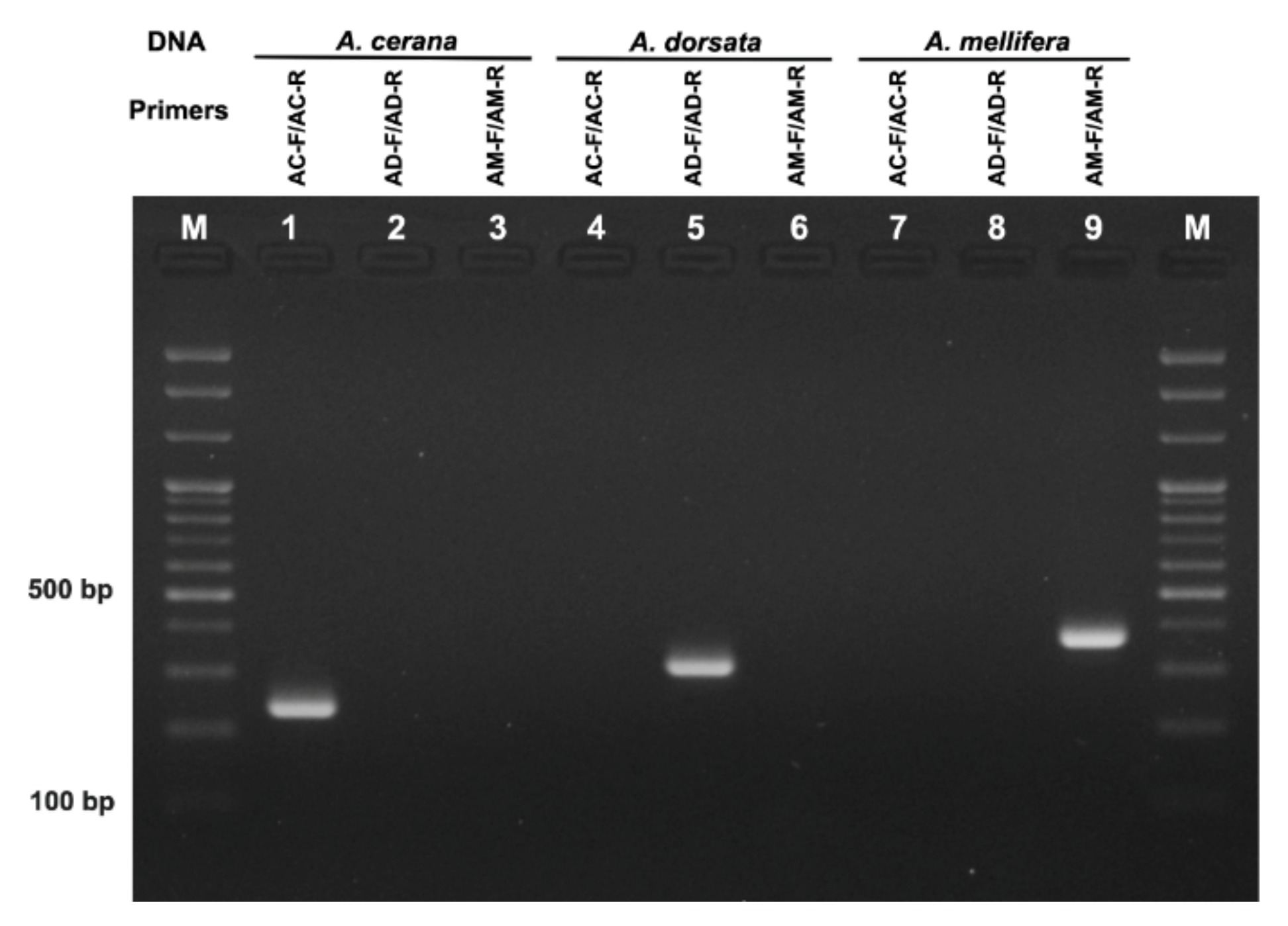
3.1. Specificity Test of Species-Specific Primers

The DNA extracts from different honeybees with different geographical origins were used to examine the specificity of the designed species-specific primers. Each primer set successfully amplified ND2 region from the DNA samples extracted from A. cerana, A. dorsata, and A. mellifera with an amplicon size of 224, 302, and 377 bp, respectively (Figure 2). None of the non-specific DNA amplification was observed with tested primer sets, suggesting these three species-specific primers can be successfully used to distinguish the origin of the honeybee at the DNA level.

3.2. Sensitivity Test of Primers Using Bee and Honey DNA

To evaluate the sensitivity of PCR-based assay, DNA samples from 3 different bees (A. cerana, A. dorsata and A. mellifera) were serially diluted (100 to 0.01 ng/μL) and used for PCR. The PCR condition using primer sets showed that all primer sets are able to amplify the specific bands (Figure 3). From the A. dorsata DNA, the AD-F/AD-R primer set successfully amplified the band with the expected size (302 bp) (Figure 3A). The intensity of characteristic bands was gradually raised as the concentration of DNA template increased, and the band could be visible when the DNA template was as low as 0.1 ng. From the A. mellifera (Figure 3B) and A. cerana DNA, similarly, AM-F/AM-R and AC-F/AC-R primer sets were also able to amplify specific bands with a detection limit of total 0.1 ng template DNA in the PCR reaction. This suggests that our species-specific primers can be used to detect the origin of honeybee samples with a low amount of DNA.
In other to test the ability to detect the target DNA among pure and adulterated honey samples, AMH was mixed with either ADH or ACH in different proportions. DNA was extracted from pure and mixed honey and used for the subsequent PCR analysis. Importantly, although the same amount of DNA (100 ng) from honey and bee samples was used for PCR, we were not able to detect the specific band from the first round of PCR with DNA from honey, unlike with DNA from bees (Figure 2 and Figure 3). This is likely due to actual amount of bee DNA being lower in DNA extracted from honey, as the honey sample contains biological tissues of other organisms (e.g., plant, microorganism, and other insect tissues). Thus, we performed another round of PCR by using 5 μL of PCR product as a template for analyzing the honey samples. In the second round of PCR, AC-F/AC-R and AD-F/AD-R primer sets produced a single band at the expected size with DNA from 100% ACD (Figure 4A, lane 1) and 100% ADH (Figure 4B, lane 1), respectively. On the contrary, the AM-F/AM-R primer set failed to amplify the band from the DNA extracted from 100% ACH (Figure 4A, lane 2) or 100% ADH (Figure 4B, lane 2). Neither AC-F/AC-R nor AD-F/AD-R primer sets amplified the specific bands from DNA extracted from 100% AMH (Figure 4A,B, lane 11).
DNA form ACH and ADH with different concentrations of AMH were also tested with species-specific primers (Figure 4A,B, lanes 3–12). In both conditions, the species-specific band of for A. mellifera was visible when the concentration of AMH was as low as 1%. The intensity of A. mellifera species-specific band gradually increased when the DNA from mixed honey with a proportion of 1 to 50% AMH were used and remained constant up to 100% AMH. To examine the possibility of using species-specific primer sets in the practical adulteration assay, 20 honey samples labeled as ADH, ACH, and AMH from different localities were tested. Analysis of the sequences of PCR products indicated that the primer sets are specific enough to detect the entomological origin of honey from different geographical localities.

3.3. Melting Curve Analysis by Real-Time PCR

To evaluate the possibility of use of melting curve analysis for detecting ACH or ADH adulteration, a real-time PCR experiment was conducted using the same PCR condition and primer sets and DNA extracted from honeybees. The result was confirmed using agarose gel electrophoresis and sequencing. Melting curve analysis of real-time PCR products demonstrated two distinct curves allowing the discrimination of A. dorsata from A. mellifera (Figure 5A) and of ADH from AMH. The melting temperature (Tm) of amplicons generated from A. dorsata (69.2 ± 0.1 °C) was distinct from the A. mellifera (72.4 ± 0.1 °C); hence, the detection of Tm could be an alternative method to detect the origin of ADH in addition to standard PCR method. Melting curve analyses of PCR products between A. cerana and A. mellifera were also performed (Figure 5B). Tm of amplicons of A. cerana (71.9 ± 0.2 °C) was distinct from A. mellifera (72.4 ± 0.1 °C) but very similar; hence, the use of Tm for distinguishing A. cerana and A. mellifera need more caution. The results of one-way ANOVA indicated that there was a significant difference between the Tm values of all three species (F value = 325.2, p-value < 0.001).

4. Discussion

Although previous attempts based on DNA barcoding of 16S rRNA and COI genes were helpful to inspect mislabeling [33], it was not functional to detect honey adulteration. In spite of the availability of species-specific primers to differentiate ACH from AMH [30], the primers developed by Kim et al. [30] were only applicable to honey originated from Korea but failed to differentiate ACH and AMH originated from China [26]. Since Soares et al. [31] only developed species-specific primers (AC1-F/AC1-R) to amplify 111 bp of tRNAlux-cox2 intergenic region of A. cerana mtDNA, the new A. mellifera species-specific primers were needed for the adulteration test. In addition, although AC1-F/AC1-R primers were useful in the discrimination of ACH from AMH, unlike ACF2/ACR2, they also amplified the non-specific band from ADH DNA extract, suggesting AC1-F/AC1-R was not enough to distinguish ACH from ADH (Supplementary Figure S1). In this study, we provided not only the first species-specific primer set to identify ADH, but also two new species-specific primer sets to identify ACH and AMH. Notably, our newly designed primers successfully amplified specific bands only from the targeted DNA sample and were able to discriminate both ACH and ADH from AMH, and vice versa, thus providing new DNA-based assay for testing entomological origin of honey (refer to Figure 4A,B).
Zhang et al. [26] developed a gDNA-based method for the identification of two different major honeys from domesticated honeybees in the market. Two sets of primers (C-F/C-R for A. cerana and M-F/M-R for A. mellifera) were designed to amplify the Major royal jelly protein 2 (MRJP2) gene, resulting in the different size of PCR product in the gel electrophoresis, making it useful to discriminate ACH from AMH. In addition, it is possible to identify the honey samples through real-time PCR-based Tm analysis. Although the predicted size of the PCR product was 212 bp for A. cerana and 393 bp for A. mellifera, but the length of amplicons for A. mellifera was 560 bp from the PCR, as the primers were designed based on complementary DNA (cDNA) without an intron. ACH and AMH samples were distinguishable using C-F/C-R and M-F/M-R primer sets; however, 560 bp tends to be long for accurate honey identification and adulteration test with relatively old honey samples, which can possibly have DNA degradation problems. Honey is a complex matrix, and its phenolic/H2O2 induced oxidative stress would lead the DNA that remained inside honey to be easily degraded as storage time increased [31,34,35,36]. Notably, Schnell et al. [37] reported the diminished rate of successful amplification of amplicon size larger than 380 bp and in the fragmented DNA, thus the amplification of short amplicons is preferable [38]. Thus our new species-specific primer sets with amplicon size ranging 224~377 bp (refer to Figure 2 and Figure 3) would provide a better chance to successfully examine the old honey samples. Although the speed of degradation of DNA inside honey is not well understood and very difficult to predict accurately as different honey have different biochemical compositions, DNA degradation problem needs to be considered while examining the entomological origin of honey via DNA-based assay.
Real-time PCR-based identification of the entomological origin of honey was successfully developed previously to discriminate ACH and AMH using species-specific primers [26,31]. Refer to the Figure 5, the primer sets developed in the present study can be used to differentiate the entomological origin of three different types of honey. Although ADH and AMH can be simply differentiated using melting curve analysis, this method should be applied to differentiate ACH and AMH with caution due to the close Tm of the amplicons. Tm-based identification method is quick and accurate without the requirement of the gel electrophoresis step. Thus, it will provide a possible high-throughput analyses method for the identification of the origin of honey.
The cost of conducting analysis for one honey sample using the combination of two-round PCR and subsequent gel electrophoresis using the methodology described in current research is 6.4$ per honey sample (DNA extraction kit, PCR master mix, agarose powder, ladder, TBE buffer, staining dye and primer cost), however, these expenses for authentication analysis using Real-Time PCR technic is about 5.2$ (DNA extraction, master mix, and primer cost). The electricity and labor cost required to run the equipment have not been considered in our calculation. Although the cost of the authentication analysis per sample is slightly lower using Real-Time PCR, it is more expensive to establish such facilities in comparison to the conventional PCR method. Furthermore, according to our calculations, the duration of analysis using Real-Time PCR is slightly longer (~35 min) than conventional PCR.

5. Conclusions

Three species-specific primer sets targeting the NADH dehydrogenase 2 (ND2) region of mtDNA were designed and successfully applied to trace the entomological origin of honey produced by different honeybees, A. cerana, A. dorsata and A. mellifera. In addition, the A. mellifera specific primer set is applicable in honey fraud detection. The possibility of using melting curve analysis in discrimination of the origin of honey using the same primer sets is also confirmed. Our preliminary studies indicated the impossibility of providing species-specific primers with a smaller size of PCR product in the mitochondrial DNA (except the one provided by Soares et al. [7] for ACH). However, further studies targeting nuclear DNA are required. PCR-based method using species-specific primers provides a rapid and cost-effective method to screen the entomological origin of honey. Therefore, the development of new primer sets to identify honey produced by other species of honeybees will be valuable. On the other hand, more studies are needed to understand the pace of DNA degradation in honey and the applicability and limitations of using molecular methods in the authentication of older honey samples.

Supplementary Materials

The following supporting information can be downloaded at: https://0-www-mdpi-com.brum.beds.ac.uk/article/10.3390/foods11070928/s1, Table S1: The honey samples and their labels and localities used for analysis. Table S2: Mitochondrial sequences of honeybees (A. cerana, A. dorsata and A. mellifera) used to design species-specific primers. Figure S1: Preliminary specificity test of ACF/ACR (Suarez et al., 2018) using DNA extracts of honeybees [39,40,41,42,43,44,45,46,47,48,49,50,51,52,53,54,55,56,57].

Author Contributions

S.M.N.: Visualization, methodology, design primers, Sequence assembling and blast, investigation, writing the original draft. F.Y.: Visualization, writing the original draft, investigation, methodology. H.W.C.: Project administration, supervision, review and editing. C.J.: Supervision, funding acquisition, resources, review and editing. All authors have read and agreed to the published version of the manuscript.

Funding

This study was supported by the BSRP through the National Research Foundation of Korea (NRF), Ministry of Education (grant number NRF-2018R1A6A1A03024862), and Rural Development Administration (RDA agenda PJ01574604 on honeybee pollination).

Institutional Review Board Statement

Not applicable.

Informed Consent Statement

Not applicable.

Data Availability Statement

All the sequences generated in this research, have been deposited in GenBank under accession numbers MW660861-MW660880.

Acknowledgments

We are grateful to Bajaree Chuttong, Chiang Mai University who provided us with honey and bee samples from Thailand. We also thank Ratna Thapa for facilitating SMA and CJ’s expedition trip to Nepal. This study was supported by the BSRP through the National Research Foundation of Korea (NRF), Ministry of Education (grant number NRF-2018R1A6A1A03024862).

Conflicts of Interest

S.M.N. and F.Y. are a research professor and Ph.D. student at ANU, respectively, and received a full-time salary for this work. C.J. has received research grants from NRF, and supervised the work. H.W.C. has served on advisory for the project. All declare no conflicts of interest on this paper.

References

  1. Ajibola, A.; Chamunorwa, J.P.; Erlwanger, K.H. Dietary supplementation with natural honey promotes growth and health of male and female rats compared to cane syrup. Sci. Res. Essays 2013, 8, 543–553. [Google Scholar]
  2. Soares, S.; Amaral, J.S.; Oliveira, M.B.; Mafra, I. A comprehensive review on the main honey authentication issues: Production and origin. Compr. Rev. Food Sci. Food Saf. 2017, 16, 1072–1100. [Google Scholar] [CrossRef] [PubMed] [Green Version]
  3. Oldroyd, B.P.; Wongsiri, S. Asian Honey Bees: Biology, Conservation and Human Interactions; Harvard University Press: Cambridge, MA, USA, 2006. [Google Scholar]
  4. Wongsiri, S.; Chanchao, C.; Lekprayoon, C.; Wattanasermkit, K.; Deowanish, S.; Leepitakrat, S. Honeybee diversity and management in the new millennium in Thailand. In Proceedings of the 7th International Conference on Tropical Bees, Chiang Mai, Thailand, 19–25 March 2000; pp. 9–14. [Google Scholar]
  5. Corlett, R.T. Honeybees in natural ecosystems. In Honeybees of Asia; Hepburn, H.R., Radloff, S.E., Eds.; Springer-Verlag: Berlin, Germany, 2011; pp. 215–226. [Google Scholar]
  6. Partap, U. The pollination role of honeybees. In Honeybees of Asia; Hepburn, H.R., Radloff, S.E., Eds.; Springer-Verlag: Berlin, Germany, 2011; pp. 227–255. [Google Scholar]
  7. Hall, T.A. BioEdit: A User-Friendly Biological Sequence Alignment Editor and Analysis Program for Windows 95/98/NT. Nucleic Acids Symp. Ser. 1999, 41, 95–98. [Google Scholar]
  8. Yong, P.L.; Othman, M.S.H. Economic Value of Honey Bees, Peninsular Malaysia; Forestry Department Peninsular Malaysia (FDPM): Kuala Lumpur, Malaysia, 2007. [Google Scholar]
  9. Moniruzzaman, M.; Khalil, M.I.; Sulaiman, S.A.; Gan, S.H. Physicochemical and antioxidant properties of Malaysian honeys produced by Apis cerana, Apis dorsata and Apis mellifera. BMC Complement. Altern. Med. 2013, 13, 43. [Google Scholar] [CrossRef] [PubMed] [Green Version]
  10. Ruttner, F. Biogeography and Taxonomy of Honeybees; Springer: Heidelberg/Berlin, Germany, 1988. [Google Scholar]
  11. Jung, C.; Lee, M. Beekeeping in Korea: Past, present, and future challenges. In Asian Beekeeping in the 21st Century; Chantawannakul, P., Williams, G., Neumann, P., Eds.; Springer: Berlin/Heidelberg, Germany, 2016; pp. 175–197. [Google Scholar]
  12. Lee, D.C.; Lee, S.Y.; Cha, S.H.; Choi, Y.S.; Rhee, H.I. Discrimination of native bee-honey and foreign bee-honey by SDS–PAGE. Korean J. Food Sci. Technol. 1998, 30, 1–5. [Google Scholar]
  13. He, X.; Wang, W.; Qin, Q.; Zeng, Z.; Zhang, S.; Barron, A.B. Assessment of flight activity and homing ability in Asian and European honey bee species, Apis cerana and Apis mellifera, measured with radio frequency tags. Apidologie 2012, 44, 38–51. [Google Scholar] [CrossRef] [Green Version]
  14. Jaafar, M.; Othman, M.; Yaacob, M.; Talip, B.; Ilyas, M.; Ngajikin, N.; Fauzi, N. A review on honey adulteration and the available detection approaches. J. Integr. Eng. 2020, 12, 125–131. [Google Scholar]
  15. Bogdanov, S.; Ruoff, K.; Oddo, L.P. Physico-chemical methods for the characterisation of unifloral honeys: A review. Apidologie 2004, 35 (Suppl. 1), S4–S17. [Google Scholar] [CrossRef] [Green Version]
  16. Sahinler, N.; Sahinler, S.; Gul, A. Biochemical composition of honeys produced in Turkey. J. Apic. Res. 2004, 43, 53–56. [Google Scholar] [CrossRef]
  17. Guler, A.; Bakan, A.; Nisbet, C.; Yavuz, O. Determination of important biochemical properties of honey to discriminate pure and adulterated honey with sucrose (Saccharum officinarum L.) syrup. Food Chem. 2007, 105, 1119–1125. [Google Scholar] [CrossRef]
  18. Chen, L.; Xue, X.; Ye, Z.; Zhou, J.; Chen, F.; Zhao, J. Determination of Chinese honey adulterated with high fructose corn syrup by near infrared spectroscopy. Food Chem. 2011, 128, 1110–1114. [Google Scholar] [CrossRef]
  19. Moore, J.C.; Spink, J.; Lipp, M. Development and application of a database of food ingredient fraud and economically motivated adulteration from 1980 to 2010. J. Food Sci. 2012, 77, R118–R126. [Google Scholar] [CrossRef]
  20. Bottero, M.T.; Dalmasso, A. Animal species identification in food products: Evolution of biomolecular methods. Vet. J. 2011, 190, 34–38. [Google Scholar] [CrossRef] [PubMed]
  21. Kumar, A.; Kumar, R.R.; Sharma, B.D.; Gokulakrishnan, P.; Mendiratta, S.K.; Sharma, D. Identification of species origin of meat and meat products on the DNA basis: A review. Crit. Rev. Food. Sci. Nutr. 2013, 55, 1340–1351. [Google Scholar] [CrossRef] [PubMed]
  22. Amaral, J.; Meira, L.; Oliveira, M.; Mafra, I. Advances in authenticity testing for meat speciation. In Advances in Food Authenticity Testing; Downey, G., Ed.; Elsevier: Amsterdam, The Netherlands, 2016; pp. 369–414. [Google Scholar]
  23. Willette, D.A.; Simmonds, S.E.; Cheng, S.H.; Esteves, S.; Kane, T.L.; Nuetzel, H.; Pilaud, N.; Rachmawati, R.; Barber, P.H. Using DNA barcoding to track seafood mislabeling in Los Angeles restaurants. Conserv. Biol. 2017, 31, 1076–1085. [Google Scholar] [CrossRef] [PubMed]
  24. Won, S.; Lee, D.; Ko, S.H.; Kim, J.; Rhee, H. Honey major protein characterization and its application to adulteration detection. Food Res. Int. 2008, 41, 952–956. [Google Scholar] [CrossRef]
  25. Zhang, Y.; Wang, S.; Chen, Y.; Wu, Y.; Tian, J.; Si, J.; Zhang, C.; Zheng, H.; Hu, F. Authentication of Apis cerana honey and Apis mellifera honey based on major royal jelly protein 2 gene. Molecules 2019, 24, 289. [Google Scholar] [CrossRef] [Green Version]
  26. Zhang, Y.-Z.; Chen, Y.-F.; Wu, Y.-Q.; Si, J.-J.; Zhang, C.-P.; Zheng, H.-O.; Hu, F.-L. Discrimination of the entomological origin of honey according to the secretions of the bee (Apis cerana or Apis mellifera). Food Res. Int. 2019, 116, 362–369. [Google Scholar] [CrossRef]
  27. Moritz, C.; Dowling, T.E.; Brown, W.M. Evolution of animal mitochondrial DNA: Relevance for population biology and systematics. Annu. Rev. Ecol. Evol. Syst. 1987, 18, 269–292. [Google Scholar] [CrossRef]
  28. Song, S.; Pursell, Z.F.; Copeland, W.C.; Longley, M.J.; Kunkel, T.A.; Mathews, C.K. DNA precursor asymmetries in mammalian tissue mitochondria and possible contribution to mutagenesis through reduced replication fidelity. Proc. Natl. Acad. Sci. USA 2005, 102, 4990–4995. [Google Scholar] [CrossRef] [Green Version]
  29. Zink, R.M.; Barrowclough, G.F. Mitochondrial DNA under siege in avian phylogeography. Mol. Ecol. 2008, 17, 2107–2121. [Google Scholar] [CrossRef] [PubMed]
  30. Kim, C.; Lee, D.; Choi, S. Detection of Korean native honey and European honey by using duplex polymerase chain reaction and Immunochromatographic assay. Food Sci. Anim. Resour. 2017, 37, 599–605. [Google Scholar] [CrossRef] [PubMed] [Green Version]
  31. Soares, S.; Grazina, L.; Mafra, I.; Costa, J.; Pinto, M.A.; Duc, H.P.; Oliviera, M.B.; Amaral, J. Novel diagnostic tools for Asian (Apis cerana) and European (Apis mellifera) honey authentication. Food Res. Int. 2018, 105, 686–693. [Google Scholar] [CrossRef] [PubMed] [Green Version]
  32. R Core Team. R: A Language and Environment for Statistical Computing; R Foundation for Statistical Computing: Vienna, Austria, 2018; Available online: https://www.R-project.org/ (accessed on 20 December 2021).
  33. Kek, S.P.; Chin, N.L.; Tan, S.W.; Yusof, Y.A.; Chua, L.S. Molecular identification of honey entomological origin based on bee mitochondrial 16S rRNA and COI gene sequences. Food Control 2017, 78, 150–159. [Google Scholar] [CrossRef]
  34. Brudzynski, K.; Abubaker, K.; Miotto, D. Unraveling a mechanism of honey antibacterial action: Polyphenol/H2O2-induced oxidative effect on bacterial cell growth and on DNA degradation. Food Chem. 2012, 133, 329–336. [Google Scholar] [CrossRef] [PubMed]
  35. Utzeri, V.J.; Ribani, A.; Fontanesi, L. Authentication of honey based on a DNA method to differentiate Apis mellifera subspecies: Application to Sicilian honey bee (A. M. siciliana) and iberian honey bee (A. M. iberiensis) honeys. Food Control 2018, 91, 294–301. [Google Scholar] [CrossRef]
  36. Bovo, S.; Utzeri, V.J.; Ribani, A.; Cabbri, R.; Fontanesi, L. Shortgun sequencing of honey DNA can describe honey bee derived environmental signatures and the honey bee hologenome complexity. Sci. Rep. 2020, 10, 9279. [Google Scholar] [CrossRef]
  37. Schnell, I.B.; Fraser, M.; Willerslev, E.; Gilbert, M.T.P. Characterisation of insect and plant origins using DNA extracted from small volumes of bee honey. Arthropod Plant Interact. 2010, 4, 107–116. [Google Scholar] [CrossRef]
  38. Jain, S.A.; Jesus, F.T.; Marchioro, G.M.; Araujo, E.D. Extraction of DNA from honey and its amplification by PCR for botanical identification. Food Sci. Technol. 2013, 33, 753–756. [Google Scholar] [CrossRef] [Green Version]
  39. Eimanifar, A.; Kimball, R.T.; Braun, E.L.; Ellis, J.D. The complete mitochondrial genome of the Cape honey bee Esch., Apis mellifera capensis (Insecta: Hymenoptera: Apidae). Mitochondrial DNA Part B 2016, 1, 817–819. [Google Scholar] [CrossRef] [Green Version]
  40. Eimanifar, A.; Kimball, R.T.; Braun, E.L.; Fuchs, S.; Grünewald, B.; Ellis, J.D. The complete mitochondrial genome of an east African honey bee, Apis mellifera monticola Smith (Insecta: Hymenoptera: Apidae). Mitochondrial DNA Part B 2017, 2, 589–590. [Google Scholar] [CrossRef] [PubMed] [Green Version]
  41. Eimanifar, A.; Kimball, R.T.; Braun, E.L.; Moustafa, D.M.; Haddad, N.; Fuchs, S.; Grunewald, B.; Ellis, J.D. The complete mitochondrial genome of the Egyptian honey bee, Apis mellifera lamarckii (Insecta: Hymenoptera: Apidae). Mitochondrial DNA Part B 2017, 2, 270–272. [Google Scholar] [CrossRef] [PubMed] [Green Version]
  42. Eimanifar, A.; Kimball, R.T.; Braun, E.L.; Fuchs, S.; Grunewald, B.; Ellis, J.D. The complete mitochondrial genome of Apis mellifera meda (Insecta: Hymenoptera: Apidae). Mitochondrial DNA Part B 2017, 2, 268–269. [Google Scholar] [CrossRef] [PubMed] [Green Version]
  43. Eimanifar, A.; Kimball, R.T.; Braun, E.L.; Ellis, J.D. Mitochondrial genome diversity and population structure of two western honeybee subspecies in the Republic of South Africa. Sci. Rep. 2018, 8, 1333. [Google Scholar] [CrossRef] [PubMed] [Green Version]
  44. Fuller, Z.L.; Nino, E.L.; Patch, H.M.; Bedoya-Reina, O.C.; Baumgarten, T.; Muli, E.; Mumoki, F.; Ratan, A.; McGraw, J.; Frazier, M.; et al. Genome-wide analysis of signatures of selection in populations of African honey bees (Apis mellifera) using new web-based tools. BMC Genom. 2015, 16, 518. [Google Scholar] [CrossRef] [Green Version]
  45. Gibson, J.D.; Hunt, G.J. The complete mitochondrial genome of the invasive Africanized honey bee, Apis mellifera scutellata (Insecta: Hymenoptera: Apidae). Mitochondrial DNA Part A 2016, 27, 561–562. [Google Scholar] [CrossRef]
  46. Haddad, N.J. Mitochondrial genome of the Levant Region honey bee, Apis mellifera syriaca (Hymenoptera: Apidae). Mitochondrial DNA Part A 2016, 27, 4067–4068. [Google Scholar] [CrossRef]
  47. Hu, P.; Lu, Z.X.; Haddad, N.; Noureddine, A.; Loucif-Ayad, W.; Wang, Y.Z.; Zhang, R.B.Z.A.L.; Guan, X.; Zhang, H.X.; Niu, H. Complete mitochondrial genome of the Algerian honey bee, Apis mellifera intermissa (Hymenoptera: Apidae). Mitochondrial DNA Part A 2016, 27, 1791–1792. [Google Scholar]
  48. Ilyasov, R.A.; Park, J.; Takahashi, J.; Kwon, H.W. Phylogenetic uniqueness of honeybee Apis cerana from the Korean peninsula inferred from the mitochondrial, nuclear, and morphological data. J. Apicul. Sci. 2018, 62, 189–214. [Google Scholar] [CrossRef] [Green Version]
  49. Nakagawaa, I.; Maedaa, M.; Chikanoa, M.; Okuyamaa, H.; Murrayb, R.; Takahashia, J. The complete mitochondrial genome of the yellow coloured honeybee Apis mellifera (Insecta: Hymenoptera: Apidae) of New Zealand. Mitochondrial DNA Part B 2018, 3, 66–67. [Google Scholar] [CrossRef] [Green Version]
  50. Okuyama, H.; Hill, J.; Martin, S.J.; Takahashi, J. The complete mitochondrial genome of a Buckfast bee, Apis mellifera (Insecta: Hymenoptera: Apidae) in Northern Ireland. Mitochondrial DNA Part B 2018, 3, 338–339. [Google Scholar] [CrossRef] [PubMed] [Green Version]
  51. Okuyama, H.; Tingek, S.; Takahashi, J. The complete mitochondrial genome of the cavity-nesting honeybee, Apis cerana (Insecta: Hymenoptera: Apidae) from Borneo. Mitochondrial DNA Part B 2017, 2, 475–476. [Google Scholar] [CrossRef] [PubMed] [Green Version]
  52. Okuyama, H.; Jimi, R.; Wakamiya, T.; Takahashi, J. Complete mitochondrial genome of the honeybee Apis cerana native to two remote islands in Japan. Conserv. Genet. Resour. 2017, 9, 557–560. [Google Scholar] [CrossRef]
  53. Takahashi, J.; Deowanish, S.; Okuyama, H. Analysis of the complete mitochondrial genome of the giant honeybee, Apis dorsata, (Hymenoptera: Apidae) in Thailand. Conserv. Genet. Resour. 2017. [Google Scholar] [CrossRef]
  54. Takahashi, J.; Wakamiya, T.; Kiyoshi, T.; Uchiyama, H.; Yajima, S.; Kimura, K.; Nomura, T. The complete mitochondrial genome of the Japanese honeybee, Apis cerana japonica (Insecta: Hymenoptera: Apidae). Mitochondrial DNA Part B 2016, 1, 156–157. [Google Scholar] [CrossRef] [PubMed]
  55. Tan, H.W.; Liu, G.H.; Dong, X.; Lin, R.Q.; Song, H.Q.; Huang, S.Y.; Yuan, Z.G.; Zhao, X.Q. The complete mitochondrial genome of the Asiatic cavity nesting honeybee Apis cerana (Hymenoptera: Apidae). PLoS ONE 2011, 6, e23008. [Google Scholar] [CrossRef] [PubMed] [Green Version]
  56. Wang, A.R.; Kim, J.S.; Kim, M.J.; Kim, H.K.; Choi, Y.S.; Kim, I. Comparative description of mitochondrial genomes of the honey bee Apis (Hymenoptera: Apidae): Four new genome sequences and Apis phylogeny using whole genomes and individual genes. J. Apic. Res. 2018, 57, 484–503. [Google Scholar] [CrossRef]
  57. Yang, J.; Xu, J.; Wu, J.; Zhang, X.; He, S. The complete mitogenome of wild honeybee Apis dorsata (Hymenoptera: Apidae) from South-Western China. Mitochondrial DNA Part B 2019, 4, 231–232. [Google Scholar] [CrossRef] [Green Version]
Figure 1. Schematic overview of the experimental design.
Foods 11 00928 g001
Figure 2. Agarose gel electrophoresis of PCR products amplified from DNA extracted of honeybees with species-specific primers. Bee DNA (Lanes 1–3: A. cerana DNA, Lanes 4–6: A. dorsata DNA, Lanes 7–9: A. mellifera DNA) Primers (Lanes 1, 4 and 7: A. cerana specific primers AC-F/AC-R, Lanes 2, 5 and 8: A. dorsata specific primers AD-F/AD-R, Lanes 3, 6 and 9: A. mellifera specific primers AM-F/AM-R), M: 100 bp ladder.
Foods 11 00928 g002
Figure 3. Sensitivity test of the designed species-specific primers using serially diluted DNA extract of A. dorsata (A), A. mellifera (B), and A. cerana (C). Lane M, DNA marker; Lane 1, 100 ng; lane 2, 10 ng; lane 3, 1 ng; lane 4, 0.1 ng; lane 5, 0.01 ng; lane 6, negative control.
Foods 11 00928 g003
Figure 4. Adulteration test with artificially mixed honey samples. PCR products amplified from DNA extracted either from mixtures of ACH and AMH (A) or ADH and AMH (B) were analyzed by DNA gel electrophoresis. The proportions of AMH inside either ACH or ADH and usage of species-specific primer sets are shown. AC-F/AC-R, A. cerana specific primers; AD-F/AD-R, A. dorsata specific primers; AM-F/AM-R, A. mellifera specific primers; M, 1 kb ladder.
Foods 11 00928 g004
Figure 5. Conventional melting curves obtained by real-time PCR amplification targeting ND2 region of mtDNA using DNA extracts from honeybees. (A) A. dorsata (Tm = 69.2 ± 0.1) and A. mellifera (Tm = 72.4 ± 0.1). (B) A. cerana (Tm = 71.9 ± 0.2) and A. mellifera (Tm = 72.4 ± 0.1).
Foods 11 00928 g005
Table 1. Specific-primers for honey identification, nucleotide sequence, primer length, and the expected length of PCR product.
SpeciesPrimer5′-3′LengthTarget Fragment
A. ceranaAC-FTCATTAGATTTTACAAAATCAGATCA26224 bp
AC-RCTTATAACTAAATATGTTAATGATCATA28
A. dorsataAD-FTATATTAATTGTTATAACTTACATAAATAA31302 bp
AD-RGGATTAAGAATATATAATATTCATATTTT29
A. melliferaAM-FCTATTAGATTTACTAAAACAGATACT26377 bp
AM-RATAATTAAATGAATATAAAATAATTATAGCA31
Publisher’s Note: MDPI stays neutral with regard to jurisdictional claims in published maps and institutional affiliations.

