Article

Hurdle Technology Approach to Control Listeria monocytogenes Using Rhamnolipid Biosurfactant

by
Lowieze Lenaerts
1,
Tathiane Ferroni Passos
2,
Elisa Gayán
3,
Chris W. Michiels
1,* and
Marcia Nitschke
2,*
1
Department of Microbial and Molecular Systems, Leuven Food Science and Nutrition Research Center (LFoRCe), KU Leuven, Kasteelpark Arenberg 22, B-3001 Leuven, Belgium
2
São Carlos Institute of Chemistry (IQSC), University of São Paulo, Trabalhador São-Carlense Av., 400, P.O. Box 780, São Carlos 13560-970, São Paulo, Brazil
3
Department of Animal Production and Food Science, AgriFood Institute of Aragon (IA2), Faculty of Veterinary, University of Zaragoza-CITA, Miguel Servet 177, 50013 Zaragoza, Spain
*
Authors to whom correspondence should be addressed.
Submission received: 12 December 2022 / Revised: 21 January 2023 / Accepted: 25 January 2023 / Published: 28 January 2023

Abstract

:
This study evaluates the combination of mild heat with a natural surfactant for the inactivation of L. monocytogenes Scott A in low-water-activity (aw) model systems. Glycerol or NaCl was used to reduce the aw to 0.92, and different concentrations of rhamnolipid (RL) biosurfactant were added before heat treatment (60 °C, 5 min). Using glycerol, RL treatment (50–250 µg/mL) reduced bacterial population by less than 0.2 log and heat treatment up to 1.5 log, while the combination of both hurdles reached around 5.0 log reduction. In the NaCl medium, RL treatment displayed higher inactivation than in the glycerol medium at the same aw level and a larger synergistic lethal effect when combined with heat, achieving ≥ 6.0 log reduction at 10–250 µg/mL RL concentrations. The growth inhibition activity of RL was enhanced by the presence of the monovalent salts NaCl and KCl, reducing MIC values from >2500 µg/mL (without salt) to 39 µg/mL (with 7.5% salt). The enhanced antimicrobial activity of RL promoted by the presence of salts was shown to be pH-dependent and more effective under neutral conditions. Overall, results demonstrate that RL can be exploited to design novel strategies based on hurdle approaches aiming to control L. monocytogenes.

1. Introduction

Heat processing is the most extensively utilized commercial food preservation method to destroy microorganisms and thereby extend the shelf life and ensure the microbiological safety of food. However, intensive heat treatments result in undesirable nutritional and organoleptic changes in the final product [1,2]. The tendency for more natural and healthy foods has prompted the development of mild processing methods that can replace traditional heat treatments or reduce their intensity while retaining the nutritional and sensory properties and safety of products [3,4]. A reduction of treatment intensity can be achieved when heat is combined with other preservation methods that together ensure the appropriate inactivation of pathogenic and spoilage microorganisms. Such combined treatments can also be used to enhance treatment efficiency when intrinsic food properties, such as low water activity (aw), tend to protect bacteria against heat [5]. Within this context, hurdle approaches resulting in a synergistic lethal effect are desired in order to significantly reduce the intensity of each individual preservation method while obtaining foods with better quality and lower processing cost [6].
On the other hand, the food industry is making increasing efforts to replace chemical food preservatives by natural alternatives [7]. Although some natural compounds, such as those from plant essential oils, have a strong antimicrobial effect, they often have low solubility and a strong impact on the sensorial properties of foods [8,9]. Therefore, hurdle approaches wherein natural antimicrobials at lower concentrations are combined with mild processing are being explored to reduce their negative impact on food products.
Microbial surfactants or biosurfactants (BS) are a natural class of surface-active compounds produced by microorganisms. The possibility of producing BS from renewable feedstocks or agricultural wastes, their biodegradability and low toxicity align well with the concept of “green chemistry”, which represents an important tool for innovation and sustainability, fulfilling actual market needs [10]. The rhamnolipids (RLs) produced by Pseudomonas aeruginosa are glycolipid biosurfactants with several useful characteristics, and they are considered promising multipurpose ingredients in food processing [11]. Rhamnolipids are approved by the US Environmental Protection Agency (EPA) as antimicrobials for the washing or spraying of agricultural crops and are classified by the EPA in the lowest toxicity category (IV), having an acute oral LD50 greater than 5000 mg/kg [12].
Listeria monocytogenes are widespread in diverse natural environments such as soil, water, plants and the gastro-intestinal tract of humans and animals [13]. They can also establish and thrive in many food production environments, and their presence and persistence in the food supply chain is a matter of concern, as they are the causative agent of a serious foodborne illness known as listeriosis [14]. The consumption of contaminated foods, including chilled meat, poultry, seafood, vegetables, fruits, dairy and ready-to-eat products, is responsible for outbreaks worldwide [15]. A factor contributing to the success of the pathogen is its ability to grow over a wide range of pHs (4.5–9.5), salt concentrations (up to 10%) and aw (down to 0.92) [16,17]. In addition, it can form biofilms and resist sanitizers [18], increasing its ability to contaminate food products. Finally, the pathogen has a remarkable aptitude for long-term survival under stressful conditions. For example, L. monocytogenes ATCC 19115 was reported to survive in culture medium at pH 4.0 and 4°C for 19 days in the presence of 21% NaCl [19].
In a previous study, it was demonstrated that a purified RL produced by P. aeruginosa PA1 inhibited the growth of L. monocytogenes ATCC 19112 and ATCC 7644 by 100% [20]. Further work, conducted with 32 L. monocytogenes isolates, revealed that over 90% were susceptible to RL with MIC values ranging from 78.1 to 2500 µg/mL. The study also demonstrated that RL had a synergistic effect when combined with nisin, probably because both molecules act on the same cell target [21]. The exact mechanism behind RLs’ antimicrobial activity remains unclear, but most researchers anticipate that they disturb the cell membrane due to their amphipathic nature, leading to increased permeability, metabolite leakage and eventually cellular lysis [21,22].
It is well known that reducing microbial contamination of low aw foods by thermal processing requires very intensive treatment conditions due to the increased heat tolerance of microorganisms [23,24]. Such treatments therefore cause heat damage, leading to unacceptable quality deterioration. Several membrane-targeting antimicrobials show a synergistic lethal effect when combined with mild heat treatment [25,26,27]; however, the effectiveness of synergistic combinations has hardly been studied in low-moisture systems. Therefore, in this work, we investigated whether the combination of RL and mild heat treatment is effective, and possibly synergistic, in inactivating L. monocytogenes in low-moisture systems. Both a nonionic (glycerol) and ionic (NaCl) solute were used for reducing the aw of a model system, and the effect of RL, heat and their combination was evaluated. Further, the influence of ionic strength on the growth-inhibitory activity of RL is also studied and discussed.

2. Materials and Methods

2.1. Biosurfactant

Commercial rhamnolipid solution (99% purity) was provided by Rhamnolipid Inc. (Tampa, FL, USA), containing approximately 54.4% RhaC10C10 and 24.2% Rha2C10C10 as major components.

2.2. Bacterial Strain and Culture Conditions

L. monocytogenes Scott A, acquired from the International Life Sciences Institute (ILSI), North America [28], was used throughout this investigation. The strain was maintained at −80 °C in Brain Heart Infusion (BHI; Oxoid, Basingstoke, UK) broth supplemented with 25% glycerol. For cell revitalization, a loopful of frozen culture was spread on BHI agar and incubated for 24 h at 30 °C. Stationary-phase cultures were prepared by inoculating a test tube containing 4 mL of BHI broth with a single colony from the stock plate and incubating it aerobically in an orbital shaker at 200 rpm (GFL 3005, Burgwedel, Germany) for 19 ± 1 h at 30 °C.

2.3. Inactivation of L. monocytogenes by Heat and RL in Reduced Moisture Systems

A 4 mL stationary phase culture of L. monocytogenes Scott A was transferred to a 10 mL tube and centrifuged at 3829× g for 5 min. After the removal of the supernatant, the cells were washed twice with 4 mL of 10 mM sodium phosphate buffer (pH 6.7) and centrifuged again. The pellet was finally suspended in 7.5 mL of buffer with the appropriate solutes to adjust the cell concentration to ~1.3 × 10⁹ CFU/mL.
The inactivation assays were conducted in 10 mM sodium phosphate buffer (pH 6.7) adjusted to aw 0.98 or 0.92 with glycerol (6.95% and 29.63% (w/w) for aw 0.98 and 0.92, respectively) or NaCl (3.63% and 14.94% (w/w) for aw 0.98 and 0.92, respectively) as model systems of low-moisture matrices. The water activity was measured using a HygroPalm AW1 hygrometer (Rotronics AG, Bassersdorf, Germany). When necessary, the buffer was supplemented with the appropriate RL concentration (1 to 250 µg/mL). For RL treatment at room temperature, 100 μL of the cell suspension (~1.3 × 10⁹ CFU/mL) was added to an Eppendorf tube containing 900 µL of the buffer with RL added and incubated for 5 min at ~20 °C. For heat treatment alone or in combination with RL, the same volume of cell suspension was added to an Eppendorf tube containing 900 µL of the buffer without or with RL that was previously prewarmed at 60 °C in a heating block (VWR, Radnor, PA, USA), and then the samples were incubated at the same temperature for 5 min. As such, the initial cell concentration for all RL, heat and the combined RL and heat treatment was ~1.3 × 10⁸ CFU/mL. After treatment, the samples were serially diluted, and the number of viable cells was determined by the drop plate method [29] (5 μL drops of each dilution) on BHI agar. Plates were incubated at 30 °C for 24 h, and then spots containing 5–50 colonies were counted, so that the quantification limit was 1000 CFU/mL. The logarithmic reduction factor (LRF, dimensionless unit) was calculated as log(N0/N), where N0 and N are the viable cell concentration (CFU/mL) at the start and at the end of the treatment, respectively.
To determine the occurrence of synergistic/additive interactions between the lethal effect of heat and RL, the LRF of the combined treatment was statistically compared to the sum of the LRFs of the individual treatments (theoretical additive inactivation), as described by Feyaerts et al. [30].

2.4. Growth Inhibitory Activity

The minimum inhibitory concentration (MIC) and minimum bactericidal concentration (MBC) of the RL were determined using the micro-broth dilution technique according to the Clinical and Laboratory Standards Institute guidelines [31], using concentrations from 2500.0 to 4.9 µg/mL. The MBC was determined by transferring 100 µL from the wells of the MIC microtiter plates where no growth was observed (after 24 h of incubation) to the surface of BHI agar plates that were subsequently incubated for 48 h at 30 °C. The MBC was considered the lowest concentration of RL at which no colonies were formed [32].

2.5. Growth Curves

Growth experiments were conducted in BHI medium in 96-well microplates covered with adhesive foil in an automated microplate reader (Multiskan Ascent, Thermo Fisher Scientific, Waltham, MA, USA) at 30 °C [33]. An aliquot of 20 µL of an overnight culture diluted to a cell population of 107 CFU/mL was added to 180 µL of BHI supplemented with 500 µg/mL of RL, and the optical density (OD, 620 nm) was measured at defined time intervals. To study the influence of NaCl or KCl on the antimicrobial activity of RL, the salts were added to the culture medium at concentrations ranging from 1.0% to 7.5% (w/v).

2.6. Microscopy

Fluorescence microscopy was performed with a Ti-Eclipse inverted microscope (Nikon, Tokyo, Japan) after cell staining with a LIVE/DEAD BacLight Bacterial Viability kit (Thermo Fisher Scientific) following supplier specifications [34]. Images were acquired and processed using NIS-Elements (Nikon, Tokyo, Japan) and visualized using the open software ImageJ (https://imagej.net/ij/index.html, version downloaded on 1 March 2019).

2.7. CMC Determination

The critical micelle concentration (CMC) was determined by surface tension measurements of surfactant dilutions using the Du Nouy ring method on a Sigma700/701 tensiometer (Attention, Helsinki, Finland), and calculated by the equipment software. The measurements were done in distilled water with different concentrations of NaCl and KCl as required. The pH of such solutions was adjusted using HCl or NaOH (0.1 M) when needed.

2.8. Statistics

Inactivation and growth curve data were expressed as the mean of at least three independent replicate experiments performed on different days. MIC values were expressed as the mode of at least five independent replicates. One-way analysis of variance (ANOVA) and t-tests were performed with the software JMP (Version 14; SAS Institute Inc., Cary, NC, USA). For the comparison of individual treatments and the determination of synergistic/additive lethal effects, differences were considered significant when p ≤ 0.05.

3. Results and Discussion

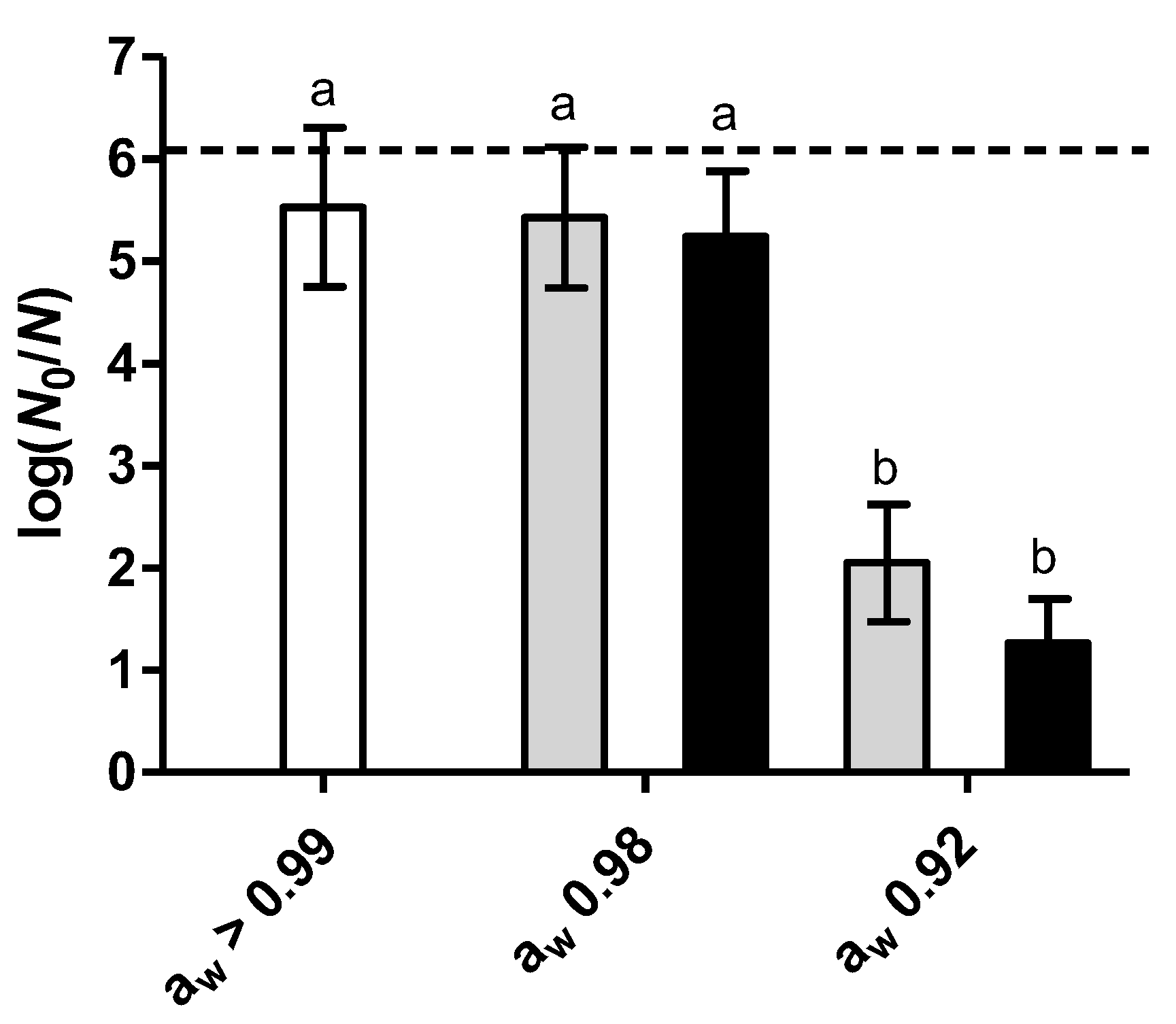
3.1. Effect of RL on Heat Inactivation at Low Water Activity

Figure 1 shows the heat inactivation (60 °C, 5 min) of L. monocytogenes Scott A in sodium phosphate buffer (aw > 0.99) and in the same buffer with reduced aw from >0.99 to 0.98 or 0.92 by the addition of the solutes. While reducing aw to 0.98 by either glycerol or NaCl did not significantly (p > 0.05) change inactivation, further reduction to aw 0.92 exerted a strong protective effect, decreasing inactivation by 3.5 to 4.3 log cycles compared to the control (aw > 0.99).
It is well known that bacterial heat resistance increases when reducing the aw of the treatment medium, as has been documented for L. monocytogenes [35]. This phenomenon is attributed to the reduced mobility of water molecules and the increased stability of proteins and ribosomes promoted by the presence of solutes, although this effect is dependent on the type of solute, treatment temperature and bacterial strain [23,24]. Due to the large protective effect against heat observed at aw 0.92, further experiments were conducted at this condition.
Subsequently, we studied the inactivation of L. monocytogenes Scott A by heat (60 °C, 5 min) combined with RL at aw 0.92 in comparison with the inactivation of each individual treatment. To verify whether RL lethality had a concentration-dependent effect, a range of concentrations from 1 to 250 µg/mL was studied. Using glycerol as a solute, less than 0.2 log cycles of inactivation was observed when RL was added, whereas the heat treatment (without RL) caused about 1.5 log reduction (Figure 2A). Notably, the combination of both hurdles considerably boosted the inactivation to around 5.0 log cycles for RL concentrations from 50 µg/mL to 250 µg/mL, indicating the existence of a synergistic lethal effect. At lower RL concentrations, the degree of synergy between RL and heat progressively decreased until it disappeared at 1 µg/mL.
Using NaCl as a solute, RL displayed a concentration-dependent bactericidal effect at room temperature, with inactivation ranging from 0.4 log cycles at 10 µg/mL to 1.8 log cycles at 250 µg/mL (Figure 2B). The synergy of RL with heat was also stronger in the presence of NaCl than of glycerol as a solute: L. monocytogenes population was reduced by ≥6.0 log cycles at RL concentrations between 25 µg/mL and 250 µg/mL. The above results suggest that RL may offer potential to make the heat treatment of foods with reduced aw more effective.
There is no data available regarding microbial inactivation by the combination of heat and RL; however, there exist similar studies using other natural antimicrobials in high-moisture matrices. For instance, the population of L. monocytogenes Scott A present in semi-skimmed milk was reduced by 4 log cycles after combining heat (60 °C, 6 min) with vanillin (1400 ppm) [36]. The combination of essential oils from Laurus mobilis and Myrtus communis L. (0.2 µL/mL) with mild heat (54 °C, 10 min) in buffer systems (pH 7.0) resulted in the synergistic inactivation of L. monocytogenes EGD-e, reducing the population by 3.5 log cycles [37]. The thermal resistance of L. monocytogenes STCC 4032 was reduced by 1–4 log cycles by 0.5 mM D-limonene at 55 °C (1 min), depending on whether the compound was added directly or in the form of a nanoemulsion [38].
It has been demonstrated that the lethality of natural antimicrobials may be reduced in low-water-activity systems, probably due to lower chances of contact between the compounds and the cell target [39,40]. As such, the inactivation of Salmonella Tennessee by cinnamaldehyde (125–500 mg/L), carvacrol (125–500 mg/L) or lauric arginate (50–200 mg/L) after 3 days of incubation (25 °C) was dramatically reduced when decreasing aw from >0.99 to 0.7–0.3 in a glycerol–sucrose model and peanut paste [40]. However, we found only one study on the effectiveness of synergistic heat and antimicrobial combinations in low-moisture systems, in which it was reported that the synergetic lethal effect between heat and oregano essential oil (1–2%) or Ԑ-polylysine (0.4%) in diluted tahini (aw > 0.99) was completely lost in undiluted tahini (aw 0.256–0.335) [39]. The reason for this effect may be that the antimicrobials are concentrated in the oil phase in undiluted tahini. Our experiments were conducted in a fat-free medium and showed a strong synergy between RL and heat at aw 0.92, suggesting that such a hurdle approach could be effective for processing low-fat low-water-activity foods.
The antimicrobial action of RL is based on disturbance of the cell membrane integrity [41], although the precise details of the mechanism are unknown. The generation of reactive oxygen species (ROS) was also proposed to contribute to the bactericidal effect of rhamnolipids [42]. As a surfactant, RL molecules can self-assemble into structures such as micelles, vesicles and lamellas [43] at concentrations above their critical micelle concentration (CMC). Actually, the role of RL aggregation in its antimicrobial activity is not well understood: while surfactant monomers can easily insert themselves into the cell membrane, the presence of molecular aggregates can enhance local surfactant concentration and charge [44]. According to Rodrigues et al. [45], the antimicrobial activity of RL against two Aspergillus species was associated with increasing micelle size and with the presence of vesicle-like molecular aggregates. Factors, such as temperature, pH, ionic strength and surfactant concentration, influence the type, size and charge of molecular self-assembly structures [46,47,48], and therefore, it is reasonable to assume that changes in these parameters will have an impact on RL antimicrobial activity. Temperature influences CMC, and such an effect is dependent on the type of surfactant. For ionic surfactants, such as RL, an increase in temperature can reduce the hydration of the hydrophilic head group favoring the formation of micelles and consequently decreasing their CMC [49].
It is important to note that the combined treatment was more effective with NaCl as a solute than with glycerol at the same aw and at all RL concentrations tested (Figure 2). These results suggest that, although heat increases the antimicrobial effect of RL in the presence of both solutes, NaCl and glycerol interact differently with RL. Possibly, the ionic nature of NaCl may enhance RL antimicrobial activity compared to glycerol. Given the strong lethal effect of RL in the presence of NaCl, we proceeded to investigate whether the combination of RL and NaCl exerted a growth inhibitory effect on L. monocytogenes.

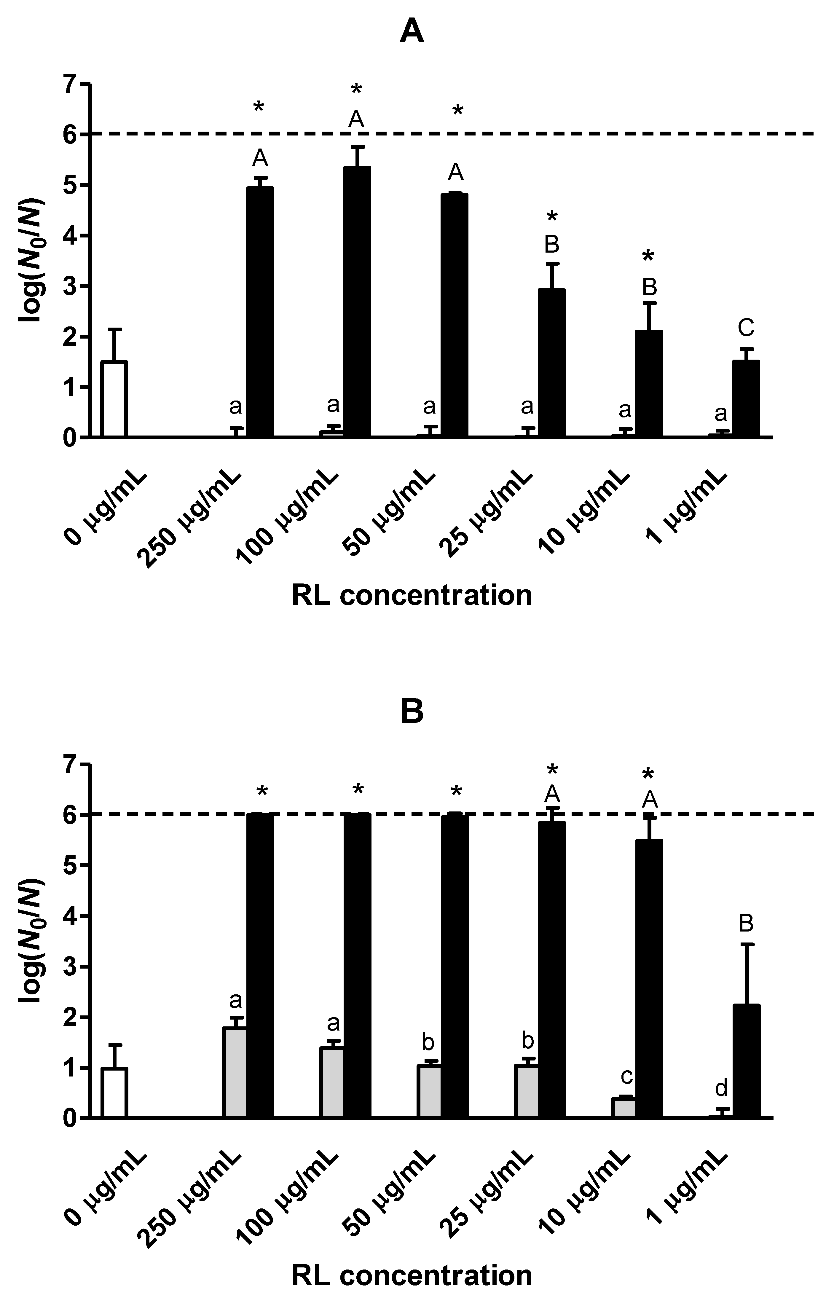
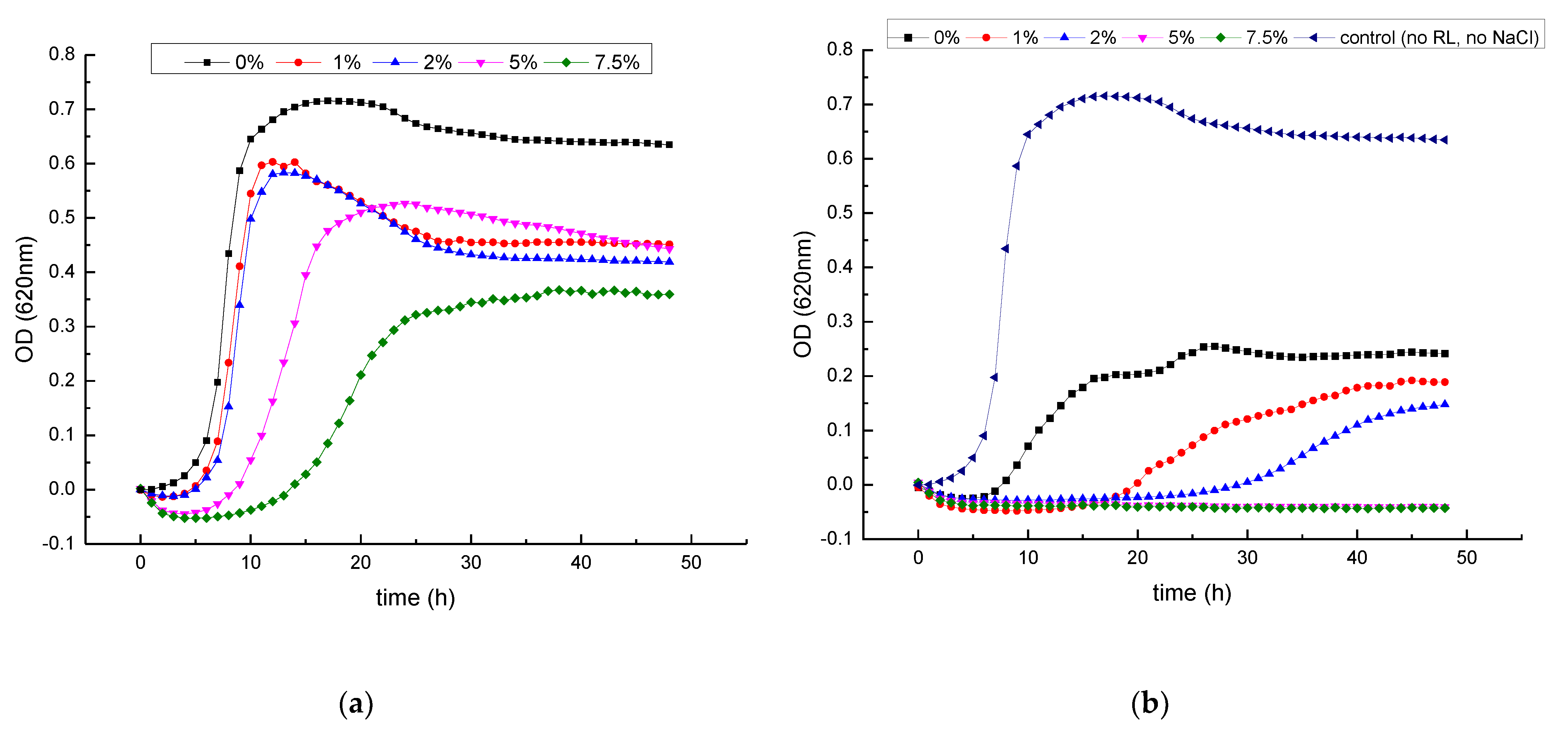
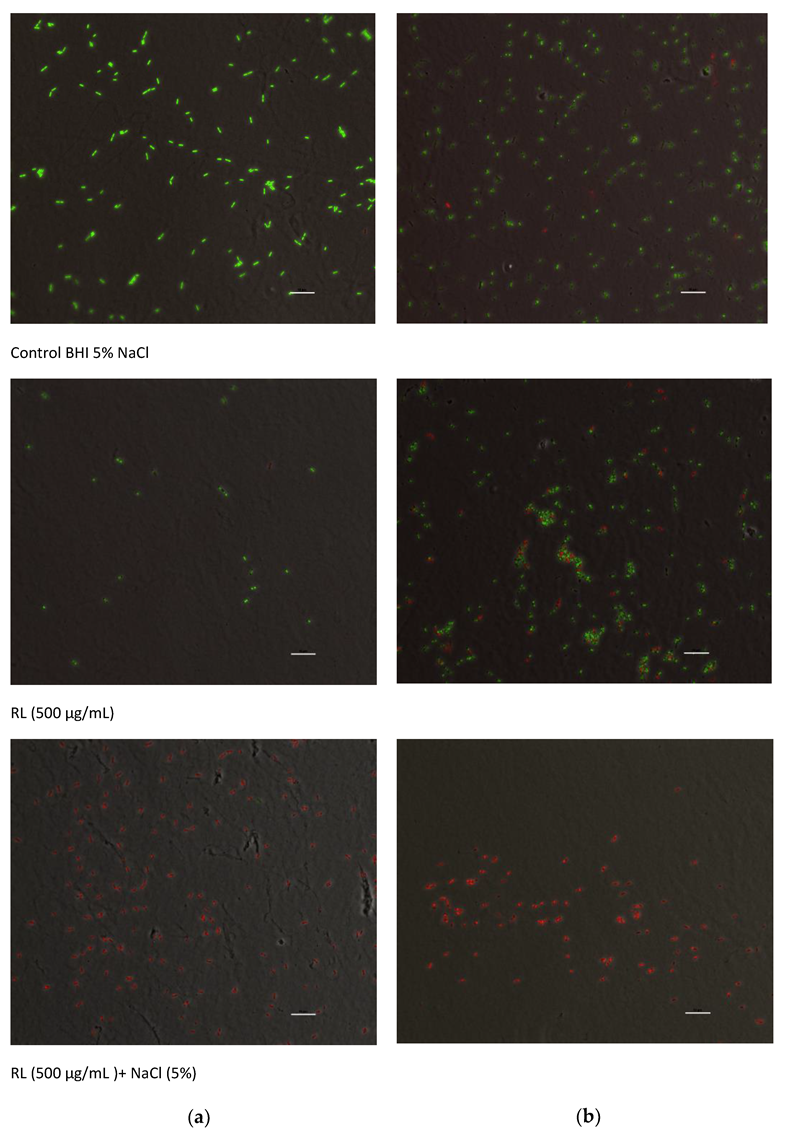
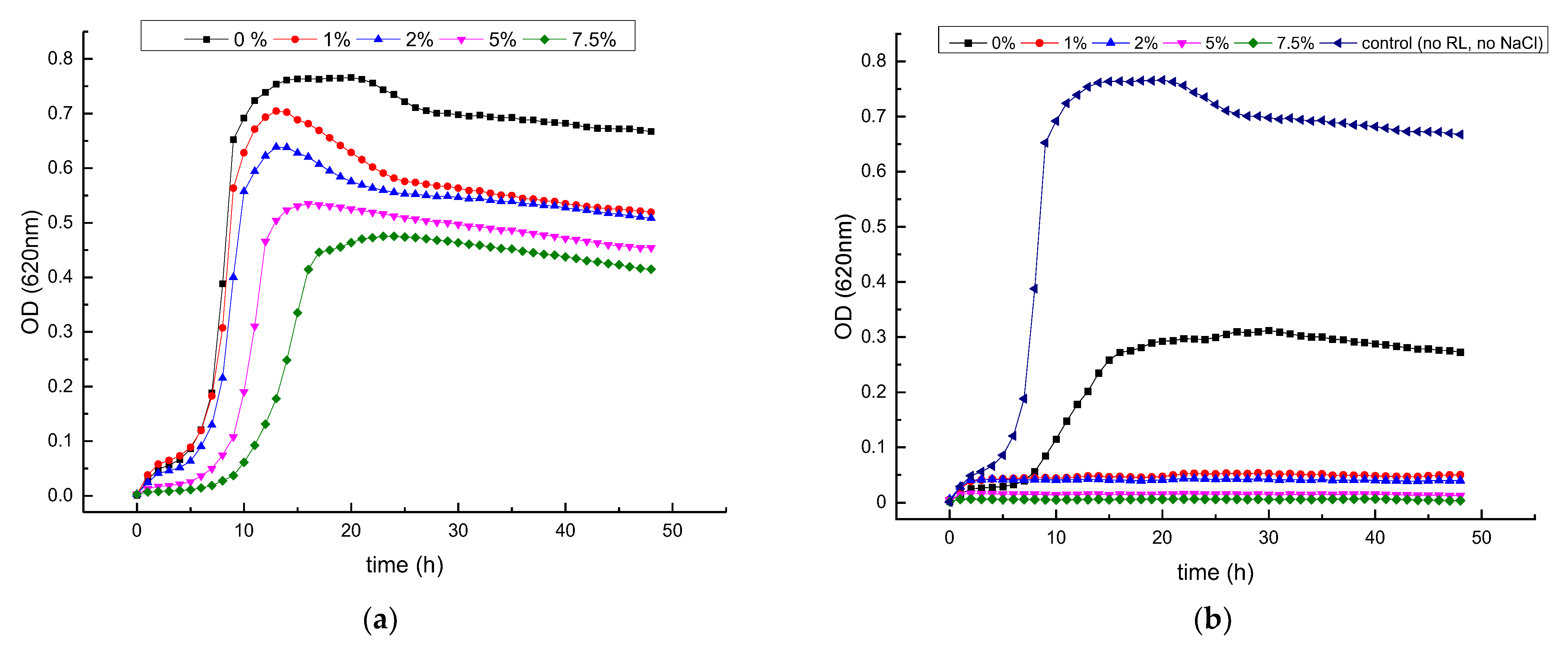
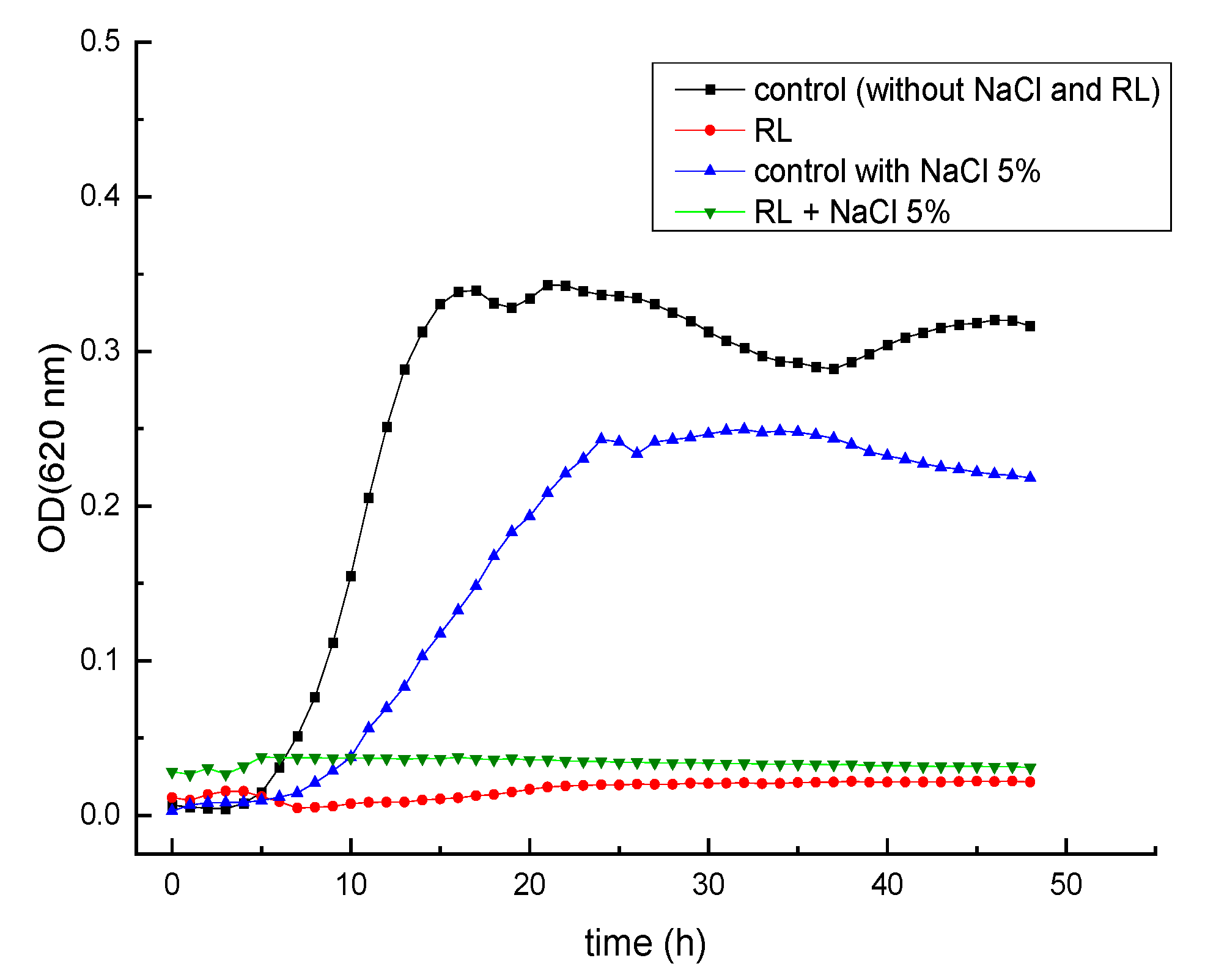
3.2. Growth Inhibitory Activity of RL in Combination with Salts

The growth kinetics of L. monocytogenes in BHI with 0.0%, 1.0%, 2.0%, 5.0% and 7.5% NaCl with and without 500 µg/mL of RL is shown in Figure 3. In the absence of RL, lag phases increased and growth rates decreased especially at 7.5% NaCl but not at lower concentrations (Figure 3A). The addition of RL (500 µg/mL) to the medium impaired bacterial growth but also rendered the bacteria more sensitive to NaCl, even at the lowest concentration used (1%) (Figure 3B). At the highest NaCl concentrations (5.0–7.5%), growth was completely inhibited. The viability staining of the bacterial cells after growing in culture medium with 5% NaCl added (control), RL (500 μg/mL) and the combination of both (RL+NaCl) is displayed in Figure 4. Green viable cells were present in control medium and also after the treatment with RL for 4 h (Figure 4A) and 24 h (Figure 4B), whereas no live cells were visible after the combined treatment even after only 4 h. Thus, it can be concluded that NaCl and RL inactivate L. monocytogenes in a synergistic manner.
While NaCl is one of the most commonly used food additives, and many foods depend partly on NaCl for their microbiological stability and safety, the growing pressure for the reduction of dietary sodium intake is pushing food producers to replace NaCl by substitutes such potassium salts [50]. Therefore, we evaluated if KCl and RL (500 µg/mL) also synergistically inhibited the growth of L. monocytogenes. Remarkably, although KCl was slightly less inhibitory than NaCl when used alone, it had a stronger synergistic effect when combined with RL than NaCl (Figure 5). Even at the lowest KCl concentration (1.0%), growth was completely inhibited in combination with RL (500 µg/mL).
The interaction between RL and NaCl or KCl was investigated in more detail by determining the MIC and MBC values of RL at different salt concentrations (Table 1). In the absence of salts, no MIC or MBC value could be determined because growth was not completely inhibited even at the highest RL concentration. At salt concentrations of 1.0% and 2.0%, the MIC and MBC values of RL were considerably lower for KCl than for NaCl, while at 5.0% and 7.5%, there was no difference. These results confirm that KCl has a stronger synergistic effect than NaCl in combination with RL.
At the neutral pH of the BHI culture medium, the RL carboxyl groups are dissociated, resulting in a negatively charged structure [51], in which strong repulsive forces exist between the head groups of RL molecules. Cations of opposite charge (Na+ or K+) shield these negative charges and thus decrease the repulsive forces and lower the CMC [48,52]. Moreover, the addition of salts increases the micelle size and aggregation number of ionic surfactants [53]. Thus, micelles are formed at lower surfactant concentration, and more surfactant molecules are present in the micelles.
Na+ and K+ cations may have a different effect because of their different charge/size ratio. In water, the radius of a hydrated Na+ cation is larger than that of a hydrated K+ cation [50], and consequently, the binding of Na+ with surfactant molecules is weaker than that of K+. Sood and Aggarwal [53] reported that the CMC of the anionic surfactant sodium dodecylbenzene sulphonate was lower in the presence of KCl than in the presence of NaCl. In line with this behavior, the CMC of RL was lower in KCl than in NaCl considering the same molar concentration of both salts (Table 2). However, our antimicrobial assays were conducted using equal percentage (w/v) concentrations of salts (at pH 7.0), and, under such conditions, the CMC of RL was lower in NaCl than in KCl (Table 2). Thus, the enhanced antimicrobial activity of the combination of KCl and RL cannot be only attributed to decreased CMC, and therefore other specific interactions of K+ cations with cell surface components or in surfactant micelle organization may account for the observed results.
The carboxyl groups of RL molecules become predominantly protonated (uncharged) when the pH is lower than the pKa, which is 5.6 and 5.9 for di and mono-RL forms, respectively [43,54]. In previous work, it was observed that the antimicrobial activity of RL increased under acidic conditions [55], and authors postulated that the electrostatic repulsion between RL and the cell surface was reduced, favoring cell–surfactant interaction.
If the increased antimicrobial activity of RL at low pH and in the presence of salts were indeed due to the reduction or shielding of negative charges, it would be predicted that the cations would have little or no effect under acidic conditions wherein RL are nonionic. To test this hypothesis, we conducted a new set of experiments in BHI medium adjusted to pH 5.0. Figure 6 shows that L. monocytogenes Scott A was able to grow in BHI adjusted to pH 5.0 when 5% NaCl was added (control). Nevertheless, when the culture broth was supplemented with RL (500 µg/mL), the growth was inhibited independently of the presence of NaCl. Furthermore, the MIC value of RL at pH 5.0 was 19.5 µg/mL for both NaCl-supplemented and -non-supplemented media (data not shown). Comparatively, when RL were utilized under neutral pH (Table 1), the MIC was reduced from >2500 (without NaCl) to 78 µg/mL (with 5% NaCl). These results suggest that the nonionic form is more effective against L. monocytogenes and confirm that the effect of salts is observed only at pH values wherein negatively charged RL molecules predominate in solution. As such, they lend support to the hypothesis that the shielding of negative charges by monovalent cations makes RL behave as nonionic surfactants favoring their interaction with cells.
Table 2 shows that the CMC of RL decreased as NaCl concentration increased in solution. The addition of 5% NaCl lowered the CMC from 94.2 to 16.0 at pH 7.0 and the MIC from >2500 to 78 µg/mL (Table 1). The effect of salt on surfactant CMC was less evident at pH 5.0, probably due to the predominance of the nonionic form of the RL as discussed above (Table 2). The increase in pH also increased the CMC of the surfactant in the absence of salt and at specific ionic strength; however, this was not observed at the highest salt concentration. Our findings are in agreement with similar reports in literature regarding the CMC behavior of RL [56,57].
An interesting question is whether the increased antimicrobial effect observed when the negative charge of RL is reduced or shielded is only due to the decreased repulsion of RL and the bacterial surface or is also affected by their molecular aggregation. The morphology of RL molecular aggregates is also governed by pH. An increase of pH from 5.0 to 7.0 induced a shift of RL self-assembly structures from vesicles to lamellas, lipid particles and micelles [43,56]. It is known that membrane vesicles and micelles can fuse with bacterial membranes under particular conditions, and it is conceivable that such fusion events can strongly enhance the antimicrobial activity of surfactants. However, further work is needed to investigate the formation of such structures in relation to environmental conditions (pH, salts) and their interaction with bacterial cells.

4. Conclusions

A hurdle technology approach combining a rhamnolipid biosurfactant and mild heat revealed a strongly synergistic effect against L. monocytogenes in a low-water-activity model using ionic and sugar solutes. Furthermore, the growth-inhibitory activity of RL was also strongly enhanced by the presence of NaCl and KCl, suggesting that the overall antilisterial properties of rhamnolipids depend on the ionic strength of the medium and the surfactant CMC. The results obtained in this work contribute to the understanding of RL antimicrobial action and may open new perspectives regarding their applications in food processing. Notably, further research exploring the efficacy of this natural bio-based surfactant in combination with salts can result in innovative and sustainable strategies to control L. monocytogenes in the food chain.

Author Contributions

Conceptualization, E.G., C.W.M. and M.N.; methodology, E.G., C.W.M. and M.N.; software, L.L., T.F.P., E.G. and M.N.; formal analysis, L.L., T.F.P., E.G. and M.N.; investigation, L.L., T.F.P. and M.N.; data curation, L.L., T.F.P., E.G. and M.N.; writing—original draft preparation, E.G., C.W.M. and M.N.; writing—review and editing, E.G., C.W.M. and M.N.; supervision, E.G., C.W.M. and M.N.; project administration, C.W.M. and M.N.; funding acquisition, C.W.M. and M.N. All authors have read and agreed to the published version of the manuscript.

Funding

This research was funded by the KU Leuven Research Fund (METH/14/03). M. Nitschke acknowledges Fundação de Amparo à Pesquisa do Estado de São Paulo (FAPESP) for providing her research scholarship (Grant nº 2017/18298-7).

Institutional Review Board Statement

Not applicable.

Informed Consent Statement

Not applicable.

Data Availability Statement

Data is contained within the article.

Acknowledgments

Authors thank to Julien Mortier for his assistance with fluorescence microscopy and to Johnson & Johnson (São José dos Campos -SP- Brazil) for the donation of the rhamnolipid biosurfactant.

Conflicts of Interest

The authors declare no conflict of interest.

References

  1. Barba, F.J.; Koubaa, M.; do Prado-Silva, L.; Orlien, V.; Sant’Ana, A.d.S. Mild processing applied to the inactivation of the main foodborne bacterial pathogens: A review. Trends Food Sci. Technol. 2017, 66, 20–35. [Google Scholar] [CrossRef]
  2. Ekonomou, S.I.; Boziaris, I.S. Non-thermal methods for ensuring the microbiological quality and safety of seafood. Appl. Sci. 2021, 11, 833. [Google Scholar] [CrossRef]
  3. De Corato, U. Improving the shelf-life and quality of fresh and minimally-processed fruits and vegetables for a modern food industry: A comprehensive critical review from the traditional technologies into the most promising advancements. Crit. Rev. Food Sci. Nutr. 2020, 60, 940–975. [Google Scholar] [CrossRef]
  4. Pasha, I.; Saeed, F.; Sultan, M.T.; Khan, M.R.; Rohi, M. Recent developments in minimal processing: A tool to retain nutritional quality of food. Crit. Rev. Food Sci. Nutr. 2014, 54, 340–351. [Google Scholar] [CrossRef]
  5. Beuchat, L.R.; Komitopoulou, E.; Beckers, H.; Betts, R.P.; Bourdichon, F.; Fanning, S.; Joosten, H.M.; Ter Kuile, B.H. Low–water activity foods: Increased concern as vehicles of foodborne pathogens. J. Food Prot. 2013, 76, 150–172. [Google Scholar] [CrossRef]
  6. Leistner, L. Basic aspects of food preservation by hurdle technology. Int. J. Food Microbiol. 2000, 55, 181–186. [Google Scholar] [CrossRef] [PubMed]
  7. Naqash, S.; Naqash, F.; Fayaz, S.; Khan, S.; Dar, B.N.; Makroo, H.A. Application of natural antimicrobial agents in different food packaging systems and their role in shelf-life extension of food: A Review. J. Packag. Technol. Res. 2022, 6, 73–89. [Google Scholar]
  8. Boskovic, M.; Djordjevic, J.; Ivanovic, J.; Janjic, J.; Zdravkovic, N.; Glisic, M.; Glamoclija, N.; Baltic, B.; Djordjevic, V.; Baltic, M. Inhibition of Salmonella by thyme essential oil and its effect on microbiological and sensory properties of minced pork meat packaged under vacuum and modified atmosphere. Int. J. Food Microbiol. 2017, 258, 58–67. [Google Scholar] [CrossRef]
  9. García-Díez, J.; Alheiro, J.; Pinto, A.L.; Soares, L.; Falco, V.; Fraqueza, M.J.; Patarata, L. Behaviour of food-borne pathogens on dry cured sausage manufactured with herbs and spices essential oils and their sensorial acceptability. Food Control 2016, 59, 262–270. [Google Scholar] [CrossRef]
  10. Mulligan, C.N.; Sharma, S.K.; Mudhoo, A.; Makhijani, K. Green chemistry and biosurfactant research. In Biosurfactants Research Trends and Applications; Mulligan, C.N., Sharma, S.K., Mudhoo, A., Eds.; CRC Press: Boca Raton, FL, USA, 2014; pp. 1–30. [Google Scholar]
  11. Nitschke, M.; Silva, S.S. Recent food applications of microbial surfactants. Crit. Rev. Food Sci. Nutr. 2018, 58, 631–638. [Google Scholar] [CrossRef]
  12. US Environmental Protection Agency Office of Pesticide Programs. Biopesticides Registration Action Document. Rhamnolipid Biosurfactant (PC Code 110029). 2004. Available online: https://www3.epa.gov/pesticides/chem_search/reg_actions/registration/decision_PC-110029_11-May-04.pdf (accessed on 11 January 2023).
  13. Hamon, M.; Bierne, H.; Cossart, P. Listeria monocytogenes: A multifaceted model. Nat. Rev. Microbiol. 2006, 4, 423–434. [Google Scholar] [CrossRef] [PubMed]
  14. Townsend, A.; Strawn, L.K.; Chapman, B.J.; Dunn, L.L. A systematic review of Listeria species and Listeria monocytogenes prevalence, persistence, and diversity throughout the fresh produce supply chain. Foods 2021, 10, 1427. [Google Scholar] [CrossRef] [PubMed]
  15. Buchanan, R.L.; Gorris, L.G.M.; Hayman, M.M.; Jackson, T.C.; Whiting, R.C. A review of Listeria monocytogenes: An update on outbreaks, virulence, dose-response, ecology, and risk assessments. Food Control 2017, 75, 1–13. [Google Scholar] [CrossRef]
  16. Tienungoon, S.; Ratkowsky, D.A.; McMeekin, T.A.; Ross, T. Growth limits of Listeria monocytogenes as a function of temperature, pH, NaCl, and lactic acid. Appl. Environ. Microbiol. 2000, 66, 4979–4987. [Google Scholar] [CrossRef] [Green Version]
  17. Miller, A.J. Combined water activity and solute effects on growth and survival of Listeria monocytogenes Scott A. J. Food Prot. 1992, 55, 414–418. [Google Scholar] [CrossRef]
  18. Pang, X.; Wong, C.; Chung, H.-J.; Yuk, H.-G. Biofilm formation of Listeria monocytogenes and its resistance to quaternary ammonium compounds in a simulated salmon processing environment. Food Control 2019, 98, 200–208. [Google Scholar] [CrossRef]
  19. Shamloo, E.; Abdimoghadam, Z.; Nazari, K.; Hosseini, S.M.; Hosseini, H.; Alebouyeh, M. Long-term survival of Listeria monocytogenes in stress conditions: High pH and salt concentrations. J. Res. Med. Dent. Sci. 2018, 6, 96–100. [Google Scholar]
  20. Araujo, L.V.; Abreu, F.; Lins, U.; Santa Anna, L.M.d.M.; Nitschke, M.; Freire, D.M.G. Rhamnolipid and surfactin inhibit Listeria monocytogenes adhesion. Food Res. Int. 2011, 44, 481–488. [Google Scholar] [CrossRef]
  21. Magalhães, L.; Nitschke, M. Antimicrobial activity of rhamnolipids against Listeria monocytogenes and their synergistic interaction with nisin. Food Control 2013, 29, 138–142. [Google Scholar] [CrossRef] [Green Version]
  22. Sotirova, A.V.; Spasova, D.I.; Galabova, D.N.; Karpenko, E.; Shulga, A. Rhamnolipid–biosurfactant permeabilizing effects on Gram-positive and Gram-negative bacterial strains. Curr. Microbiol. 2008, 56, 639–644. [Google Scholar] [CrossRef]
  23. Smelt, J.P.P.M.; Brul, S. Thermal inactivation of microorganisms. Crit. Rev. Food Sci. Nutr. 2014, 54, 1371–1385. [Google Scholar] [CrossRef] [PubMed]
  24. Syamaladevi, R.M.; Tang, J.; Villa-Rojas, R.; Sablani, S.; Carter, B.; Campbell, G. Influence of water activity on thermal resistance of microorganisms in low-moisture foods: A review. Compr. Rev. Food. Sci. Food Saf. 2016, 15, 353–370. [Google Scholar] [CrossRef] [PubMed]
  25. Gayán, E.; Geens, E.; Berdejo, D.; García-Gonzalo, D.; Pagán, R.; Aertsen, A.; Michiels, C.W. Combination of mild heat and plant essential oil constituents to inactivate resistant variants of Escherichia coli in buffer and in coconut water. Food Microbiol. 2020, 87, 103388. [Google Scholar]
  26. Espina, L.; Somolinos, M.; Ouazzou, A.A.; Condón, S.; García-Gonzalo, D.; Pagán, R. Inactivation of Escherichia coli O157:H7 in fruit juices by combined treatments of citrus fruit essential oils and heat. Int. J. Food Microbiol. 2012, 159, 9–16. [Google Scholar] [PubMed]
  27. Arioli, S.; Montanari, C.; Magnani, M.; Tabanelli, G.; Patrignani, F.; Lanciotti, R.; Mora, D.; Gardini, F. Modelling of Listeria monocytogenes Scott A after a mild heat treatment in the presence of thymol and carvacrol: Effects on culturability and viability. J. Food Eng. 2019, 240, 73–82. [Google Scholar] [CrossRef]
  28. Fugett, E.; Fortes, E.; Nnoka, C.; Wiedmann, M. International Life Sciences Institute North America Listeria monocytogenes strain collection: Development of standard Listeria monocytogenes strain sets for research and validation studies. J. Food Prot. 2006, 69, 2929–2938. [Google Scholar] [CrossRef] [PubMed]
  29. Sieuwerts, S.; de Bok, F.A.M.; Mols, E.; de Vos, W.M.; van Hylckama Vlieg, J.E.T. A simple and fast method for determining colony forming units. Lett. Appl. Microbiol. 2008, 47, 275–278. [Google Scholar] [CrossRef]
  30. Feyaerts, J.; Rogiers, G.; Corthouts, J.; Michiels, C.W. Thiol-reactive natural antimicrobials and high-pressure treatment synergistically enhance bacterial inactivation. Innov. Food Sci. Emerg. Technol. 2015, 27, 26–34. [Google Scholar] [CrossRef]
  31. Clinical and Laboratory Standards Institute (CLSI). Methods for Dilution Antimicrobial Susceptibility Tests for Bacteria That Grow Aerobically, 9th ed.; Approved Standard; CLSI Document M07-A9; Clinical and Laboratory Standards Institute (CLSI): Wayne, PA, USA, 2012. [Google Scholar]
  32. Rodríguez-Melcón, C.; Alonso-Calleja, C.; García-Fernández, C.; Carballo, J.; Capita, R. Minimum Inhibitory Concentration (MIC) and Minimum Bactericidal Concentration (MBC) for twelve antimicrobials (biocides and antibiotics) in eight strains of Listeria monocytogenes. Biology 2022, 11, 46. [Google Scholar] [CrossRef]
  33. Sun, L.; Rogiers, G.; Courtin, P.; Chapot-Chartier, M.P.; Bierne, H.; Michiels, C.W. AsnB mediates amidation of meso-diaminopimelic acid residues in the peptidoglycan of Listeria monocytogenes and affects bacterial surface properties and host cell invasion. Front. Microbiol. 2021, 12, 760253. [Google Scholar] [CrossRef]
  34. LIVE/DEAD BacLight Bacterial Viability Kit Protocol. Available online: https://www.thermofisher.com/br/en/home/references/protocols/cell-and-tissue-analysis/protocols/live-dead-baclight-bacterial-viability-protocol.html (accessed on 9 January 2023).
  35. Fernández, A.; López, M.; Bernardo, A.; Condón, S.; Raso, J. Modelling thermal inactivation of Listeria monocytogenes in sucrose solutions of various water activities. Food Microbiol. 2007, 24, 372–379. [Google Scholar] [CrossRef] [PubMed]
  36. Cava-Roda, R.M.; Taboada, A.; Palop, A.; López-Gómez, A.; Marin-Iniesta, F. Heat resistance of Listeria monocytogenes in semi-skim milk supplemented with vanillin. Int. J. Food Microbiol. 2012, 157, 314–318. [Google Scholar] [CrossRef] [PubMed]
  37. Cherrat, L.; Espina, L.; Bakkali, M.; García-Gonzalo, D.; Pagán, R.; Laglaoui, A. Chemical composition and antioxidant properties of Laurus nobilis L. and Myrtus communis L. essential oils from Morocco and evaluation of their antimicrobial activity acting alone or in combined processes for food preservation. J. Sci. Food Agric. 2014, 94, 1197–1204. [Google Scholar] [CrossRef] [PubMed]
  38. Maté, J.; Periago, P.M.; Palop, A. When nanoemulsified, d-limonene reduces Listeria monocytogenes heat resistance about one hundred times. Food Control 2016, 59, 824–828. [Google Scholar] [CrossRef] [Green Version]
  39. Xu, Y.; Guan, X.; Lin, B.; Li, R.; Wang, S. Oregano oil, epsilon-polylysine and citric acid assisted inactivation of Salmonella in two kinds of tahini during thermal treatment and storage. Foods 2021, 10, 1272. [Google Scholar] [CrossRef]
  40. Chen, W.; Golden, D.A.; Critzer, F.J.; Davidson, P.M. Antimicrobial activity of cinnamaldehyde, carvacrol, and lauric arginate against Salmonella Tennessee in a glycerol-sucrose model and peanut paste at different fat concentrations. J. Food Prot. 2015, 78, 1488–1495. [Google Scholar]
  41. Ortiz, A.; Teruel, J.A.; Espuny, M.J.; Marqués, A.; Manresa, Á.; Aranda, F.J. Effects of dirhamnolipid on the structural properties of phosphatidylcholine membranes. Int. J. Pharma. 2006, 325, 99–107. [Google Scholar] [CrossRef]
  42. Gaur, V.K.; Tripathi, V.; Gupta, P.; Dhiman, N.; Regar, R.K.; Gautam, K.; Srivastava, J.K.; Patnaik, S.; Patel, D.K.; Manickam, N. Rhamnolipids from Planococcus spp. and their mechanism of action against pathogenic bacteria. Bioresour. Technol. 2020, 307, 123206. [Google Scholar] [CrossRef]
  43. Yutaka, I.; Yasuo, G.; Hitoshi, N.; Muneo, Y.; Hisae, N.; Toshio, K. The pH-Sensitive conversion of molecular aggregates of rhamnolipid biosurfactant. Chem. Lett. 1987, 16, 763–766. [Google Scholar]
  44. Zhou, C.; Wang, F.; Chen, H.; Li, M.; Qiao, F.; Liu, Z.; Hou, Y.; Wu, C.; Fan, Y.; Liu, L.; et al. Selective antimicrobial activities and action mechanism of micelles self-assembled by cationic oligomeric surfactants. ACS Appl. Mat. Interfaces 2016, 8, 4242–4249. [Google Scholar] [CrossRef] [PubMed]
  45. Rodrigues, A.I.; Gudiña, E.J.; Teixeira, J.A.; Rodrigues, L.R. Sodium chloride effect on the aggregation behaviour of rhamnolipids and their antifungal activity. Sci. Rep. 2017, 7, 12907. [Google Scholar] [CrossRef] [PubMed] [Green Version]
  46. Silva, S.S.; Carvalho, J.W.P.; Aires, C.P.; Nitschke, M. Disruption of Staphylococcus aureus biofilms using rhamnolipid biosurfactants. J. Dairy Sci. 2017, 100, 7864–7873. [Google Scholar] [CrossRef] [PubMed] [Green Version]
  47. Zhong, H.; Yang, L.; Yang, X.; Zeng, G.; Liu, Z.; Liu, Y.; Yuan, X. Aggregation of low-concentration dirhamnolipid biosurfactant in electrolyte solution. RSC Adv. 2015, 5, 88578–88582. [Google Scholar] [CrossRef] [Green Version]
  48. Helvacı, Ş.Ş.; Peker, S.; Özdemir, G. Effect of electrolytes on the surface behavior of rhamnolipids R1 and R2. Colloids Surf. B Biointerfaces 2004, 35, 225–233. [Google Scholar] [CrossRef] [PubMed]
  49. Demissie, H.; Duraisamy, R. Effects of electrolytes on the surface and micellar characteristics of sodium dodecyl sulphate surfactant solution. J. Sci. Innov. Res. 2016, 5, 208–214. [Google Scholar] [CrossRef]
  50. Cepanec, K.; Vugrinec, S.; Cvetković, T.; Ranilović, J. Potassium chloride-based salt substitutes: A critical review with a focus on the patent literature. Compr. Rev. Food. Sci. Food Saf. 2017, 16, 881–894. [Google Scholar] [CrossRef] [Green Version]
  51. Lebrón-Paler, A.; Pemberton, J.E.; Becker, B.A.; Otto, W.H.; Larive, C.K.; Maier, R.M. Determination of the acid dissociation constant of the biosurfactant monorhamnolipid in aqueous solution by potentiometric and spectroscopic methods. Anal. Chem. 2006, 78, 7649–7658. [Google Scholar] [CrossRef] [PubMed]
  52. Ahmadi, M.A.; Shadizadeh, S.R.; Salari, Z. Dependency of critical micellization concentration of an anionic surfactant on temperature and potassium chloride salt. Pet. Sci. Technol. 2014, 32, 1913–1920. [Google Scholar] [CrossRef]
  53. Sood, A.K.; Aggarwal, M. Evaluation of micellar properties of sodium dodecylbenzene sulphonate in the presence of some salts. J. Chem. Sci. 2018, 130, 39. [Google Scholar] [CrossRef] [Green Version]
  54. Abbasi, H.; Noghabi, K.A.; Hamedi, M.M.; Zahiri, H.S.; Moosavi-Movahedi, A.A.; Amanlou, M.; Teruel, J.A.; Ortiz, A. Physicochemical characterization of a monorhamnolipid secreted by Pseudomonas aeruginosa MA01 in aqueous media. An experimental and molecular dynamics study. Colloids Surf. B Biointerfaces 2013, 101, 256–265. [Google Scholar] [CrossRef]
  55. Ferreira, J.F.; Vieira, E.A.; Nitschke, M. The antibacterial activity of rhamnolipid biosurfactant is pH dependent. Food Res. Int. 2019, 116, 737–744. [Google Scholar] [CrossRef] [PubMed]
  56. Raza, Z.A.; Khalid, Z.M.; Khan, M.S.; Banat, I.M.; Rehman, A.; Naeem, A.; Saddique, M.T. Surface properties and sub-surface aggregate assimilation of rhamnolipid surfactants in different aqueous systems. Biotechnol. Lett. 2010, 32, 811–816. [Google Scholar] [CrossRef] [PubMed]
  57. Ciesla, J.; Koczanska, M.; Bieganowski, A. An interaction of rhamnolipids with Cu2+ ions. Molecules 2018, 23, 488. [Google Scholar] [CrossRef] [PubMed]
Figure 1. Logarithmic reduction factor (LRF; log(N0/N)) of L. monocytogenes Scott A by heat treatment (60 °C, 5 min) in sodium phosphate buffer (pH 6.7) at aw > 0.99 (control, white bars), 0.98 or 0.92, adjusted with glycerol (gray bars) or NaCl (black bars). Error bars represent the standard deviation of the means of triplicates. Different letters indicate statistically significant differences (p ≤ 0.05) in the inactivation at different aw levels and using different solutes. The dashed line indicates the LRF corresponding to the quantification limit of surviving cells (1000 CFU/mL).
Foods 12 00570 g001
Figure 2. Logarithmic reduction factor (LRF; log(N0/N)) of L. monocytogenes Scott A in sodium phosphate buffer (pH 6.7) of aw 0.92 with (A) glycerol or (B) NaCl by heat, RL at different concentrations and the combination of heat and RL. The white bar represents heat inactivation alone (60 °C, 5 min; without RL added). For each RL concentration, the gray bar presents inactivation by RL at room temperature (~20 °C, 5 min), while the black bar indicates inactivation by the combination of RL and heat (60 °C, 5 min). The dashed line indicates the maximum detectable LRF corresponding to the quantification limit (1000 CFU/mL). Error bars represent the standard deviations of the means of triplicates. Asterisk indicates statistically significant differences (p ≤ 0.05) between the theoretical additive inactivation by heat and RL and the experimental inactivation obtained by the combined treatment, and therefore the occurrence of a synergistic lethal effect. Lowercase and capital letters indicate statistically significant differences (p ≤ 0.05) in the inactivation by RL and by the combination of heat and RL treatment, respectively, among the different RL concentrations tested.
Foods 12 00570 g002
Figure 3. Growth kinetics of L. monocytogenes Scott A in BHI medium supplemented with (a) different concentrations of NaCl, without RL and (b) in combination with 500 µg/mL of RL at 30 °C. Data points represent the mean values of three replicates. Error bars have been omitted for clarity.
Foods 12 00570 g003
Figure 4. Fluorescence microscopy images of L. monocytogenes Scott A after incubation in BHI medium supplemented with NaCl (5%), rhamnolipids (500 µg/mL) and the combination of both for (a) 4 h and (b) 24 h at 30 °C. Green and red cells correspond to live and dead cells, respectively. Scale bar 10 µm.
Foods 12 00570 g004
Figure 5. Growth kinetics of L. monocytogenes Scott A in BHI medium supplemented with (a) different concentrations of KCl, without RL and (b) in combination with 500 µg/mL of RL at 30 °C. Data points represent the mean values of three replicates. Error bars have been omitted for clarity.
Foods 12 00570 g005
Figure 6. Growth kinetics of L. monocytogenes Scott A in BHI medium at pH 5.0 with and without the addition of 500 µg/mL of RL and 5% NaCl at 30 °C. Data points represent the mean values of three replicates. Error bars have been omitted for clarity.
Foods 12 00570 g006
Table 1. MIC and MBC (between brackets) values for RL (µg/mL) against L. monocytogenes Scott A at different NaCl and KCl concentrations.
Salt Concentration0.0%1.0%2.0%5.0%7.5%
NaCl>2500 (-)2500 (-)312 (-)78 (78)39 (39)
KCl>2500 (-)312 (625)156 (156)78 (78)39 (39)
-: no MBC observed.
Table 2. Critical micelle concentration values of RL solutions under different pH and salt levels.
%NaCl0.0%1.0% (171 mM)2.0% (342 mM)5.0% (855 mM)
pH 5.019.611.510.411.9
pH 6.093.232.017.414.2
pH 7.094.235.826.316.0
pH 8.0121.439.125.615.5
% KCl0.0%1.0% (134 mM)2.0% (268 mM)5.0% (670 mM)
pH 7.094.246.938.321.7
% KCl 1.3% (171mM)2.5% (342 mM)6.4% (855 mM)
pH 7.0 25.519.513.4
Disclaimer/Publisher’s Note: The statements, opinions and data contained in all publications are solely those of the individual author(s) and contributor(s) and not of MDPI and/or the editor(s). MDPI and/or the editor(s) disclaim responsibility for any injury to people or property resulting from any ideas, methods, instructions or products referred to in the content.

