Andean Sacha Inchi (Plukenetia Volubilis L.) Leaf-Mediated Synthesis of Cu2O Nanoparticles: A Low-Cost Approach


Abstract
:
1. Introduction
2. Materials and Methods
2.1. Materials
2.2. Preparation of Cu2O Nanoparticles
2.3. Characterization of Cu2O Nanoparticles
2.4. Photocatalytic Effects
3. Results and Discussion
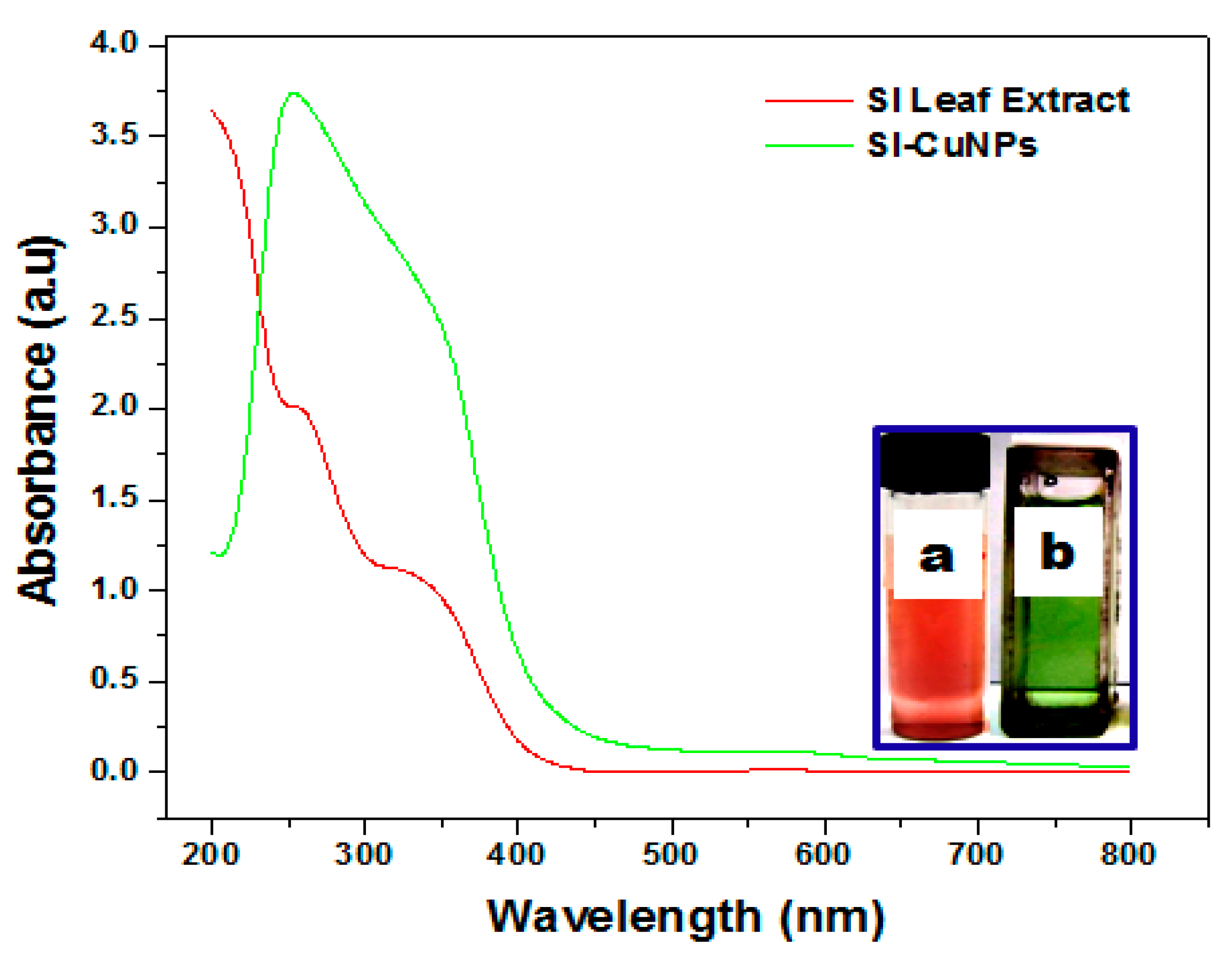
3.1. UV-Vis Spectroscopy Analysis
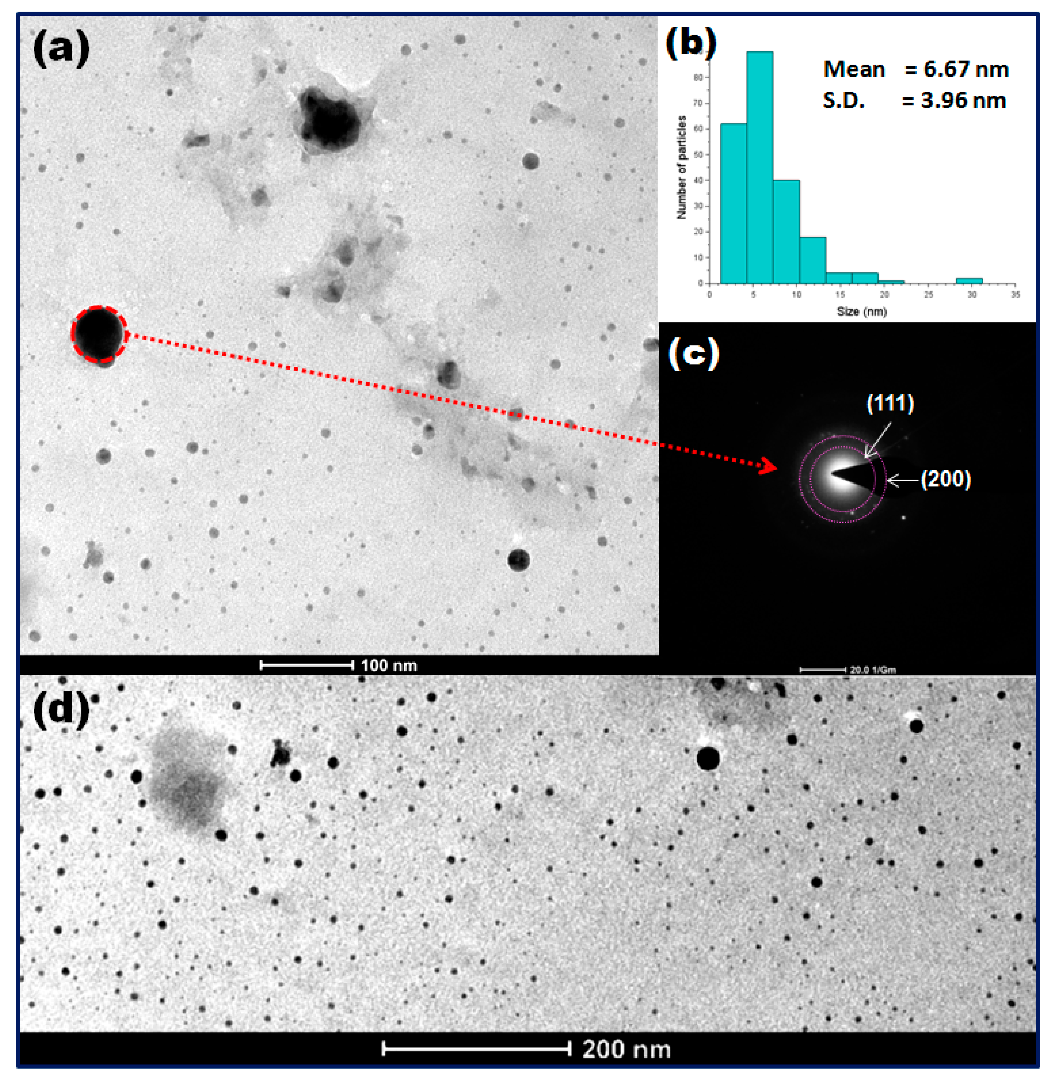
3.2. TEM and SAED Analysis
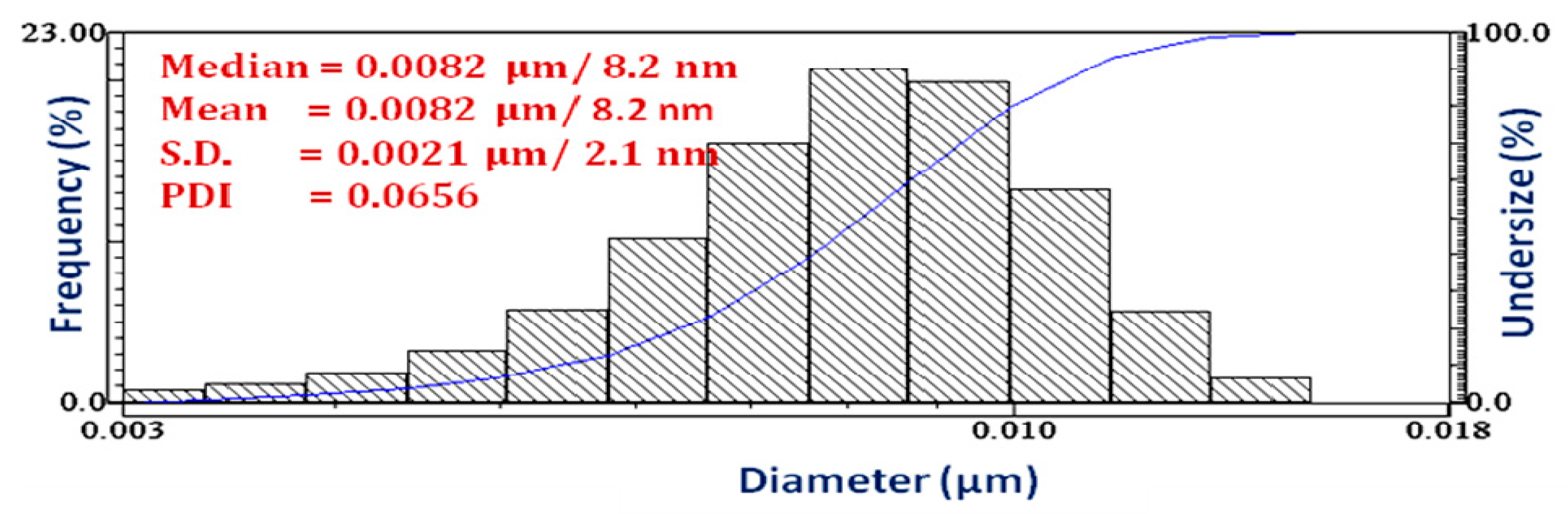
3.3. DLS Analysis
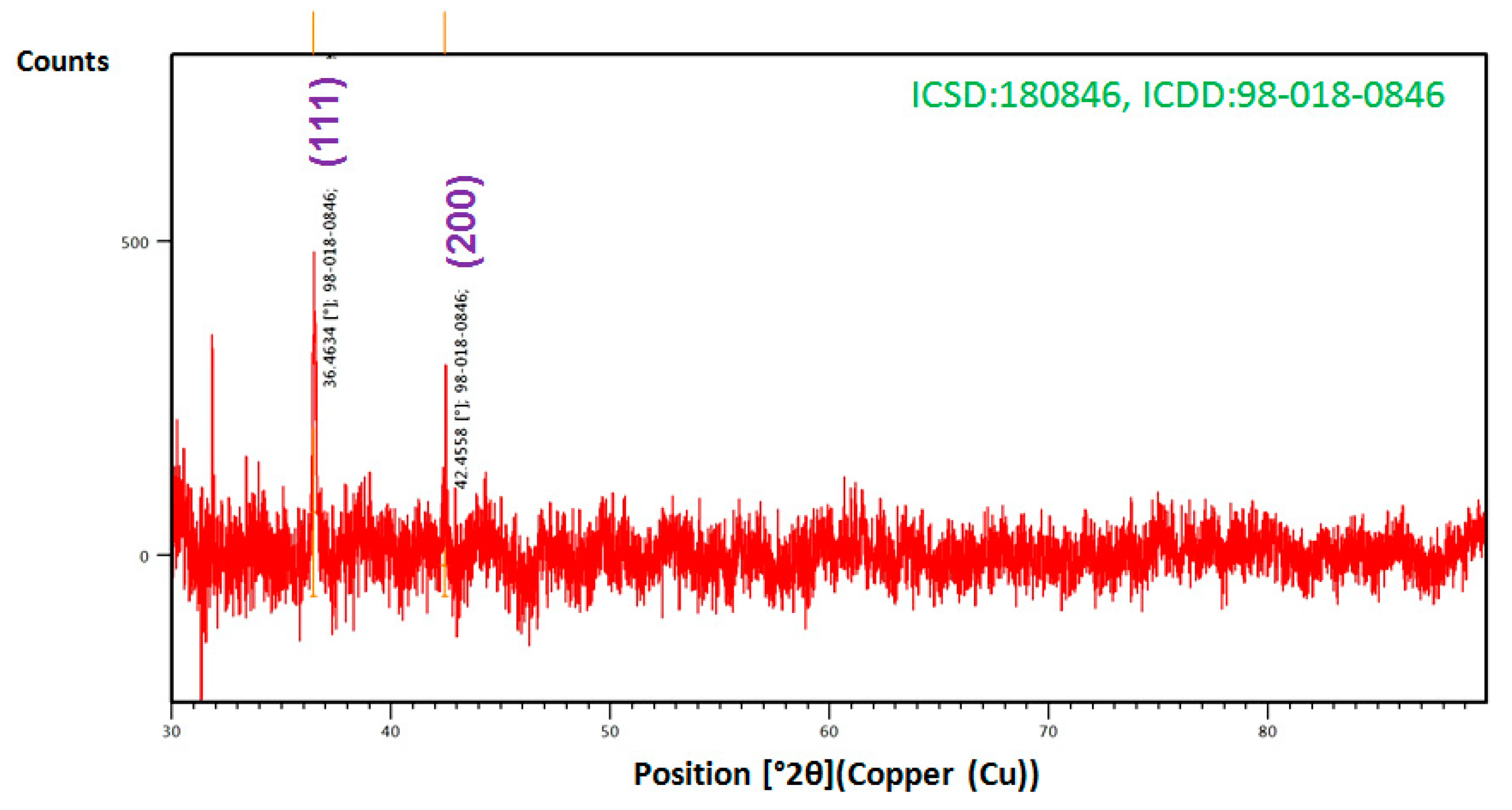
3.4. XRD Analysis
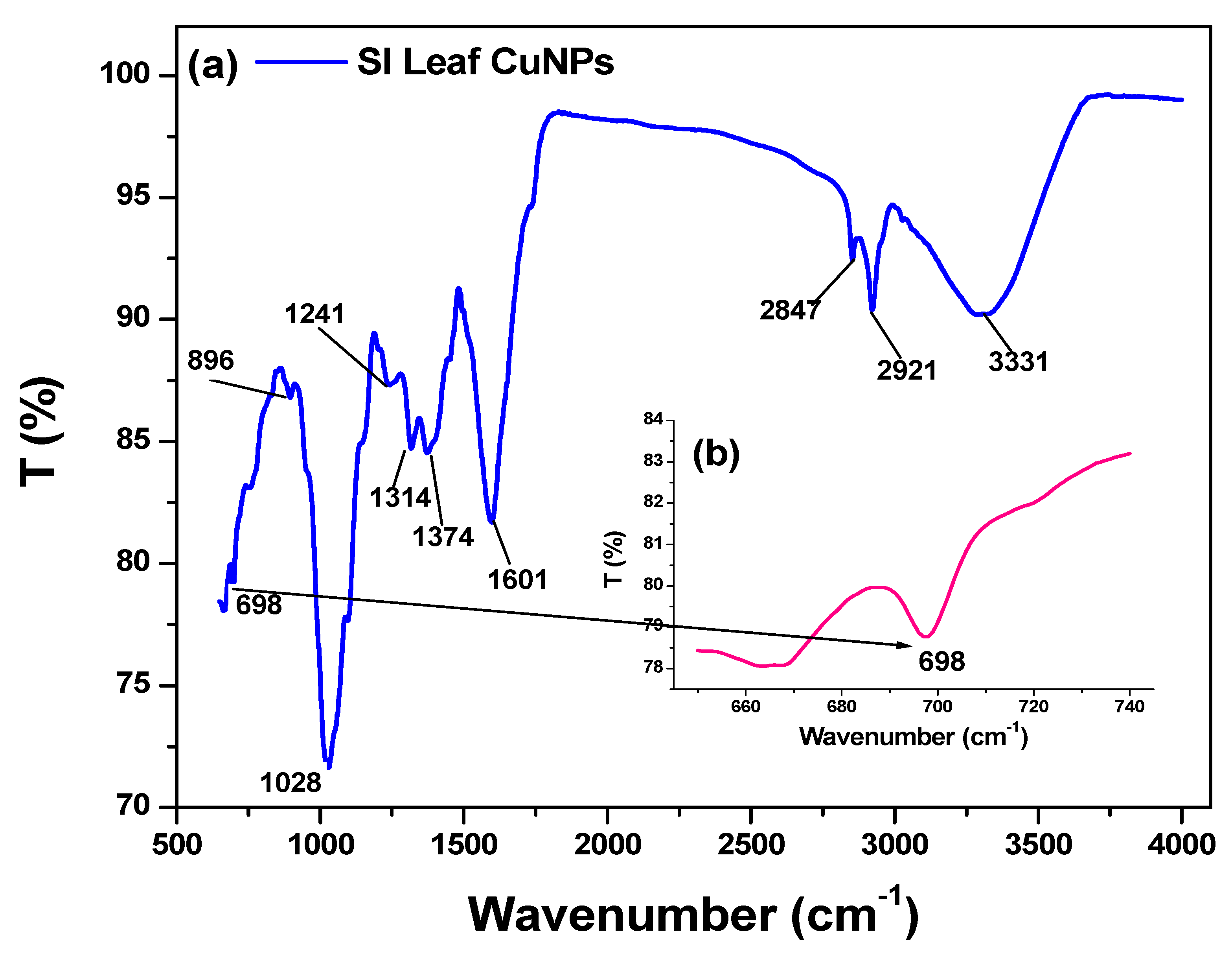
3.5. FTIR Analysis
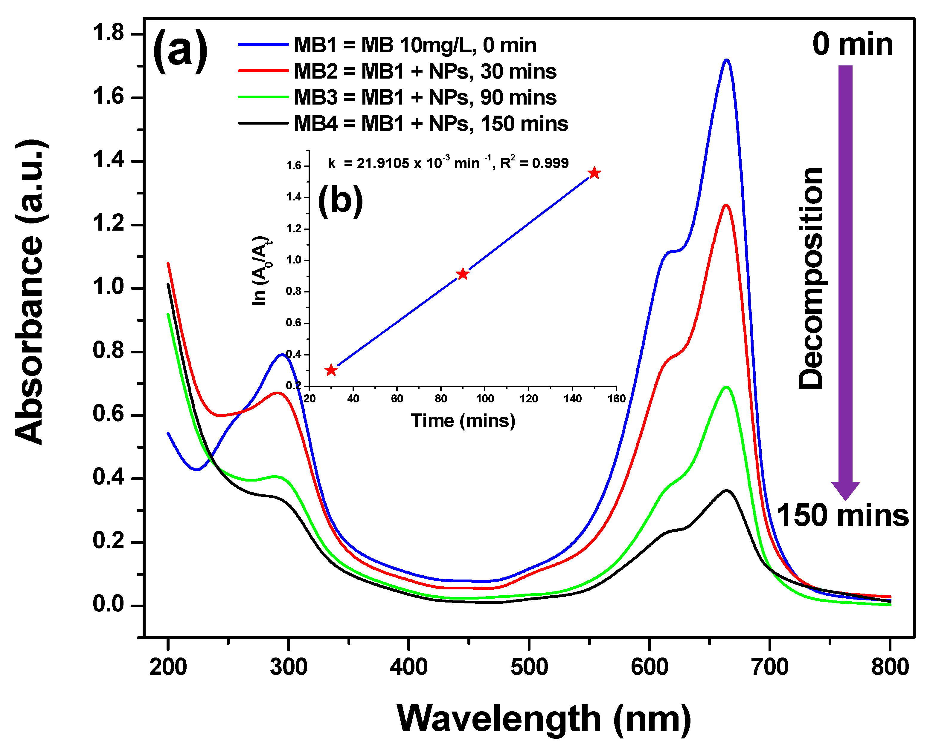
3.6. Photocatalytic Activity
4. Conclusions
Author Contributions
Funding
Acknowledgments
Conflicts of Interest
References
- Din, M.I.; Arshad, F.; Hussain, Z.; Mukhtar, M. Green adeptness in the synthesis and stabilization of copper nanoparticles: Catalytic, Antibacterial, Cytotoxicity, and Antioxidant Activities. Nanoscale Res. Lett 2017, 12, 638. [Google Scholar] [CrossRef] [PubMed] [Green Version]
- Mehdizadeh, T.; Zamani, A.; Froushani, M.A. Preparation of Cu nanoparticles fixed on cellulosic walnut shell material and investigation of its antibacterial, antioxidant and anticancer effects. Heliyon 2020, 6, e03528. [Google Scholar] [CrossRef] [PubMed]
- Poizot, P.; Laruelle, S.; Grugeon, S.; Dupont, L.; Tarascon, J.M. Nano-sized Transition-Metal Oxides as Negative-Electrode Materials for Lithium-Ion Batteries. Nature 2000, 407, 496–499. [Google Scholar] [CrossRef] [PubMed]
- Gawande, M.B.; Goswami, A.; Felpin, F.-X.; Asefa, T.; Huang, X.; Silva, R.; Zou, X.; Zboril, R.; Varma, R.S. Cu and Cu-based nanoparticles: Synthesis and applications in catalysis. Chem. Rev. 2016, 116, 3722–3811. [Google Scholar] [CrossRef] [Green Version]
- Li, Y.; Qian, F.; Xiang, J.; Charles, M.L. Nanowire electronics and opteoelectronics devices. Mater. Today 2006, 9, 18–27. [Google Scholar] [CrossRef]
- Lee, W.; Lim, Y.S.; Kim, S.; Jung, J.; Han, Y.-K.; Yoon, S.; Pioa, L.; Kim, S.-H. Crystal-to crystal conversion of Cu2O nanoparticles to Cu crystals and applications in printed electronics. J. Mater. Chem. 2011, 21, 6928–6933. [Google Scholar] [CrossRef]
- Prasad, P.R.; Kanchi, S.; Naidoo, E.B. In-vitro evaluation of copper nanoparticles cytotoxicity on prostate cancer cell lines and their antioxidant, sensing and catalytic activity: One-pot green approach. J. Photochem. Photobiol. B Biol. 2016, 161, 375–382. [Google Scholar] [CrossRef]
- Patel, B.H.; Channiwala, M.Z.; Chaudhari, S.B.; Mandot, A.A. Biosynthesis of copper nanoparticles; Its characterization and efficacy against human pathogenic bacterium. J. Environ. Chem. Eng. 2016, 4, 2163–2169. [Google Scholar] [CrossRef]
- Xiao, G.; Gao, P.; Wang, L.; Chen, Y.; Wang, Y.; Zhang, G. Ultrasonochemical-assisted synthesis of CuO nanorods with high hydrogen storage ability. J. Nanomater. 2011, 201, 1–6. [Google Scholar] [CrossRef] [Green Version]
- Zhu, H.; Wang, J.; Xu, G. Fast Synthesis of Cu2O hollow microspheres and their application in DNA biosensor of hepatitis B virus. Cryst. Growth Des. 2009, 9, 633–638. [Google Scholar] [CrossRef]
- Saravanan, S.; Sivasankar, T. Effect of ultrasound power and calcination temperature on the sonochemical synthesis of copper oxide nanoparticles for textile dyes treatment. Environ. Prog. Sustain. Energy 2016, 35, 669–679. [Google Scholar] [CrossRef]
- Zhang, J.; Liu, J.; Peng, Q.; Wang, X.; Li, Y. Nearly monodisperse Cu2O and CuO nanospheres: Preparation and applicationfs or sensitive gas sensors. Chem. Mater. 2006, 18, 867–871. [Google Scholar] [CrossRef]
- Pan, L.; Zou, J.-J.; Zhang, T.; Wang, S.; Li, Z.; Wang, L.; Zhang, X. Cu2O film via hydrothermal redox approach: Morphology and photocatalytic performance. J. Phys. Chem. C 2014, 118, 16335–16343. [Google Scholar] [CrossRef]
- Wang, Z.; Wang, H.; Wang, L.; Pan, L. Controlled synthesis of Cu2O cubic and octahedral nano- and microcrystals. Cryst. Res. Technol. 2009, 44, 624–628. [Google Scholar] [CrossRef]
- Gou, L.; Murphy, C.J. Solution-phase synthesis of Cu2O nanocubes. Nano Lett. 2003, 3, 231–234. [Google Scholar] [CrossRef]
- Bhosale, M.A.; Bhanage, B.M. A simple approach for sonochemical synthesis of Cu2O nanoparticles with high catalytic properties. Adv. Powder Technol. 2016, 27, 238–244. [Google Scholar] [CrossRef]
- Wang, W.-W.; Zhu, Y.-J.; Cheng, G.-F.; Huang, Y.-H. Microwave-assisted synthesis of cupric oxide nanosheets and nanowhiskers. Mater. Lett. 2006, 60, 609–612. [Google Scholar] [CrossRef]
- Swarnkar, R.K.; Singh, S.C.; Gopal, R. Effect of aging on copper nanoparticles synthesized by pulsed laser ablation in water: Structural and optical characterizations. Bull. Mater. Sci. 2011, 34, 1363–1369. [Google Scholar] [CrossRef]
- Khayati, G.R.; Nourafkan, E.; Karimi, G.; Moradgholi, J. Synthesis of cuprous oxide nanoparticles by mechanochemical oxidation of copper in high planetary energy ball mill. Adv. Powder Technol. 2013, 24, 301–305. [Google Scholar] [CrossRef]
- Kuppusamy, P.; Ilavenil, S.; Srigopalram, S.; Maniam, G.P.; Yusoff, M.M.; Govindan, N.; Choi, K.C. Treating of palm oil mill effluent using Commelina nudiflora mediated copper nanoparticles as a novel bio-control agent. J. Clean. Prod. 2017, 141, 1023–1029. [Google Scholar] [CrossRef] [Green Version]
- Ghorbani, H.R. Biological and non-biological methods for fabrication of copper nanoparticles. Chem. Eng. Commun. 2015, 202, 1463–1467. [Google Scholar] [CrossRef]
- Nagar, N.; Devra, V. Green synthesis and characterization of copper nanoparticles using Azadirachta indica leaves. Mater. Chem. Phys. 2018, 213, 44–51. [Google Scholar] [CrossRef]
- Angajala, G.; Pavan, P.; Subashini, R. One-step biofabrication of copper nanoparticles from Aegle Marmelos correa aqueous leaf extract and evaluation of its anti-inflammatory and mosquito larvicidal efficacy. RSC Adv. 2014, 4, 51459–51470. [Google Scholar] [CrossRef]
- Gunalan, S.; Sivaraj, R.; Venckatesh, R. Aloe barbadensis Miller mediated green synthesis of mono-disperse copper oxide nanoparticles: Optical properties. Spectrochim. Acta Part A Mol. Biomol. Spectrosc. 2012, 97, 1140–1144. [Google Scholar] [CrossRef] [PubMed]
- Sivaraj, R.; Rahman, P.K.S.M.; Rajiv, P.; Narendhran, S.; Venckatesh, R. Biosynthesis and characterization of Acalypha indica mediated copper oxide nanoparticles and evaluation of its antimicrobial and anticancer activity. Spectrochim. Acta Part A Mol. Biomol. Spectrosc. 2014, 129, 255–258. [Google Scholar] [CrossRef]
- Ghidan, A.Y.; Al-Antary, T.M.; Awwad, A.M. Green synthesis of copper oxide nanoparticles using Punica granatum peels extract: Effect on green peach Aphid. Environ. Nanotechnol. Monit. Manag. 2016, 6, 95–98. [Google Scholar] [CrossRef]
- Yallappa, S.; Manjanna, J.; Sindhe, M.A.; Satyanarayan, N.D.; Pramod, S.N.; Nagaraja, K. Microwave assisted rapid synthesis and biological evaluation of stable copper nanoparticles using T. arjuna bark extract. Spectrochim Acta A Mol. Biomol. Spectrosc. 2013, 110, 108–115. [Google Scholar] [CrossRef]
- Khatami, M.; Heli, H.; Jahani, P.M.; Marcos, H.A.; Nobre, A.L. Copper/copper oxide nanoparticles synthesis using Stachys lavandulifolia and its antibacterial activity. IET Nanobiotechnol. 2017, 11, 709–713. [Google Scholar] [CrossRef]
- Viswadevarayalu, A.; Ramana, P.V.; Kumar, G.S.; Sumalatha, J.; Reddy, S.A. Fine ultrasmall copper nanoparticle (UCuNPs) synthesis by using Terminalia bellirica fruit extract and its antimicrobial activity. J. Clust. Sci. 2016, 27, 155–168. [Google Scholar] [CrossRef]
- Kumar, B.; Smita, K.; Cumbal, C.; Debut, A.; Angulo, Y. Biofabrication of copper oxide nanoparticles using Andean blackberry (Rubus glaucus Benth.) fruit and leaf. J. Saudi Chem. Soc. 2017, 21, S475–S480. [Google Scholar] [CrossRef] [Green Version]
- Kodahl, N. Sacha inchi (Plukenetia volubilis L.)—From lost crop of the Incas to part of the solution to global challenges? Planta 2020, 251, 80. [Google Scholar] [CrossRef] [PubMed]
- Nascimento, A.K.L.; Melo-Silveira, R.F.; Dantas-Santos, N.; Fernandes, J.M.; Zucolotto, S.M.; Rocha, H.A.O.; Scortecci, K.C. Antioxidant and antiproliferative activities of leaf extracts from Plukenetia volubilis Linneo (Euphorbiaceae). Evid. Based Complement. Altern. Med. 2013, 2013, 950272. [Google Scholar] [CrossRef] [PubMed] [Green Version]
- Wang, S.; Zhub, F.; Kakuda, Y. Sacha inchi (Plukenetia volubilis L.): Nutritional composition, biological activity, and uses. Food Chem. 2018, 265, 316–328. [Google Scholar] [CrossRef] [PubMed]
- Kumar, B.; Smita, K.; Cumbal, C.; Debut, A. Synthesis of silver nanoparticles using Sacha inchi (Plukenetia volubilis L.) leaf extracts. Saudi J. Biol. Sci. 2014, 21, 605–609. [Google Scholar] [CrossRef] [Green Version]
- Kumar, B.; Smita, K.; Cumbal, C.; Debut, A. Sacha inchi (Plukenetia volubilis L.) oil for one pot synthesis of silver nanocatalyst: An ecofriendly approach. Ind. Crop. Prod. 2014, 58, 238–243. [Google Scholar] [CrossRef]
- Kumar, B.; Smita, K.; Cumbal, C.; Debut, A. One pot synthesis and characterization of gold nanocatalyst using Sacha inchi (Plukenetia volubilis) oil: Green approach. J. Photochem. Photobiol. B Biol. 2016, 158, 55–60. [Google Scholar] [CrossRef]
- Kumar, B.; Smita, K.; Cumbal, C.; Debut, A. Sacha inchi (Plukenetia volubilis L.) shell biomass for synthesis of silver nanocatalyst. J. Saudi Chem. Soc. 2017, 21, S293–S298. [Google Scholar] [CrossRef] [Green Version]
- Kumar, B.; Smita, K.; Sánchez, E.; Stael, C.; Cumbal, C. Andean Sacha inchi (Plukenetia volubilis L.) shell biomass as new biosorbents for Pb2+ and Cu2+ ions. Ecol. Eng. 2016, 93, 152–158. [Google Scholar] [CrossRef]
- Abboud, Y.; Saffaj, T.; Chagraoui, A.; El Bouari, A.; Brouzi, K.; Tanane, O.; Ihssane, B. Biosynthesis, characterization and antimicrobial activity of copper oxide nanoparticles (CONPs) produced using brown alga extract (Bifurcaria bifurcata). Appl. Nanosci. 2014, 4, 571–576. [Google Scholar] [CrossRef] [Green Version]
- Borgohain, K.; Murase, N.; Mahamuni, S. Synthesis and properties of Cu2O quantum particles. J. Appl. Phys. 2002, 92, 1292–1297. [Google Scholar] [CrossRef]
- Bhattacharjee, S. DLS and zeta potential—What they are and what they are not? J. Control. Release 2016, 235, 337–351. [Google Scholar] [CrossRef] [PubMed]
- Meghana, S.; Kabra, P.; Chakraborty, S.; Padmavathy, N. Understanding the pathway of antibacterial activity of copper oxide nanoparticles. RSC Adv. 2015, 5, 12293–12299. [Google Scholar] [CrossRef]
- Lin, H.; Huang, C.P.; Li, W.; Ni, C.; Shah, S.I.; Tseng, Y.H. Size dependency of nanocrystalline TiO2 on its optical property and photocatalytic reactivity exemplified by 2-chlorophenol. Appl. Catal. B Environ. 2006, 68, 1–11. [Google Scholar] [CrossRef]
- Moniri, S.; Ghoranneviss, M.; Hantehzadeh, M.R.; Asadabad, M.A. Synthesis and optical characterization of copper nanoparticles prepared by laser ablation. Bull. Mater. Sci. 2017, 40, 37–43. [Google Scholar] [CrossRef]
- Fathima, J.B.; Pugazhendhi, A.; Oves, M.; Venis, R. Synthesis of eco-friendly copper nanoparticles for augmentation of catalytic degradation of organic dyes. J. Mol. Liq. 2018, 260, 1–8. [Google Scholar] [CrossRef]


© 2020 by the authors. Licensee MDPI, Basel, Switzerland. This article is an open access article distributed under the terms and conditions of the Creative Commons Attribution (CC BY) license (http://creativecommons.org/licenses/by/4.0/).
Share and Cite
Kumar, B.; Smita, K.; Debut, A.; Cumbal, L. Andean Sacha Inchi (Plukenetia Volubilis L.) Leaf-Mediated Synthesis of Cu2O Nanoparticles: A Low-Cost Approach. Bioengineering 2020, 7, 54. https://0-doi-org.brum.beds.ac.uk/10.3390/bioengineering7020054
Kumar B, Smita K, Debut A, Cumbal L. Andean Sacha Inchi (Plukenetia Volubilis L.) Leaf-Mediated Synthesis of Cu2O Nanoparticles: A Low-Cost Approach. Bioengineering. 2020; 7(2):54. https://0-doi-org.brum.beds.ac.uk/10.3390/bioengineering7020054
Chicago/Turabian StyleKumar, Brajesh, Kumari Smita, Alexis Debut, and Luis Cumbal. 2020. "Andean Sacha Inchi (Plukenetia Volubilis L.) Leaf-Mediated Synthesis of Cu2O Nanoparticles: A Low-Cost Approach" Bioengineering 7, no. 2: 54. https://0-doi-org.brum.beds.ac.uk/10.3390/bioengineering7020054