
Autopsy and Cardiac Magnetic Resonance Image Case of Bevacizumab-Related Cardiomyopathy


 , , and
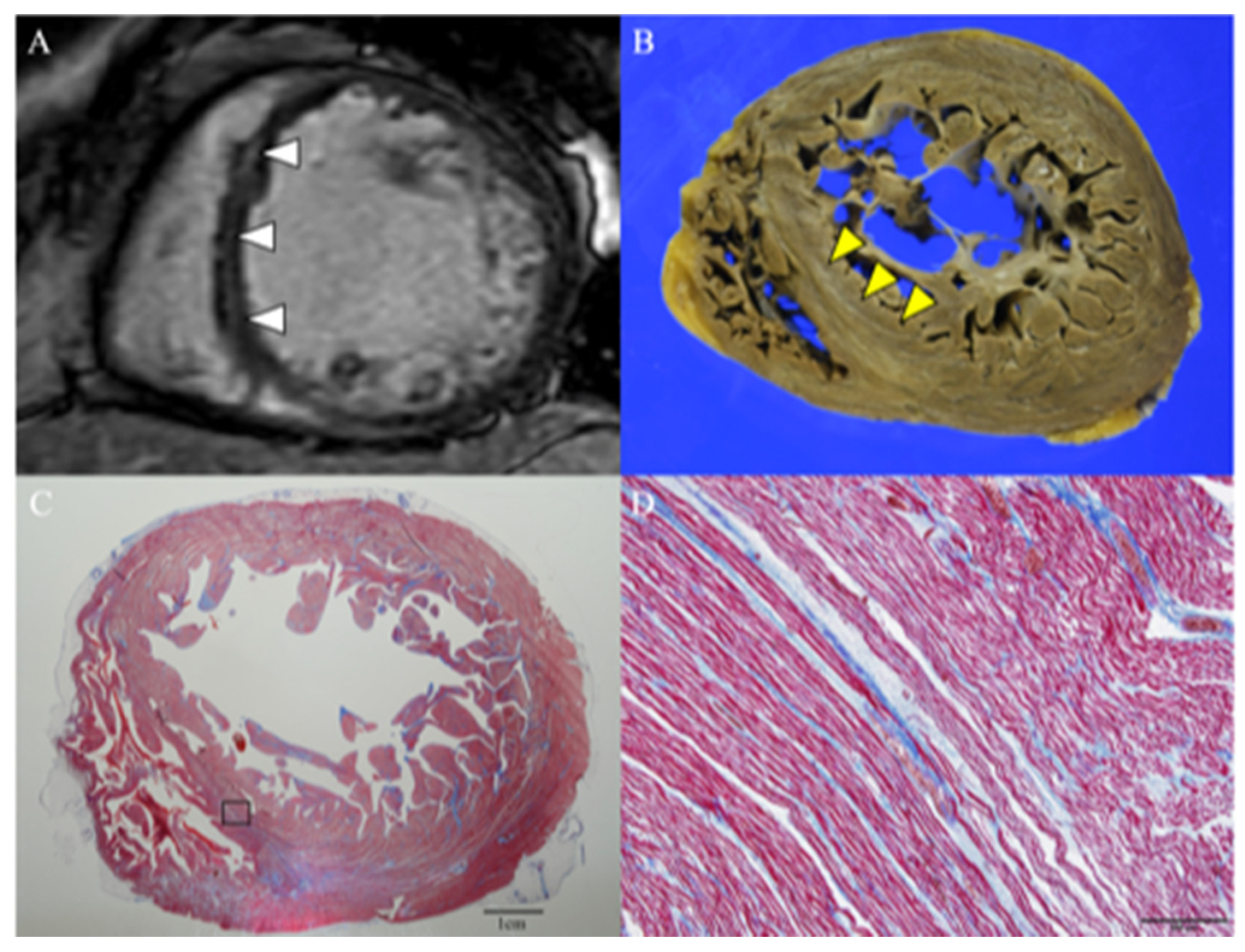
Abstract
:1. Introduction
2. Case Presentation
3. Discussion
4. Conclusions
Supplementary Materials
Author Contributions
Funding
Institutional Review Board Statement
Informed Consent Statement
Data Availability Statement
Acknowledgments
Conflicts of Interest
References
- Yeh, E.T.H.; Bickford, C.L. Cardiovascular Complications of Cancer Therapy: Incidence, Pathogenesis, Diagnosis, and Management. J. Am. Coll. Cardiol. 2009, 53, 2231–2247. [Google Scholar] [CrossRef] [PubMed] [Green Version]
- Qi, W.-X.; Fu, S.; Zhang, Q.; Guo, X.-M. Bevacizumab Increases the Risk of Severe Congestive Heart Failure in Cancer Patients: An up-to-Date Meta-Analysis with a Focus on Different Subgroups. Clin. Drug Investig. 2014, 34, 681–690. [Google Scholar] [CrossRef]
- des Guetz, G.; Uzzan, B.; Chouahnia, K.; Morère, J.-F. Cardiovascular Toxicity of Anti-Angiogenic Drugs. Target. Oncol. 2011, 6, 197–202. [Google Scholar] [CrossRef] [PubMed]
- Dobbin, S.J.H.; Petrie, M.C.; Myles, R.C.; Touyz, R.M.; Lang, N.N. Cardiotoxic Effects of Angiogenesis Inhibitors. Clin. Sci. 2021, 135, 71–100. [Google Scholar] [CrossRef]
- Izumiya, Y.; Shiojima, I.; Sato, K.; Sawyer, D.B.; Colucci, W.S.; Walsh, K. Vascular Endothelial Growth Factor Blockade Promotes the Transition from Compensatory Cardiac Hypertrophy to Failure in Response to Pressure Overload. Hypertension 2006, 47, 887–893. [Google Scholar] [CrossRef] [PubMed]
- Arnold, J.R.; McCann, G.P. Cardiovascular Magnetic Resonance: Applications and Practical Considerations for the General Cardiologist. Heart 2020, 106, 174–181. [Google Scholar] [CrossRef] [PubMed]
- De Angelis, G.; De Luca, A.; Merlo, M.; Nucifora, G.; Rossi, M.; Stolfo, D.; Barbati, G.; De Bellis, A.; Masè, M.; Santangeli, P.; et al. Prevalence and Prognostic Significance of Ischemic Late Gadolinium Enhancement Pattern in Non-Ischemic Dilated Cardiomyopathy. Am. Heart J. 2022, 246, 117–124. [Google Scholar] [CrossRef] [PubMed]
- Isner, J.M.; Ferrans, V.J.; Cohen, S.R.; Witkind, B.G.; Virmani, R.; Gottdiener, J.S.; Beck, J.R.; Roberts, W.C. Clinical and Mor-phologic Cardiac Findings after Anthracycline Chemotherapy. Analysis of 64 Patients Studied at Necropsy. Am. J. Cardiol. 1983, 51, 1167–1174. [Google Scholar] [CrossRef]

Publisher’s Note: MDPI stays neutral with regard to jurisdictional claims in published maps and institutional affiliations. |
© 2022 by the authors. Licensee MDPI, Basel, Switzerland. This article is an open access article distributed under the terms and conditions of the Creative Commons Attribution (CC BY) license (https://creativecommons.org/licenses/by/4.0/).
Share and Cite
Hashimoto, N.; Kitano, D.; Tamaki, T.; Koyama, Y.; Yamada, A.; Hatakeyama, K.; Hao, H.; Okumura, Y. Autopsy and Cardiac Magnetic Resonance Image Case of Bevacizumab-Related Cardiomyopathy. J. Cardiovasc. Dev. Dis. 2022, 9, 208. https://0-doi-org.brum.beds.ac.uk/10.3390/jcdd9070208
Hashimoto N, Kitano D, Tamaki T, Koyama Y, Yamada A, Hatakeyama K, Hao H, Okumura Y. Autopsy and Cardiac Magnetic Resonance Image Case of Bevacizumab-Related Cardiomyopathy. Journal of Cardiovascular Development and Disease. 2022; 9(7):208. https://0-doi-org.brum.beds.ac.uk/10.3390/jcdd9070208
Chicago/Turabian StyleHashimoto, Naoki, Daisuke Kitano, Takehiro Tamaki, Yutaka Koyama, Akimasa Yamada, Kinta Hatakeyama, Hiroyuki Hao, and Yasuo Okumura. 2022. "Autopsy and Cardiac Magnetic Resonance Image Case of Bevacizumab-Related Cardiomyopathy" Journal of Cardiovascular Development and Disease 9, no. 7: 208. https://0-doi-org.brum.beds.ac.uk/10.3390/jcdd9070208