Article

Impact of Media Heat Treatment on Cell Morphology and Stability of L. acidophilus, L. johnsonii and L. delbrueckii subsp. delbrueckii during Fermentation and Processing

1
Department Bioprocess Engineering and Applied Microbiology, Research and Teaching Institute for Brewing in Berlin, Seestrasse 13, 13353 Berlin, Germany
2
Department Food Chemistry and Toxicology, Institute of Food Technology and Food Chemistry, TU Berlin, Straße des 17. Juni 135, 10623 Berlin, Germany
*
Author to whom correspondence should be addressed.
Submission received: 10 September 2020 / Revised: 23 September 2020 / Accepted: 24 September 2020 / Published: 25 September 2020
(This article belongs to the Special Issue Industrial Fermentation)

Abstract

:
Manufacturers of starter cultures and probiotics aim to provide preparations with the highest possible amount of living cells and assurance of long-term storage stability. Thereby the industrial economy and thus an efficient outcome of the processes is of utmost importance. Earlier research has shown that the sterilization procedure of the microbial culture medium tremendously impacts growth performance of heating product-sensitive Lactobacillus strains. Thus, three different strains, i.e., L. acidophilus NCFM, L. johnsonii La-2801 and L. delbrueckii subsp. delbrueckii La-0704, were investigated for the influence of media heat pretreatment on cell morphology and stability during fermentation and further freeze drying and storage. The data indicate a relationship between the heating time of the culture medium, which is associated with an increase in browning reactions, and the cultural characteristics of the three strains. The resulting characteristic cell sizes of the cultures could be a major reason for the different stability properties during processing and storage that were observed. Besides the obvious relevance of the results for the production of starter cultures and probiotics, the pleomorphic phenomenon described here could also be a subject for other biotechnological processes, where heat-mediated media conversions, and thereby related cellular effects, could be a topic. Future studies have to show if further functional properties are influenced by the cell morphology and which cellular mechanisms lead to the observed pleomorphism.

1. Introduction

The complex genus of Lactobacillus currently comprises 261 species (March 2020; [1]). Some of them are very important for the food industry. Homofermentative representatives, such as members of the Lactobacillus delbrueckii group, are widely applied as starter cultures in the dairy industry [2] as well as probiotics [3,4] and biopreservatives [5,6]. In this context, high viability of bacteria is of utmost importance to ensure their performance within the subsequent application. So, there is an urgent need and interest to optimize the manufacturing processes in order to generate bacterial cultures with outstanding viability and stability shares.
One pivotal factor is the choice of the right culture medium for propagation, whereby the complex nutrient medium MRS, according to De Man, Rogosa and Sharpe, refers as standard medium for cultivation of lactic acid bacteria [7]. The medium contains d-glucose as a reducing monosaccharide and also amino-rich components from rather poorly defined yeast extract, beef extract and peptone. This unequivocally leads to the formation of Maillard reaction products (MRP; e.g., Amadori-products, melanoidins) during non-enzymatic browning when sterilizing the medium by heat treatment [8]. These generated MRP represent a highly diverse group of chemical compounds. The biological effects caused by MRP on lactobacilli, reviewed in [9], are also diverse and range from growth promoting [10,11,12,13] to antimicrobial [14,15,16,17] activities.
There are many reviews dealing with the relationship between nutrient availability, cell morphology and growth rate for bacteria in general as well as lactic acid bacteria, but the exact mechanisms are still largely unknown. The appearance of unusually filamentous cell shapes of various lactobacilli as a result of nutrient deficiency (e.g., Vitamin B12 [18,19,20], DNA-precursors [21,22,23] has been observed in many studies.
In previous investigations, various Lactobacillus species were tested with regard to their susceptibility to MRP in nutrient media, with five strains showing characteristic pleomorphic properties after cultivation in terms of the total cell concentration and the average cell size [13]. For Lactobacillus acidophilus NCFM, the interrelation between nutrient medium, cell size and cell stability was preserved even through further processing steps up to the freeze-dried product [12]. With these observations in mind, this study aimed to evaluate processing stability of the three lactobacilli strains previously also identified as Maillard susceptible regarding their pleomorphic properties. Yet for the three strains L. acidophilus, L. johnsonii and L. delbrueckii subsp. delbrueckii, all used as starter cultures and probiotics in industry, the growth properties were investigated in depth when grown in differently heat-treated nutrient media and potential stability properties were investigated during the production of freeze-dried preparations and subsequent storage.

2. Materials and Methods

2.1. Bacterial Strains

Three different strains of lactobacilli (L. acidophilus NCFM, L. johnsonii La-2801 and L. delbrueckii subsp. delbrueckii La-0704) were obtained from the internal strain collection of the Department Bioprocess Engineering and Applied Microbiology of the Research and Teaching Institute for Brewing (VLB) in Berlin. For long-time storage, glycerol stocks (33% v/v) of a stationary phase culture were prepared and maintained at −80 °C.

2.2. Culture Media

Culture media were used as described before [13]. Briefly, MRS medium according to De Man et al. (1960) [7] from DifcoTM (Becton Dickinson GmbH (BD), Heidelberg, Germany), here called MRSD, was used for cultivation. The MRSD contained 10 g proteose peptone no. 3, 10 g beef extract, 5 g yeast extract, 20 g dextrose, 1 g polysorbate 80, 2 g ammonium citrate, 5 g sodium acetate, 0.1 g magnesium sulfate, 0.05 g manganese sulfate, and 2 g dipotassium phosphate per liter at pH = 6.5. For the fermentation studies, MRSD was either sterilized in 1 L bottles at 121 °C for 20 min (according to manufacturer specifications), here designated as MRSDa, or sterile-filtered at a pore size of 0.22 µm (VWR International GmbH, Darmstadt, Germany), designated as MRSDf. In a subset of experiments, MRSD heated at 100 °C under atmospheric pressure for a defined time (1 h, 2 h, 3 h, 4 h, 5 h, 6 h, 7 h, 8 h, 10 h) was used.

2.3. Culture Conditions

A two-stage pre-culture was set up to obtain bacterial cultures for main batch fermentations. First, 14 mL of MRSDa medium were inoculated with 1 mL of a glycerol stock culture and incubated for 72 h at 37 °C in gas-tight tubes. For the second pre-culture, 15 mL MRSDa medium were inoculated with 107 cells/mL from the first pre-culture and incubated for 24 h at 37 °C as stand cultures in gas-tight tubes. For the main batch fermentations, 50 mL culture medium were inoculated with 107 cells/mL from the second pre-culture and incubated for 24 h at 37 °C as stand cultures in gas-tight tubes.

2.4. Sample Preparation for Freeze Drying and Storage Conditions

Samples were harvested and separated via centrifugation (5 min, 4800× g) and washed with 0.9% (w/v) NaCl solution. Afterwards, the cells were resuspended in the same volume of the vegan cryo- and lyoprotective matrix LyoF [24], containing (w/w) 1% glycerol (AppliChem GmbH, Darmstadt, Germany), 5% maltodextrin (Glucidex 12®, Roquette Frères S.A., France) and 5% lactose monohydrate (AppliChem GmbH, Darmstadt, Germany). The mixtures were distributed in 1 mL aliquots in 5 mL glass vials and lyophilized (Sublimator 15, Zirbus Technology GmbH, Germany) for 42 h with an automated profile consisting of a freezing rate of 0.64 °C/min to a minimal pressure of 0.02 mbar with the following shelf-temperature profiles: 7 h −25 °C, 0.67 h −40 °C, 6.5 h −20 °C, 6.5 h −15 °C, 7 h −5 °C, 8.7 h +20 °C, 2 h +4 °C. The freeze-dried preparations were stored in darkness in glass vials closed with gas-tight caps at +4 °C. The lyophilized samples were rehydrated for 30 min in 0.9% (w/v) NaCl solution prior to determine cell concentration and bacterial viability.

2.5. Determination of the Total Cell Concentration and Cell Volume Distribution

The total cell concentration (cc; in cells/mL) and cell volumes (cv; in µm3) were determined with a Beckman Multisizer™ 3 Coulter Counter® (Beckman Coulter GmbH, Germany) with a capillary diameter of 30 µm. The pulse data were converted to size features using the Multisizer™ 3 software version 3.53 (Beckman Coulter GmbH, Germany). Between 30,000 and 50,000 cells were measured per measurement. Since the cell size within the population follows a cell size distribution, the mean cell volume (mcv; in µm3) is shown to evaluate the characteristic cell size of the population.

2.6. Determination of Cell Viability via Colony Forming Units

The number of colony-forming units (cfu) was determined using the plate count method. Samples were diluted in 0.9% NaCl solution. Then, 100 µL of the appropriate dilution were plated on MRS-agar (Th. Geyer GmbH & Co. KG, Renningen, Germany) and incubated aerobically at 37 °C for 48–72 h before counting cfu. The bacterial viability was achieved from the ratio of the cfu per mL to the total cell concentration (cc) per mL.

2.7. Determination of Cell Viability via Flow Cytometry

The LIVE/DEAD BacLight Bacterial Viability Kit (Thermo Fisher Scientific, Germany) was used to differentiate viable and nonviable bacterial cells based on membrane permeability. The kit contains two nucleic acid staining fluorescent probes, SYTO9 (Ex485nm⁄Em498nm when bound to DNA) and propidium iodide (Ex535nm⁄Em617nm when bound to DNA). One milliliter of each bacteria sample containing approximately 106 cells was mixed with 1.5 µL SYTO9 and 1.5 µL PI. After 15 min incubation in the dark at room temperature, the sample was examined via flow cytometry (CyFlow® Cube 8, Sysmex Inc., Germany). All data files from the flow cytometry instrument were analyzed using the software FCS Express, Version 5. Furthermore, a compensation of the fluorescence signals (2.7% FL3 signal (PI) of FL1 signal (SYTO9)) was performed. The green-fluorescent dye SYTO9 can cross all bacterial cell membranes, whereas PI is only incorporated into bacteria with damaged cell membrane. Bacteria with intact cell membrane (SYTO9+/PI) were classified as viable cells.

2.8. Statistical Analysis

The cultivation studies in differently heated MRS media were carried out in independent duplicates and the characteristic data were given as mean value ± mean deviation. The stability experiments were conducted in biological triplicates and the data are presented as mean ± standard deviation (SD). The number of cfu was additionally determined as technical replicates (one to six countable plates per sample). An unpaired t-test was carried out to assess differences (significance level α = 0.05) in mean cell volume, cell concentration and viability between the bacterial populations grown in MRSDa and MRSDf.

3. Results

3.1. Influence of Media Heat Treatment on the Growth Characteristics of Three Sensitive Lactobacilli

Previous studies showed the variability in cell concentration, as well as mean cell volumes, for species of the phylogenetic L. delbrueckii group, namely L. acidophilus NCFM, L. johnsonii La-2801 and L. delbrueckii subsp. delbrueckii La-0704, as a consequence of media heat treatment during media preparation [12,13]. There, it was hypothesized that the appearance of Maillard reaction products was the main reason for this phenomenon. To further substantiate the pleomorphic occurrence of those putative MRP-sensitive strains and to corroborate the impact of the thermal media pretreatment, L. acidophilus NCFM, L. johnsonii La-2801 and L. delbrueckii subsp. delbrueckii La-0704 were cultivated in MRSD medium that was sterile-filtered or heat-treated for different durations (Figure 1). With increasing heating time, a stronger browning of the media was observed, indicating the progress of the Maillard reaction. L. acidophilus NCFM and L. johnsonii La-2801 were responsive to media pretreatment showing higher cell concentrations and lower mean cellular volumes with ongoing media heat pretreatment. This trend was not as pronounced for L. delbrueckii subsp. delbrueckii La-0704, where the cell concentration was the highest in intermediately heated MRSD; however, this strain also showed lower cell sizes when grown in heat-treated media compared to sterile-filtered medium. Thus, sensitivity of those three strains to heat products was substantiated. Further studies are under-way to characterize media after heating, focusing on the qualitative and quantitative assessment of Maillard intermediates and products. Given that the differences in cell volumes were most consistent between bacteria cultured in MRSDa and MRSDf, only these two media variants were applied in further experiments.

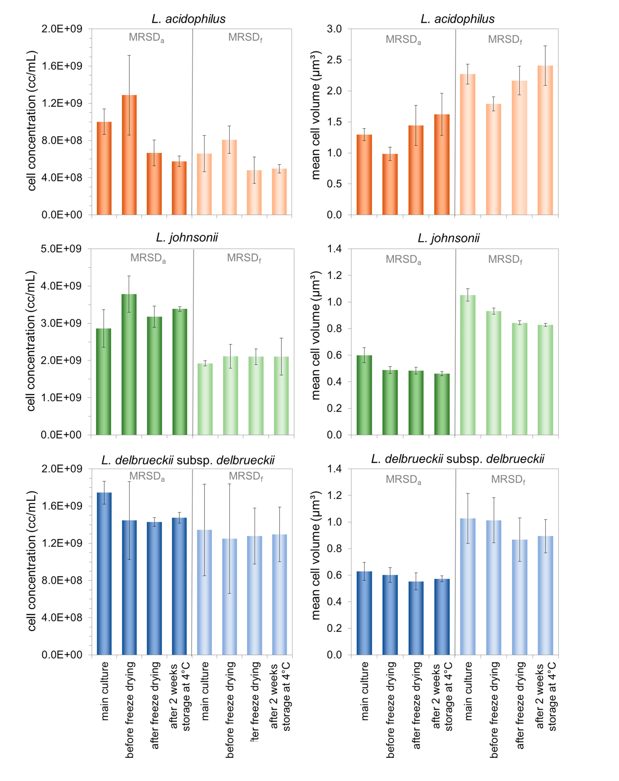
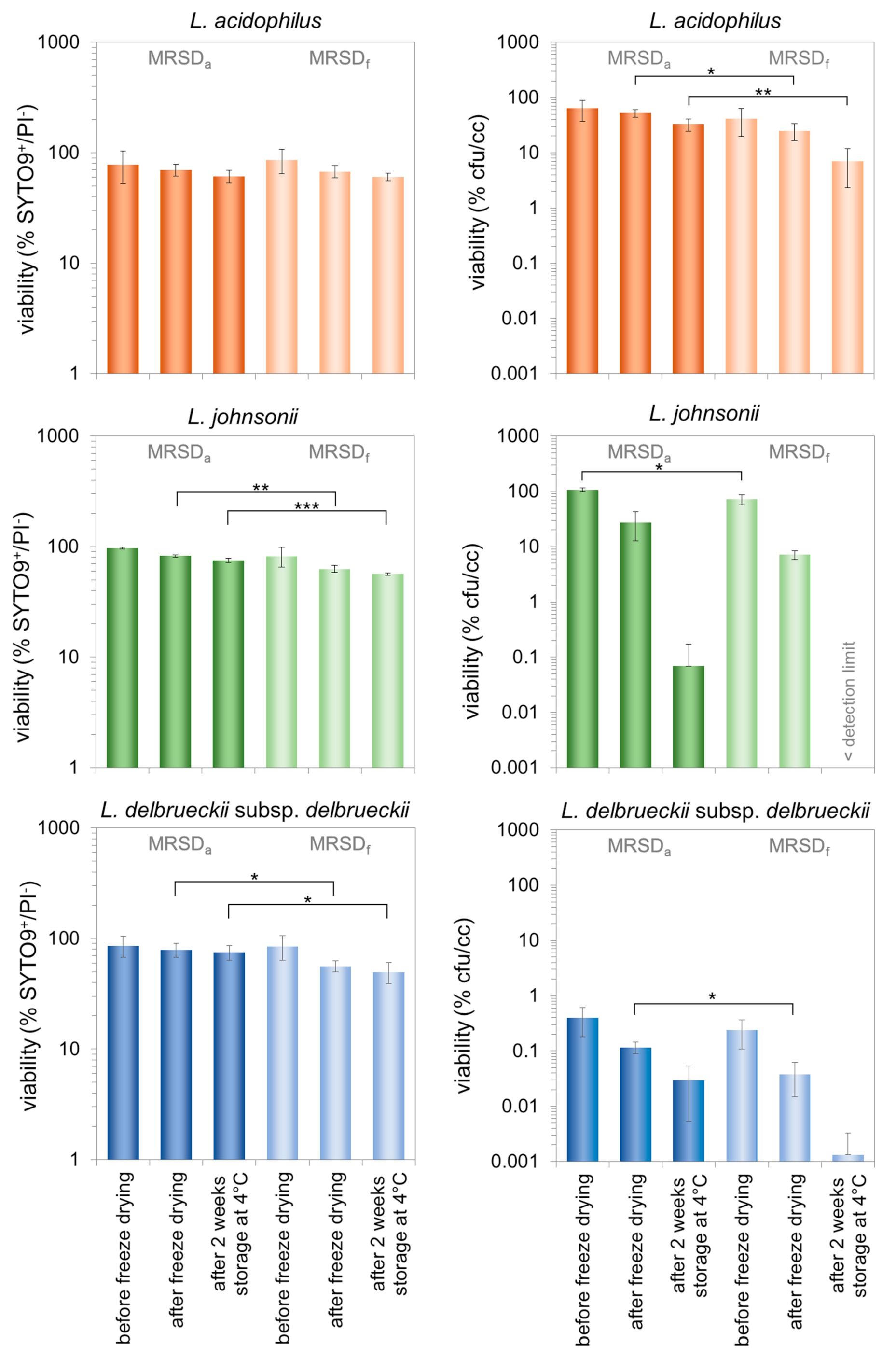
3.2. Characterization of Morphologically Different Populations of the Three Lactobacilli during the Production of Freeze-Dried Preparations and Subsequent Storage

It was previously shown that phenotypically short L. acidophilus NCFM cells were more stable with regard to viability, assessed by colony forming ability, during industrially relevant processing steps, such as freeze drying, compared to filamentous bacteria [12]. In order to systematically investigate whether the media-dependent phenotypic characteristics of the different cell populations obtained during fermentation can be maintained during industrial drying process, L. acidophilus NCFM, L. johnsonii La-2801 and L. delbrueckii subsp. delbrueckii La-0704 were grown in either MRSDa or MRSDf and then freeze dried followed by a storage period.
As shown in Figure 2, the pleomorphic effects already seen in the bacteria suspensions after fermentation in the differently treated nutrient media were also significantly present in relation to the two different media types during further processing steps. Short cells obtained from cultivation in MRSDa remained short in size even when they were freeze dried and rehydrated. The same was seen vice versa for more filamentous cells out of MRSDf fermentation for all three investigated Lactobacillus strains. In the next set of experiments, we evaluated the relationship between these morphological properties and the survival rate during processing.
As another industrially relevant feature, the viability of those three strains was studied during processing in order to examine the effect of the cell shape predefined in the course of the fermentation in the differently treated culture media. Therefore, on the one hand, viability was assessed using the standard method of determining the colony forming units relative to the total number of cells at the end of the fermentation (cfu/cc). In addition, a flow cytometric assay evaluating the cell membrane integrity was used for the valuation (Figure 3). Both methods showed the same trends, however, absolute bacterial viabilities were much lower based on the results of cfu/cc assay compared to the flow cytometry evaluation, especially for L. delbrueckii subsp. delbrueckii. By trend, bacterial preparations grown in MRSDa, thus starting with lower mean cell volumes and higher cell concentrations, were more stable during freeze drying and storage compared to MRSDf-grown counterparts. This effect was seen for all investigated processing steps (see Figure 3) and was statistically significant in half of the tested pairs. These results provide strong evidence that the cultivation of lactobacilli in MRP-enriched growth medium is beneficial for stability of susceptible Lactobacillus strains, further beyond the stage of fermentation also in industrial processing.
The flow cytometry assay further allowed us to unravel different subpopulations within a pool of equally treated bacterial cells (Figure 4). The cellular SYTO9 and/or PI dye accessibility was the criterion used to discriminate different subpopulations. Thereby, PI is supposed to penetrate only cells with disrupted membranes whereas SYTO9 enters living as well as dead cells. If both dyes are present in the cell, PI has a higher affinity for nucleic acids than SYTO9, and as a consequence, SYTO9 is replaced by PI [25].
Considering all stages, it was seen that the portion of cells with damaged membranes (SYTO9/PI+) was the lowest for L. acidophilus (up to 4%) whereas L. johnsonii and L. delbrueckii subsp. delbrueckii showed higher amounts (up to 38%) (Figure 4). This confirms the results obtained here, that L. acidophilus NCFM generally appears to be the most robust of the three strains. Interestingly, there is always a portion of cells showing signals for both used stains indicating an incomplete replacement of the green fluorescent SYTO9 by PI. This phenomenon could be caused by only minor damages of the membrane. Hence, this portion of cells, which tends to increase with increasing processing steps and is particularly visible in the dry product here, is of high interest for further investigation as those cells might be still metabolically active and thus contribute to the functional/probiotic character but probably lost their colony-forming capability.

4. Discussion

4.1. Relation between Media Preparation and Growth Characteristics of L. acidophilus, L. johnsonii and L. delbrueckii subsp. delbrueckii

The presented results regarding the interrelation between pre-heat exposure of culture media and cell morphology, as well as cell concentration, are consistent with previously published results [13]. Senz et al. (2015) hypothesized that MRP present in the medium may cause the transformation of media derived (e.g., delivered by peptone) small double-stranded DNA fragments into biofavorable DNA-precursors for the microorganisms [12]. Several studies [21,26,27,28] showed the great importance of sufficient availability of DNA precursors for optimal growth of lactobacilli. In general, bacterial populations characterized by a low cell concentration and long cells can be assumed to be restricted in certain aspects in cell division [29]. Regardless of whether the restricted DNA synthesis and/or cell separation is the reason for different phenotypes, it can be assumed that this possible (nutrient) limitation tends to be rather disadvantageous for the population [29,30]. Recently published data [31] also suggest that the choice of a suitable yeast extract (as a source of nucleobases) is very important for optimal growth of the lactobacilli. It is subject to further investigations as to whether and which kinds of MRP may have a more generalistic benefit on the nutritional requirements and highlights the importance of deeper research on this topic, especially regarding the identification of specific bio/redox active chemical components.

4.2. Effect of the Cell Properties Induced by the Growth Medium on the Stability Properties of the Three Investigated Lactobacilli

The cell damage that occurs during freezing is caused by intracellular ice crystal formation or osmotic stress due to the concentration of the dissolved substances. Damage that occurs during dehydration primarily affects the cell membrane and proteins of the bacterial cell as a consequence of the removal of their hydration shell. Both process steps, freezing and drying, can thus lead to leakage of the cell membrane, which in turn causes loss of viability [32]. Differences in loss of viability can possibly be explained by different cell morphologies of the respective populations. The influence of cell morphology on the stability of microorganisms during downstream processing steps has already been discussed several times in the literature. In accordance with the data shown here, Miyamoto-Shinohara et al. (2010) observed that yeast survival rate after freeze drying was decreased as cell size of the used specimen for freeze drying increased [33]. In addition, enterococci (small spherical cells) were more resistant during storage in dried state than lactobacilli (larger rods) [34]. Fonseca et al. (2000) described that Streptococcus thermophilus (smaller cells) showed greater resistance to freezing than Lactobacillus strains (larger rods). Moreover, Lactococcus lactis cells were much smaller and more resistant than Lactobacillus bulgaricus cells [35].
A larger cell surface, and thus an increased potential target area for external adverse influences, might explain the phenomenon of why cells larger in size are rather instable during processing. By increasing this parameter, e.g., by increased longitudinal growth, the likeliness of cell membrane damage in the course of freeze drying increases [35]. The surface is influenced by the cell size and also defines the transfer surface for intracellular heat and water. The balance between heat transfer and water outflow is crucial for bacterial viability. Either the cells are cooled too fast, i.e., heat transfer is high and thus ice crystal formation inside the cell is favored leading to intracellular damage or they are cooled too slow, i.e., heat transfer is low and ice crystal formation is favored outside the cells leading to dehydration [36]. As an outstandingly important observation, this study’s experiments show that the loss of viability in the course of freeze drying can be controlled by the choice of media pretreatment across species. The cultivation of lactobacilli in autoclaved MRSD led to improved viability compared to cultivation in sterile-filtered MRSD. Thereby, lactobacilli cultivated in MRSDa had a significantly lower cell volume than bacteria cultivated in MRSDf. Thus, it can be assumed that smaller cells showing higher surface-to-volume-ratios were not as susceptible to intracellular ice crystal formation as longer rods, which were handled at the same freezing rate. Consequently, it would be interesting to investigate whether the freezing rate could counteract the effect of viability loss of longer cells. On the other hand, larger intracellular ice crystals lead to larger pores in the cake structure of the lyophilisate and can be expected to facilitate water-vapor transmission during primary drying, which in turn can reduce cell damage [37]. Moreover, the described effects should have been minimized by the applied cryoprotection matrix LyoF that should avoid dehydration due to its osmotic activity and thus prevent uneven freeze drying [38].
Another important fact is that the cells, once they are dried, are more susceptible to oxidation processes especially concerning the membrane lipids [39,40]. The antioxidative capacity, i.e., the presence of antioxidative substances proven for MRSDa [12], might have had a positive effect on cells cultivated in this medium in contrast to cells grown in MRSDf, which may then also have perpetuated during freeze drying and storage. It remains open if and to what amount the described phenomena were all contributing to the overall advantageous features of small cell sizes leaving potential for further research.

4.3. The Role of the Viability Assessment for the Process Evaluation

In this study, two different methods (cultivability-dependent, cultivability-independent) were used to assess the viability of lactobacilli: determining either the number of cfu in relation to the present total cell concentration or detection of the cell membrane integrity via live/dead fluorescent staining. These methods are based on fundamentally different principles and have different criteria according to which bacterial viability is defined. It was therefore to be expected that the two methods would lead to different results with regard to the viability actually measured. Flow cytometry applying dual fluorescent staining is a rapid single-cell analysis with high informative value, potential high throughput and less labor and space requirement compared with conventional plating techniques [41]. In contrast to the determination of the number of living cells via the colony-forming ability, which only records the cultivable bacteria, all cells with an intact cell membrane are recorded by flow cytometry after differential staining with SYTO9 and PI.
The determination of the viable cells via cfu is usually considered to be the “gold standard” [41,42] for industrial producers as well as users of starter cultures, since only those bacteria are recorded that are capable of replication and formation of a colony under the given cultivation conditions. Or in other words, only these are paid for. However, it can be assumed that the plate count method often gives an underestimation of the true quantity of alive or metabolically active and thus potentially functional cells in a sample [41,43].
In fact, it is obvious that the determined viability strongly depends on the method of choice. Plate counting results showed lower relative viability than flow cytometry results irrespective of the Lactobacillus strain and processing step. In particular, L. delbrueckii subsp. delbrueckii showed very low viability rates estimated as cfu/cc ratio. Since the detected viability was very low, an environmental influence can be assumed here at some point, clearly influencing the ability to form colonies and for a relatively high proportion of non-cultivatable, possibly also a high proportion of dormant cells. One explanation might be an effect of a certain oxygen sensitivity of this strain, even though this species is known to be aerotolerant. A hint is provided by Beshkova et al. (2002) who published that the cultivation of L. delbrueckii subsp. bulgaricus in oxygen-rich culture medium resulted in decreased cell numbers and acid formation in contrast to cultivation under anaerobic conditions [44]. Nevertheless, relatively low viabilities of this species were also detected in other studies on freeze drying [45,46,47] indicating also a low intrinsic robustness for the processing. Since a positive influence on cell stability could be shown here, the results provide an example for process improvement via culture media treatment for labile species.

4.4. Practical Relevance of the Identified Relations between Growth Medium and Cell Properties

The phenomenon of different cell morphologies and cell viability based on the growth medium examined here automatically raises various questions about the underlying mechanisms, the relevance to other industrially momentous microorganisms, but also about the possible influence of medium components on other functional properties of lactobacilli. Rajab et al. (2020) recently examined the effect of the cell size of various Lactobacillus strains on their probiotic characteristics. They showed that long Lactobacillus cells often had higher antioxidant and aggregation activities, but were usually more sensitive to acid and bile conditions and resulted in a lower cfu yield compared to short cells [48].
The impact of MRP on microorganisms can be diverse. Species-specific effects have already been shown for lactobacilli in differently treated, i.e., sterile-filtered or autoclaved, MRS media [13]. Even though manufacturers of starter cultures and probiotics have established standardized manufacturing processes, the importance of the heat pretreatment of the nutrient media can also be relevant for such processes, but particularly when changing processes, e.g., when transferring to new strains or when changing/restricting raw materials. Particularly in the case of lactobacilli with extremely high nutritional requirements, the choice of the complex and thus vitamin and nucleobases supplying components becomes relevant for vegan-compliant processes [49]. One way of achieving this adequate intake of nutrients and growth factors is to use improved yeast extracts. Here, the manufacturers have already recognized the need to respond to the specific requirements of industrially highly important lactobacilli, which is reflected in extensive ongoing research [31,50] as well as in the product portfolio that has been expanded and specialized in recent years by different manufacturers, e.g., Yeast Extract YB (Organotechnie SAS, La Courneuve France), X-SEED® Nucleo Advanced (Ohly GmbH, Hamburg, Germany) or NuCel® (Lesaffre, Maisons-Alfort, France).
As mentioned above, due to different requirements, there are often discrepancies between the culture media used in research (e.g., MRS) and those used in the industrial production of starter cultures and probiotics [49]. Thus, it is of utmost importance for ongoing research and advancement of production processes to consciously consider not only the nutrient media components but also preparation aspects, especially those of thermal pretreatment. As various studies have already shown positive effects of nutrient media heat treatment on bacterial viability and stability, filtration of the culture medium as a general-purpose approach of sterilization cannot be recommended.

5. Conclusions

In the present study, the influence of nutrient media sterilization, more precisely heat treatment compared to sterile filtration, on the growth and stability properties of three industrially relevant Lactobacillus species, L. acidophilus NCFM, L. johnsonii La-2801 and L. delbrueckii subsp. delbrueckii La-0704, was investigated. When using MRS culture media prepared in different ways, a clear dependency of the cell concentration and the average cell size on the degree of heat treatment could be shown, whereas for all strains, sterile filtration led to populations with increased mean cell sizes and lowered cell concentrations. These properties of the respective culture also persisted during further processing into correspondingly manufactured dry preparations. From an economic point of view, the detected circumstance of cultures out of differently treated media differing in their stability properties during freeze drying and storage are of high relevance. Candidates that were cultivated in heat-sterilized nutrient media tended to show higher stabilities. In the case of the viability determined via the ability to form colonies, this could be detected both after freeze drying (for L. acidophilus and L. delbrueckii) and after storage (L. acidophilus). The assessment of the cell membrane integrity of the differently grown cultures over the processing procedure also showed a tendency towards higher viability after freeze drying and storage (for L. johnsonii and L. delbrueckii, respectively). The effects shown here illustrate the momentousness of the nutrient media sterilization methodology that should be considered in detail for each Lactobacillus strain to be produced and should raise the awareness of manufacturers of starter cultures and probiotics to this possible relationship. Future research should address the molecular change in the nutrient medium caused by heat treatment and thus provide valuable conclusions about the mechanisms that mediate pleomorphism shown in this study.

Supplementary Materials

The following figure is available online at https://0-www-mdpi-com.brum.beds.ac.uk/2311-5637/6/4/94/s1, Figure S1: Exemplary dot-plot data of the flow cytometric analyses during the different stages of processing.

Author Contributions

M.S. (Martin Senz), M.S. (Maximilian Schmacht), and C.K., conceptualized the study. M.S. (Martin Senz), acquired the funding and was responsible for project administration. M.L., conducted and analyzed the experiments. M.S. (Martin Senz), M.S. (Maximilian Schmacht), C.K., and H.H., supervised and analyzed the experiments. All authors wrote and edited the manuscript. All authors have read and agreed to the published version of the manuscript.

Funding

This research was funded by the German Federal Ministry for Economic Affairs and Energy and the Euronorm GmbH (program INNO-KOM OST; research project MikroMail; funding no.: VF150026).

Acknowledgments

The authors thank Sophie Palinski for experimental support.

Conflicts of Interest

The authors declare no conflict of interest. The funders had no role in the design of the study; in the collection, analyses, or interpretation of data; in the writing of the manuscript, or in the decision to publish the results.

References

  1. Zheng, J.; Wittouck, S.; Salvetti, E.; Franz, C.M.A.P.; Harris, H.M.B.; Mattarelli, P.; O’Toole, P.W.; Pot, B.; Vandamme, P.; Walter, J.; et al. A taxonomic note on the genus Lactobacillus: Description of 23 novel genera, emended description of the genus Lactobacillus Beijerinck 1901, and union of Lactobacillaceae and Leuconostocaceae. Int. J. Syst. Evol. Microbiol. 2020, 70, 2782–2858. [Google Scholar] [CrossRef] [PubMed]
  2. Bintsis, T. Lactic acid bacteria: Their applications in foods. JBMOA 2018, 6, 89–94. [Google Scholar] [CrossRef] [Green Version]
  3. Sanders, M.E.; Klaenhammer, T.R. Invited review: The scientific basis of Lactobacillus acidophilus NCFM functionality as a probiotic. J. Dairy Sci. 2001, 84, 319–331. [Google Scholar] [CrossRef]
  4. Sanders, M.E.; Merenstein, D.J.; Reid, G.; Gibson, G.R.; Rastall, R.A. Probiotics and prebiotics in intestinal health and disease: From biology to the clinic. Nat. Rev. Gastroenterol. Hepatol. 2019, 16, 605–616. [Google Scholar] [CrossRef] [PubMed]
  5. Bull, M.; Plummer, S.; Marchesi, J.; Mahenthiralingam, E. The life history of Lactobacillus acidophilus as a probiotic: A tale of revisionary taxonomy, misidentification and commercial success. FEMS Microbiol. Lett. 2013, 349, 77–87. [Google Scholar] [CrossRef] [Green Version]
  6. Russo, P.; Fares, C.; Longo, A.; Spano, G.; Capozzi, V. Lactobacillus plantarum with broad antifungal activity as a protective starter culture for bread production. Foods 2017, 6, 110. [Google Scholar] [CrossRef] [PubMed] [Green Version]
  7. De Man, J.C.; Rogosa, M.; Sharpe, M. A medium for the cultivation of lactobacilli. J. Appl. Microbiol. 1960, 23, 130–135. [Google Scholar] [CrossRef]
  8. Wang, H.-Y.; Qian, H.; Yao, W.-R. Melanoidins produced by the Maillard reaction: Structure and biological activity. Food Chem. 2011, 128, 573–584. [Google Scholar] [CrossRef]
  9. Helou, C.; Marier, D.; Jacolot, P.; Abdennebi-Najar, L.; Niquet-Léridon, C.; Tessier, F.J.; Gadonna-Widehem, P. Microorganisms and Maillard reaction products: A review of the literature and recent findings. Amino Acids 2014, 46, 267–277. [Google Scholar] [CrossRef]
  10. Rose, D.; Peterson, R. Influence of the amino acid-dextrose reaction on growth of lactic acid bacteria. Can. J. Res. 1949, 27, 428–436. [Google Scholar] [CrossRef]
  11. Furukawa, H.; Matsuo, H.; Deguchi, T.; Shiga, A.; Samejima, H. Growth-promoting factors for Lactobacillus bifidus var. pennsylvanicus produced by the Amino-Carbonyl Reaction of l -Lysine and d -Glucose. Agric. Biol. Chem. 2014, 32, 617–623. [Google Scholar] [CrossRef]
  12. Senz, M.; van Lengerich, B.; Bader, J.; Stahl, U. Control of cell morphology of probiotic Lactobacillus acidophilus for enhanced cell stability during industrial processing. Int. J. Food Microbiol. 2015, 192, 34–42. [Google Scholar] [CrossRef] [PubMed]
  13. Senz, M.; Keil, C.; Schmacht, M.; Palinski, S.; Cämmerer, B.; Hageböck, M. Influence of media heat sterilization process on growth performance of representative strains of the genus Lactobacillus. Fermentation 2019, 5, 20. [Google Scholar] [CrossRef] [Green Version]
  14. Einarsson, H.; Eklund, T.; Nes, I. Inhibitory mechanisms of Maillard reaction products. Microbios 1988, 53, 27–36. [Google Scholar] [PubMed]
  15. Ames, J.M.; Wynne, A.; Hofmann, A.; Plos, S.; Gibson, G.R. The effect of a model melanoidin mixture on faecal bacterial populations in vitro. Brit. J. Nutr. 1999, 82, 489–495. [Google Scholar] [CrossRef] [Green Version]
  16. Rurián-Henares, J.A.; Morales, F.J. Antimicrobial activity of melanoidins against Escherichia coli is mediated by a membrane-damage mechanism. J. Agric. Food Chem. 2008, 56, 2357–2362. [Google Scholar] [CrossRef] [PubMed] [Green Version]
  17. Hauser, C.; Müller, U.; Sauer, T.; Augner, K.; Pischetsrieder, M. Maillard reaction products as antimicrobial components for packaging films. Food Chem. 2014, 145, 608–613. [Google Scholar] [CrossRef]
  18. Deibel, R.H.; Downing, M.; Niven, C.F., Jr.; Schweigert, B.S. Filament formation by Lactobacillus leichmannii when desoxyribosides replace vitamin B12 in the growth medium. J. Bacteriol. 1956, 71, 255–256. [Google Scholar] [CrossRef] [Green Version]
  19. Beck, W.S.; Hook, S.; Barnett, B.H. The metabolic functions of vitamin B12: Distinctive modes of unbalanced growth behavior in Lactobacillus leichmannii. Biochim. Biophys. Acta 1962, 55, 455–469. [Google Scholar] [CrossRef]
  20. Kusaka, I.; Kitahara, K. Effect of several vitamins on the cell division and the growth of Lactobacillus delbrueckii. J. Vitaminol. 1962, 8, 115–120. [Google Scholar] [CrossRef]
  21. Jeener, H.; Jeener, R. Cytological study of Thermobacterium acidophilus R 26 cultured in absence of deoxyribonucleosides or uracil. Exp. Cell Res. 1952, 3, 675–680. [Google Scholar] [CrossRef]
  22. Beck, W.S.; Levin, M. Purification, kinetics, and repression control of bacterial trans-N-deoxyribosylase. J. Biol. Chem. 1963, 238, 702–709. [Google Scholar] [PubMed]
  23. Ives, D.H.; Ikeda, S. Life on the salvage path: Deoxynucleoside kinases of Lactobacillus acidophilus R-26. Prog. Nucleic Acid Res. Mol. Biol. 1997, 59, 205–255. [Google Scholar] [CrossRef]
  24. Köhler, S.; Schmacht, M.; Malchow, S.; Wolff, L.; Senz, M. Preparation of freeze dried and vacuum dried yeast starter cultures: Evaluation of relevant viability detection analyses. Brew. Sci. 2020, 73, 41–50. [Google Scholar] [CrossRef]
  25. Arndt-Jovin, D.J.; Jovin, T.M. Fluorescence Labeling and Microscopy of DNA. In Fluorescence Microscopy of Living Cells in Culture Part B. Quantitative Fluorescence Microscopy—Imaging and Spectroscopy; Taylor, D.L., Wang, Y.-L., Eds.; Elsevier: Amsterdam, The Netherlands, 1989; pp. 417–448. ISBN 9780080859286. [Google Scholar]
  26. Bringel, F.; Hubert, J.-C. Extent of genetic lesions of the arginine and pyrimidine biosynthetic pathways in Lactobacillus plantarum, L. paraplantarum, L. pentosus, and L. casei: Prevalence of CO(2)-dependent auxotrophs and characterization of deficient arg genes in L. plantarum. Appl. Environ. Microbiol. 2003, 69, 2674–2683. [Google Scholar] [CrossRef] [Green Version]
  27. Siedler, A.J.; Nayder, F.A.; Schweigert, B.S. Studies on improvements in the medium for Lactobacillus acidophilus in the assay for deoxyribonucleic acid. J. Bacteriol. 1957, 73, 670–675. [Google Scholar] [CrossRef] [Green Version]
  28. Reich, J.; Soska, J. Thymineless death in Lactobacillus acidophilus R-26. II. Factors determining the rate of the reproductive inactivation. Folia Microbiol. 1973, 18, 361–367. [Google Scholar] [CrossRef]
  29. Chien, A.-C.; Hill, N.S.; Levin, P.A. Cell size control in bacteria. Curr. Biol. 2012, 22, R340–R349. [Google Scholar] [CrossRef] [Green Version]
  30. Parlindungan, E.; Dekiwadia, C.; Tran Khanh, T.M.; Jones, O.A.H.; May, B.K. Morphological and ultrastructural changes in Lactobacillus plantarum B21 as an indicator of nutrient stress. LWT 2018, 92, 556–563. [Google Scholar] [CrossRef]
  31. Somani, A.; Boran, B.; Bekers, K.M. Ohly White Paper, Impact of Nucleotides on L. bulgaricus Growth, Viability and Performance; Ohly GmbH: Hamburg, Germany, 2020. [Google Scholar]
  32. Aschenbrenner, M.; Först, P.; Kulozik, U. Freeze-Drying of Probiotics. In Advances in Probiotic Technology, 1st ed.; Foerst, P., Santivarangkna, C., Eds.; CRC Press: Boca Raton, FL, USA, 2015. [Google Scholar]
  33. Miyamoto-Shinohara, Y.; Nozawa, F.; Sukenobe, J.; Imaizumi, T. Survival of yeasts stored after freeze-drying or liquid-drying. J. Gen. Appl. Microbiol. 2010, 56, 107–119. [Google Scholar] [CrossRef] [Green Version]
  34. Carvalho, A.S.; Silva, J.; Ho, P.; Teixeira, P.; Malcata, F.X.; Gibbs, P. Protective effect of sorbitol and monosodium glutamate during storage of freeze-dried lactic acid bacteria. Lait 2003, 83, 203–210. [Google Scholar] [CrossRef] [Green Version]
  35. Fonseca, F.; Béal, C.; Corrieu, G. Method of quantifying the loss of acidification activity of lactic acid starters during freezing and frozen storage. J. Dairy Res. 2000, 67, 83–90. [Google Scholar] [CrossRef] [PubMed]
  36. Mazur, P. The Role of Intracellular Freezing in the Death of Cells Cooled at Supraoptimal Rates. Cryobiology 1977, 14, 251–272. [Google Scholar] [CrossRef]
  37. Patapoff, T.W.; Overcashier, D.E. The importance of freezing on Lyophilization cycle development. BioPharm 2002, 15, 16–21. [Google Scholar]
  38. Santivarangkna, C.; Higl, B.; Foerst, P. Protection mechanisms of sugars during different stages of preparation process of dried lactic acid starter cultures. Food Microbiol. 2008, 25, 429–441. [Google Scholar] [CrossRef] [PubMed]
  39. Meng, X.C.; Stanton, C.; Fitzgerald, G.F.; Daly, C.; Ross, R.P. Anhydrobiotics: The challenges of drying probiotic cultures. Food Chem. 2008, 106, 1406–1416. [Google Scholar] [CrossRef]
  40. Santivarangkna, C.; Kulozik, U.; Foerst, P. Storing Lactic Acid Bacteria: Current Methodologies and Physiological Implications. In Stress Response in Lactic Acid Bacteria; Tsakalidou, E., Papadimitriou, K., Eds.; Springer: New York, NY, USA; London, UK, 2010. [Google Scholar]
  41. Wilkinson, M.G. Flow cytometry as a potential method of measuring bacterial viability in probiotic products: A review. Trends Food Sci. Technol. 2018, 78, 1–10. [Google Scholar] [CrossRef]
  42. Davey, H.M. Life, death, and in-between: Meanings and methods in microbiology. Appl. Environ. Microbiol. 2011, 77, 5571–5576. [Google Scholar] [CrossRef] [Green Version]
  43. Liu, J.; Deng, Y.; Soteyome, T.; Li, Y.; Su, J.; Li, L.; Li, B.; Shirtliff, M.E.; Xu, Z.; Peters, B.M. Induction and recovery of the viable but nonculturable state of hop-resistance Lactobacillus brevis. Front. Microbiol. 2018, 9, 2076. [Google Scholar] [CrossRef]
  44. Beshkova, D.M.; Simova, E.D.; Frengova, G.I.; Simov, Z.; Spasov, Z.N. Effect of oxygen on batch yogurt cultures. World J. Microbiol. Biotechnol. 2002, 18, 361–365. [Google Scholar] [CrossRef]
  45. Martos, G.I.; Minahk, C.J.; Font de Valdez, G.; Morero, R. Effects of protective agents on membrane fluidity of freeze-dried Lactobacillus delbrueckii ssp. bulgaricus. Lett. Appl. Microbiol. 2007, 45, 282–288. [Google Scholar] [CrossRef] [PubMed]
  46. Meneghel, J.; Passot, S.; Dupont, S.; Fonseca, F. Biophysical characterization of the Lactobacillus delbrueckii subsp. bulgaricus membrane during cold and osmotic stress and its relevance for cryopreservation. Appl. Microbiol. Biotechnol. 2017, 101, 1427–1441. [Google Scholar] [CrossRef] [PubMed]
  47. Otero, M.C.; Espeche, M.C.; Nader-Macías, M.E. Optimization of the freeze-drying media and survival throughout storage of freeze-dried Lactobacillus gasseri and Lactobacillus delbrueckii subsp. delbrueckii for veterinarian probiotic applications. Proc. Biochem. 2007, 42, 1406–1411. [Google Scholar] [CrossRef]
  48. Rajab, S.; Tabandeh, F.; Shahraky, M.K.; Alahyaribeik, S. The effect of lactobacillus cell size on its probiotic characteristics. Anaerobe 2020, 62, 102103. [Google Scholar] [CrossRef] [PubMed]
  49. Ummadi, M.S.; Bawden-Curic, M. Use of Protein Hydrolysates in Industrial Starter Culture Fermentations. In Protein Hydrolysates in Biotechnology; Pasupuleti, V.K., Demain, A.L., Eds.; Springer: Dordrecht, The Netherlands; London, UK, 2010; pp. 91–114. [Google Scholar]
  50. Proust, L.; Sourabié, A.; Pedersen, M.; Besançon, I.; Haudebourg, E.; Monnet, V.; Juillard, V. Insights into the complexity of yeast extract peptides and their utilization by Streptococcus thermophilus. Front. Microbiol. 2019, 10, 906. [Google Scholar] [CrossRef] [PubMed] [Green Version]
Figure 1. Growth characteristics of the three investigated lactobacilli cultured in either sterile-filtered or pre-heated MRS media. Properties of the corresponding cultures are shown as ratio of the cell concentration (ratio cc; dark bars) or mean cell volume (ratio mean cell volume (mcv); light bars) between cultivation in the respective heat-treated and the sterile-filtered medium. Thereby, a value >1 indicates a higher cell concentration or mean cell volume, respectively, in heat-treated MRSD compared to MRSDf, whereas values <1 designate a lower cell concentration or mean cell volume in heat-treated MRSD compared to MRSDf. Data represent mean values ± mean deviation of independent duplicates.
Fermentation 06 00094 g001
Figure 2. Cell concentration and mean cell volume of the three investigated lactobacilli cultivated in either standardly autoclaved (MRSDa; dark bars) or sterile-filtered (MRSDf; light bars) medium and during different stages of processing and storage. The data represent the mean values ± standard deviation (SD) of three biological replicates. Comparison of the mcv of the populations at each stage of processing by statistical analyses (t-test, α = 0.05) revealed significant differences (p ≤ 0.05) between cells grown in MRSDa and MRSDf at each stage for all three organisms.
Fermentation 06 00094 g002
Figure 3. Viability of the three investigated lactobacilli, grown in either standardly autoclaved (MRSDa; dark bars) or sterile-filtered (MRSDf; light bars) medium, during different stages of processing and storage. Viability of the bacteria was assessed by two different assays. Left: live/dead staining and measurement via flow cytometry; right: colony-forming units relative to the total number of cells (see materials and methods section). The data represent the mean values ± SD of three biological replicates. Significant differences of the culture-viability at the same stage of processing are labeled by asterisks (t-test, p: * ≤ 0.05; ** ≤ 0.01; *** ≤ 0.001).
Fermentation 06 00094 g003
Figure 4. Data of flow cytometry analyses obtained from bacterial samples during different stages of processing (see Figure 3) illustrating the different subpopulations detectable after dual staining with the two nucleic acid-staining fluorescent dyes SYTO9 and PI (see also Supplementary Figure S1). SYTO9+/PI: fully intact cell membrane; SYTO9+/PI+: cells with damaged cell membrane; SYTO9/PI+: dead cells with lost cell membrane integrity; SYTO9/PI: non-stained particles. The data represent the mean values ± SD of three biological replicates.
Fermentation 06 00094 g004

