Article

Influence of Light Conditions and Medium Composition on Morphophysiological Characteristics of Stevia rebaudiana Bertoni In Vitro and In Vivo

by
Alla A. Shulgina
1,
Elena A. Kalashnikova
1,
Ivan G. Tarakanov
2,
Rima N. Kirakosyan
1,
Mikhail Yu. Cherednichenko
1,
Oksana B. Polivanova
1,
Ekaterina N. Baranova
3,4 and
Marat R. Khaliluev
1,5,*
1
Department of Biotechnology, Russian State Agrarian University–Moscow Timiryazev Agricultural Academy, Timiryazevskaya, 49, 127550 Moscow, Russia
2
Department of Plant Physiology, Russian State Agrarian University–Moscow Timiryazev Agricultural Academy, Timiryazevskaya, 49, 127550 Moscow, Russia
3
Laboratory of Cell Biology, All-Russia Research Institute of Agricultural Biotechnology, Timiryazevskaya, 42, 127550 Moscow, Russia
4
Laboratory of Plant Protection, N.V. Tsitsin Main Botanical Garden of Russian Academy of Sciences, Botanicheskaya, 4, 127276 Moscow, Russia
5
Laboratory of Plant Cell Engineering, All-Russia Research Institute of Agricultural Biotechnology, Timiryazevskaya, 42, 127550 Moscow, Russia
*
Author to whom correspondence should be addressed.
Submission received: 24 May 2021 / Revised: 10 July 2021 / Accepted: 11 July 2021 / Published: 15 July 2021

Abstract

:
We investigated the influence of different conditions (light composition and plant growth regulators (PGRs) in culture media) on the morphophysiological parameters of Stevia rebaudiana Bertoni in vitro and in vivo. Both PGRs and the light spectra applied were found to significantly affect plant morphogenesis. During the micropropagation stage of S. rebaudiana, optimal growth, with a multiplication coefficient of 15, was obtained in an MS culture medium containing 2,4-epibrassinolide (Epin) and indole-3-acetic acid (IAA) at concentrations of 0.1 and 0.5 mg L−1, respectively. During the rooting stage, we found that the addition of 0.5 mg L−1 hydroxycinnamic acid (Zircon) to the MS medium led to an optimal root formation frequency of 85% and resulted in the formation of strong plants with well-developed leaf blades. Cultivation on media containing 0.1 mg L−1 Epin and 0.5 mg L−1 IAA and receiving coherent light irradiation on a weekly basis resulted in a 100% increase in the multiplication coefficient, better adventitious shoot growth, and a 33% increase in the number of leaves. S. rebaudiana microshoots, cultured on MS media containing 1.0 mg L−1 6-benzylaminopurine (BAP) and 0.5 mg L−1 IAA with red monochrome light treatments, increased the multiplication coefficient by 30% compared with controls (white light, media without PGRs).

1. Introduction

Sucrose is the most commonly used food sweetener and is extracted from sugar beet or sugarcane [1]. However, excessive sugar consumption can undermine human health. As problems related to excess weight continue to increase worldwide, studies on native sugar substitutes are becoming more and more important. For this reason, interest in plant secondary metabolites and their biologically active substances is increasing. Diterpenoids are a large class of natural metabolic compounds belonging to higher plants [2,3]. Many diterpenoids are considered valuable substances, functioning as, for example, plant growth regulators (PGRs) [4], insecticides [5], antibiotics [6,7], and tumor formation inhibitors [8,9].
Among those particularly useful are sweet diterpenic glycosides, found in the perennial herbaceous plant Stevia rebaudiana Bertoni [10,11,12]. The most valuable of them is stevioside, which has a strong sweet taste, about 200–300 times sweeter than sucrose. Importantly, however, it does not increase the blood sugar level [1,2,13]. Stevioside is widely used as a low-caloric sugar substitute [14] and is recognized as safe [15]. In addition, its chemical structure remains intact at high temperatures, so it can be used in hot preparation. Other qualities of stevioside include its ability to accelerate wound healing and its anti-allergenic and anti-inflammatory effects [16,17,18]. Knowledge of the morphogenesis pattern of the promising crop from which stevioside is derived is thus important to realize the full potential of the plant during its intensive in vitro culture.
The biosynthesis of secondary metabolites in plants is directly correlated with growth intensity [19] and, therefore, we focused on the morphogenetic responses in S. rebaudiana under different in vitro culture conditions, explanted sources, and other factors [20,21,22,23,24,25,26,27,28,29,30,31,32].
Cultures of isolated cells and tissues serve as a universal model for the in vitro study of patterns of formation during plant morphogenesis [33]. Moreover, in the conditions of in vitro culture, the biosynthetic potential can be controlled, and super-producing strains can be obtained, which may be relevant with respect to S. rebaudiana [21,24]. Several biotechnological approaches have been applied to increase steviol glycoside production in in vitro culture; the most extensive studies have explored the influence of phytohormones and PGRs. For example, adventitious roots, cultured in media supplemented with gibberellic acid (2 mg/L), accumulated steviosides in high concentrations [34]. Accumulation of steviosides and other secondary metabolites, such as phenols and flavonoids, under the influence of BAP and other PGRs has also been studied in Stevia callus cultures [35,36,37]. However, S. rebaudiana calli and suspension cultures have a limited ability to produce steviol glycosides, and have been assumed to be tightly dependent on their level of differentiation [38]. Secondary metabolite accumulation in both differentiated and dedifferentiated tissues can be influenced by various stress factors and elicitors. It has been noted that drought and salinity stresses tend to increase the accumulation of steviol glycosides and the upregulation of several genes encoding key enzymes of the steviol glycoside biosynthesis pathways [39,40,41]. However, the influence of stress factors is controversial and can be determined by the level of exposure, e.g., salt type and concentration [39,42,43]. Application of elicitors such as alginate and yeast extract also increase the biosynthesis of steviol glycosides [44]. Treatment of S. rebaudiana adventitious roots with copper and gold nanoparticles, in combination with PGRs in vitro, had a stimulating effect on secondary metabolite production [45].
Physical factors are important regulators of morphogenesis. Light-emitting diodes (LED) provide unique possibilities for manipulating plant photosynthesis, photomorphogenesis, and the synthesis of secondary metabolites [46,47,48]. The last directly depends on the intensity of photosynthesis and growth rate. Therefore, the morphogenetic responses of S. rebaudiana, specifically, have been studied. Recently, researchers have been interested in innovative LEDs [32,47], as well as exposure to low-intensity radiation with an injection laser. However, studies on the effects of physical factors, especially light treatment, on in vitro S. rebaudiana cell culture are limited [29,48,49]. Cultivation of S. rebaudiana adventitious roots under different light spectra showed that violet light had a stimulating effect, and red light an inhibitory one on biomass growth. At the same time, the blue spectrum contributed to the maximum accumulation of phenolic compounds [50]. The influence of the spectral composition of light has also been studied on callus cultures of Stevia [51]. However, there are no data on the effects of light on the accumulation of steviosides in Stevia microplants. Further study of light’s influence on differentiated tissues is promising because such tissues have significant potential to accumulate steviosides. Considering the positive impact of light on callus and adventitious roots, an additional stimulating effect on microplants can be expected.
Based on the above, the aim of our research was to study the influence of culture conditions (light factors and PGRs in culture media) on the morphophysiological parameters of S. rebaudiana plants in vitro and in vivo.

2. Materials and Methods

2.1. Plant Material

All experiments were carried out at the department of biotechnology of Russian State Agrarian University–Moscow Timiryazev Agricultural Academy. The explant source was aseptically cultured microplants of S. rebaudiana that were provided by Nikolay I. Bondarev (PhD of Timiryazev Institute of Plant Physiology of Russian Academy of Sciences, Moscow, Russia). We used a node with two axillary buds as an explant for in vitro experiments.

2.2. Influence of PGRs on S. rebaudiana Morphogenesis In Vitro

We used various PGRs to induce the formation of adventitious buds, axillary shoots, and rooting. To examine the influence of PGRs on plant growth, isolated S. rebaudiana microshoots were clonally propagated from cuttings and cultured in vitro on Murashige and Skoog basal media (MS) [52] (PGRs-free MS media), as well as MS media supplemented with various concentrations of PGRs: 1 mg L−1 kinetin and 0.5 mgL−1 indole-3-acetic acid (IAA); 1 mg L−1 6-benzylaminopurine (BAP) and 0.5 mg L−1 IAA; 0.1 mg L−1 2,4-epibrassinolide (Epin) and 0.5 mg L−1 IAA; and 0.5 mg L−1 hydroxycinnamic acids (Zircon). A PGRs-free MS medium was used as a control. The multiplication coefficient was measured after 60 days.
Explants and microshoots were cultured in a light room where the temperature was maintained at 22–24 °C, lighting was provided with an OSRAM L36/25 fluorescent white lamps with an illumination intensity of 3000 lx under a 16/8 h photoperiod and 150–180 mmol/m2 s−1 photon flux density.

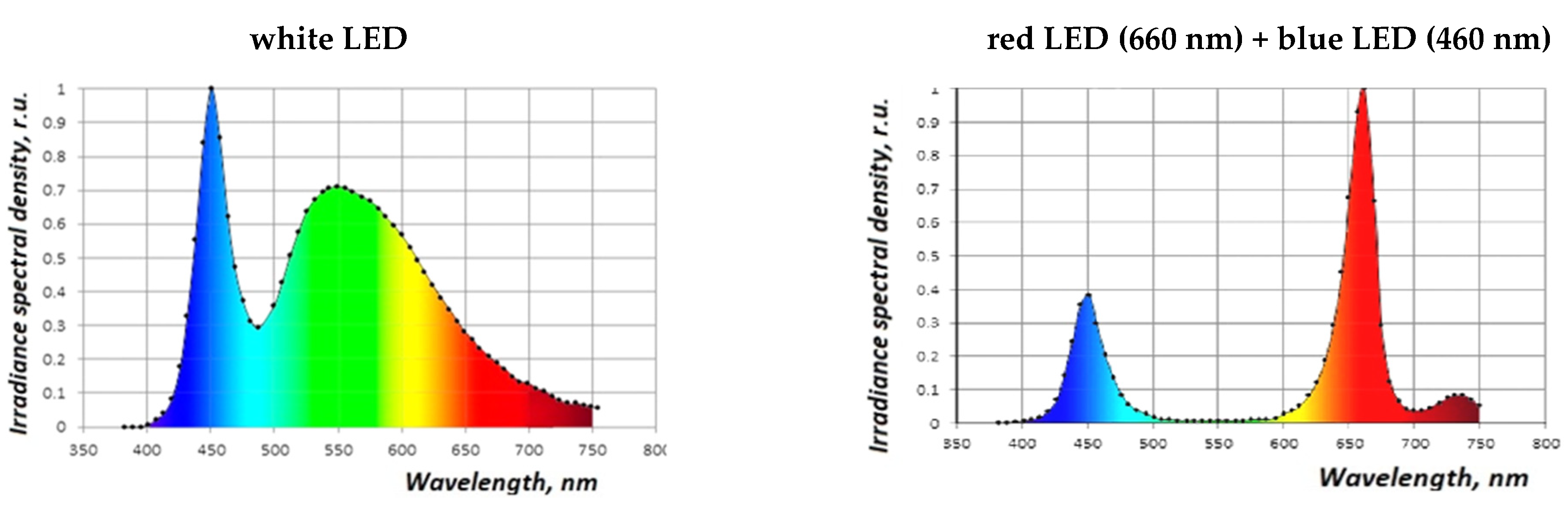
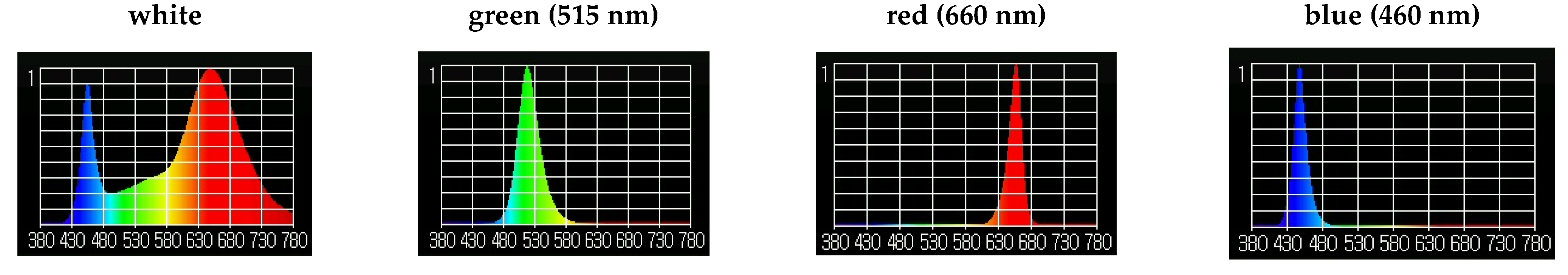
2.3. Influence of Light Factors on the Morphophysiological Parameters of S. rebaudiana In Vitro

We chose light treatment as a physical factor, and we studied the influence of different kits of light spectra on the morphogenesis of S. rebaudiana in vitro. First, we examined white and red-blue LED lighting (Figure 1). Fluorescent white lamps were used as a control. The study lasted 60 days.
Second, LED lighting was evaluated with light from the blue (B) and different parts of the red spectra, including short-wave red (SR), long-wave red (LR), and far red (FR). The control included all types of diodes (SR + LR + FR + B); in each of our variants, one of the spectra was excluded (Figure 2). Third, we studied monochrome light spectra; white (5000 K) as a control, green (515 nm), red (660 nm), and blue (460 nm) (Figure 3). The study lasted 55 days. Finally, coherent light, supplemented with irradiation of low-intensity, was evaluated with an injection laser LPI-2 device (Russia). The wavelength was 650 nm, the oscillation frequency 2000 Hz, and the light beam’s power was 2–4 W/m2. Plants were exposed either once or weekly to irradiation for 30, 60, 120, or 240 s. Plants without any light treatment were used as a control. The study lasted 4 weeks.

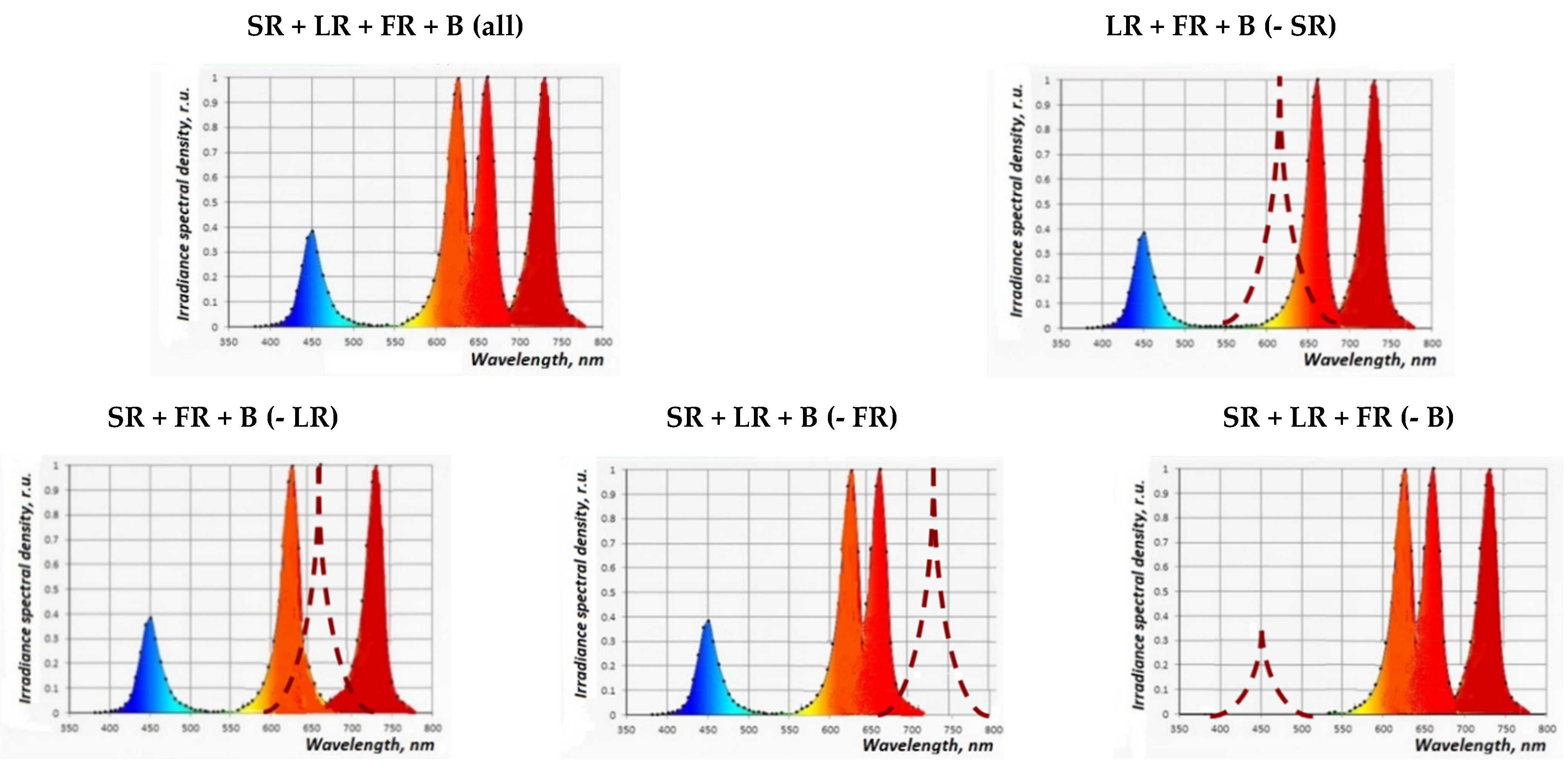
2.4. The Combined Effect of Physical and PGRs Factors on the Morphophysiological Parameters of In Vitro S. rebaudiana Microshoot Growth

The combined effect of physical (light) and chemical (PGRs combination) factors on the morphophysiological parameters of in vitro S. rebaudiana microshoot growth was studied. Microshoots were cultured on three variants of MS media: MS without PGRs, (PGRs-free media); MS supplemented with 0.1 mgL−1 of Epin and 0.5 mgL−1 of IAA; and MS supplemented with BAP and IAA at concentrations of 1.0 and 0.5 mg L−1, respectively. Different light spectra (white, green, red, and blue) were used as light treatments (Figure 3). Also, coherent light treatment was conducted after 6 weeks.

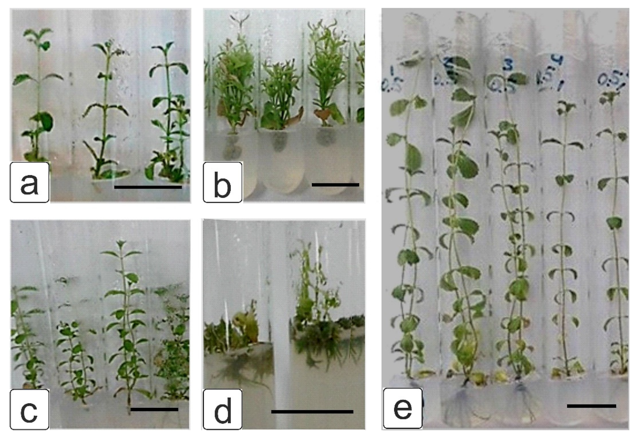
2.5. Phenotypic Analysis of Plants

Phenotypic assays of microshoots, cultured using various MS media and light settings, were carried out. The height of the main shoot (cm), total number of leaves (units), and multiplication coefficient were measured. In the case of in vivo plants, also leaf area (cm2) and the fresh/dry weight of leaves were measured. The leaf area was determined using the photoplanimeter Li-Cor LI-3100 (USA, Nebraska).

2.6. Quantitative Determination of Photosynthetic Pigments of S. rebaudiana In Vivo

The levels of chlorophylls a, b, (Ca, Cb) and carotenoids (Ccar) were characterized in the leaves of S. rebaudiana plants grown under different light spectra. The concentrations of pigments were determined using an SF-104 spectrophotometer. Measurements were taken at wavelengths of 662, 644, and 440.5 nm, from which the absolute number of pigments was determined. The pigment concentration was calculated using the following formulae:
Ca = 9.784 D662 − 0.990 D644
Cb = 21.426 D644 − 4.650 D662
Ccar = 4.695 D440,5 − 0.268 Ca + Cb

2.7. Qualitative and Semi-Quantitative Determination of Diterpene Glycosides in Dry Leaves of S. rebaudiana In Vivo

Qualitative and semi-quantitative determination of diterpene glycosides were achieved using thin-layer chromatography (TLC) [53]. The objects of the study individually received an application, with a micro-syringe of 10 µL to the start line of the Sorbfil STX-1A plate, of an aluminum substrate that had been pre-activated by heating, on a tile, at 80 °C for 15 min. The solvent system was comprised of chloroform, ethanol, and water (10:5:1). After raising the solvent front, the product was determined in a chamber with a UV-illuminator at a wavelength of 254 nm. Then the plate was developed with a solution of iodine in 95% alcohol; the staining of spots with an Rf = 0.33 zone in a dark brown color was noted.

2.8. Quantitative Determination of Steviosides by High-Performance Liquid Chromatography (HPLC) of Dry Leaves of S. rebaudiana In Vivo

Quantitative determination of steviosides by HPLC was performed on an Agilent 1100 device with a diode matrix detector (Zorbox C-1854,6-150 column). Samples of 0.01 g were taken, with 1 mL of deionized water, placed in an ultrasonic bath at a temperature of 60 °C for 3 h. Then samples were centrifuged for 5 min at 2000 rpm. After that the supernatant was evaporated dry under vacuum at 45–50 °C. The dry residue was analyzed on a liquid chromatograph in 1 mL of deionized water. The solvent gradient was 15% acetonitrile and 85% deionized water. The flow rate was 0.2 mL min−1 at normal pressure, the volume of the injected sample was 10 µL, and the analysis time was 15 min. Samples were introduced by washing the input of the polar solvent. Samples were detected at wavelengths of 193, 200, 210, 220, and 230 nm. Assays were performed at a wavelength of 210 nm. For quantitative and qualitative determinations, a calibration graph of a control standard of the substance was used, in addition to literary data [11,36].

2.9. Statistical Analysis of Experimental Data

Experimental data were statistically compared using analysis of variance (ANOVA) with MS Excel software and AGROS software (version 2.11, Russia). Differences were considered statistically significant at p < 0.05. The experiments were carried out using three biological and 5–10 analytical replicates.

3. Results and Discussion

3.1. Influence of PGRs on In Vitro S. rebaudiana Clonal Propagation and Microshoot Morphology

Clonal micropropagation of S. rebaudiana using in vitro techniques has been studied by many researchers to develop an efficient and economic protocol in a semisolid culture medium [20,24,28,54,55]. Several studies suggest that the use of a temporary immersion bioreactor system is effective for in vitro S. rebaudiana propagation because it combines a liquid and semisolid culture technique [30,56]. In vitro S. rebaudiana micropropagation has been reported from nodes [20,56], leaf segments [25,27], suspension cultures [22], and shoot apices [24]. Shoots are formed by cultivating explants on cultured media with the addition of various types, concentrations, and combinations of PGRs, such as kinetin, BAP, thidiazuron, IAA, IBA, and NAA [20,21,22,23,24,25,54,55,56,57]. In our study, for induction of adventitious buds and axillary shoots, MS culture media included IAA and cytokinins (kinetin, BAP) at various concentrations. In addition, we studied PGRs in the brassinosteroid class—2,4-epibrassinolide (Epin) and hydroxycinnamic acids (Zircon)—at concentrations of 0.05–1.0 mg L−1.
It was found that the formation of both lateral and adventitious shoots differed dependently upon the PGRs added, and their concentrations. The culture of microshoots on a PGRs-free MS medium (Figure 4a) led to the formation of 1–2 adventitious shoots with slightly elongated internodes, but without root system formation; the multiplication coefficient was 3.9. Subculturing on this medium led to a decrease in the multiplication coefficient, slowed shoot growth, and underdeveloped shoot formation.
Single shoot formation, with elongated internodes, was observed when microshoots were cultured on an MS medium containing 1 mg L−1 kinetin and 0.5 mg L−1 IAA (Figure 4b); the multiplication coefficient was 4.2. Microshoots cultured on an MS medium containing 1 mg L−1 BAP and 0.5 mg L−1 IAA (Figure 4c) formed a proliferating callus tissue in their basal parts, and “spindle-shaped” leaf blades; the multiplication coefficient in this treatment was 3.7.
Microshoots formed 2–3 large shoots with shortened internodes when cultured on MS media containing Epin and IAA (Figure 4d); the multiplication coefficient was 9.4. The frequency of root formation was 45%; notably, root formation was not observed for any of the treatments described above. Subculturing on this MS medium led to an increase in the multiplication coefficient and, by the third passage, it reached 14.4.
The inclusion of Zircon (Figure 4e) in culture media led to the formation of rapidly growing lateral and adventitious shoots, the average heights of which were double that of the control. However, with increased Zircon concentration in the media of 1 mg L−1, significant shoot growth inhibition occurred; the optimal concentration of Zircon was 0.5 mg L−1. In this PGRs treatment, both active growth of shoots and a strong root system formation, in 85% of cases, was demonstrated. The multiplication coefficient was 12–15 and was stable during subculturing.
Thus, the optimal PGRs concentrations were 0.5 mg L−1 Zircon, or 0.1 mg L−1 Epin and 0.5 mg L−1 IAA, which maximized the maximum multiplication coefficient and the number of adventitious shoots, leading to a well-developed root system.

3.2. Influence of Physical Factors on the Morphophysiological Parameters of S. rebaudiana In Vitro

Few studies have assessed the effect of the photoperiod [57] and the light spectral composition on the growth of S. rebaudiana, nor biosynthesis and the accumulation of steviosides [26,29,48,49,56,58]. These studies were performed both on intact- [26,58,59] and in vitro-grown plants [26,48,49]. Bondarev et al. (2008) [49] demonstrated the influence of photoperiod and light intensity on both shoot development and steviol glycoside synthesis in S. rebaudiana in vitro, where a light intensity of 35–45 W m—2 was found to be optimal, and was 2.5–3 times higher for intact plants. In addition, it was previously observed that the cultivation of plantlets using an LED source with an intensity of 75 to 230 μmol/(m2 s) promoted the development of S. rebaudiana plantlets in terms of a number of morphological (plant height, number and length of internodes, and fresh and dry weight of plantlet) and mesostructural (leaf thickness and chloroplast number in palisade and spongy mesophyll cells) parameters that are important for microclonal reproduction and/or adaptation to soil conditions [48].

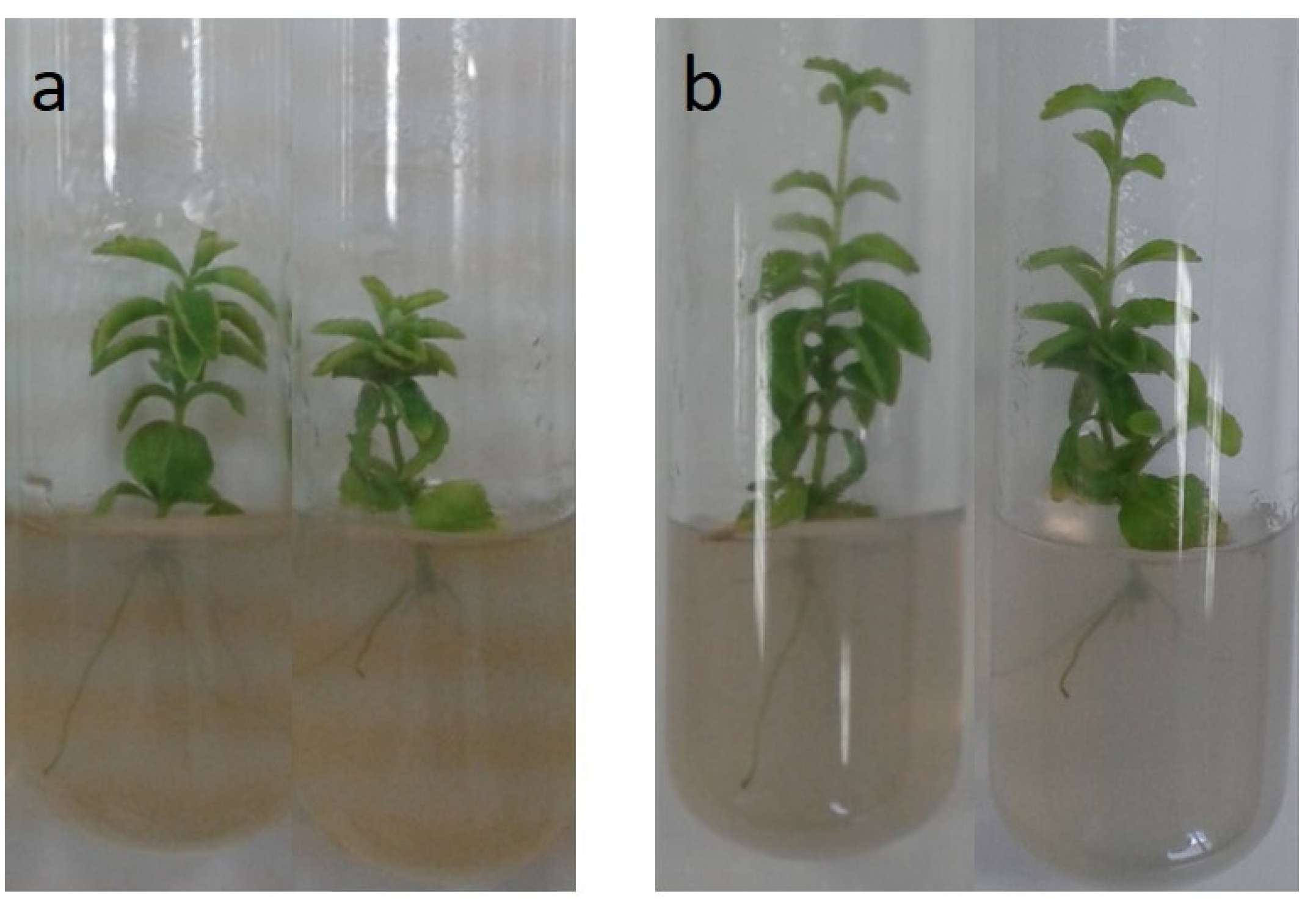
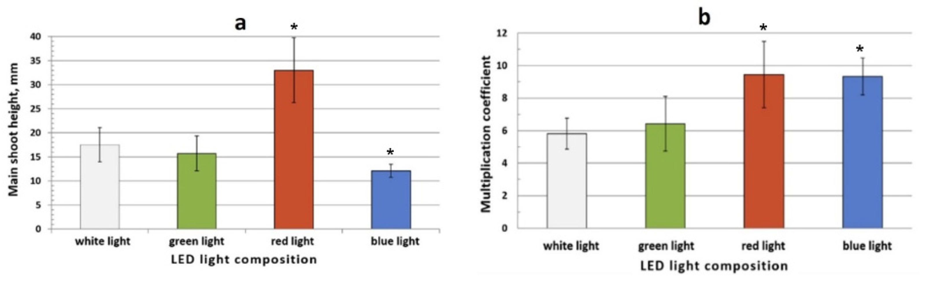
3.2.1. Effect of Red-Blue LED Lighting on Morphophysiological Parameters of S. rebaudiana Microshoots

Red-blue spectrum treatment has an inhibitory effect on the formation and growth of shoots. This manifested as shoot formation with shortened internodes as compared with the control (Figure 5a) (fluorescent white lamps). Moreover, root formation was observed under these conditions. In the case of the shoots returning to the control treatment, the elongation of the upper, newly formed internodes was observed (Figure 5b).

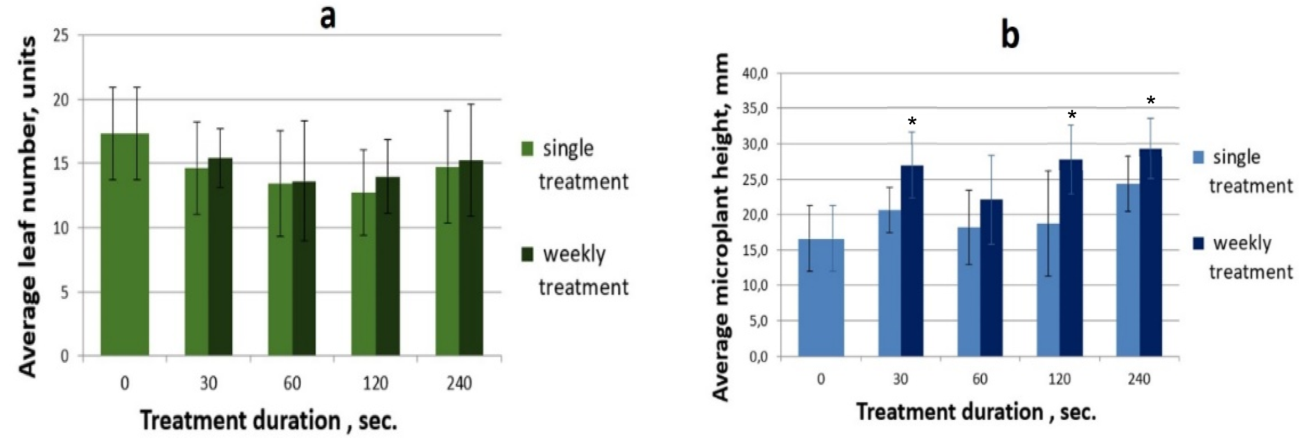
3.2.2. The Effect of Coherent Light Treatment on the Morphophysiological Characteristics of S. rebaudiana Microshoots In Vitro

Coherent light treatments did not significantly affect the morphophysiological parameters (newly formed shoots, total number of leaves, multiplication coefficient, and the frequency of root formation) of S. rebaudiana microshoots when compared with controls, according to ANOVA tests, with one exception—weekly exposure to coherent light for 120 and 240 s stimulated microshoot growth on MS culture media without PGRs. The average height (11 cm) and number of leaves (11.6) under coherent light treatments of 120 and 240 s were approximately twice as high as those of the controls.
A possible explanation for the lack of statistically significant differences in morphological characteristics of cultured S. rebaudiana microshoots is the fact that the microshoots were cultured in a PGRs-free medium; that is, the factors regulating morphogenesis were absent. Previous studies have shown that the presence of Epin in culture media has a stimulating effect on morphophysiological characteristics. Therefore, in the following series of experiments, we examined the possible synergistic effect of monochrome and coherent light treatments in combination with Epin.

3.2.3. The Effect of Monochrome Light Treatment on the Morphophysiological Characteristics of S. rebaudiana Microshoots In Vitro

S. rebaudiana microshoots cultured in vitro under red light treatment exhibited intensive growth, which manifested as the formation of a taller main shoot. When monochrome LEDs in the blue spectrum were used, stunted plants were formed. As regards the green and white (control) spectra, the plants had comparable average values under these conditions (Figure 6a). Despite the significantly different plant height between the treatment with red and blue spectra, the multiplication coefficient was the same, and averaged 9.5 (Figure 6b). This indicates that the internodes elongate in response to red light, while under blue light more compact microshoots were formed. As for green light, the multiplication coefficient was the same as the control.

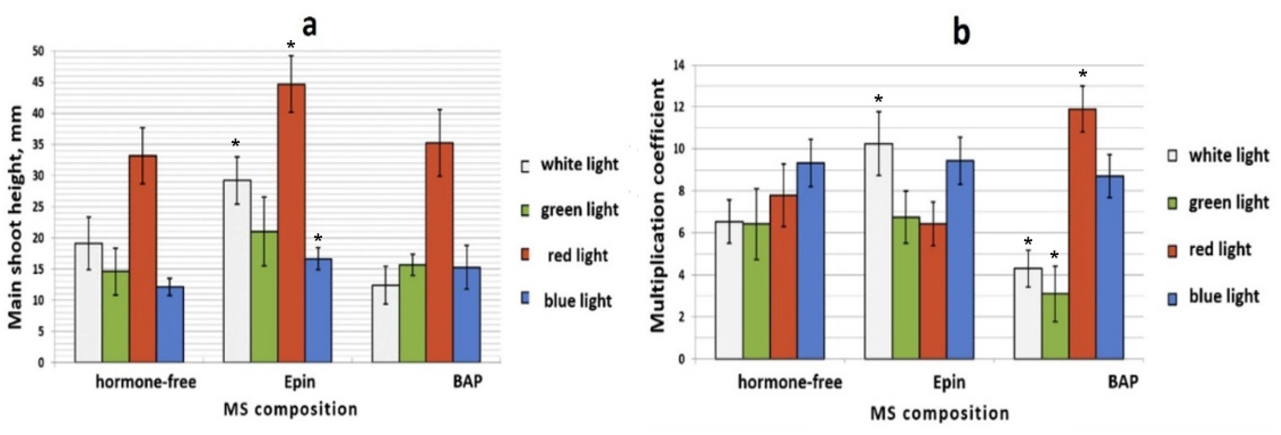
3.2.4. The Combined Effect of Physical and PGRs Factors on the Morphogenesis of S. rebaudiana Microshoots Culture In Vitro

Monochrome Light Treatment in Combination with PGRs under In Vitro Conditions

To study the combined effect of physical (monochrome light) and chemical (PGRs) factors on the morphological characteristics of S. rebaudiana microshoots in vitro, an extended experiment was conducted. We found that all variants of culture media, under red LED treatment, promoted intensive shoot growth, which occurred due to internode extension. A more obvious visible effect was observed in the variant supplemented with 0.1 mg L−1 Epin in combination with 0.5 mg L−1 IAA in culture on MS media (Figure 7a, Epin). In culture media with 1.0 mg L−1 BAP and IAA (Figure 7a, BAP), red light also enhanced the biometric indicators of S. rebaudiana microshoot cultures compared with controls (MS media without PGRs).
It was experimentally established that the multiplication coefficient was controlled by both PGRs and the spectral composition of provided light sources. The biggest differences were obtained on culture media supplemented with BAP (Figure 7b, BAP). In the case of light from the white and green spectra, the multiplication coefficient decreased to 3–4. The multiplication coefficient for the blue spectrum was 8.5–9.5 and did not depend on PGRs composition in culture media. The maximum multiplication coefficient was observed under red light treatments, on culture media supplemented with BAP (11.9) (Figure 7b, BAP), and under white light treatments, on culture media supplemented with Epin (10.5) (Figure 7b, Epin). The lowest multiplication coefficient was observed in green light treatments on culture media containing BAP.

Coherent Light Treatment in Combination with PGRs under In Vitro Conditions

Coherent light treatment was conducted on microshoots cultured on MS media containing 0.1 mg L−1 Epin and 0.5 mg L−1 IAA, and was performed either once or weekly. The results showed that intensive microshoot growth occurred, while the number of leaves was comparable to the control (Figure 8a).
It should be noted that in the variants given weekly coherent light treatments, the average height reached 27–29 mm and exceeded controls by 58–70% (Figure 8b). Moreover, significant differences between variants with 30, 120, and 240 s exposure were found. In the variant given a 60 s exposure, the average microplant height was comparable to controls. Thus, PGRs and light treatment exhibited a synergistic effect on the growth and development of S. rebaudiana microshoots.

3.3. Effects of Red-Blue LED Lighting on the Biochemical Characteristics of S. rebaudiana Plants In Vivo

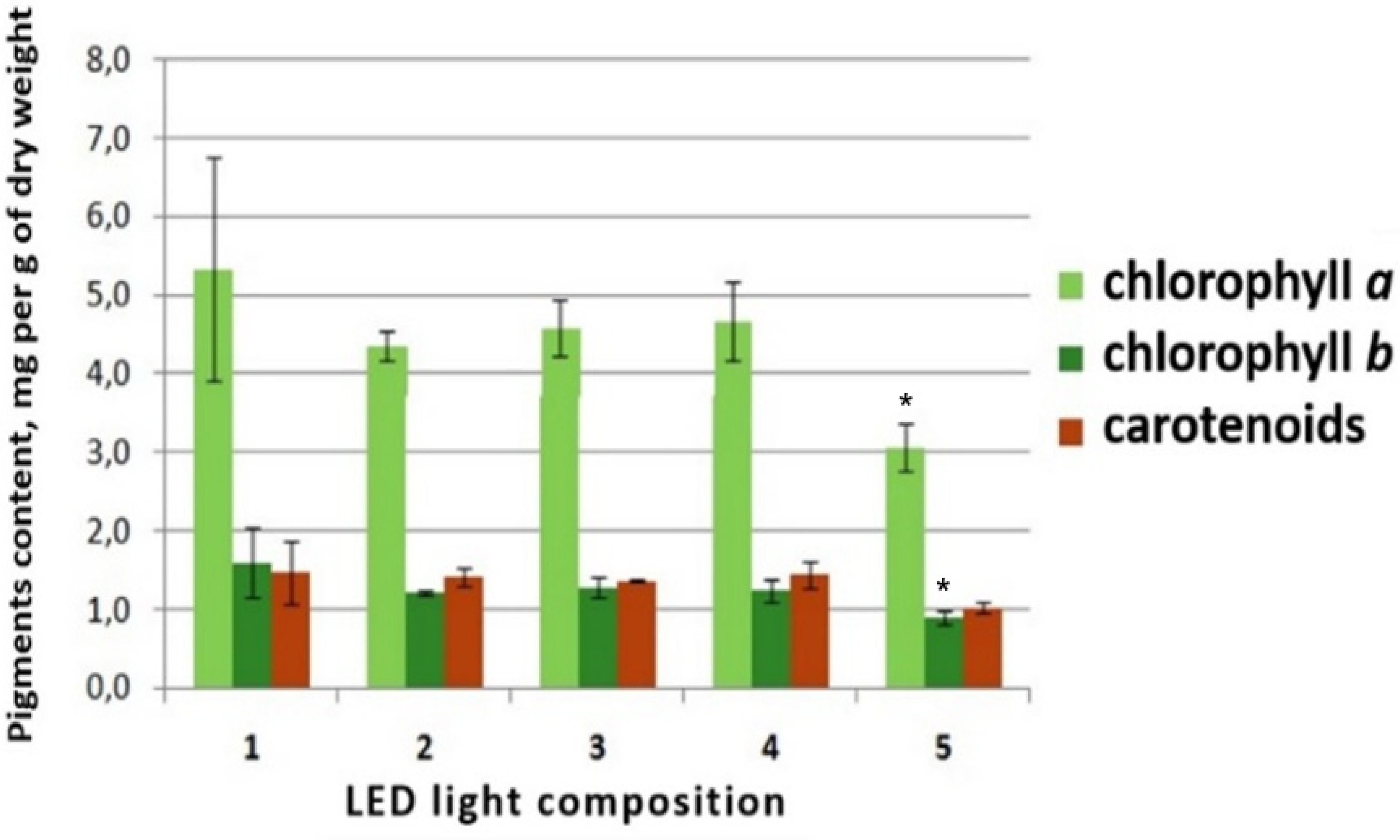
3.3.1. Quantitative Determination of Photosynthetic Pigments

In treatment five, from which the blue spectrum was excluded, the amount of chlorophyll a and b was significantly lower than in control treatments (Figure 9), which confirmed that maximum visible light absorption by chlorophylls occurs in the blue part of the spectrum.

3.3.2. Qualitative and Semi-Quantitative Determination of Diterpene Glycosides by TLC

The studied samples were found to contain diterpene glycosides, as determined by the Rf = 0.33 zone, which is consistent with findings of a previous study [59]. In addition, the spots that appeared had different color intensities. In the experimental samples, the intensity of staining was pronounced, which indicates that there was a greater number of glycosides in these variants.

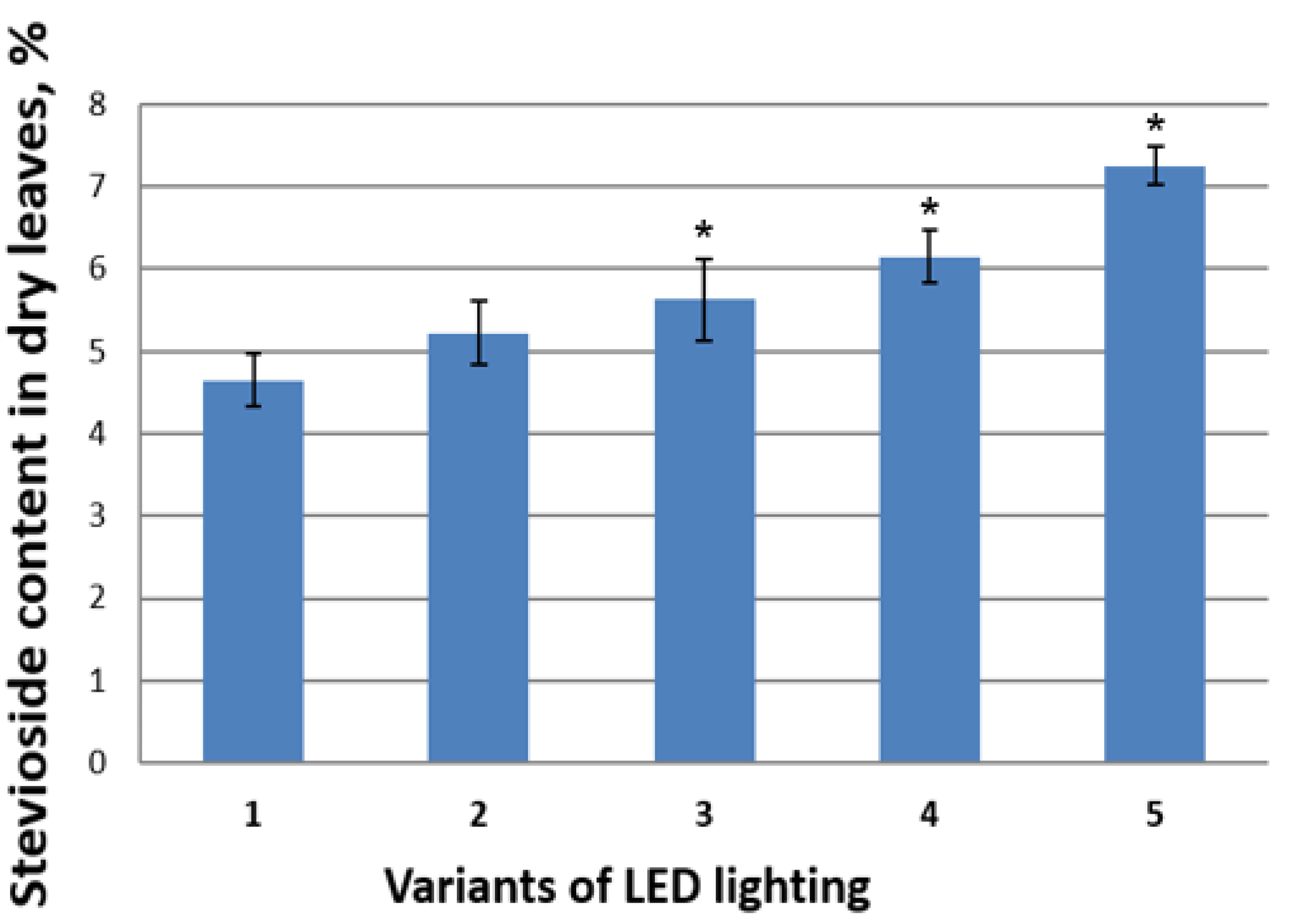
3.3.3. Quantitative Determination of Stevioside by HPLC

The highest content of stevioside in dry S. rebaudiana leaves was found in treatment five, from which the blue spectrum was excluded from provided lighting (Figure 10). The results support the notion that it is indeed the red part of the spectrum that induces secondary metabolite synthesis. Thus, we can conclude that the PGRs composition of a culture medium and spectral composition of its lighting have a significant regulating effect on the morphophysiological processes of S. rebaudiana plants, grown both in vitro and in vivo.

4. Conclusions

PGRs are widely used in in vitro plant techniques, especially in micropropagation. The nature and concentration of PGRs in culture media has a significant effect, both on morphogenesis and secondary metabolite biosynthesis. Application of PGRs in the micropropagation of S. rebaudiana is traditionally limited to the use of BAP, NAA, 2,4-D, and combinations thereof. In this study we used, for the first time, such PGRs as 2,4-epibrassinolide (Epin) and hydroxycinnamic acid (Zircon), and found that they have strong positive impacts on shoot multiplication and rooting, as well as their promotion of the formation of strong plants with well-developed leaf blades. We have also demonstrated that cultivation of S. rebaudiana microshoots on a medium containing Epin and IAA, combined with weekly coherent light irradiation, doubles their multiplication coefficient, stimulates adventitious shoot growth, and increases their number of leaves. This multiplication coefficient can also be increased by the use of media containing BAP and IAA in combination with red monochrome light conditions. In addition, it has been shown that the highest stevioside content in leaves of S. rebaudiana was observed after the treatment from which blue light was excluded.

Author Contributions

A.A.S. and R.N.K. conducting experiments on Stevia culture in vitro and statistical analysis of experimental data. I.G.T. providing experiments for spectral light composition. M.Y.C., O.B.P. and E.N.B. conducting experiments on TLC, HPLC and determination of photosynthetic pigments. E.A.K. and M.R.K. participated in manuscript conceptualization; writing, reviewing, and editing of manuscript; approved the final manuscript for publication, and agreed to be accountable for all aspects of the manuscript. All authors have read and agreed to the published version of the manuscript.

Funding

The reported study was supported by assignments 0574-2019-0002 and 18-118021490111-5 of the Ministry of Science and Higher Education of the Russian Federation.

Institutional Review Board Statement

Not applicable.

Informed Consent Statement

Not applicable.

Data Availability Statement

Data sharing is not applicable to this article.

Acknowledgments

The authors are grateful to Nikolay I. Bondarev (Timiryazev Institute of Plant Physiology Russian Academy of Sciences) for providing intact plant material; and Alina S. Ivanitskikh (Russian State Agrarian University–Moscow Timiryazev Agricultural Academy) for assistance in TLC and HPLC providing.

Conflicts of Interest

The authors declare no conflict of interest.

References

  1. Saraiva, A.; Carrascosa, C.; Raheem, D.; Ramos, F.; Raposo, A. Natural sweeteners: The relevance of food naturalness for consumers, food security aspects, sustainability and health impacts. Int. J. Environ. Res. Public Health 2020, 17, 6285. [Google Scholar] [CrossRef]
  2. Bergman, M.E.; Davis, B.; Phillips, M.A. Medically useful plant terpenoids: Biosynthesis, occurrence, and mechanism of action. Molecules 2019, 24, 3961. [Google Scholar] [CrossRef] [Green Version]
  3. Lanzotti, V. Diterpenes for Therapeutic Use, in Natural Products; Springer: Berlin/Heidelberg, Germany, 2013; p. 3173. [Google Scholar]
  4. Goel, M.K.; Kukreja, A.K.; Singh, A.K.; Khanuja, S.P.S. In vitro plant growth promoting activity of phyllocladane diterpenoids isolated from Callicarpa macrophylla Vahl. in shoot cultures of Rauwolfia serpentina. Nat. Prod. Commun. 2007, 2, 1934578X0700200802. [Google Scholar] [CrossRef]
  5. Abbaszadeh, G.; Srivastava, C.; Walia, S. Insecticidal and antifeedant activities of clerodane diterpenoids isolated from the Indian bhant tree, Clerodendron infortunatum, against the cotton bollworm, Helicoverpa armigera. J. Insect Sci. 2014, 14, 1. [Google Scholar] [CrossRef]
  6. Urzúa, A.; Rezende, M.C.; Mascayano, C.; Vásquez, L. A structure-activity study of antibacterial diterpenoids. Molecules 2008, 13, 882–891. [Google Scholar] [CrossRef] [Green Version]
  7. Bozov, P.; Girova, T.; Prisadova, N.; Hristova, Y.; Gochev, V. Antimicrobial activity of neo-clerodane diterpenoids isolated from Lamiaceae species against pathogenic and food spoilage microorganisms. Nat. Prod. Commun. 2015, 10, 1934578X1501001101. [Google Scholar] [CrossRef] [Green Version]
  8. Islam, M.T. Diterpenes and their derivatives as potential anticancer agents. Phytother. Res. 2017, 31, 691–712. [Google Scholar] [CrossRef]
  9. Jian, B.; Zhang, H.; Han, C.; Liu, J. Anti-cancer activities of diterpenoids derived from Euphorbia fischeriana Steud. Molecules 2018, 23, 387. [Google Scholar] [CrossRef] [Green Version]
  10. Koyama, E.; Kitazawa, K.; Ohori, Y.; Izawa, O.; Kakegawa, K.; Fujino, A.; Ui, M. In vitro metabolism of the glycosidic sweeteners, stevia mixture and enzymatically modified stevia in human intestinal microflora. Food Chem. Toxicol. 2003, 41, 359–374. [Google Scholar] [CrossRef]
  11. Bondarev, N.I.; Sukhanova, M.A.; Reshetnyak, O.V.; Nosov, A.M. Steviol glycoside content in different organs of Stevia rebaudiana and its dynamics during ontogeny. Biol. Plant. 2003, 47, 261–264. [Google Scholar] [CrossRef]
  12. Muanda, F.N.; Soulimani, R.; Diop, B.; Dicko, A. Study on chemical composition and biological activities of essential oil and extracts from Stevia rebaudiana Bertoni leaves. LWT 2011, 44, 1865–1872. [Google Scholar] [CrossRef]
  13. Chen, T.H.; Chen, S.C.; Chan, P.; Chu, Y.; Yang, H.Y.; Cheng, J.T. Mechanism of the hypoglycemic effect of stevioside, a glycoside of Stevia rebaudiana. Planta Med. 2005, 71, 108–113. [Google Scholar] [CrossRef] [PubMed]
  14. Megeji, N.W.; Kumar, J.K.; Singh, V.; Kaul, V.K.; Ahuja, P.S. Introducing Stevia rebaudiana, a natural zero-calorie sweetener. Curr. Sci. 2005, 88, 801–804. [Google Scholar]
  15. Gupta, E.; Purwar, S.; Sundaram, S.; Rai, G.K. Nutritional and therapeutic values of Stevia rebaudiana: A review. J. Med. Plants Res. 2013, 7, 3343–3353. [Google Scholar]
  16. Jeppesen, P.B.; Gregersen, S.; Alstrup, K.K.; Hermansen, K. Stevioside induces antihyperglycaemic, insulinotropic and glucagonostatic effects in vivo: Studies in the diabetic Goto-Kakizaki (GK) rats. Phytomedicine 2002, 9, 9–14. [Google Scholar] [CrossRef] [PubMed]
  17. Starratt, A.N.; Kirby, C.W.; Pocs, R.; Brandle, J.E.; Rebaudioside, F. A diterpene glycoside from Stevia rebaudiana. Phytochemistry 2002, 59, 367–370. [Google Scholar] [CrossRef]
  18. Tadhani, M.B.; Patel, V.H.; Subhash, R. In vitro antioxidant activities of Stevia rebaudiana leaves and callus. J. Food Compos. Anal. 2007, 20, 323–329. [Google Scholar] [CrossRef]
  19. Brandle, J.E.; Starratt, A.N.; Gijzen, M. Stevia rebaudiana: Its agricultural, biological, and chemical properties. Can. J. Plant Sci. 1998, 78, 527–536. [Google Scholar] [CrossRef] [Green Version]
  20. Ahmed, M.B.; Salahin, M.; Karim, R.; Razvy, M.A.; Hannan, M.M.; Sultana, R.; Hossain, M.; Islam, R. An efficient method for in vitro clonal propagation of a newly introduced sweetener plant (Stevia rebaudiana Bertoni.) in Bangladesh. Am. Eurasian J. Sci. Res. 2007, 2, 121–125. [Google Scholar]
  21. Bondarev, N.; Reshetnyak, O.; Nosov, A. Peculiarities of diterpenoid steviol glycoside production in in vitro cultures of Stevia rebaudiana Bertoni. Plant Sci. 2001, 161, 155–163. [Google Scholar] [CrossRef]
  22. Ferreira, C.M.; Handro, W. Production, maintenance and plant regeneration from cell suspension cultures of Stevia rebaudiana (Bert.) Bertoni. Plant Cell Rep. 1988, 7, 123–126. [Google Scholar] [CrossRef]
  23. Gantait, S.; Das, A.; Mandal, N. Stevia: A comprehensive review on ethnopharmacological properties and in vitro regeneration. Sugar Tech. 2015, 17, 95–106. [Google Scholar] [CrossRef]
  24. Giridhar, P.; Sowmya, K.S.; Ramakrishna, A.; Ravishankar, G.A. Rapid clonal propagation and stevioside profiles of Stevia rebaudiana Bertoni. Int. J. Plant Dev. Biol. 2010, 4, 47–52. [Google Scholar]
  25. Guruchandran, V.; Sasikumar, C. Organogenic plant regeneration via callus induction in Stevia rebaudiana Bert. Int. J. Curr. Microbiol. Appl. Sci. 2013, 2, 56–61. [Google Scholar]
  26. Jarma-Orozco, A.; Combatt-Caballero, E.; Jaraba-Navas, J. Growth and development of Stevia rebaudiana Bert., in high and low levels of radiation. Curr. Plant Biol. 2020, 22, 100144. [Google Scholar] [CrossRef]
  27. Kalpana, M.; Anbazhagan, M.; Natarajan, V.; Dhanavel, D. Improved micropropagation method for the enhancement of biomass in Stevia rebaudiana Bertoni. Rec. Res. Sci. Tech. 2010, 2, 8–13. [Google Scholar]
  28. Karim, M.A.; Jannat, R.; Rahman, M.S.; Haque, M.S. Micropropagation of stevia plant from nodal segments. Prog. Agric. 2008, 19, 21–26. [Google Scholar] [CrossRef] [Green Version]
  29. Kulchin, Y.N.; Nakonechnaya, O.V.; Gafitskaya, I.V.; Grishchenko, O.V.; Epifanova, T.Y.; Orlovskaya, I.Y.; Zhuravlev, Y.N.; Subbotin, E.P. Plant morphogenesis under different light intensity. Defect Diffus. Forum Trans. Tech. Publ. 2018, 386, 201–206. [Google Scholar] [CrossRef]
  30. Melviana, A.C.; Esyanti, R.R.; Mel, M.; Setyobudi, R.H. Biomass enhancement of Stevia rebaudiana Bertoni Shoot culture in temporary immersion system (TIS) RITA® bioreactor optimized in two different immersion periods. In Proceedings of the 1st International Conference on Bioenergy and Environmentally Sustainable Agriculture Technology (ICoN BEAT 2019), East Java, Indonesia, 7–8 November 2019; p. 00007. [Google Scholar]
  31. Razak, U.N.A.A.; Ong, C.B.; Yu, T.S.; Lau, L.K. In vitro micropropagation of Stevia rebaudiana Bertoni in Malaysia. Braz. Arch. Biol. Technol. 2014, 57, 23–28. [Google Scholar] [CrossRef] [Green Version]
  32. Muthu, S.; Schuurmans, F.J.; Pashley, M.D. Red, green, and blue LEDs for white light illumination. IEEE J. Sel. Top. Quantum Electron. 2002, 8, 333–338. [Google Scholar] [CrossRef] [Green Version]
  33. Sivaram, L.; Mukundan, U. In vitro culture studies on Stevia rebaudiana. Vitro Cell. Dev. Biol. Plant 2003, 39, 520–523. [Google Scholar] [CrossRef]
  34. Ahmad, A.; Ali, H.; Khan, H.; Begam, A.; Khan, S.; Ali, S.S.; Ahmad, N.; Fazal, H.; Ali, M.; Hano, C.; et al. Effect of Gibberellic Acid on Production of Biomass, Polyphenolics and Steviol Glycosides in Adventitious Root Cultures of Stevia rebaudiana (Bert.). Plants 2020, 9, 420. [Google Scholar] [CrossRef] [Green Version]
  35. Blinstrubienė, A.; Burbulis, N.; Juškevičiūtė, N.; Vaitkevičienė, N.; Žūkienė, R. Effect of Growth Regulators on Stevia rebaudianaBertoni Callus Genesis and Influence of Auxin and Proline to Steviol Glycosides, Phenols, Flavonoids Accumulation, and Antioxidant Activity In Vitro. Molecules 2020, 25, 2759. [Google Scholar] [CrossRef] [PubMed]
  36. Bondarev, N.; Reshetnyak, O.; Bondareva, T.; Il’in, M.; Nosov, A. Impact of Cultivation Factors in vitro on the Growth and the Biosynthesis of Steviol Glycosides in Stevia rebaudiana cell Cultures. Physiol. Mol. Biol. Plants 2019, 25, 1091–1096. [Google Scholar] [CrossRef] [PubMed]
  37. Gupta, P.; Sharma, S.; Saxena, S. Biomass Yield and Steviol Glycoside Production in Callus and Suspension Culture of Stevia rebaudiana Treated with Proline and Polyethylene Glycol. Appl. Biochem. Biotechnol. 2015, 176, 863–874. [Google Scholar] [CrossRef] [PubMed]
  38. Libik-Konieczny, M.; Capecka, E.; Tuleja, M.; Konieczny, R. Synthesis and production of steviol glycosides: Recent research trends and perspectives. Appl. Microbiol. Biotechnol. 2021, 105, 3883–3900. [Google Scholar] [CrossRef] [PubMed]
  39. Rameeh, V.; Gerami, M.; Omran, V.G.; Ghavampour, S. Impact of glycine betaine on salinity tolerance of stevia (Stevia rebaudiana Bertoni) under in vitro condition. Cercet. Agron. Mold. 2017, 3, 95–105. [Google Scholar] [CrossRef] [Green Version]
  40. Gupta, P.; Sharma, S.; Saxena, S. Effect of abiotic stress on growth parameters and steviol glycoside content in Stevia rebaudiana (Bertoni) raised in vitro. J. Appl. Res. Med. Aromat. Plants 2016, 3, 160–167. [Google Scholar] [CrossRef]
  41. Lucho, S.R.; do Amaral, M.N.; Auler, P.A.; Bianchi, V.J.; María Ángeles Ferrer, M.A.; Calderón, A.A.; Bolacel Braga, E.J. Salt stress-induced changes in in vitro cultured Stevia rebaudiana Bertoni: Effect on metabolite contents, antioxidant capacity and expression of steviol glycosides-related biosynthetic genes. J. Plant. Growth Regul. 2019, 38, 1341–1353. [Google Scholar] [CrossRef]
  42. Fallah, F.; Nokhasi, F.; Ghaheri, M.; Kahrizi, D.; Beheshti, A.A.A.; Ghorbani, T.; Kazemi, E.; Ansarypou, Z. Effect of salinity on gene expression, morphological and biochemical characteristics of Stevia rebaudiana Bertoni under in vitro conditions. Cell Mol. Biol. 2017, 63, 102–103. [Google Scholar] [CrossRef]
  43. Pandey, M.; Chikara, S.K. Effect of salinity and drought stress on growth parameters, glycoside content and expression level of vital genes in steviol glycosides biosynthesis pathway of Stevia rebaudiana (Bertoni). Int. J. Genet. 2015, 7, 153–160. [Google Scholar]
  44. Bayraktar, M.; Naziri, E.; Akgun, I.H.; Karabey, F.; Ilhan, E.; Akyol, B.; Bedir, E.; Gurel, A. Elicitor induced stevioside production, in vitro shoot growth, and biomass accumulation in micropropagated Stevia rebaudiana. Plant Cell Tissue Organ Cult. 2016, 127, 289–300. [Google Scholar] [CrossRef]
  45. Ghazal, B.; Saif, S.; Farid, K.; Khan, A.; Rehman, S.; Reshma, A.; Fazal, H.; Ali, M.; Ahmad, A.; Rahman, L.; et al. Stimulation of secondary metabolites by copper and gold nanoparticles in submerge adventitious root cultures of Stevia rebaudiana (Bert.). IET Nanobiotechnol. 2018, 12, 569–573. [Google Scholar] [PubMed]
  46. Tarakanov, I.G. Light control of growth and development in vegetable plants with various life strategies. Acta Hortic. 2006, 711, 315–321. [Google Scholar] [CrossRef]
  47. Tarakanov, I.; Yakovleva, O.; Konovalova, I.; Paliutina, G.; Anisimov, A. Light-emitting diodes: On the way to combinatorial lighting technologies for basic research and crop production. Acta Hortic. 2012, 956, 171–178. [Google Scholar] [CrossRef]
  48. Nakonechnaya, O.V.; Gafitskaya, I.V.; Burkovskaya, E.V.; Khrolenko, Y.A.; Grishchenko, O.V.; Zhuravlev, Y.N.; Sussotin, E.P.; Kulchin, Y.N. Effect of light intensity on the morphogenesis of Stevia rebaudiana under in vitro conditions. Russ. J. Plant Physiol. 2019, 66, 656–663. [Google Scholar] [CrossRef]
  49. Bondarev, N.I.; Reshetnyak, O.V.; Nosov, A.M. Influence of photoperiod and irradiation intensity on the development of Stevia rebaudiana shoots in vitro and synthesis of steviol glycosides. Izv. TSKhA 2008, 4, 102–107. [Google Scholar]
  50. Idrees, M.; Sania, B.; Hafsa, B.; Kumari, S.; Khan, H.; Fazal, H.; Ahmad, I.; Akbar, F.; Ahmad, N.; Ali, S.; et al. Spectral lights trigger biomass accumulation and production of antioxidant secondary metabolites in adventitious root cultures of Stevia rebaudiana (Bert.). C. R. Biol. 2018, 341, 334–342. [Google Scholar] [CrossRef]
  51. Ahmad, N.; Rab, A.; Ahmad, N. Light-induced biochemical variations in secondary metabolite production and antioxidant activity in callus cultures of Stevia rebaudiana (Bert). J. Photochem. Photobiol. B 2016, 154, 6–51. [Google Scholar] [CrossRef]
  52. Murashige, T.; Skoog, F. A revised medium for rapid growth and bio assays with tobacco tissue cultures. Physiol. Plant. 1962, 15, 473–497. [Google Scholar] [CrossRef]
  53. Jaitak, V.; Gupta, A.P.; Kaul, V.K.; Ahuja, P.S. Validated high-performance thin-layer chromatography method for steviol glycosides in Stevia rebaudiana. J. Pharm. Biomed. Anal. 2008, 47, 790–794. [Google Scholar] [CrossRef]
  54. Rokosa, M.T.; Kulpa, D. Micropropagation of Stevia rebaudiana plants. Ciência Rural 2020, 50, e20181029. [Google Scholar] [CrossRef] [Green Version]
  55. Nower, A.A. In vitro propagation and synthetic seeds production: An efficient method for Stevia rebaudiana Bertoni. Sugar Tech. 2014, 16, 100–108. [Google Scholar] [CrossRef]
  56. Bayraktar, M. Micropropagation of Stevia rebaudiana Bertoni using RITA®® bioreactor. HortScience 2019, 54, 725–731. [Google Scholar] [CrossRef] [Green Version]
  57. Lata, H.; Chandra, S.; Wang, Y.H.; ElSohly, M.A.; Khan, I.A. Polyhouse cultivation of in vitro raised elite Stevia rebaudiana Bertoni: An assessment of biochemical and photosynthetic characteristics. Int. J. Trop. Agric. 2015, 33, 2381–2389. [Google Scholar]
  58. Ceunen, S.; Werbrouck, S.; Geuns, J.M. Stimulation of steviol glycoside accumulation in Stevia rebaudiana by red LED light. J. Plant Physiol. 2012, 169, 749–752. [Google Scholar] [CrossRef]
  59. Nikolova-Damyanova, B.; Bankova, V.; Popov, S. Separation and quantitation of stevioside and rebaudioside a in plant extracts by normal-phase high performance liquid chromatography and thin-layer chromatography: A comparison. Phytochem. Anal. 1994, 5, 81–85. [Google Scholar] [CrossRef]
Figure 1. Graphs of LED light spectra used to evaluate the effect of exposure to light of white and red-blue spectra on S. rebaudiana microshoot growth.
Horticulturae 07 00195 g001
Figure 2. Graphs of LED light spectra used to evaluate the effects of their exposure (blue and various red spectra) on S. rebaudiana growth. Short-wave red (SR), long-wave red (LR), far red (FR), and blue (B) spectra are depicted.
Horticulturae 07 00195 g002
Figure 3. Graphs of LED light spectra used to evaluate the effects of monochrome LED on S. rebaudiana microshoot growth.
Horticulturae 07 00195 g003
Figure 4. Morphological differences of S. rebaudiana microshoots cultured in vitro on various media. Figured depict results of enculturation on: (a) PGRs-free MS media and on MS media supplemented with different PGRs; (b) 1 mg L−1 kinetin and 0.5 mgL−1 IAA; (c) 1 mg L−1 BAP and 0.5 mg L−1 IAA; (d) 0.1 mg L−1 Epin and 0.5 mg L−1 IAA; and (e) 0.5 mg L−1 Zircon. Scale 1 cm.
Horticulturae 07 00195 g004
Figure 5. Morphological changes of S. rebaudiana microshoots (a) after 20 days being cultured on MS media without PGRs under red-blue LEDs, and (b) after 5 days being returned to fluorescent white lamps.
Horticulturae 07 00195 g005
Figure 6. (a) Main shoot heights and (b) multiplication coefficients of S. rebaudiana microshoots in in vitro cultures under different spectra of monochrome LED lighting. Means ± standard errors at α = 0.05 according to the ANOVA test (n = 60). *-variants that had significant differences between control treatments (white light).
Horticulturae 07 00195 g006
Figure 7. The influence of different LED lighting and PGRs combinations on (a) main shoot height and (b) the multiplication coefficient of S. rebaudiana microshoots in vitro. Means ± standard errors at α = 0.05 according to ANOVA tests (n = 60). *-variants of culture media that have significant differences between controls (MS media without PGRs).
Horticulturae 07 00195 g007
Figure 8. The influence of coherent light treatments on (a) the average number of leaves per microshoot and (b) the microshoot height of plants cultured on an MS medium containing 0.1 mg L−1 Epin and 0.5 mg L−1 IAA. Means ± standard errors at α = 0.05 according to ANOVAs test (n = 60). *-variants that have significant differences between control treatments (without treatment).
Horticulturae 07 00195 g008
Figure 9. The effect of spectra composition on concentrations of chlorophylls a, b and carotenoids in S. rebaudiana plants grown in vivo under various red-blue LED lightings: (1) SR + LR + FR + B; (2) LR + FR + B; (3) SR + FR + B; (4) SR + LR + B; and (5) SR + LR + FR (Figure 2). Means ± standard errors at α = 0.05 according to ANOVA tests (n = 10). *-variants that have significant differences between control treatments (SR + LR + FR + B).
Horticulturae 07 00195 g009
Figure 10. The effect of spectral light composition on stevioside content in S. rebaudiana plants grown in vivo under different treatments of red-blue LED lighting: (1) SR + LR + FR + B; (2) LR + FR + B; (3) SR + FR + B; (4) SR + LR + B; and (5) SR + LR + FR (Figure 2). Means ± standard errors at α = 0.05 according to ANOVA tests (n = 10). *-variants that have significant differences between control treatments (SR + LR + FR + B).
Horticulturae 07 00195 g010
Publisher’s Note: MDPI stays neutral with regard to jurisdictional claims in published maps and institutional affiliations.

