Next Issue
Volume 5, May
Previous Issue
Volume 5, March


J. Imaging, Volume 5, Issue 4 (April 2019) – 8 articles

Cover Story (view full-size image): 
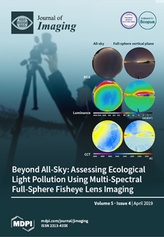
A new method to measure ecological light pollution, a novel anthropogenic stressor that affects flora and fauna on many scales is presented. We used a commercial digital camera with a fisheye lens to acquire at least two vertical-plane multispectral (RGB) images to cover the full solid angle, not only the all-sky view. This simple procedure provides a comprehensive way to characterize nocturnal light and light pollution. To make the method accessible to a broad audience, we give a step-by-step explanation of the technical and practical procedure and software to process luminance and correlated color temperature maps. The image shows data obtained in winter near the arctic circle in Finland. View this paper.
  • Issues are regarded as officially published after their release is announced to the table of contents alert mailing list.
  • You may sign up for e-mail alerts to receive table of contents of newly released issues.
  • PDF is the official format for papers published in both, html and pdf forms. To view the papers in pdf format, click on the "PDF Full-text" link, and use the free Adobe Reader to open them.
Order results
Result details
Section
Select all
Export citation of selected articles as:
12 pages, 2118 KiB  
Article
Evaluating Human Photoreceptoral Inputs from Night-Time Lights Using RGB Imaging Photometry
by Alejandro Sánchez de Miguel, Salvador Bará, Martin Aubé, Nicolás Cardiel, Carlos E. Tapia, Jaime Zamorano and Kevin J. Gaston
J. Imaging 2019, 5(4), 49; https://0-doi-org.brum.beds.ac.uk/10.3390/jimaging5040049 - 16 Apr 2019
Cited by 9 | Viewed by 7272
Abstract
Night-time lights interact with human physiology through different pathways starting at the retinal layers of the eye; from the signals provided by the rods; the S-, L- and M-cones; and the intrinsically photosensitive retinal ganglion cells (ipRGC). These individual photic channels combine in [...] Read more.
Night-time lights interact with human physiology through different pathways starting at the retinal layers of the eye; from the signals provided by the rods; the S-, L- and M-cones; and the intrinsically photosensitive retinal ganglion cells (ipRGC). These individual photic channels combine in complex ways to modulate important physiological processes, among them the daily entrainment of the neural master oscillator that regulates circadian rhythms. Evaluating the relative excitation of each type of photoreceptor generally requires full knowledge of the spectral power distribution of the incoming light, information that is not easily available in many practical applications. One such instance is wide area sensing of public outdoor lighting; present-day radiometers onboard Earth-orbiting platforms with sufficient nighttime sensitivity are generally panchromatic and lack the required spectral discrimination capacity. In this paper, we show that RGB imagery acquired with off-the-shelf digital single-lens reflex cameras (DSLR) can be a useful tool to evaluate, with reasonable accuracy and high angular resolution, the photoreceptoral inputs associated with a wide range of lamp technologies. The method is based on linear regressions of these inputs against optimum combinations of the associated R, G, and B signals, built for a large set of artificial light sources by means of synthetic photometry. Given the widespread use of RGB imaging devices, this approach is expected to facilitate the monitoring of the physiological effects of light pollution, from ground and space alike, using standard imaging technology. Full article
(This article belongs to the Special Issue Light Pollution Assessment with Imaging Devices)
Show Figures

Figure 1

25 pages, 6680 KiB  
Review
Degraded Historical Document Binarization: A Review on Issues, Challenges, Techniques, and Future Directions
by Alaa Sulaiman, Khairuddin Omar and Mohammad F. Nasrudin
J. Imaging 2019, 5(4), 48; https://0-doi-org.brum.beds.ac.uk/10.3390/jimaging5040048 - 12 Apr 2019
Cited by 61 | Viewed by 9147
Abstract
In this era of digitization, most hardcopy documents are being transformed into digital formats. In the process of transformation, large quantities of documents are stored and preserved through electronic scanning. These documents are available from various sources such as ancient documentation, old legal [...] Read more.
In this era of digitization, most hardcopy documents are being transformed into digital formats. In the process of transformation, large quantities of documents are stored and preserved through electronic scanning. These documents are available from various sources such as ancient documentation, old legal records, medical reports, music scores, palm leaf, and reports on security-related issues. In particular, ancient and historical documents are hard to read due to their degradation in terms of low contrast and existence of corrupted artefacts. In recent times, degraded document binarization has been studied widely and several approaches were developed to deal with issues and challenges in document binarization. In this paper, a comprehensive review is conducted on the issues and challenges faced during the image binarization process, followed by insights on various methods used for image binarization. This paper also discusses the advanced methods used for the enhancement of degraded documents that improves the quality of documents during the binarization process. Further discussions are made on the effectiveness and robustness of existing methods, and there is still a scope to develop a hybrid approach that can deal with degraded document binarization more effectively. Full article
Show Figures

Figure 1

14 pages, 5269 KiB  
Article
Qualitative Methods for the Inverse Obstacle Problem: A Comparison on Experimental Data
by Martina T. Bevacqua and Roberta Palmeri
J. Imaging 2019, 5(4), 47; https://0-doi-org.brum.beds.ac.uk/10.3390/jimaging5040047 - 10 Apr 2019
Cited by 14 | Viewed by 4672
Abstract
Qualitative methods are widely used for the solution of inverse obstacle problems. They allow one to retrieve the morphological properties of the unknown targets from the scattered field by avoiding dealing with the problem in its full non-linearity and considering a simplified mathematical [...] Read more.
Qualitative methods are widely used for the solution of inverse obstacle problems. They allow one to retrieve the morphological properties of the unknown targets from the scattered field by avoiding dealing with the problem in its full non-linearity and considering a simplified mathematical model with a lower computational burden. Very many qualitative approaches have been proposed in the literature. In this paper, a comparison is performed in terms of performance amongst three different qualitative methods, i.e., the linear sampling method, the orthogonality sampling method, and a recently introduced method based on joint sparsity and equivalence principles. In particular, the analysis is focused on the inversion of experimental data and considers a wide range of (distinct) working frequencies and different kinds of scattering experiments. Full article
(This article belongs to the Special Issue Microwave Imaging and Electromagnetic Inverse Scattering Problems)
Show Figures

Figure 1

17 pages, 4327 KiB  
Article
Beyond All-Sky: Assessing Ecological Light Pollution Using Multi-Spectral Full-Sphere Fisheye Lens Imaging
by Andreas Jechow, Christopher C.M. Kyba and Franz Hölker
J. Imaging 2019, 5(4), 46; https://0-doi-org.brum.beds.ac.uk/10.3390/jimaging5040046 - 09 Apr 2019
Cited by 58 | Viewed by 10997
Abstract
Artificial light at night is a novel anthropogenic stressor. The resulting ecological light pollution affects a wide breadth of biological systems on many spatio-temporal scales, from individual organisms to communities and ecosystems. However, a widely-applicable measurement method for nocturnal light providing spatially resolved [...] Read more.
Artificial light at night is a novel anthropogenic stressor. The resulting ecological light pollution affects a wide breadth of biological systems on many spatio-temporal scales, from individual organisms to communities and ecosystems. However, a widely-applicable measurement method for nocturnal light providing spatially resolved full-spectrum radiance over the full solid angle is still missing. Here, we explain the first step to fill this gap, by using a commercial digital camera with a fisheye lens to acquire vertical plane multi-spectral (RGB) images covering the full solid angle. We explain the technical and practical procedure and software to process luminance and correlated color temperature maps and derive illuminance. We discuss advantages and limitations and present data from different night-time lighting situations. The method provides a comprehensive way to characterize nocturnal light in the context of ecological light pollution. It is affordable, fast, mobile, robust, and widely-applicable by non-experts for field work. Full article
(This article belongs to the Special Issue Light Pollution Assessment with Imaging Devices)
Show Figures

Figure 1

26 pages, 659 KiB  
Article
Zig-Zag Based Single-Pass Connected Components Analysis
by Donald G. Bailey and Michael J. Klaiber
J. Imaging 2019, 5(4), 45; https://0-doi-org.brum.beds.ac.uk/10.3390/jimaging5040045 - 06 Apr 2019
Cited by 16 | Viewed by 5617
Abstract
Single-pass connected components analysis (CCA) algorithms suffer from a time overhead to resolve labels at the end of each image row. This work demonstrates how this overhead can be eliminated by replacing the conventional raster scan by a zig-zag scan. This enables chains [...] Read more.
Single-pass connected components analysis (CCA) algorithms suffer from a time overhead to resolve labels at the end of each image row. This work demonstrates how this overhead can be eliminated by replacing the conventional raster scan by a zig-zag scan. This enables chains of labels to be correctly resolved while processing the next image row. The effect is faster processing in the worst case with no end of row overheads. CCA hardware architectures using the novel algorithm proposed in this paper are, therefore, able to process images at higher throughput than other state-of-the-art methods while reducing the hardware requirements. The latency introduced by the conversion from raster scan to zig-zag scan is compensated for by a new method of detecting object completion, which enables the feature vector for completed connected components to be output at the earliest possible opportunity. Full article
(This article belongs to the Special Issue Image Processing Using FPGAs)
Show Figures

Figure 1

15 pages, 32826 KiB  
Article
Street Sign Recognition Using Histogram of Oriented Gradients and Artificial Neural Networks
by Kh Tohidul Islam, Sudanthi Wijewickrema, Ram Gopal Raj and Stephen O’Leary
J. Imaging 2019, 5(4), 44; https://0-doi-org.brum.beds.ac.uk/10.3390/jimaging5040044 - 03 Apr 2019
Cited by 9 | Viewed by 7368
Abstract
Street sign identification is an important problem in applications such as autonomous vehicle navigation and aids for individuals with vision impairments. It can be especially useful in instances where navigation techniques such as global positioning system (GPS) are not available. In this paper, [...] Read more.
Street sign identification is an important problem in applications such as autonomous vehicle navigation and aids for individuals with vision impairments. It can be especially useful in instances where navigation techniques such as global positioning system (GPS) are not available. In this paper, we present a method of detection and interpretation of Malaysian street signs using image processing and machine learning techniques. First, we eliminate the background from an image to segment the region of interest (i.e., the street sign). Then, we extract the text from the segmented image and classify it. Finally, we present the identified text to the user as a voice notification. We also show through experimental results that the system performs well in real-time with a high level of accuracy. To this end, we use a database of Malaysian street sign images captured through an on-board camera. Full article
(This article belongs to the Special Issue Trends in Machine Learning for Visual Computing)
Show Figures

Figure 1

17 pages, 1939 KiB  
Article
Knee-Cartilage Segmentation and Thickness Measurement from 2D Ultrasound
by Prajna Desai and Ilker Hacihaliloglu
J. Imaging 2019, 5(4), 43; https://0-doi-org.brum.beds.ac.uk/10.3390/jimaging5040043 - 02 Apr 2019
Cited by 13 | Viewed by 7041
Abstract
Ultrasound (US) could become a standard of care imaging modality for the quantitative assessment of femoral cartilage thickness for the early diagnosis of knee osteoarthritis. However, low contrast, high levels of speckle noise, and various imaging artefacts hinder the analysis of collected data. [...] Read more.
Ultrasound (US) could become a standard of care imaging modality for the quantitative assessment of femoral cartilage thickness for the early diagnosis of knee osteoarthritis. However, low contrast, high levels of speckle noise, and various imaging artefacts hinder the analysis of collected data. Accurate, robust, and fully automatic US image-enhancement and cartilage-segmentation methods are needed in order to improve the widespread deployment of this imaging modality for knee-osteoarthritis diagnosis and monitoring. In this work, we propose a method based on local-phase-based image processing for automatic knee-cartilage image enhancement, segmentation, and thickness measurement. A local-phase feature-guided dynamic-programming approach is used for the fully automatic localization of knee-bone surfaces. The localized bone surfaces are used as seed points for automating the seed-guided segmentation of the cartilage. We evaluated the Random Walker (RW), watershed, and graph-cut-based segmentation methods from 200 scans obtained from ten healthy volunteers. Validation against manual expert segmentation achieved a mean dice similarity coefficient of 0.90, 0.86, and 0.84 for the RW, watershed, and graph-cut segmentation methods, respectively. Automatically segmented cartilage regions achieved 0.18 mm localization accuracy compared to manual expert thickness measurement. Full article
Show Figures

Figure 1

24 pages, 42148 KiB  
Article
Real-Time In Vivo Imaging of the Developing Pupal Wing Tissues in the Pale Grass Blue Butterfly Zizeeria maha: Establishing the Lycaenid System for Multiscale Bioimaging
by Kanako Hirata and Joji M. Otaki
J. Imaging 2019, 5(4), 42; https://0-doi-org.brum.beds.ac.uk/10.3390/jimaging5040042 - 28 Mar 2019
Cited by 8 | Viewed by 7273
Abstract
To systematically analyze biological changes with spatiotemporal dynamics, it is important to establish a system that is amenable for real-time in vivo imaging at various size levels. Herein, we focused on the developing pupal wing tissues in the pale grass blue butterfly, Zizeeria [...] Read more.
To systematically analyze biological changes with spatiotemporal dynamics, it is important to establish a system that is amenable for real-time in vivo imaging at various size levels. Herein, we focused on the developing pupal wing tissues in the pale grass blue butterfly, Zizeeria maha, as a system of choice for a systematic multiscale approach in vivo in real time. We showed that the entire pupal wing could be monitored throughout development using a high-resolution bright-field time-lapse imaging system under the forewing-lift configuration; we recorded detailed dynamics of the dorsal and ventral epithelia that behaved independently for peripheral adjustment. We also monitored changes in the dorsal hindwing at the compartmental level and directly observed evaginating scale buds. We also employed a confocal laser microscopy system with multiple fluorescent dyes for three-dimensional observations at the tissue and cellular levels. We discovered extensive cellular clusters that may be functionally important as a unit of cellular communication and differentiation. We also identified epithelial discal and marginal dents that may function during development. Together, this lycaenid forewing system established a foundation to study the differentiation process of epithelial cells and can be used to study biophysically challenging mechanisms such as the determination of color patterns and scale nanoarchitecture at the multiscale levels. Full article
(This article belongs to the Special Issue In-vivo Imaging)
Show Figures

Figure 1

Previous Issue
Next Issue
Back to TopTop