
A One-Stop Protocol to Assess Myocardial Fibrosis in Frozen and Paraffin Sections


Abstract
:1. Introduction
2. Experimental Design
2.1. Tissue Fixation and Embedding
2.1.1. Embedding in Paraffin
- Phosphate-buffered saline (PBS)
- 4% Paraformaldehyde (PFA) in PBS
- Ethanol gradients: 100%, 85%, 70%, 50%, 30%, 10% ethanol in water
- Xylene
- Xylene-ethanol solution: Mix equal volumes of ethanol and xylene
- Paraffin-ethanol mixture: Melt paraffin at 60 °C. Mix equal volumes of melted paraffin and ethanol and place the mixture at 60 °C.
- Paraffin, melted at 60 °C.
- Tissue embedding cassettes
- Tissue embedding molds
- Heart slicer matrix (2 mm)
- Blades
- Forceps
- Reagent reservoirs
- Glass beakers
- Hot plate
- Heat protective gloves
- Bucket of ice
- Water bath
- Slide warmer
2.1.2. Embedding in Optimal Cutting Temperature (OCT) Compound
- PBS
- OCT compound
- Cryomolds
- Forceps
- Dry ice or liquid nitrogen
2.2. Tissue Sectioning
- Superfrost Plus slides (Cat # 12-550-15, Fisherbrand)
- Microtome (paraffin-embedded samples) or cryostat (frozen samples)
2.3. Reagents for MTS
- MTS Kit (Cat #/HT15-KT Sigma Aldrich, Saint Louis, MO, USA)
- 5% Phosphomolybdic acid
- 5% Phosphotungstic acid
- Biebrich Scarlet-Acid Fuchsin Solution
- Aniline Blue Solution
- Bouin’s Solution
- Weigert’s Iron Hematoxylin Solution Kit:
- -
- Stock Solution A
- -
- Stock Solution B
- 1% Acetic Acid Solution in distilled water
2.4. Other Materials
- Ethanol: 100%, 85%, 70%, and 50% (v/v) in distilled water.
- Xylene
- Acetone, cold (stored at −20 °C
- Hydrophobic pen
- Distilled water
- Slide holder
- Glass coverslips
- Glass Coplin jars
- Mounting solution (ClearVue Mountant XYL, cat# 8312-4, ThermoFisher Scientific, Waltham, MA, USA)
- Rubber tube for connecting the faucet to allow continuous rinsing
- Heart matrix
- Razor blades
3. Procedure
3.1. Tissue Embedding
- Euthanize the animals using CO2 per animal protocol guidelines.
- Dissect out the hearts and thoroughly wash in PBS to remove all blood (3 times).
- Cut the tissue into 2 mm thick cross-section or longitudinal sections using a heart slicer matrix and razor blades.
3.1.1. Paraffin-Embedding and Sectioning
- Place one slice into each tissue processing cassette.
- Place the cassettes in 4% PFA solution for 1 h.
- Place the cassettes in a beaker containing distilled water for 5 min.
- Transfer the cassettes through the following solutions in sequence as shown in Table 1.
- Gently with forceps, remove the tissue section from the cassette and transfer it to a sectioning mold. Pour molten paraffin to overfill the mold slightly. Lightly press down on the section to ensure it is flat in the mold.
- Remove any air bubbles using forceps.
- Allow the paraffin to solidify at room temperature on a level surface.
- Prior to sectioning, place the paraffin block on ice for 2–3 h with the tissue surface facing the ice (bottom of the mold).
- Mount the paraffin block in the microtome and cut sections of the desired thickness.
- Using a pair of forceps, transfer the sections onto a 37 °C water bath.
- Allow the sections to stretch completely and pick them up using a glass slide.
- Place the slides for drying on a slide warmer at 37 °C overnight.
- Store slides at room temp until ready to stain.
3.1.2. Cryo-Embedding and Sectioning
- Place a small amount of OCT compound in the tissue embedding mold.
- Place one slice atop the OCT compound in the mold and lightly press down to ensure the section is flat.
- Fill the mold entirely with OCT compound. Remove any air bubbles using a needle.
- Incubate the sections in the OCT compound at room temperature for 15 min.
- Snap-freeze the OCT compound by placing the molds on dry ice.
- Store at −80 °C until sectioned.
- For sectioning, mount the OCT block onto the cryostat and cut sections of the desired thickness.
- Transfer the sections onto pre-chilled glass slides.
- Store the slides at −80 °C until use.
3.2. Slide Preparation: Deparaffinization
3.3. Fixation of Slides (Paraffin and Cryogenic)
3.4. Staining with MTC
- Rinse fixed slides with running tap water for 10 min to completely wash off the Bouin’s solution [see Appendix A, List items 3 and 4].
- Place paraffin-embedded slides in cold acetone (−20 °C) for 3 min. Skip this step for cryogenic slides.
- Mark the tissue sections with a hydrophobic pen to minimize reagent volumes to be used.
- Make a working solution of Weigert’s Iron Hematoxylin by mixing equal volumes of Solution A and Solution B [see Appendix A, List item 5].
- Add a working solution of Weigert’s Iron hematoxylin directly onto the tissue sections and incubate for 10 min [see Appendix A, List item 6].
- Gently rinse the slides under running tap water for 5 min [see Appendix A, List item 4].
- Add Biebrich scarlet-acid fuchsin solution to the slides for 10 min [see Appendix A, List items 6 and 7].
- Wash off the stain by dipping the slides 3–5 times in distilled water for 2–3 s.
- Make a phosphomolybdic-phosphotungstic acid solution by mixing phosphomolybdic acid solution, phosphotungstic acid solution, and distilled water at a ratio of 1:1:2, respectively [see Appendix A, List item 5].
- Incubate the slides in the phosphomolybdic-phosphotungstic acid solution for 15 min [see Appendix A, List item 8].
- Remove excess solution by gently tapping the slide on a paper towel. Do not rinse with distilled water.
- Add aniline blue solution on the slide and incubate for 5 min.
- Repeat step 8.
- Incubate the slide in 1% acetic acid solution for 1 min.
- Repeat step 8.
- Dip the slides in 85% ethanol, 100% ethanol, and xylene for 5 s each [see Appendix A, List item 9].
- Add a few drops of ClearVue Mountant Xyl (non-aqueous, xylene-based) mounting medium on each slide [see Appendix A, List item 10].
- Place coverslip gently, ensuring no air bubbles are formed.
- Store slides at room temperature.
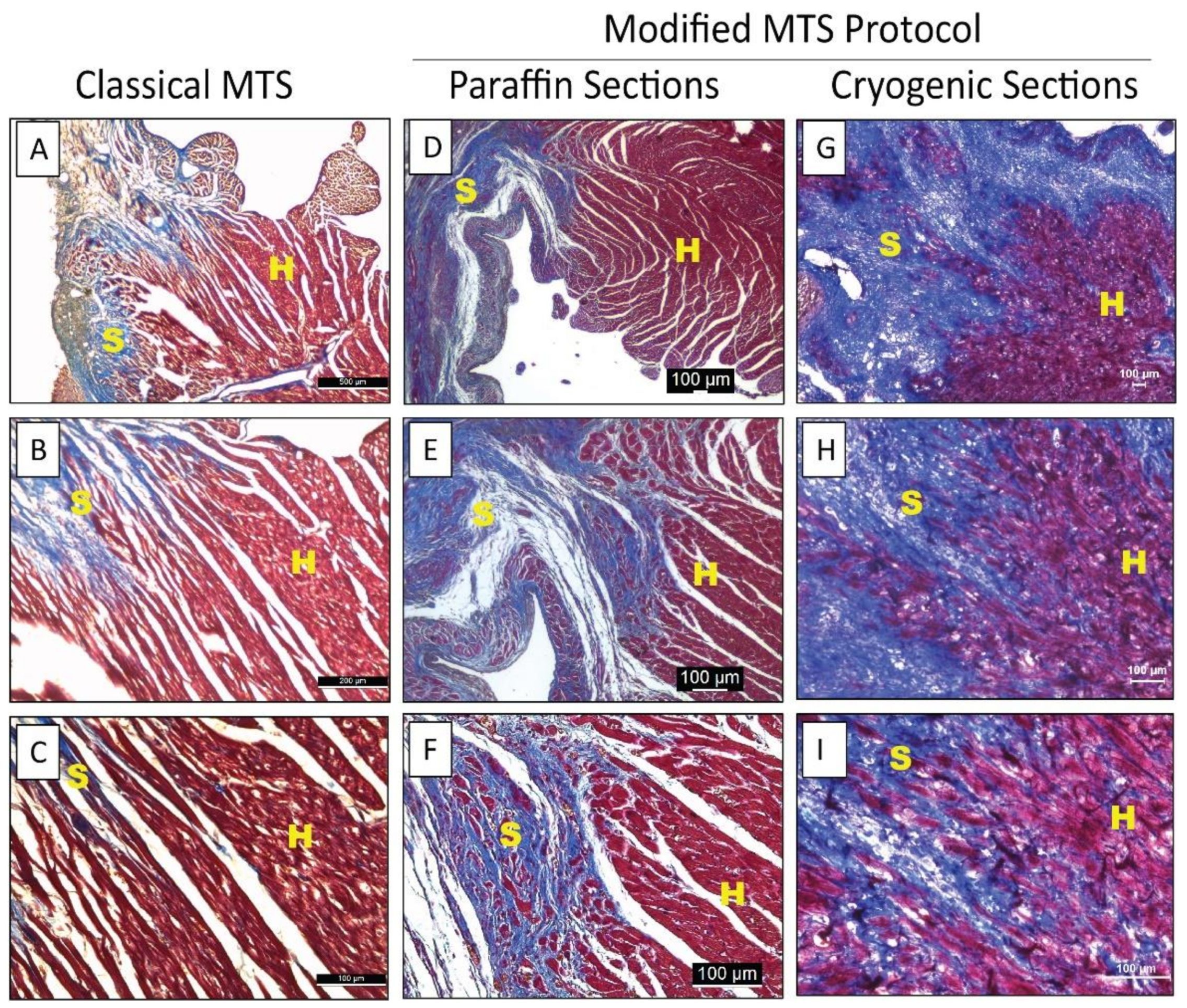
- Image the slides under a bright-field microscope (See Figure 1).
4. Expected Results
Author Contributions
Funding
Institutional Review Board Statement
Informed Consent Statement
Data Availability Statement
Conflicts of Interest
Appendix A
- Inconsistent deparaffinization may result in improper staining. Ensure that solutions for deparaffinization are fresh and the slides are uniformly deparaffinized.
- Incubating with Bouin’s solution can be tricky. If heated, it must be maintained at 56 °C. If kept at room temperature, incubation times should not exceed 6 h. Longer incubation times leave a yellowish tinge on the samples.
- While rinsing the slides under running tap water, water pressure must be regulated. In addition, ensure that the tap water is not directly onto the sections. Excess pressure or water added directly to the sample can cause the sample to detach from the slide. Nevertheless, it is essential to ensure that the slide is fully immersed in water to wash off the stain completely. If a remnant stain is observed on the slide, increase the washing time.
- In some instances, if a very faint tinge is observed after incubating the slides in Bouin’s Solution or Weigert’s Solution, the staining process can be continued further.
- While staining solutions can be prepared in advance, making fresh staining solutions each time for best results is recommended.
- At least 200 μL of stain must be used for each section to ensure complete coverage of the section. Add staining solutions dropwise onto the sections to ensure uniform coverage and consistent staining.
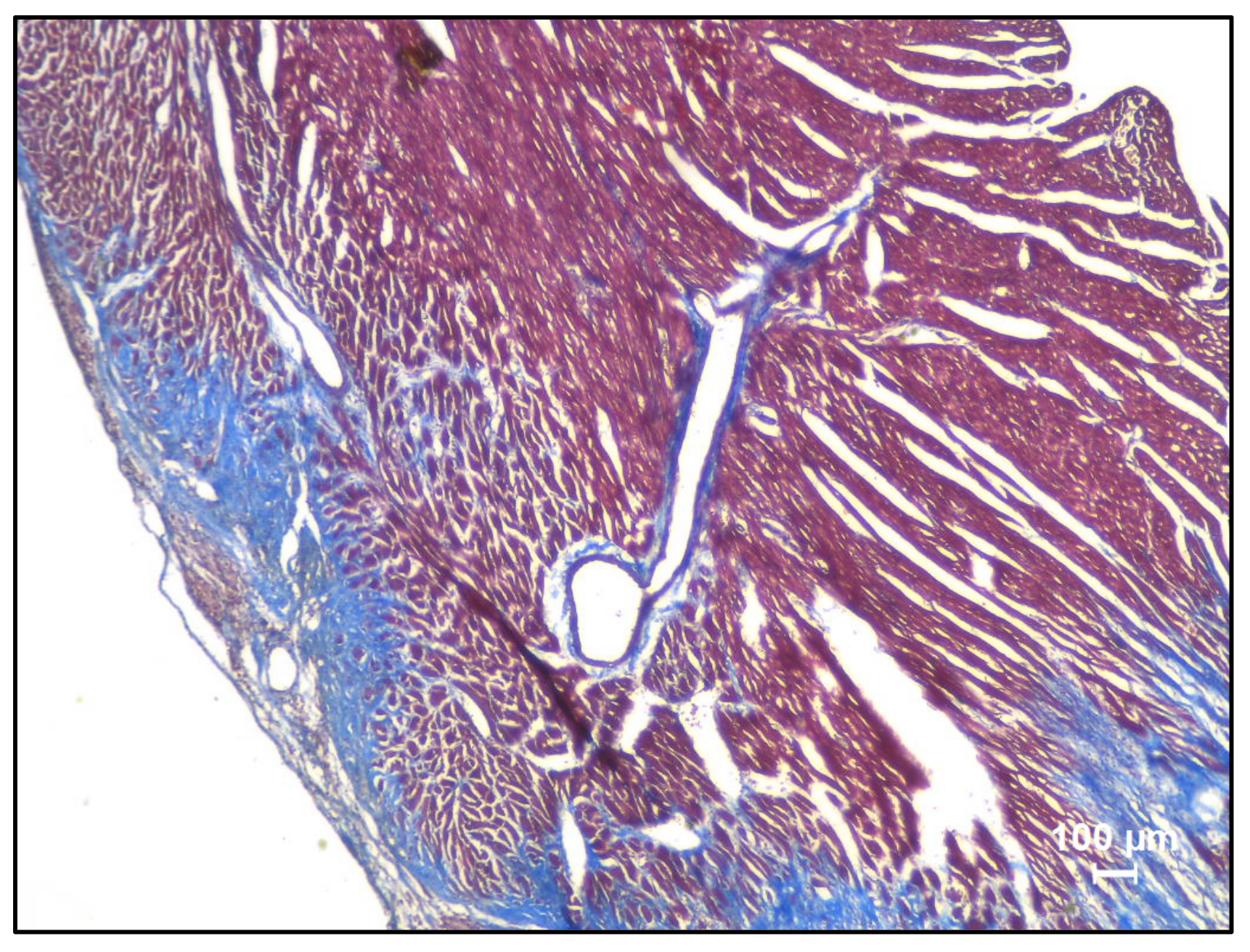
- Depending on the sample type, longer incubation times may be required for deeper red color (Figure A1).
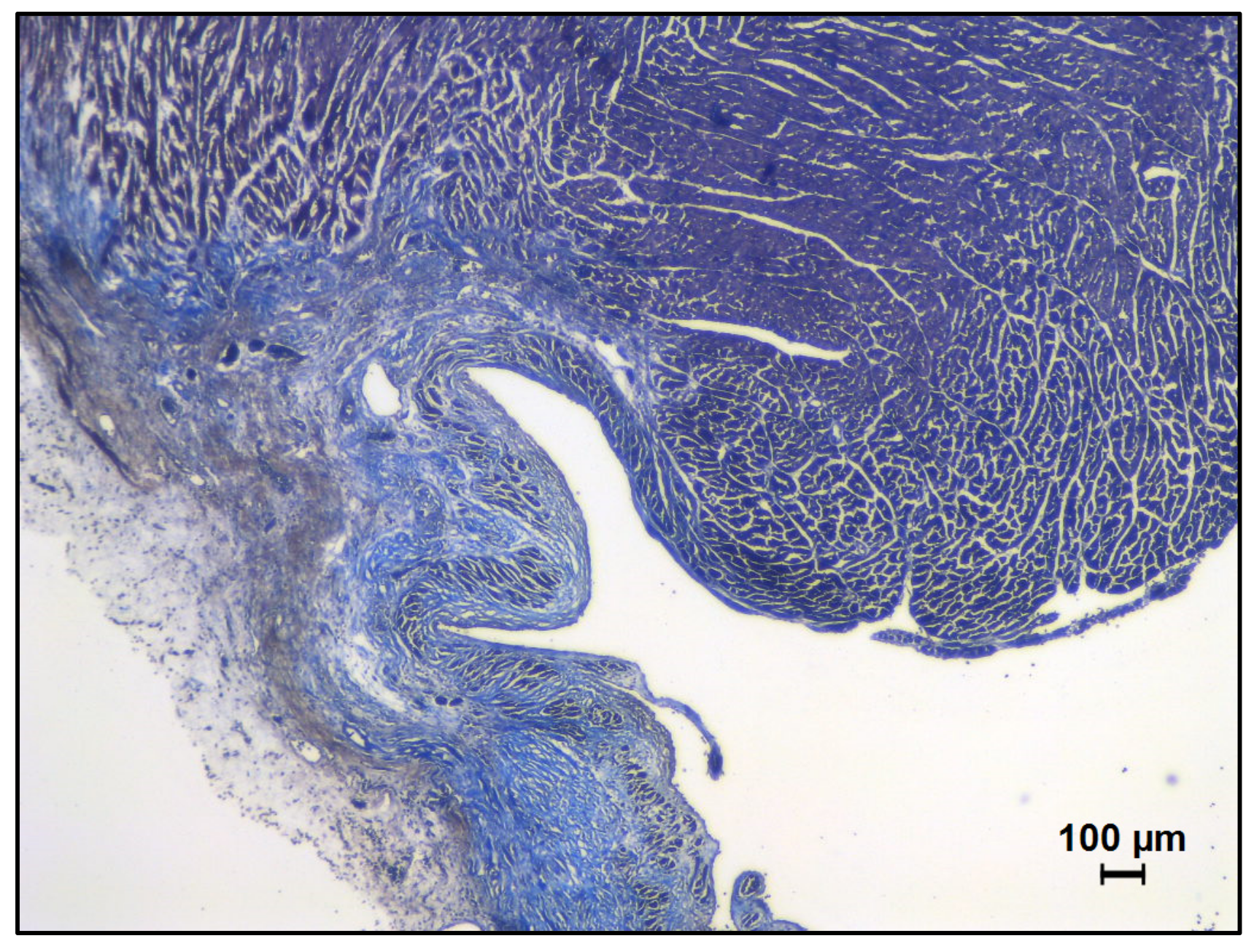
- Staining in the phosphomolybdic-phosphotungstic acid solution for longer durations can wash off the scarlet-acid fuchsin. This step must be standardized for each tissue type (Figure A2).
- Excess hydrophobic pen residue can be carefully wiped off after dipping.
- The aqueous mounting medium must not be used for MTS samples.


References
- Saleh, M.; Ambrose, J.A. Understanding myocardial infarction. F1000Research 2018, 7. [Google Scholar] [CrossRef] [PubMed]
- Richardson, W.J.; Clarke, S.A.; Quinn, T.A.; Holmes, J.W. Physiological Implications of Myocardial Scar Structure. Compr. Physiol. 2015, 5, 1877–1909. [Google Scholar] [CrossRef] [PubMed] [Green Version]
- Lin, Z.; Pu, W.T. Strategies for cardiac regeneration and repair. Sci. Transl. Med. 2014, 6, 239rv1. [Google Scholar] [CrossRef] [PubMed] [Green Version]
- Liang, C.; Wang, K.; Li, Q.; Bai, J.; Zhang, H. Influence of the distribution of fibrosis within an area of myocardial infarction on wave propagation in ventricular tissue. Sci. Rep. 2019, 9, 14151. [Google Scholar] [CrossRef] [PubMed]
- Li, W.; Tang, R.; Ouyang, S.; Ma, F.; Liu, Z.; Wu, J. Folic acid prevents cardiac dysfunction and reduces myocardial fibrosis in a mouse model of high-fat diet-induced obesity. Nutr. Metab. 2017, 14, 68. [Google Scholar] [CrossRef] [PubMed] [Green Version]
- Yu, Y.; Yin, G.; Bao, S.; Guo, Z. Kinetic alterations of collagen and elastic fibres and their association with cardiac function in acute myocardial infarction. Mol. Med. Rep. 2018, 17, 3519–3526. [Google Scholar] [CrossRef] [PubMed]
- Hinderer, S.; Schenke-Layland, K. Cardiac fibrosis—A short review of causes and therapeutic strategies. Adv. Drug Deliv. Rev. 2019, 146, 77–82. [Google Scholar] [CrossRef] [PubMed]
- Tag, H.M. Hepatoprotective effect of mulberry (Morus nigra) leaves extract against methotrexate induced hepatotoxicity in male albino rat. BMC Complement. Altern. Med. 2015, 15, 252. [Google Scholar] [CrossRef] [PubMed] [Green Version]
- Koyama, S.; Wu, H.J.; Easwaran, T.; Thopady, S.; Foley, J. The nipple: A simple intersection of mammary gland and integument, but focal point of organ function. J. Mammary Gland Biol. Neoplasia 2013, 18, 121–131. [Google Scholar] [CrossRef] [PubMed]
- Kulac, M.; Aktas, C.; Tulubas, F.; Uygur, R.; Kanter, M.; Erboga, M.; Ceber, M.; Topcu, B.; Ozen, O.A. The effects of topical treatment with curcumin on burn wound healing in rats. J. Mol. Histol. 2013, 44, 83–90. [Google Scholar] [CrossRef] [PubMed]
- Xia, Y.; Zhou, P.; Wang, F.; Qiu, C.; Wang, P.; Zhang, Y.; Zhao, L.; Xu, S. Degradability, biocompatibility, and osteogenesis of biocomposite scaffolds containing nano magnesium phosphate and wheat protein both in vitro and in vivo for bone regeneration. Int. J. Nanomed. 2016, 11, 3435–3449. [Google Scholar] [CrossRef] [Green Version]
- Shi, K.; Zhao, W.; Chen, Y.; Ho, W.T.; Yang, P.; Zhao, Z.J. Cardiac hypertrophy associated with myeloproliferative neoplasms in JAK2V617F transgenic mice. J. Hematol. Oncol. 2014, 7, 25. [Google Scholar] [CrossRef] [PubMed] [Green Version]


| Sequence | Procedure | Time (Minutes) |
|---|---|---|
| (i) | 10% ethanol | 2 × 15 min |
| (ii) | 30% ethanol | 2 × 15 min |
| (iii) | 50% ethanol | 2 × 15 min |
| (iv) | 70% ethanol | 2 × 15 min |
| (v) | 85% ethanol | 2 × 15 m |
| (vi) | 100% ethanol | 2 × 15 min |
| (vii) | Ethanol:xylene (1:1) | 2 × 20 min |
| (viii) | Xylene | 2 × 20 min |
| (ix) | Xylene:paraffin (1:1) | 1 × 60 min (60 °C) |
| (x) | Paraffin | 2 × 60 min (60 °C) |
| Sequence | Procedure | Time (Min) |
|---|---|---|
| (i) | Xylene | 2 × 3 min |
| (ii) | Ethanol:Xylene (1:1) | 3 min |
| (iii) | 100% Ethanol | 3 min |
| (iv) | 85% Ethanol | 3 min |
| (v) | 70% Ethanol | 3 min |
| (vi) | 50% Ethanol | 3 min |
Publisher’s Note: MDPI stays neutral with regard to jurisdictional claims in published maps and institutional affiliations. |
© 2022 by the authors. Licensee MDPI, Basel, Switzerland. This article is an open access article distributed under the terms and conditions of the Creative Commons Attribution (CC BY) license (https://creativecommons.org/licenses/by/4.0/).
Share and Cite
Sridharan, D.; Pracha, N.; Dougherty, J.A.; Akhtar, A.; Alvi, S.B.; Khan, M. A One-Stop Protocol to Assess Myocardial Fibrosis in Frozen and Paraffin Sections. Methods Protoc. 2022, 5, 13. https://0-doi-org.brum.beds.ac.uk/10.3390/mps5010013
Sridharan D, Pracha N, Dougherty JA, Akhtar A, Alvi SB, Khan M. A One-Stop Protocol to Assess Myocardial Fibrosis in Frozen and Paraffin Sections. Methods and Protocols. 2022; 5(1):13. https://0-doi-org.brum.beds.ac.uk/10.3390/mps5010013
Chicago/Turabian StyleSridharan, Divya, Nooruddin Pracha, Julie A. Dougherty, Ali Akhtar, Syed Baseeruddin Alvi, and Mahmood Khan. 2022. "A One-Stop Protocol to Assess Myocardial Fibrosis in Frozen and Paraffin Sections" Methods and Protocols 5, no. 1: 13. https://0-doi-org.brum.beds.ac.uk/10.3390/mps5010013