
Plasma-Generated X-ray Pulses: Betatron Radiation Opportunities at EuPRAXIA@SPARC_LAB

 , , , , , , , , , , and
Abstract
:1. Introduction
2. Results
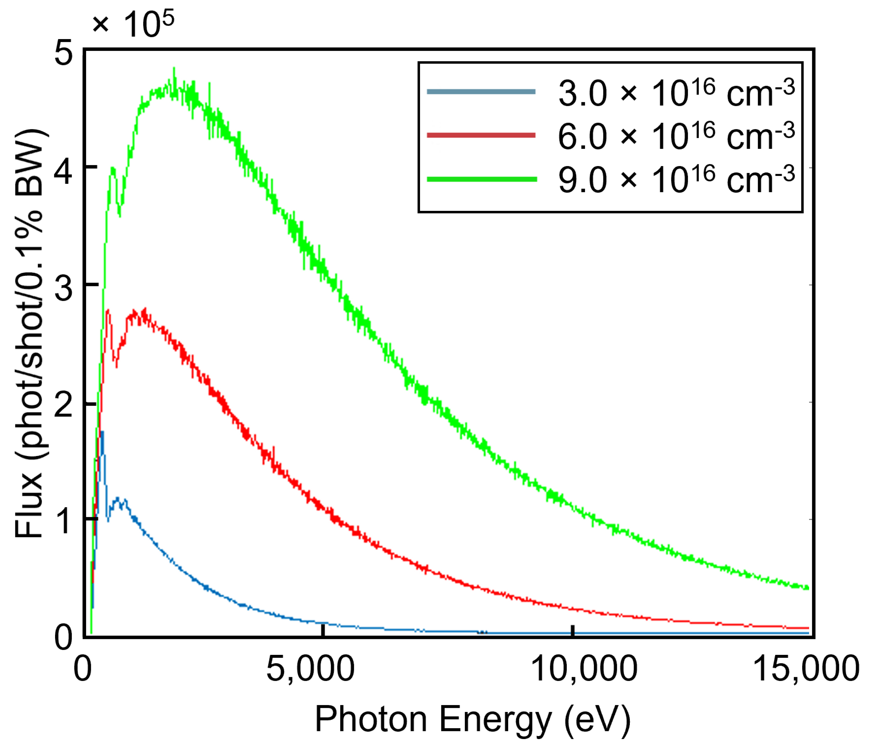
2.1. Simulations
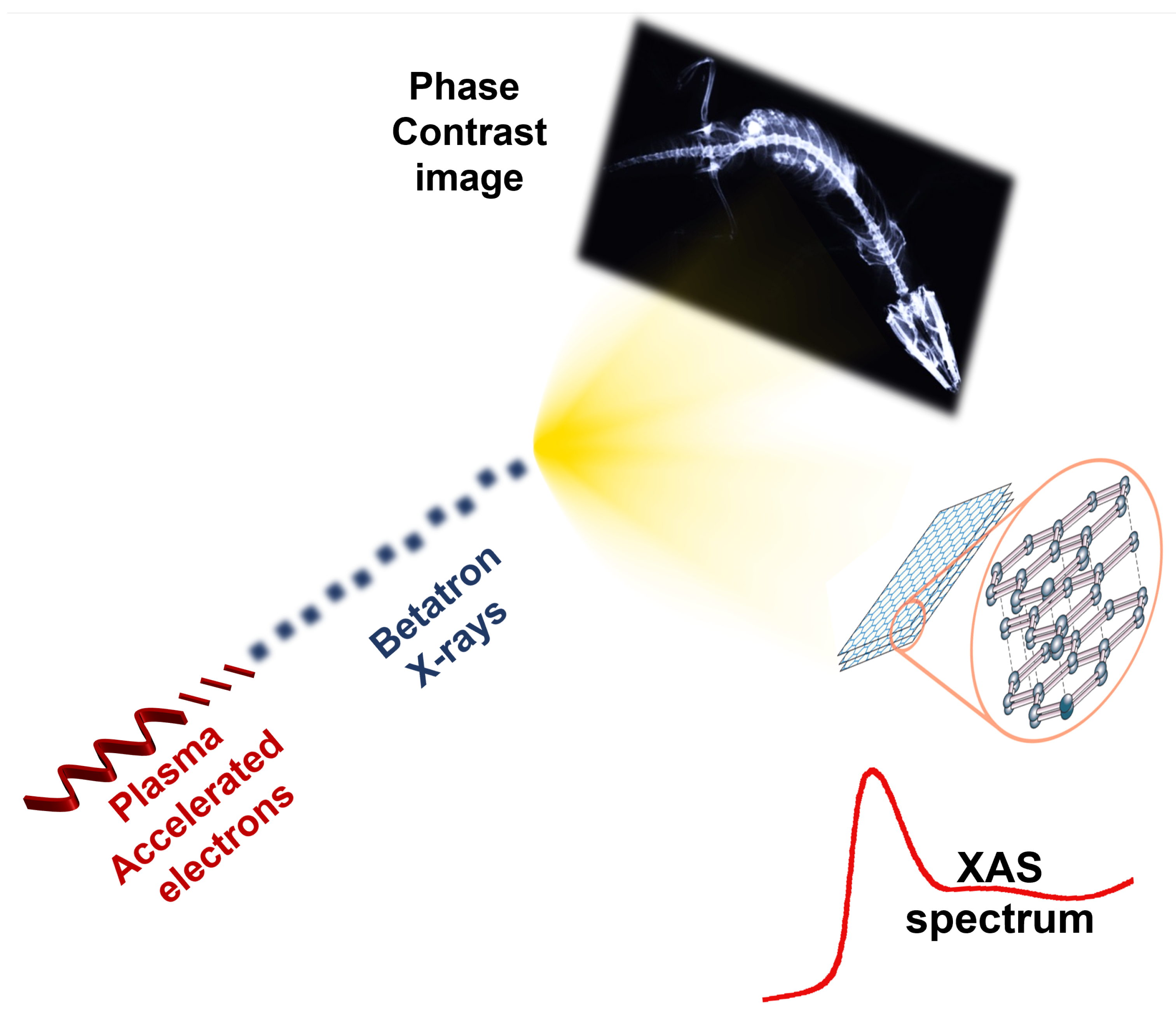
2.2. Betatron Imaging
2.3. Betatron X-ray Spectroscopy
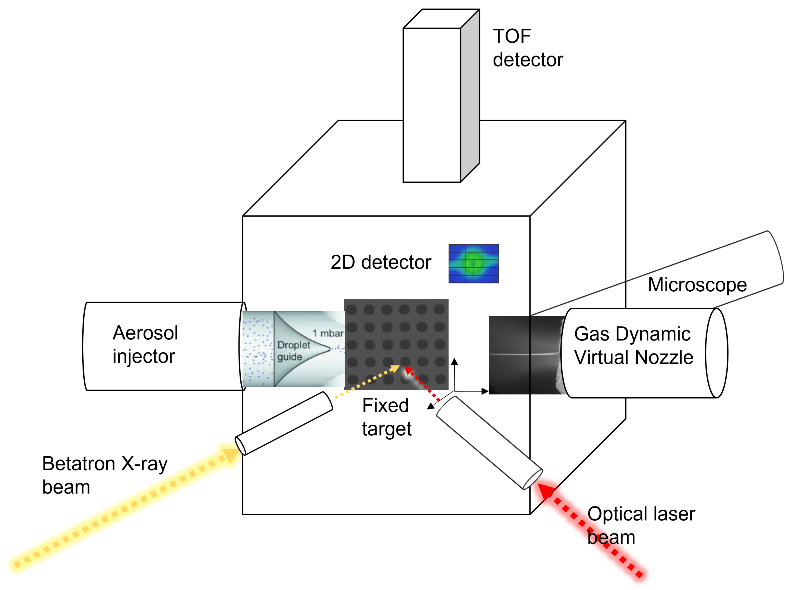
2.4. Betatron Test Chamber
3. Discussion
Author Contributions
Funding
Institutional Review Board Statement
Informed Consent Statement
Data Availability Statement
Conflicts of Interest
Abbreviations
| CT | Computer Tomography |
| FEL | Free Electron Laser |
| LCLS | Linac Coherent Light Source |
| PCI | Phase Contrast Imaging |
| XAS | X-Ray Absorption Spectroscopy |
References
- Assmann, R.; Weikum, M.; Akhter, T.; Alesini, D.; Alexandrova, A.; Anania, M.; Andreev, N.; Andriyash, I.; Artioli, M.; Aschikhin, A.; et al. EuPRAXIA conceptual design report. Eur. Phys. J. Spec. Top. 2020, 229, 3675–4284. [Google Scholar] [CrossRef]
- Ferrario, M.; Alesini, D.; Anania, M.; Artioli, M.; Bacci, A.; Bartocci, S.; Bedogni, R.; Bellaveglia, M.; Biagioni, A.; Bisesto, F.; et al. EuPRAXIA@ SPARC_LAB Design study towards a compact FEL facility at LNF. Nucl. Instrum. Methods Phys. Res. Sect. A Accel. Spectrometers Detect. Assoc. Equip. 2018, 909, 134–138. [Google Scholar] [CrossRef]
- Balerna, A.; Bartocci, S.; Batignani, G.; Cianchi, A.; Chiadroni, E.; Coreno, M.; Cricenti, A.; Dabagov, S.; Di Cicco, A.; Faiferri, M.; et al. The Potential of EuPRAXIA@ SPARC_LAB for Radiation Based Techniques. Condens. Matter 2019, 4, 30. [Google Scholar] [CrossRef] [Green Version]
- Paroli, B.; Chiadroni, E.; Ferrario, M.; Mostacci, A.; Petrillo, V.; Potenza, M.; Rossi, A.; Serafini, L. Coherence properties and diagnostics of betatron radiation emitted by an externally-injected electron beam propagating in a plasma channel. Nucl. Instrum. Methods Phys. Res. Sect. B Beam Interact. Mater. Atoms 2015, 355, 217–220. [Google Scholar] [CrossRef]
- Guo, B.; Xu, X.; Hua, J.; Wu, Y.; Pai, C.H.; Lu, W. Generation of Coherent Monochromatic Betatron Radiation by Laser-triggered Ionization Injection in Plasma Accelerators. In Proceedings of the 2018 IEEE Advanced Accelerator Concepts Workshop (AAC), Breckenridge, CO, USA, 12–17 August 2018; pp. 1–4. [Google Scholar]
- Curcio, A.; Anania, M.; Bisesto, F.; Chiadroni, E.; Cianchi, A.; Ferrario, M.; Filippi, F.; Giulietti, D.; Marocchino, A.; Mira, F.; et al. First measurements of betatron radiation at FLAME laser facility. Nucl. Instrum. Methods Phys. Res. Sect. B Beam Interact. Mater. Atoms 2017, 402, 388–392. [Google Scholar] [CrossRef]
- Curcio, A.; Anania, M.; Bisesto, F.; Chiadroni, E.; Cianchi, A.; Ferrario, M.; Filippi, F.; Giulietti, D.; Marocchino, A.; Petrarca, M.; et al. Trace-space reconstruction of low-emittance electron beams through betatron radiation in laser-plasma accelerators. Phys. Rev. Accel. Beams 2017, 20, 012801. [Google Scholar] [CrossRef] [Green Version]
- Corde, S.; Phuoc, K.T.; Lambert, G.; Fitour, R.; Malka, V.; Rousse, A.; Beck, A.; Lefebvre, E. Femtosecond x rays from laser-plasma accelerators. Rev. Mod. Phys. 2013, 85, 1. [Google Scholar] [CrossRef]
- Albert, F.; Thomas, A.G. Applications of laser wakefield accelerator-based light sources. Plasma Phys. Control. Fusion 2016, 58, 103001. [Google Scholar] [CrossRef]
- Esarey, E.; Shadwick, B.; Catravas, P.; Leemans, W. Synchrotron radiation from electron beams in plasma-focusing channels. Phys. Rev. E 2002, 65, 056505. [Google Scholar] [CrossRef] [Green Version]
- Leemans, W.P.; Esarey, E.; van Tilborg, J.; Michel, P.A.; Schroeder, C.B.; Tóth, C.; Geddes, C.G.; Shadwick, B.A. Radiation from laser accelerated electron bunches: Coherent terahertz and femtosecond X-rays. IEEE Trans. Plasma Sci. 2005, 33, 8–22. [Google Scholar] [CrossRef] [Green Version]
- Croton, L.C.; Morgan, K.S.; Paganin, D.M.; Kerr, L.T.; Wallace, M.J.; Crossley, K.J.; Miller, S.L.; Yagi, N.; Uesugi, K.; Hooper, S.B.; et al. In situ phase contrast X-ray brain CT. Sci. Rep. 2018, 8, 11412. [Google Scholar] [CrossRef] [PubMed]
- Guo, B.; Zhang, X.; Zhang, J.; Hua, J.; Pai, C.H.; Zhang, C.; Chu, H.H.; Mori, W.; Joshi, C.; Wang, J.; et al. High-resolution phase-contrast imaging of biological specimens using a stable betatron X-ray source in the multiple-exposure mode. Sci. Rep. 2019, 9, 7796. [Google Scholar] [CrossRef] [PubMed]
- Wenz, J.; Schleede, S.; Khrennikov, K.; Bech, M.; Thibault, P.; Heigoldt, M.; Pfeiffer, F.; Karsch, S. Quantitative X-ray phase-contrast microtomography from a compact laser-driven betatron source. Nat. Commun. 2015, 6, 7568. [Google Scholar] [CrossRef] [PubMed]
- Cole, J.M.; Symes, D.R.; Lopes, N.C.; Wood, J.C.; Poder, K.; Alatabi, S.; Botchway, S.W.; Foster, P.S.; Gratton, S.; Johnson, S.; et al. High-resolution μCT of a mouse embryo using a compact laser-driven X-ray betatron source. Proc. Natl. Acad. Sci. USA 2018, 115, 6335–6340. [Google Scholar] [CrossRef] [Green Version]
- Kneip, S.; McGuffey, C.; Dollar, F.; Bloom, M.; Chvykov, V.; Kalintchenko, G.; Krushelnick, K.; Maksimchuk, A.; Mangles, S.; Matsuoka, T.; et al. X-ray phase contrast imaging of biological specimens with femtosecond pulses of betatron radiation from a compact laser plasma wakefield accelerator. Appl. Phys. Lett. 2011, 99, 093701. [Google Scholar] [CrossRef]
- van Tilborg, J.; Ostermayr, T.; Tsai, H.E.; Schenkel, T.; Geddes, C.; Schroeder, C.; Esarey, E. Phase-contrast imaging with laser-plasma-accelerator betatron sources. In Proceedings of the International Conference on X-ray Lasers 2020, Rome, Italy, 6–11 September 2020; Volume 11886, p. 118860Q. [Google Scholar]
- Najmudin, Z.; Kneip, S.; Bloom, M.; Mangles, S.; Chekhlov, O.; Dangor, A.; Döpp, A.; Ertel, K.; Hawkes, S.; Holloway, J.; et al. Compact laser accelerators for X-ray phase-contrast imaging. Philos. Trans. R. Soc. A Math. Phys. Eng. Sci. 2014, 372, 20130032. [Google Scholar] [CrossRef]
- Brenner, G.; Dziarzhytski, S.; Miedema, P.S.; Rösner, B.; David, C.; Beye, M. Normalized single-shot X-ray absorption spectroscopy at a free-electron laser. Opt. Lett. 2019, 44, 2157–2160. [Google Scholar] [CrossRef] [Green Version]
- Kroll, T.; Kern, J.; Kubin, M.; Ratner, D.; Gul, S.; Fuller, F.D.; Löchel, H.; Krzywinski, J.; Lutman, A.; Ding, Y.; et al. X-ray absorption spectroscopy using a self-seeded soft X-ray free-electron laser. Opt. Express 2016, 24, 22469–22480. [Google Scholar] [CrossRef] [Green Version]
- Mitzner, R.; Rehanek, J.; Kern, J.; Gul, S.; Hattne, J.; Taguchi, T.; Alonso-Mori, R.; Tran, R.; Weniger, C.; öder, H.; et al. L-edge x-ray absorption spectroscopy of dilute systems relevant to metalloproteins using an x-ray free-electron laser. J. Phys. Chem. Lett. 2013, 4, 3641–3647. [Google Scholar] [CrossRef] [Green Version]
- Levantino, M.; Lemke, H.; Schirò, G.; Glownia, M.; Cupane, A.; Cammarata, M. Observing heme doming in myoglobin with femtosecond X-ray absorption spectroscopy. Struct. Dyn. 2015, 2, 041713. [Google Scholar] [CrossRef]
- Mahieu, B.; Jourdain, N.; Phuoc, K.T.; Dorchies, F.; Goddet, J.P.; Lifschitz, A.; Renaudin, P.; Lecherbourg, L. Probing warm dense matter using femtosecond x-ray absorption spectroscopy with a laser-produced betatron source. Nat. Commun. 2018, 9, 3276. [Google Scholar] [CrossRef] [PubMed]
- Mo, M.; Chen, Z.; Fourmaux, S.; Saraf, A.; Kerr, S.; Otani, K.; Masoud, R.; Kieffer, J.C.; Tsui, Y.; Ng, A.; et al. Measurements of ionization states in warm dense aluminum with betatron radiation. Phys. Rev. E 2017, 95, 053208. [Google Scholar] [CrossRef] [PubMed]
- Albert, F.; Barbrel, B.; Condamine, F.; Dorchies, F.; Falcone, R.; Fernandez, A.; Fletcher, L.; Fry, A.; Galtier, E.; Gamboa, E.; et al. Time-resolved X-ray absorption spectroscopy of warm dense matter with betatron X-ray radiation. In Proceedings of the APS Division of Plasma Physics Meeting Abstracts, Portland, OR, USA, 5–9 November 2018; Volume 2018, p. JO8-003. [Google Scholar]
- Juranić, P.; Stepanov, A.; Ischebeck, R.; Schlott, V.; Pradervand, C.; Patthey, L.; Radović, M.; Gorgisyan, I.; Rivkin, L.; Hauri, C.; et al. High-precision x-ray FEL pulse arrival time measurements at SACLA by a THz streak camera with Xe clusters. Opt. Express 2014, 22, 30004–30012. [Google Scholar] [CrossRef] [Green Version]
- Harmand, M.; Coffee, R.; Bionta, M.R.; Chollet, M.; French, D.; Zhu, D.; Fritz, D.; Lemke, H.; Medvedev, N.; Ziaja, B.; et al. Achieving few-femtosecond time-sorting at hard X-ray free-electron lasers. Nat. Photonics 2013, 7, 215–218. [Google Scholar] [CrossRef]
- Han, H.; Round, E.; Schubert, R.; Gül, Y.; Makroczyová, J.; Meza, D.; Heuser, P.; Aepfelbacher, M.; Barák, I.; Betzel, C.; et al. The XBI BioLab for life science experiments at the European XFEL. J. Appl. Crystallogr. 2021, 54, 7–21. [Google Scholar] [CrossRef] [PubMed]


| Parameter [Units] | Value |
|---|---|
| Beam energy [MeV] | 600–950 |
| Plasma density [cm] | 3.0 × 10 |
| RMS transverse beam size [m] | 3.0 |
| Charge [pC] | 45 |
| Capillary (plasma) length [m] | 0.6 |
| Pulse duration [fs] | 10 |
Publisher’s Note: MDPI stays neutral with regard to jurisdictional claims in published maps and institutional affiliations. |
© 2022 by the authors. Licensee MDPI, Basel, Switzerland. This article is an open access article distributed under the terms and conditions of the Creative Commons Attribution (CC BY) license (https://creativecommons.org/licenses/by/4.0/).
Share and Cite
Stellato, F.; Anania, M.P.; Balerna, A.; Botticelli, S.; Coreno, M.; Costa, G.; Galletti, M.; Ferrario, M.; Marcelli, A.; Minicozzi, V.; et al. Plasma-Generated X-ray Pulses: Betatron Radiation Opportunities at EuPRAXIA@SPARC_LAB. Condens. Matter 2022, 7, 23. https://0-doi-org.brum.beds.ac.uk/10.3390/condmat7010023
Stellato F, Anania MP, Balerna A, Botticelli S, Coreno M, Costa G, Galletti M, Ferrario M, Marcelli A, Minicozzi V, et al. Plasma-Generated X-ray Pulses: Betatron Radiation Opportunities at EuPRAXIA@SPARC_LAB. Condensed Matter. 2022; 7(1):23. https://0-doi-org.brum.beds.ac.uk/10.3390/condmat7010023
Chicago/Turabian StyleStellato, Francesco, Maria Pia Anania, Antonella Balerna, Simone Botticelli, Marcello Coreno, Gemma Costa, Mario Galletti, Massimo Ferrario, Augusto Marcelli, Velia Minicozzi, and et al. 2022. "Plasma-Generated X-ray Pulses: Betatron Radiation Opportunities at EuPRAXIA@SPARC_LAB" Condensed Matter 7, no. 1: 23. https://0-doi-org.brum.beds.ac.uk/10.3390/condmat7010023