Water Uptake in PHBV/Wollastonite Scaffolds: A Kinetics Study

 , , ,
, , ,
Abstract
:1. Introduction
2. Materials and Methods
2.1. Materials
2.2. Production of PHBV/WOL Films
2.3. Production of PHBV/WOL Scaffolds
2.4. Characterization
2.4.1. X-ray Diffraction (XRD)
2.4.2. Zeta Potential
2.4.3. Field Emission Gun—Scanning Electron Microscopy (FEG-SEM) and Scanning Electron Microscopy (SEM)
2.4.4. Fourier Transform Infrared Spectroscopy (FT-IR)
2.4.5. Raman Spectroscopy
2.4.6. Contact Angle
2.4.7. Cell Viability
2.4.8. Water Uptake
3. Results
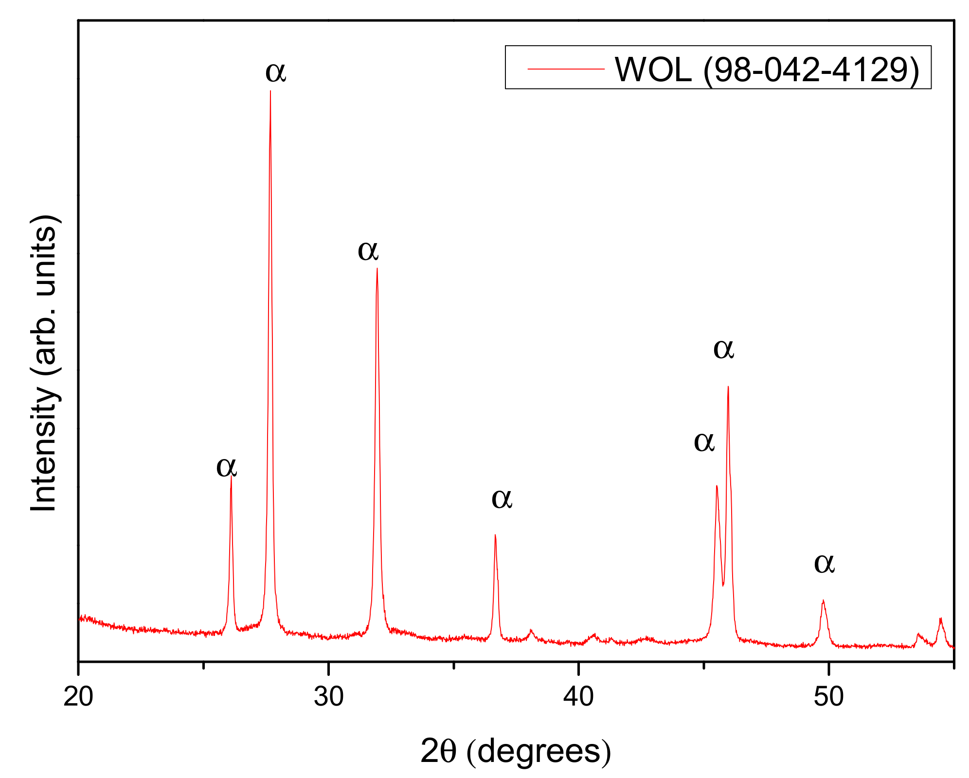
3.1. X-ray Diffraction
3.2. Zeta Potential
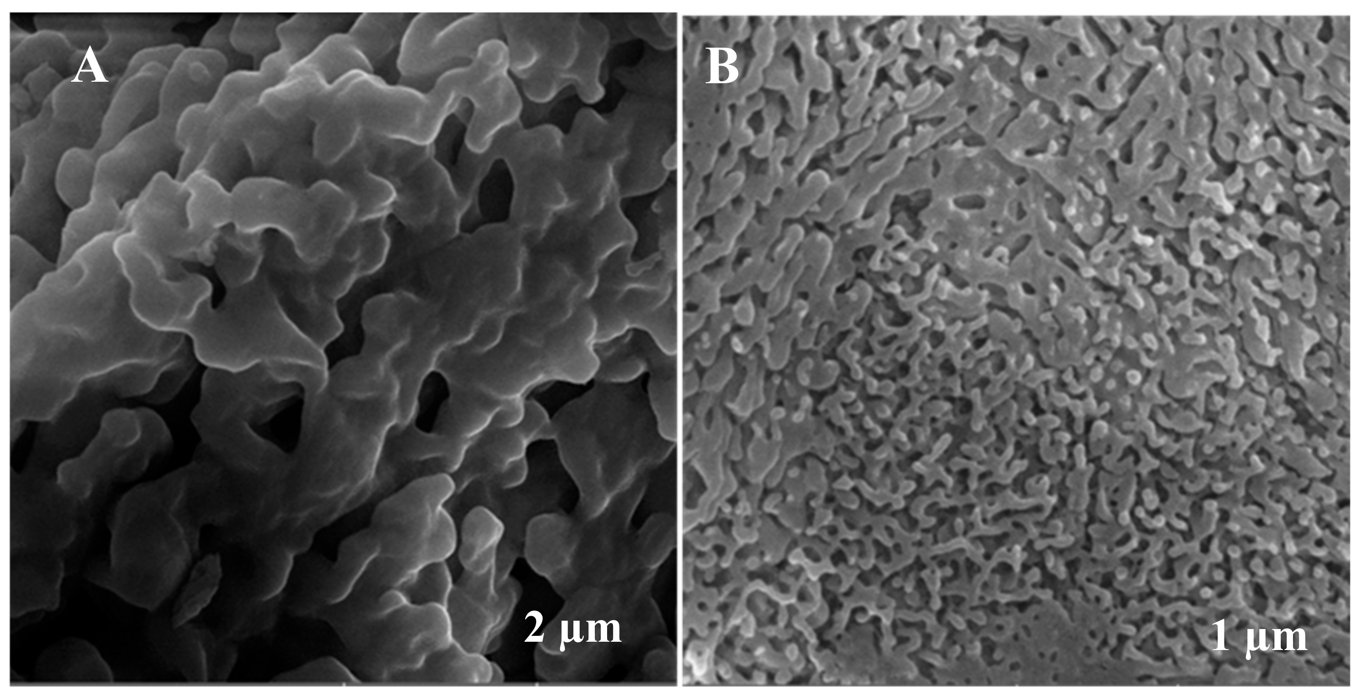
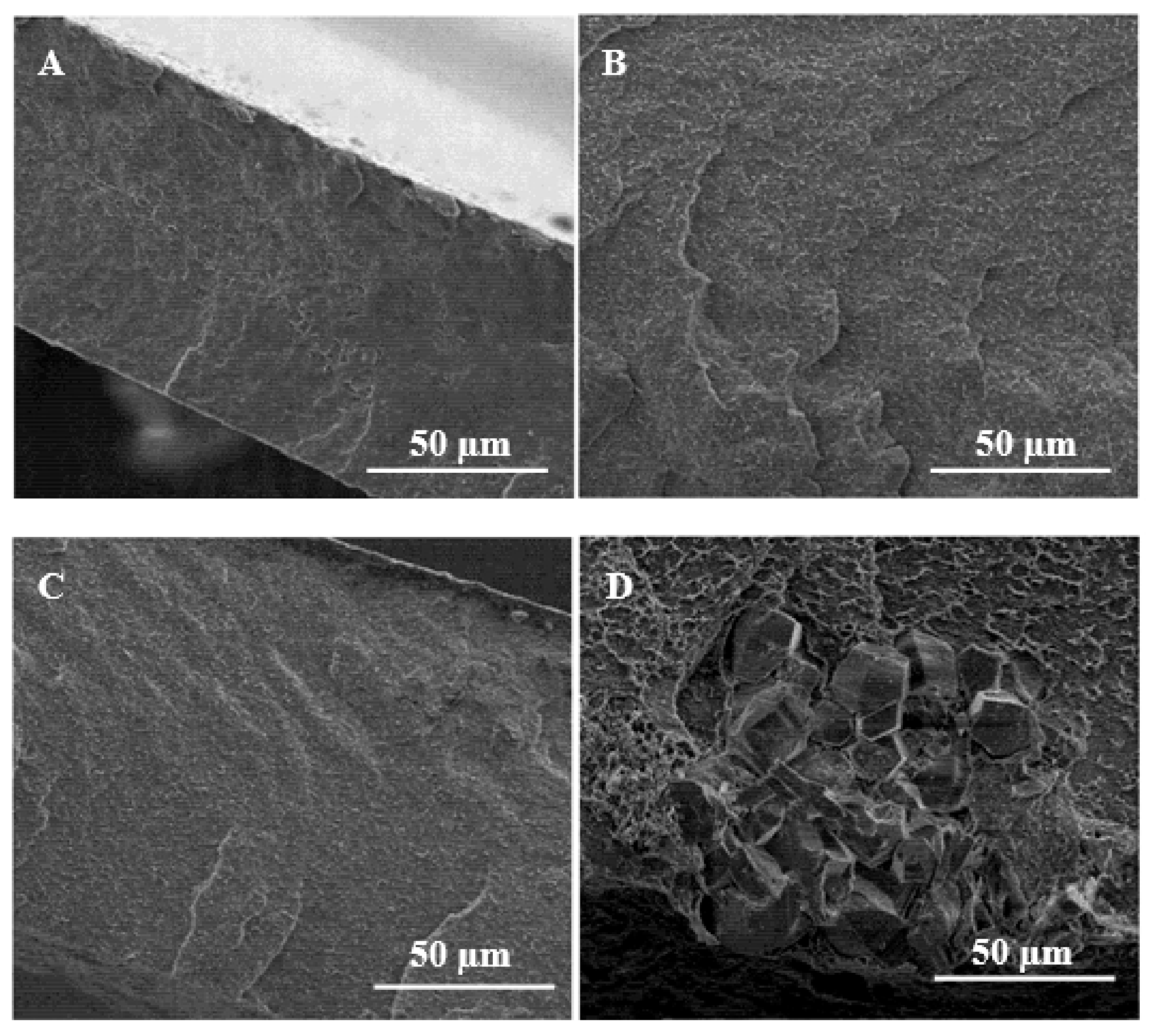
3.3. Field-Emission Scanning Electron Microscopy (FEG-SEM)
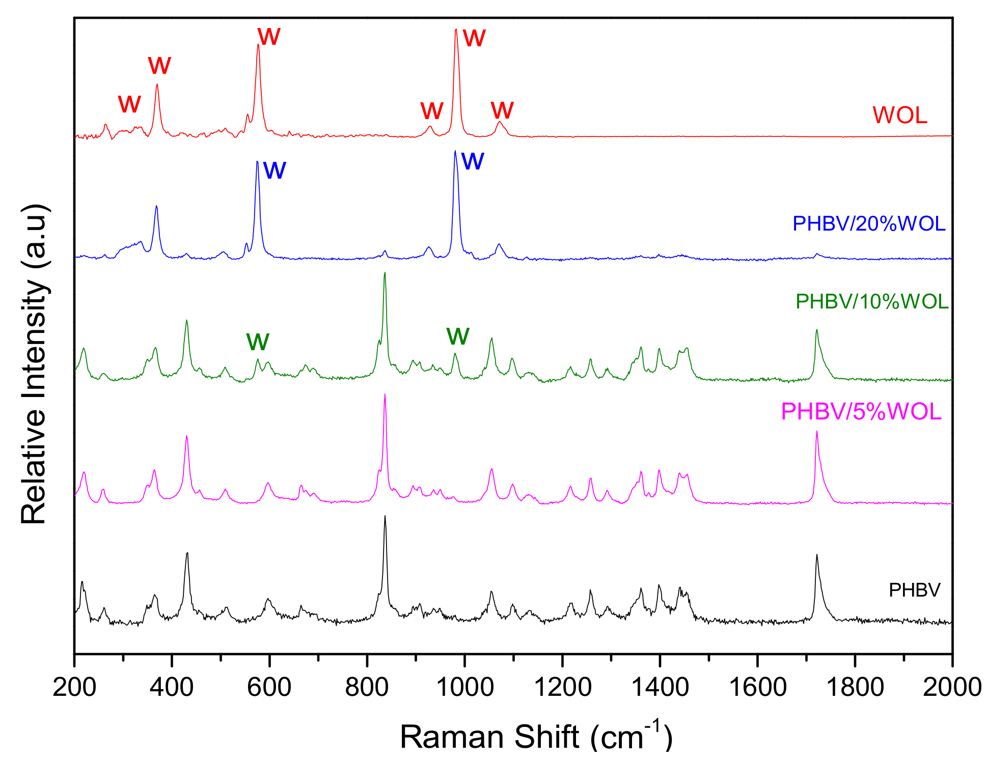
3.4. Raman Spectroscopy
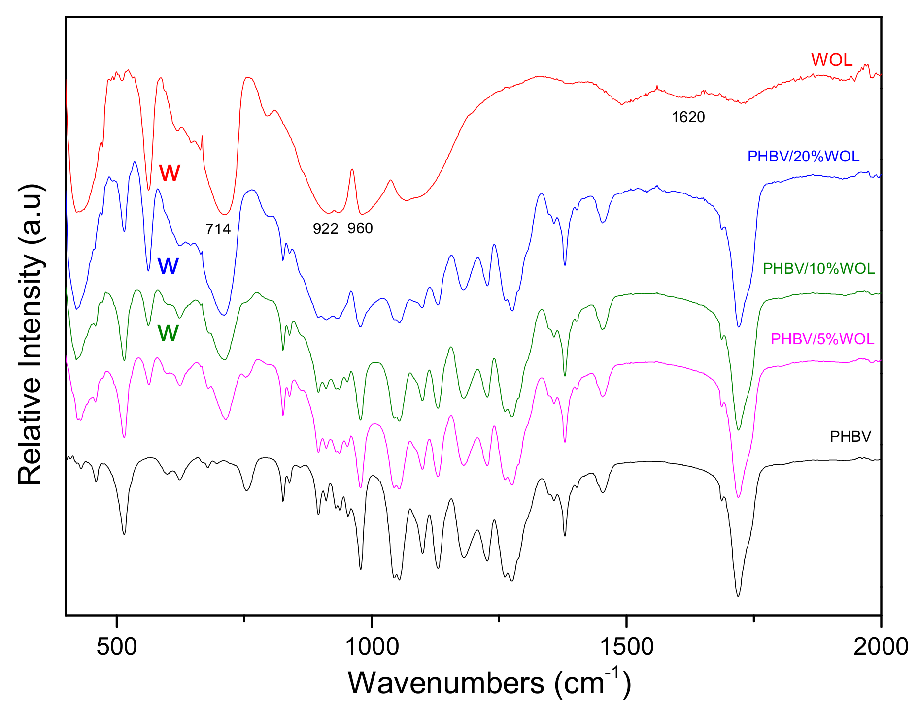
3.5. FT-IR Spectroscopy
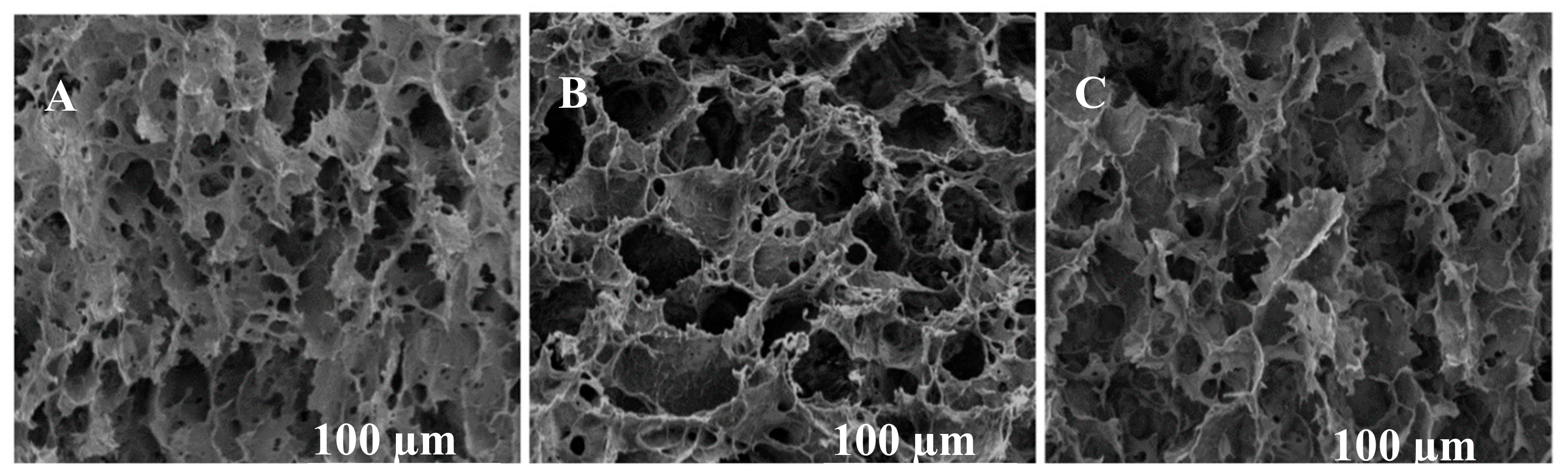
3.6. SEM Micrographs
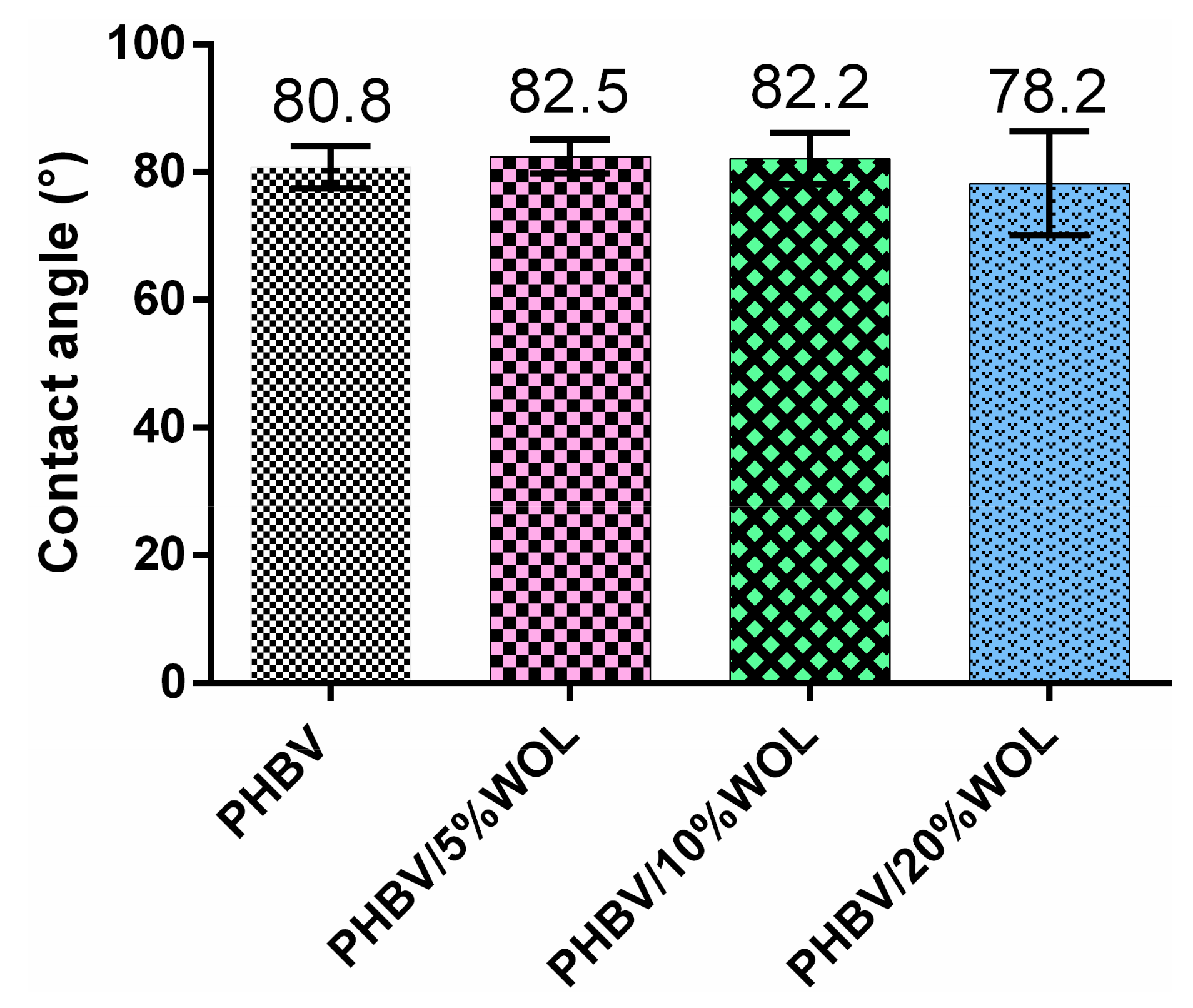
3.7. Contact Angle
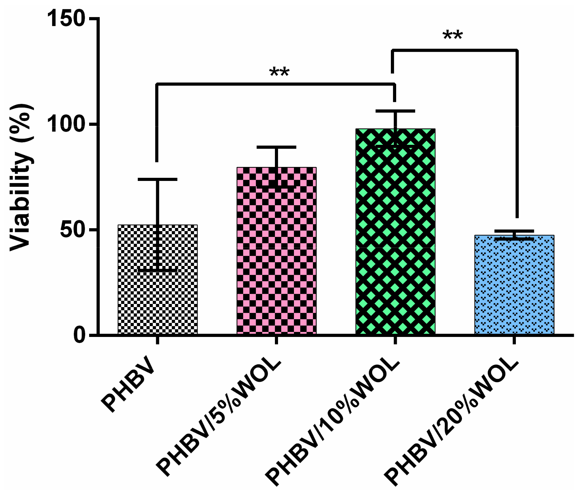
3.8. Cell Viability
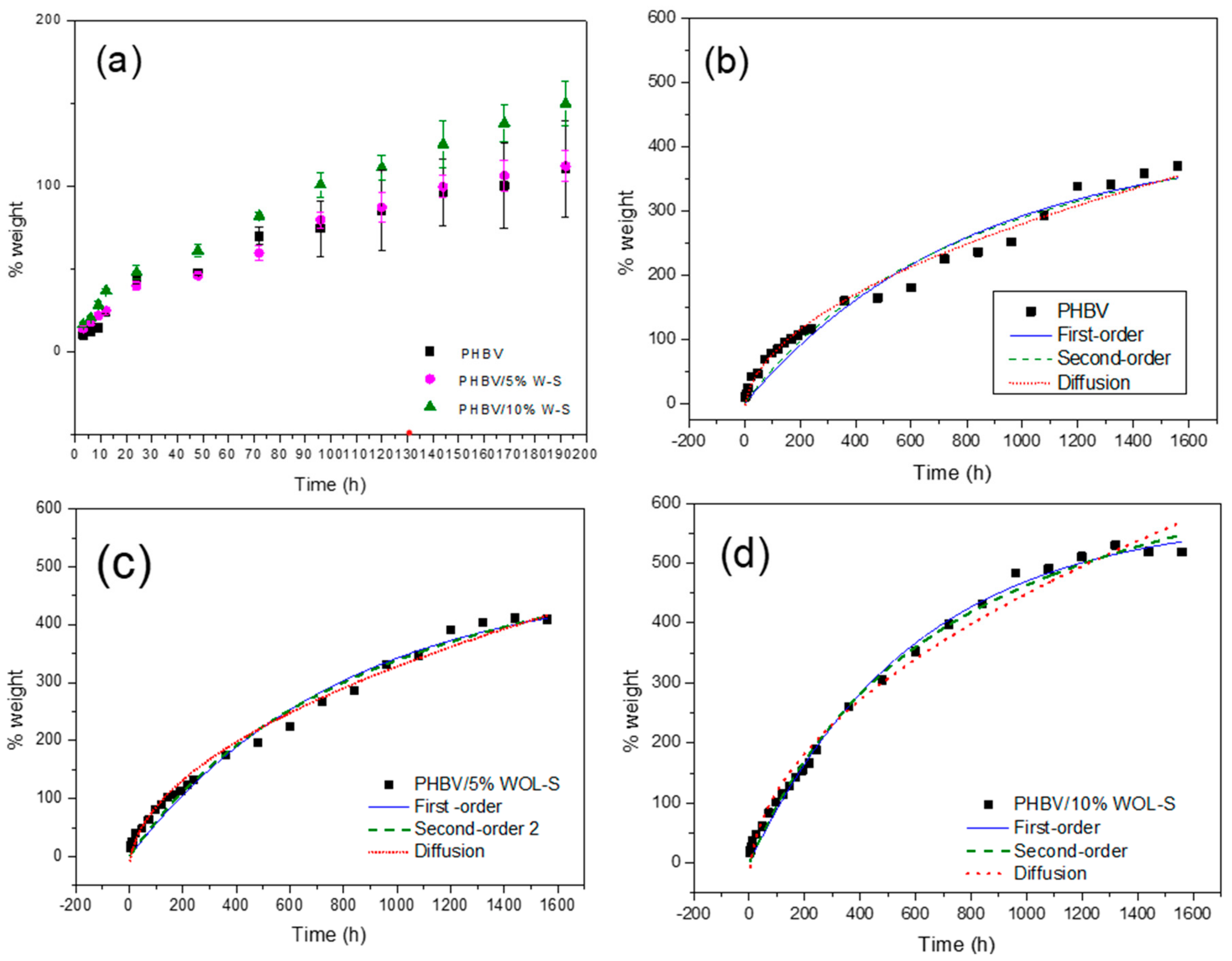
3.9. Water Uptake
4. Conclusions
Author Contributions
Funding
Conflicts of Interest
References
- Monma, H.; Ueno, S.; Kanazawaa, T. Properties of hydroxyapatite prepared by the hydrolysis of tricalcium phosphate. J. Chem. Technol. Biotechnol. 1981, 31, 15–24. [Google Scholar] [CrossRef]
- Johnell, O.; Kanis, J.A. An estimate of the worldwide prevalence and disability associated with osteoporotic fractures. Osteoporos. Int. 2006, 17, 1726–1733. [Google Scholar] [CrossRef] [PubMed]
- Eisman, J.A.; Bogoch, E.R.; Dell, R.; Harrington, J.T.; McKinney, R.E.; McLellan, A.; Mitchell, P.J.; Silverman, S.; Singleton, R.; Siris, E. Making the first fracture the last fracture: ASBMR task force report on secondary fracture prevention. J. Bone Miner. Res. 2012, 27, 2039–2046. [Google Scholar] [CrossRef] [PubMed]
- Montagna, L.S.; Montanheiro, T.L.D.A.; Machado, J.P.B.; Passador, F.R.; Lemes, A.P.; Rezende, M.C. Effect of Graphite Nanosheets on Properties of Poly (3-hydroxybutyrate-co-3-hydroxyvalerate). Int. J. Polym. Sci. 2017, 2017, 9. [Google Scholar] [CrossRef]
- Lenz, R.W.; Marchessault, R.H. Bacterial polyesters: Biosynthesis, biodegradable plastics and biotechnology. Biomacromolecules 2005, 6, 1–8. [Google Scholar] [CrossRef] [PubMed]
- Zinn, M.; Witholt, B.; Egli, T. Occurrence, synthesis and medical application of bacterial polyhydroxyalkanoate. Adv. Drug Deliv. Rev. 2001, 53, 5–21. [Google Scholar] [CrossRef]
- Zhijiang, C.; Yi, X.; Haizheng, Y.; Jia, J.; Liu, Y. Poly(hydroxybutyrate)/cellulose acetate blend nanofiber scaffolds: Preparation, characterization and cytocompatibility. Mater. Sci. Eng. C 2016, 58, 757–767. [Google Scholar] [CrossRef]
- Amaral Montanheiro, T.L.; Montagna, L.S.; Machado, J.P.B.; Lemes, A.P. Covalent functionalization of MWCNT with PHBV chains: Evaluation of the functionalization and production of nanocomposites. Polym. Compos. 2017, 40, 288–295. [Google Scholar] [CrossRef] [Green Version]
- Jiang, L.; Huang, J.; Qian, J.; Chen, F.; Zhang, J.; Wolcott, M.P.; Zhu, Y. Study of Poly(3-hydroxybutyrate-co-3-hydroxyvalerate) (PHBV)/Bamboo Pulp Fiber Composites: Effects of Nucleation Agent and Compatibilizer. J. Polym. Environ. 2008, 16, 83–93. [Google Scholar] [CrossRef]
- Li, H.; Zhai, W.; Chang, J. In vitro biocompatibility assessment of PHBV/Wollastonite composites. J. Mater. Sci. Mater. Med. 2008, 19, 67–73. [Google Scholar] [CrossRef]
- Srithep, Y.; Ellingham, T.; Peng, J.; Sabo, R.; Clemons, C.; Turng, L.; Pilla, S. Melt compounding of poly (3-hydroxybutyrate-co-3-hydroxyvalerate)/nanofibrillated cellulose nanocomposites. Polym. Degrad. Stab. 2013, 98, 1439–1449. [Google Scholar] [CrossRef]
- Ten, E.; Turtle, J.; Bahr, D.; Jiang, L.; Wolcott, M. Thermal and mechanical properties of poly(3-hydroxybutyrate-co-3-hydroxyvalerate)/cellulose nanowhiskers composites. Polymer 2010, 51, 2652–2660. [Google Scholar] [CrossRef]
- Kouhi, M.; Fathi, M.; Prabhakaran, M.P.; Shamanian, M.; Ramakrishna, S. Enhanced proliferation and mineralization of human fetal osteoblast cells on PHBV-bredigite nanofibrous scaffolds. Mater. Today Proc. 2018, 5, 15702–15709. [Google Scholar] [CrossRef]
- Li, W.; Nooeaid, P.; Roether, J.A.; Schubert, D.W.; Boccaccini, A.R. Preparation and characterization of vancomycin releasing PHBV coated 45S5 Bioglass®-based glass–ceramic scaffolds for bone tissue engineering. J. Eur. Ceram. Soc. 2014, 34, 505–514. [Google Scholar] [CrossRef]
- Kodal, M.; Erturk, S.; Sanli, S.; Ozkoc, G. Properties of Talc/Wollastonite/Polyamide 6 Hybrid Composites. Polym. Compos. 2015, 6, 739–746. [Google Scholar] [CrossRef]
- De Aza, P.N.; Luklinska, Z.B.; Anseau, M.R.; Guitian, F.; De Aza, S. Bioactivity of pseudowollastonite in human saliva. J. Dent. 1999, 27, 107–113. [Google Scholar] [CrossRef]
- De Aza, P.N.; Luklinska, Z.B.; Anseau, M.; Guitian, F.; De Aza, S. Morphological studies of pseudowollastonite for biomedical application. J. Microsc. 1996, 182, 24–31. [Google Scholar] [CrossRef]
- Turnbull, G.; Clarke, J.; Picard, F.; Riches, P.; Jia, L.; Han, F.; Li, B.; Shu, W. 3D bioactive composite scaffolds for bone tissue engineering. Bioact. Mater. 2018, 3, 278–314. [Google Scholar] [CrossRef]
- Xu, S.; Liu, J.; Li, Q. Mechanical properties and microstructure of multi-walled carbon nanotube-reinforced cement paste. Constr. Build. Mater. 2015, 76, 16–23. [Google Scholar] [CrossRef]
- Rea, S.M.; Brooks, R.A.; Best, S.M.; Kokubo, T.; Bonfield, W. Proliferation and differentiation of osteoblast-like cells on apatite-wollastonite/polyethylene composites. Biomaterials 2004, 25, 4503–4512. [Google Scholar] [CrossRef]
- Kotela, I.; Podporska, J.; Soltysiak, E.; Konsztowicz, K.J.; Blazewicz, M. Polymer nanocomposites for bone tissue substitutes. Ceram. Int. 2009, 35, 2475–2480. [Google Scholar] [CrossRef]
- Li, H.; Chang, J. In vitro degradation of porous degradable and bioactive PHBV/wollastonite composite scaffolds. Polym. Degrad. Stab. 2005, 87, 301–307. [Google Scholar] [CrossRef]
- Li, H.; Chang, J. Fabrication and characterization of bioactive wollastonite/PHBV composite scaffolds. Biomaterials 2004, 25, 5473–5480. [Google Scholar] [CrossRef] [PubMed]
- Li, H.; Zhai, W.; Chang, J. Effects of wollastonite on proliferation and differentiation of human bone marrow-derived stromal cells in PHBV/Wollastonite composite scaffolds. J. Biomater. Appl. 2009, 24, 231–246. [Google Scholar] [CrossRef] [PubMed]
- Zhu, H.; Shen, J.; Feng, X.; Zhang, H.; Guo, Y.; Chen, J. Fabrication and characterization of bioactive silk fibroin/wollastonite composite scaffolds. Mater. Sci. Eng. C 2010, 30, 132–140. [Google Scholar] [CrossRef]
- Gobin, A.S.; Froude, V.E.; Mathur, A.B. Structural and mechanical characteristics of silk fibroin and chitosan blend scaffolds for tissue regeneration. J. Biomed. Mater. Res. Part A 2005, 74, 465–473. [Google Scholar] [CrossRef] [PubMed]
- Rivera-Briso, A.; Serrano-Aroca, Á. Poly(3-Hydroxybutyrate-co-3-Hydroxyvalerate): Enhancement Strategies for Advanced Applications. Polymers 2018, 10, 732. [Google Scholar] [CrossRef] [PubMed]
- Campos, T.M.B. Estudo da influencia do etileno glicol na cristalização da mulita obtida por método sol-gel. Master’s Thesis, Instituto Tecnológico de Aeronáutica, São José dos Campos, São Paulo, Brazil, 2016. [Google Scholar]
- Rosa, A.L.; Paulo, T.; Mario, T.J. Human alveolar bone cell proliferation, expression of osteoblastic phenotype, and matrix mineralization on porous titanium produced by powder metallurgy. Clin. Oral Implant. Res. 2009, 20, 472–481. [Google Scholar] [CrossRef] [Green Version]
- Perez, D.; Andrade, D.; Marotta, L.; De Vasconcellos, R.; Chaves, I.; Carvalho, S.; Ferraz, L.; Penna, D.B.; Luzia, E.; Santos, D.S.; et al. Titanium–35niobium alloy as a potential material for biomedical implants: In vitro study. Mater. Sci. Eng. C 2015, 56, 538–544. [Google Scholar]
- Hossain, S.S.; Roy, P.K. Study of physical and dielectric properties of bio-waste-derived synthetic wollastonite. J. Asian Ceram. Soc. 2018, 6, 289–298. [Google Scholar] [CrossRef] [Green Version]
- Wang, H.; Zhang, Q.; Yang, H.; Sun, H. Synthesis and microwave dielectric properties of CaSiO 3 nanopowder by the sol–gel process. Ceram. Int. 2008, 34, 1405–1408. [Google Scholar] [CrossRef]
- Kargarzadeh, H.; Sheltami, R.M.; Ahmad, I.; Abdullah, I.; Dufresne, A. Cellulose nanocrystal: A promising toughening agent for unsaturated polyester nanocomposite. Polymer 2015, 56, 346–357. [Google Scholar] [CrossRef]
- Elimelech, M.; Zhu, X.; Childress, A.E.; Hong, S. Role of membrane surface morphology in colloidal fouling of cellulose acetate and composite aromatic polyamide reverse osmosis membranes. J. Membr. Sci. 1997, 127, 101–109. [Google Scholar] [CrossRef]
- Paluszkiewicz, C.; Blazewicz, M.; Podporska, J.; Gumuła, T.; Paluszkiewicz, C.; Blaz, M. Nucleation of hydroxyapatite layer on wollastonite material surface: FTIR studies. Vib. Spectrosc. 2008, 48, 263–268. [Google Scholar] [CrossRef]
- Huang, E.; Chen, C.H.; Huang, T.; Lin, E.H.; Xu, J.A. Raman spectroscopic characteristics of Mg-Fe-Ca pyroxenes. Am. Mineral. 2000, 85, 473–479. [Google Scholar] [CrossRef]
- Izumi, C.M.S.; Temperini, M.L.A. Vibrational Spectroscopy FT-Raman investigation of biodegradable polymers: Poly (3-hydroxybutyrate) and poly (3-hydroxybutyrate-co-3-hydroxyvalerate). Vib. Spectrosc. 2010, 54, 127–132. [Google Scholar] [CrossRef]
- Sun, Z.; Bai, Z.; Shen, H.; Zheng, S.; Frost, R.L. Electrical property and characterization of nano-SnO 2/wollastonite composite materials. Mater. Res. Bull. 2013, 48, 1013–1019. [Google Scholar] [CrossRef]
- Olesov, B.A.K.; Eiger, C.A.G.; Kolesov, B.A.; Geiger, C.A. Behavior of H2O molecules in the channels of natrolite and scolecite: A Raman and IR spectroscopic investigation of hydrous microporous silicates. Am. Mineral. 2006, 91, 1039–1048. [Google Scholar] [CrossRef]
- Montanheiro, T.L.A.; Passador, F.R.; Oliveira, M.P.; Durán, N.; Lemes, A.P. Preparation and Characterization of Maleic Anhydride Grafted Poly(Hydroxyburirate-CO-Hydroxyvalerate) -PHBV-g-MA. Mater. Res. 2016, 19, 229–235. [Google Scholar] [CrossRef]
- Al-Abadleh, H.A.; Grassian, V.H. FT-IR study of water adsorption on aluminum oxide surfaces. Langmuir 2003, 19, 341–347. [Google Scholar] [CrossRef]
- Lenza, R.F.S.S.; Nunes, E.H.M.M.; Vasconcelos, D.C.L.L.; Vasconcelos, W.L. Preparation of sol-gel silica samples modified with drying control chemical additives. J. Non-Cryst. Solids 2015, 423, 35–40. [Google Scholar] [CrossRef]
- Bheemaneni, G.; Saravana, S.; Kandaswamy, R. Processing and Characterization of Poly (butylene adipate-co-terephthalate)/Wollastonite Biocomposites for Medical Applications. Mater. Today Proc. 2018, 5, 1807–1816. [Google Scholar] [CrossRef]
- Wei, J.; Chen, F.; Shin, J.W.; Hong, H.; Dai, C.; Su, J.; Liu, C. Preparation and characterization of bioactive mesoporous wollastonite-Polycaprolactone composite scaffold. Biomaterials 2009, 30, 1080–1088. [Google Scholar] [CrossRef] [PubMed]
- Cheng, W.; Li, H.; Chang, J. Fabrication and characterization of β-dicalcium silicate/poly (d, l-lactic acid) composite scaffolds. Mater. Lett. 2005, 59, 2214–2218. [Google Scholar] [CrossRef]
- Alvares, L.; Tiago, R.; Bastos, M. Phenol removal from aqueous solution by carbon xerogel. J. Sol-Gel Sci. Technol. 2012, 63, 202–210. [Google Scholar]
- Senturk, H.B.; Ozdes, D.; Gundogdu, A.; Duran, C.; Soylak, M. Removal of phenol from aqueous solutions by adsorption onto organomodified Tirebolu bentonite: Equilibrium, kinetic and thermodynamic study. J. Hazard. Mater. 2009, 172, 353–362. [Google Scholar] [CrossRef]
- Salama, A.; Aljohani, H.A.; Shoueir, K.R. Oxidized cellulose reinforced silica gel: New hybrid for dye adsorption. Mater. Lett. 2018, 230, 293–296. [Google Scholar] [CrossRef]


| Wollastonite | PHBV | ||
|---|---|---|---|
| cm−1 | Assignments | cm−1 | Assignments |
| 237, 337 | Ca-O Stretch | 678 | γC=O |
| 321, 337 | Ca-O Stretch | 693 | γC=O |
| 400, 412 | Ca-O Stretch | 840 | υC–COO |
| 485 | O-Si-O bend | 980 | rCH3, υC–C (C) |
| 581 | O-Si-O bend | 1220 | Helical conf. (C) |
| 636 | Si-O-Si bend | 1262 | Helical conf. (C) |
| 688 | Si-O-Si bend | 1364 | δCH, wCH2, δsCH3 |
| 883 | Si-O(br) stretch | 1380 | δsCH3 |
| 970 | Si-O(br) stretch | 1443/1458 | δCH2, δasCH3 (C) |
| 997 | Si-O(br) stretch | 1725 | υC=O (C) |
| 1020 | Si-O(br) stretch | ||
| 1044 | Si-O(br) stretch | ||
| Wollastonite | PHBV | ||
|---|---|---|---|
| cm−1 | Assignments | cm−1 | Assignments |
| ~1640 | Bending water | 2975 | Symmetric stretching of CH3 group |
| 1092 | Streching bridging Si-O(Si) | 2937 | Asymmetric stretching of CH3 group |
| 1072 | 1725 | Carbonyl stretching (C=O) of PHBV | |
| ~960 | Si-OH | ||
| 985 | Streching non-bridging Si-O(Si) | 1500–900 | CH3 and CH vibrations and C-O-C and C-C stretching |
| 938 | |||
| 922 | |||
| 714 | Streching bridging Si-O(Si) | ||
| Samples | Pseudo First-Order Model | Pseudo Second-Order Model | Interparticle Diffusion Model | |||||
|---|---|---|---|---|---|---|---|---|
| k1 (h−1) | qe | R2 | k2 (h−1) | qe | R2 | kin | R2 | |
| PHBV | 1.25 × 10⁻³ | 408.65 | 0.96035 | 1.81 × 10⁻⁶ | 569.55 | 0.96707 | 9.39035 | 0.98374 |
| PHBV/5%WOL | 1.25 × 10⁻³ | 478.60 | 0.98229 | 1.40 × 10⁻⁶ | 689.05 | 0.98547 | 11.1993 | 0.98809 |
| PHBV/10%WOL | 1.68 × 10⁻³ | 578.22 | 0.99428 | 1.64 × 10⁻⁶ | 811.9 | 0.99381 | 15.26236 | 0.98577 |
© 2019 by the authors. Licensee MDPI, Basel, Switzerland. This article is an open access article distributed under the terms and conditions of the Creative Commons Attribution (CC BY) license (http://creativecommons.org/licenses/by/4.0/).
Share and Cite
Ribas, R.G.; Montanheiro, T.L.A.; Montagna, L.S.; Prado, R.F.d.; Lemes, A.P.; Bastos Campos, T.M.; Thim, G.P. Water Uptake in PHBV/Wollastonite Scaffolds: A Kinetics Study. J. Compos. Sci. 2019, 3, 74. https://0-doi-org.brum.beds.ac.uk/10.3390/jcs3030074
Ribas RG, Montanheiro TLA, Montagna LS, Prado RFd, Lemes AP, Bastos Campos TM, Thim GP. Water Uptake in PHBV/Wollastonite Scaffolds: A Kinetics Study. Journal of Composites Science. 2019; 3(3):74. https://0-doi-org.brum.beds.ac.uk/10.3390/jcs3030074
Chicago/Turabian StyleRibas, Renata G., Thaís L. A. Montanheiro, Larissa S. Montagna, Renata Falchete do Prado, Ana Paula Lemes, Tiago M. Bastos Campos, and Gilmar P. Thim. 2019. "Water Uptake in PHBV/Wollastonite Scaffolds: A Kinetics Study" Journal of Composites Science 3, no. 3: 74. https://0-doi-org.brum.beds.ac.uk/10.3390/jcs3030074