Article

Lignin-Mediated Biosynthesis of ZnO and TiO2 Nanocomposites for Enhanced Antimicrobial Activity

by
Kanchan M. Samb-Joshi
1,
Yogesh A. Sethi
2,
Anuradha A. Ambalkar
2,
Hiralal B. Sonawane
3,
Suresh P. Rasale
1,
Rajendra P. Panmand
2,
Rajendra Patil
4,
Bharat B. Kale
2,* and
Manohar G. Chaskar
1,*
1
Department of Chemistry, Prof. Ramkrishna More Arts, Commerce and Science College, Pune, Maharashtra 411044, India
2
Nanocrystalline Laboratory, Centre for Material for Electronic Technology (CMET), Pune, Maharashtra 411008, India
3
Department of Botany, Prof. Ramkrishna More Arts, Commerce and Science College, Pune, Maharashtra 411044, India
4
Department of Biotechnology, Savitribai Phule Pune University, Pune, Maharashtra 411007, India
*
Authors to whom correspondence should be addressed.
Submission received: 30 June 2019 / Revised: 28 August 2019 / Accepted: 9 September 2019 / Published: 13 September 2019
(This article belongs to the Special Issue Nanocomposites for Biomedical Implants and Tissue Engineering)

Abstract

:
In this work, we report the synthesis of fragmented lignin (FL) assisted zinc oxide (ZnO) and titanium oxide (TiO2) nanocomposites. The fragmented lignin synthesized from biomass (sugarcane bagasse) was used as a template to generate the morphology and crystallite structure of metal oxide nanomaterial. The nanocomposites were synthesized by a simple precipitation method, wherein fragmented lignin is used in alkaline medium as a template. X-ray diffraction (XRD) analysis shows the phase formation of hexagonal wurtzite ZnO and mixed phase formation of TiO2 as rutile and anatase. The morphology was studied by using field emission scanning electron microscopy (FE-SEM) and high-resolution transmission electron microscopy (HRTEM). The FE-SEM of pristine ZnO nanocomposites showed a cluster of particles whereas FL–ZnO NPs showed self-aligned nanoparticles in the form of rod shaped having average size 30–70 nm. Pristine TiO2 nanoparticles showed clusters of particles and FL–TiO2 nanocomposites showed well crystalline 41nm size nanocomposites. The FL acts as a surfactant which restrict the cluster formations. The band gap determined by diffuse reflectance spectra is 3.10 eV and 3.20 eV for FL–ZnO and FL–TiO2 nanocomposites, respectively. Photoluminescence spectra of both nanocomposites showed structural defects in the visible region. Further, the antimicrobial activity of pristine ZnO and TiO2 nanoparticles, and FL–ZnO and FL–TiO2 nanocomposites against Escherichia coli (ATCC25922), Staphylococcus aureus (ATCC25923) were studied under UV-A (315-400 nm) (8W) for 30min.

1. Introduction

One of the greatest challenges of the twenty-first century is the spread of multidrug resistance in microorganisms [1]. Microbial infections are common in humans, but the development and spread of drug resistance in microorganisms has made the present antimicrobial therapy ineffective [2]. Infections that were once easily treatable have become increasingly more difficult to treat and result in higher morbidity and mortality. Therefore, in order to overcome the inability of antibiotics to deal with the rising issue of resistance in microorganisms, the need for the development of novel, broad spectrum antimicrobials have arisen [3,4]. Various contemporary novel approaches including combination drug therapy, bacteriophage therapy, fecal microbial transplantation, antimicrobial adjuvants, and antimicrobial peptides have been tried to address the issues of resistance in microbes, however, either these have met with limited success or have failed to achieve the goal [5,6]. Recently, the advancement in field of nanotechnology has contributed towards the synthesis various antimicrobial nanomaterials with the aim to develop meta and metal oxide-based antimicrobials [7]. The nanomaterials are generally considered to be particles with at least one dimension measuring around 1–100 nm and show unique properties which change significantly with size and differ from bulk material properties, due to the great increase in surface area to volume ratio, leading to an increased number of surface atom interactions with their surroundings. Amongst all nanomaterials, semiconductor-based nanomaterials have showed wide applications including antimicrobial agents [8]. Among them, one dimensional (1D) nanostructures such as nanowires, nanorods and nanobelts have shown potentials antimicrobial activities due to their distinctive geometrical morphologies, novel physical and chemical properties [9]. One dimensional ZnO nanomaterials with a characteristic direct wide band gap of about 3.37 eV and large excitation binding energy of 60 meV at room temperature have been shown to be nontoxic and good chemical stabilities were widely explored for antimicrobial applications due to its photo catalytically phenomenon associated with it [10]. To date, several approaches have been developed for the synthesis of 1D ZnO nanostructures, such as sputtering method, physical, chemical, and pulsed laser vapor deposition [11]. However, most of these methods often faced with problems such as complex procedures, high temperature, and cost. The other problem being the aggregations behaviors and difficult dispersibility which reduces the interface compatibility of nanomaterials with other surfaces [12]. Thus, the template-based chemical-based method which has an advantage of simplicity and low cost, has become the most promising methods for synthesis of ZnO nanomaterials [13]. In chemical-based method, ZnO are synthesized on the template of organic polymer to achieve polydispersity and increase the interface compatibility of nanomaterials with other surfaces. Several natural polymers such as DNA, silk, albumen, orange juice, pea starch, peptide structures, etc., have been used as templates for the synthesis of ZnO [14]. However, one of the natural polymers, called lignin, which is available on earth in a huge quantity is relatively unexplored for the synthesis of ZnO nanomaterials [15]. The lignin is a “renewable chemical resource” formed from the assembling of functionalized aromatic entities with phenolic hydroxyl, alcoholic hydroxyl, carboxyl, or methoxy groups [16,17]. Lignin has many potential value-added applications with significant impact on industry. For example, derivation of lignin leads to functional polymers with a role of dispersant for pesticide, surfactants, additive in oil drilling, stabilizers in colloidal suspensions, antioxidants, antiviral, antibiotic, and/or anticarcinogenic agents, etc. [18,19,20]. Importantly, over 50 million tons of industrial lignin is produced from the paper-making industry as a by-product every year [21]. However, only 2% of lignin is effectively unitized and more than 98% of lignin is combusted as fuel, which not only causes environmental problems but also causes a huge waste of the resources [22]. Therefore, in the present work, a simple and environmentally friendly in-situ template fragmented lignin (FL)-assisted synthesis method is reported for controllable preparation of FL–ZnO and FL–TiO2 nanocomposites. The FL–ZnO and FL–TiO2 nanocomposites were characterized by UV-Visible, high resolution mass spectrometry (HRMS), X-ray diffraction (XRD), field emission scanning electron microscopy (FE-SEM), TEM and photoluminescence (PL) spectroscopy. Finally, the antimicrobial potential of the FL–ZnO and FL–TiO2 nanocomposites were explored by using model organisms, Escherichia coli (ATCC25922) and Staphylococcus aureus (ATCC25923). The main objective of this research is to determine the functional significance of fragmented lignin (template) assisted synthesis of ZnO and TiO2 nanocomposites for improving the antimicrobial property of ZnO and TiO2 nanoparticles.

2. Materials and Methods

2.1. Material

The materials in this work are Bagasse, Zinc acetate, Titanium isoproxide, Sodium hydroxide, and Escherichia coli (ATCC25922), and Staphylococcus aureus (ATCC25923).

2.1.1. Fragmentation of Lignin

Commercial lignin (Sigma-Aldrich, St. Louis, MO, USA) was used to prepare fragments of lignin. 5 g of purified and air-dried lignin was dissolved in 50 mL 0.1M sodium hydroxide (NaOH) solution (pH 12) then stirred for 30 min at temperature 40–50 °C. After this, 100 mL 1% hydrogen peroxide (H2O2) solution was added to above solution in a drop wise manner over the period of 1 h. The resultant solution was filtered, cooled and acidified with sulphuric acid (H2SO4) up to pH 4.5–5 and the ivory colour precipitation used as FL.

2.1.2. Synthesis of FL–ZnO Nanocomposites and Pristine ZnO Nanoparticles.

All reagents are of analytical grade without further purification, supplied by Loba Chemie Pvt. Ltd. (Mumbai, India). 0.1 g FL and 0.1 M 100 mL NaOH were sonicate for 1 h to get the homogenous solution. After sonication the solution was heated as well as stirred and maintaining temperature between 80 to 90 °C. Then 2 g of zinc acetate was added to the solution over a period of 30 min. again stirred for 1 h. A white color precipitate was obtained. It was wash with water followed by absolute ethanol to remove the residual fragmented lignin and sodium hydroxide. The precipitate was dried in an oven at 90 °C for 1 h, followed by calcination at 450 °C in muffle furnace for 3h. Similar procedure was followed to prepare pristine ZnO nanoparticles except the addition of FL.

2.1.3. Synthesis of FL–TiO2 Nanocomposites and Pristine TiO2 Nanoparticles

4 mL Titanium isopropoxide and 25 mL isopropanol mixture was added slowly in the solution of 1 N 100 mL NaOH and 0.1 g of FL at 0 °C up to 3 h. A precipitation formed was washed several times with 0.01 N hydrochloric acid, water and finally with ethanol. Then it was dried in an oven at 110 °C for 1 h followed calcination at 450 °C for 3 h. Similar procedure was followed to prepare pristine TiO2 nanoparticles except the addition of FL.

3. Characterization

The phase purity and crystallinity of the nanocomposites and nanoparticles were performed by X-ray diffraction studies by using Cu Kα1 (1.5406 angstrom) radiation scanned in the 2θ range from 10° to 80°, and photoluminescence (PL) spectroscopy optical properties by using UV-visible spectrophotometer in the range from 300-800 nm, morphology by using field emission scanning electron microscopy (FE-SEM) and transmission electron microscopy (TEM).

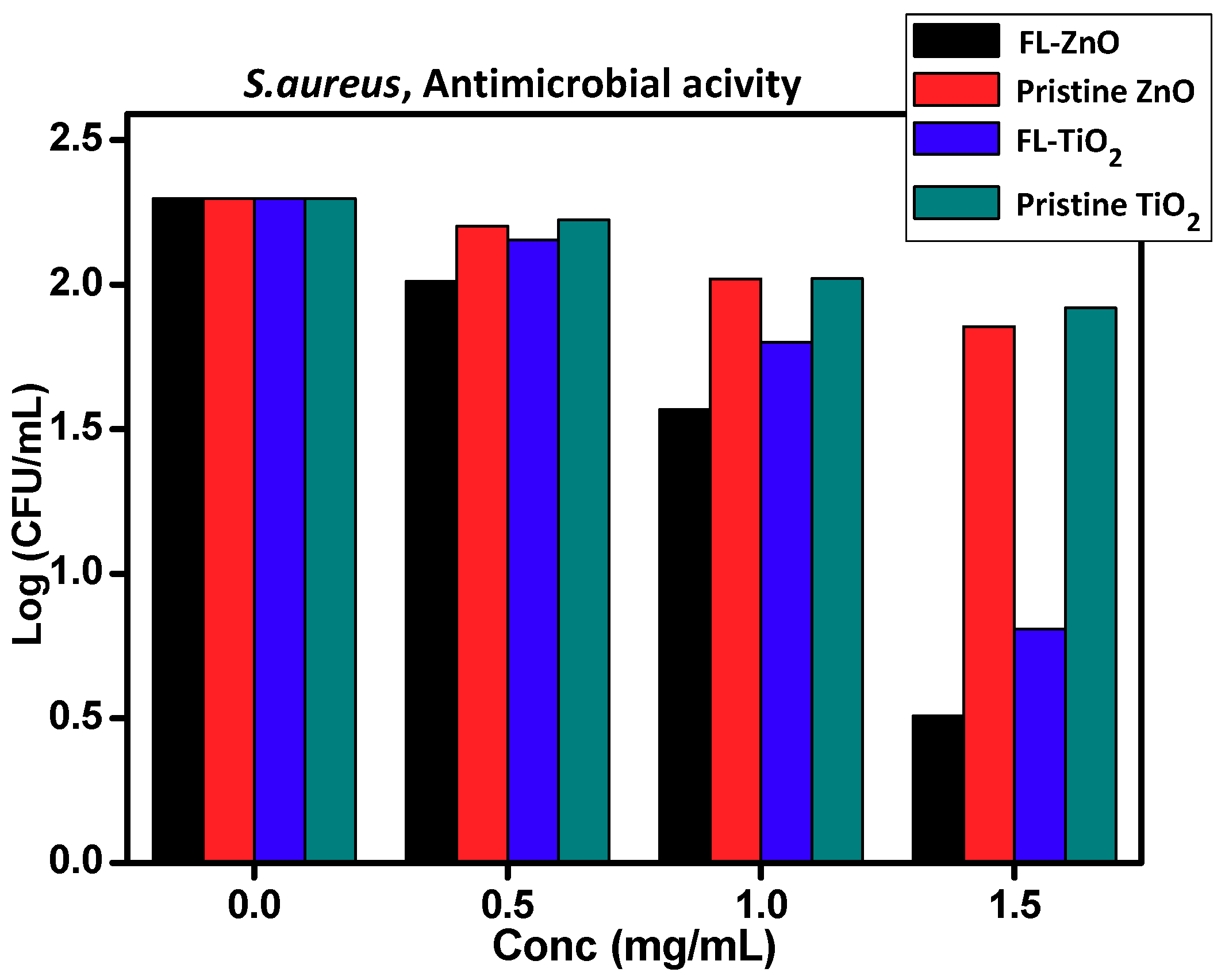
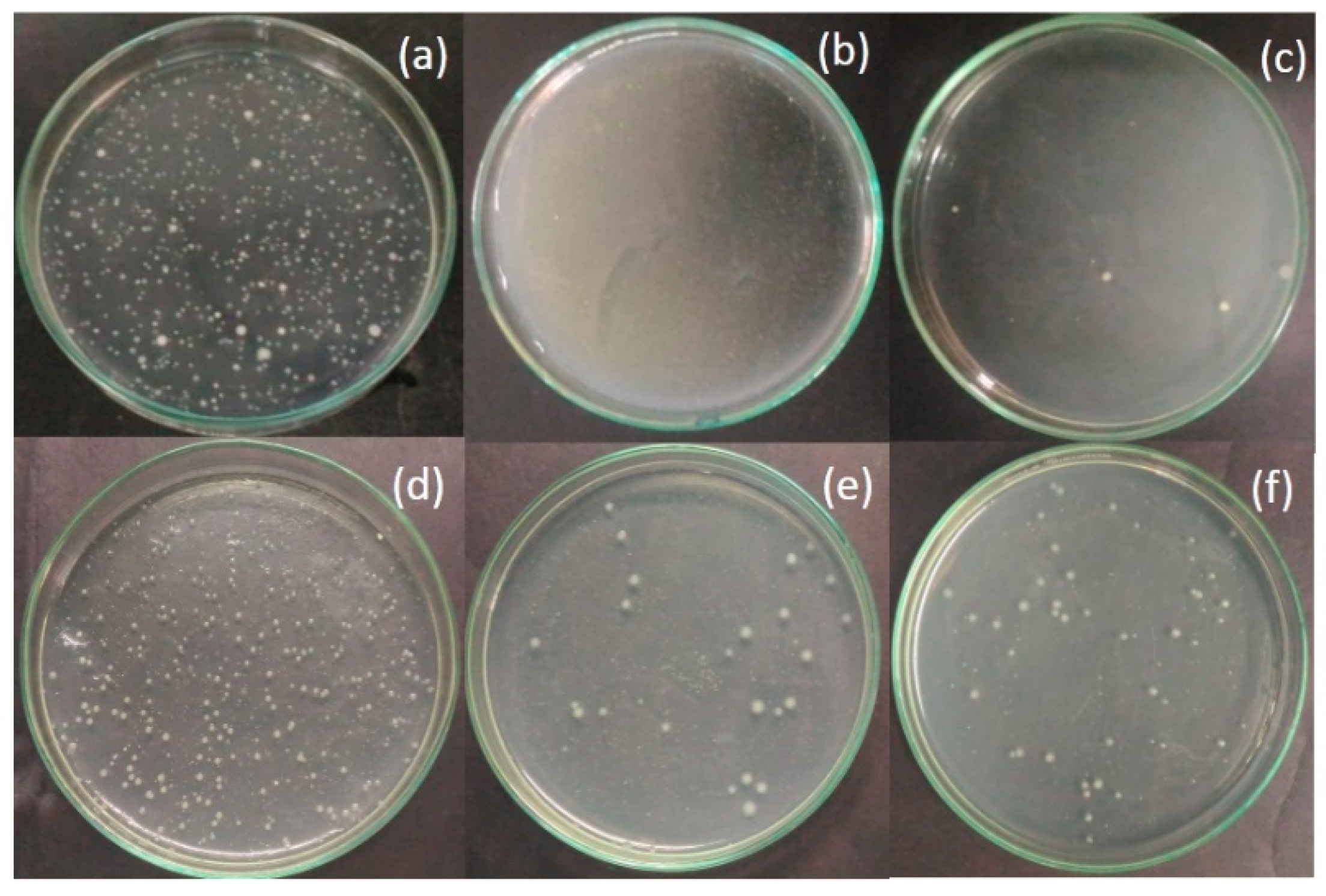
Antimicrobial Property of Biosynthesis FL–ZnO and FL–TiO2 Nanomaterial with Pristine ZnO and TiO2 Material

The standard strain of Escherichia coli (ATCC 25922), Staphylococcus aureus (ATCC 25923) obtain from Microbiology Laboratory Fergusson College Pune were used for antimicrobial studies. The antimicrobial activity was performed as per the guideline of Clinical and Laboratory Standards Institute [23]. It was performed by measuring the minimum bactericidal concentration (MBC) values of composites and pristine nanoparticles against standard strains of microorganisms. In short, 0.1 mL of 1 × 10⁴ cells/mL was inoculated into 1 mL volume of nutrient broth containing different concentrations of nanocomposites and pristine nanoparticles (0.5–1.5 mg/mL). After inoculation all sets were irradiated in UV-A for 30 min. Then each sample was spread on nutrient agar plate and incubated at 35 ± 2 °C for 24 h. Colonies appeared were counted to calculate the MBC value. Nutrient agar plate with microorganism and without nanoparticles served as control. Similar procedure with standard antibiotics, amoxicillin, served as positive control. The experiments were performed in triplicate and the average measurement were reported.

4. Results and Discussions

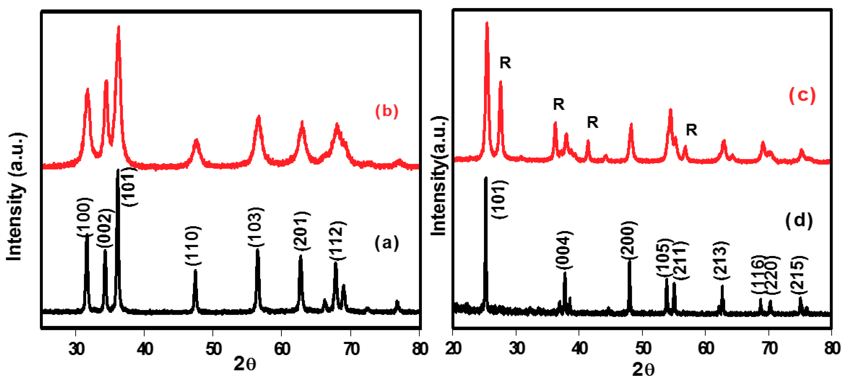
4.1. Structural Study

Crystalline phase and lattice parameters of FL–ZnO and FL–TiO2 nanocomposites, and pristine ZnO and TiO2 nanoparticle were obtained from recorded XRD spectra (Figure 1). The crystal size was calculated by using the Debye–Scherrer equation:
β ( 2 θ ) = K λ L C o s θ ,
where K—Scherrer constant. K varies from 0.68 to 2.08. K = 0.94 for spherical crystallites with cubic symmetry, L—volume average of the crystal thickness in the directional normal to the reflecting planes, λ—X-ray wavelength. For Mini XRD, Cu Kα average = 1.54178 Å, θ—XRD peak position, one half of 2θ.
The major peaks in XRD of FL–ZnO nanocomposites and pristine ZnO do not show any difference in peak positions but an appreciable difference in peak intensities were observed. The observed peak at 2θ = 31.610, 34.320, 36.140, 47.370,56. 480, 62.710 and 68.280 correspond to (100), (002), (101), (110), (103) and (201) planes these are characteristic peaks of hexagonal crystalline structure of ZnO (JCPDS file 36-1451 of ZnO). Sharp and intense peaks in the XRD analyses of both F–ZnO nanocomposites and pristine ZnO indicate high crystallinity and the polycrystalline nature of ZnO nanoparticles (curves a and b Figure 1). Moreover, no extra peaks other than hexagonal crystalline phase of ZnO were found in XRD. Pristine ZnO has average crystallite size 43.76. High intensity of peak at (101) plane shows a weak preferential growth in FL–ZnO nanoparticles along c-axis [24] with an average crystallite size 25.34 nm. The XRD of FL–TiO2 nanocomposites showed a mix phase of anatase and rutile (JCPDS anatase 33-1381, and Rutile 34-0180) which shows the major peaks at 2θ = 25.30,27.380, 36.08 37.730, 41.24 48.020, 54.320, 55.068, 62.840, 68.780, and 70.250 (curve c in Figure 1). The observed peaks (27.23, 36.08 41.24, and 54.32) at (110) (101), (111) and (211) can be indexed is a typical characteristic of rutile phase TiO2. The peaks 25.30, 37.730, 48.020, 53.068, 62.11, 62.840, and 68.780, and 70.250 at (101), (004), (200), (105), (211), (213), (116) and (220) is typical of anatase phase TiO2 (curve d in Figure 1). The pristine TiO2 showed pure anatase phase (curve d in Figure 1) with major peaks 25.380, 37.730, 48.910, 53.980, 55.980, 62.840, 68.830, 70.160 and 75.030 indexed at (101), (004), (200), (105), (211), (213), (116), (220), and (215) planes. The FTIR and HRMS spectra of FL gives the information of functional groups present in the FL such as 679, 850, 1029–1150, 1629, 2829–2926, and 3354 cm1 for aromatic ring, aromatic ring (Para Substituted), CNO stretching, C–O–C stretching, aromatic compound, CH2 stretching, hydroxyl group broad band (Intramolecular Hydrogen Bonding), HRMS showed five different fragments as (1) m/z 276 gives 4-(4-(hydroxymethyl)-3-methoxyphenoxy)-2-methoxyphenol, (2) m/z 290 suggest the β-etheral linkage of phenolic structure, (3) m/z 322 is for Sinapic acid (3-(4-hydroxy 3, 5-dimethyl phenyl), (4) m/z 405 is for dimeric fragments, (5)534m/z for β-O-4 dimer with attachment of different carbon atom or –CH2 group present in the molecule, 1H NMR of FL. 1H NMR (400MHz, MeOD): δ0.84, δ1.2, δ1.52, δ2, δ2.3, δ3.8, and δ6.5–7.8 showed presence of methylene proton, ethylene proton, acetylene proton and aromatic proton (SEI-1 FTIR of Fragmented Lignin, SEI-2 1H-NMR of Fragmented Lignin and SEI-3 HRMS of Fragmented Lignin in Supplementary Materials).

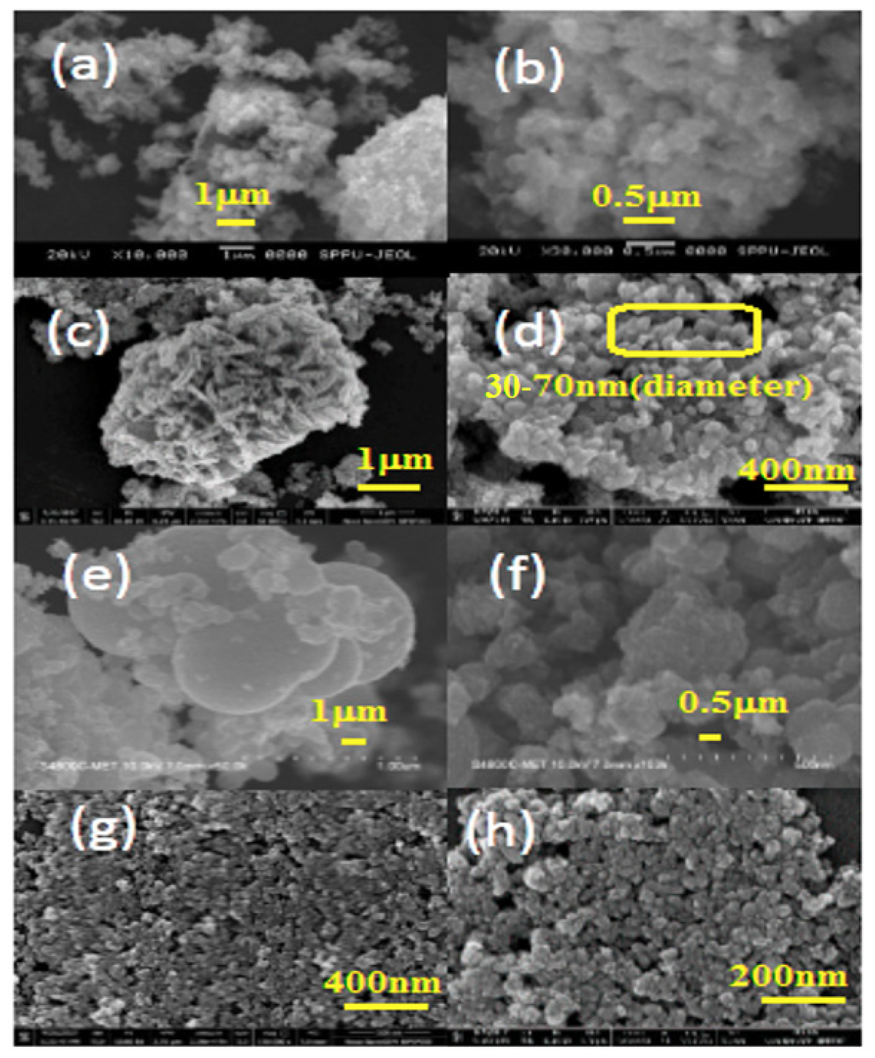
4.2. Surface Morphological Studies
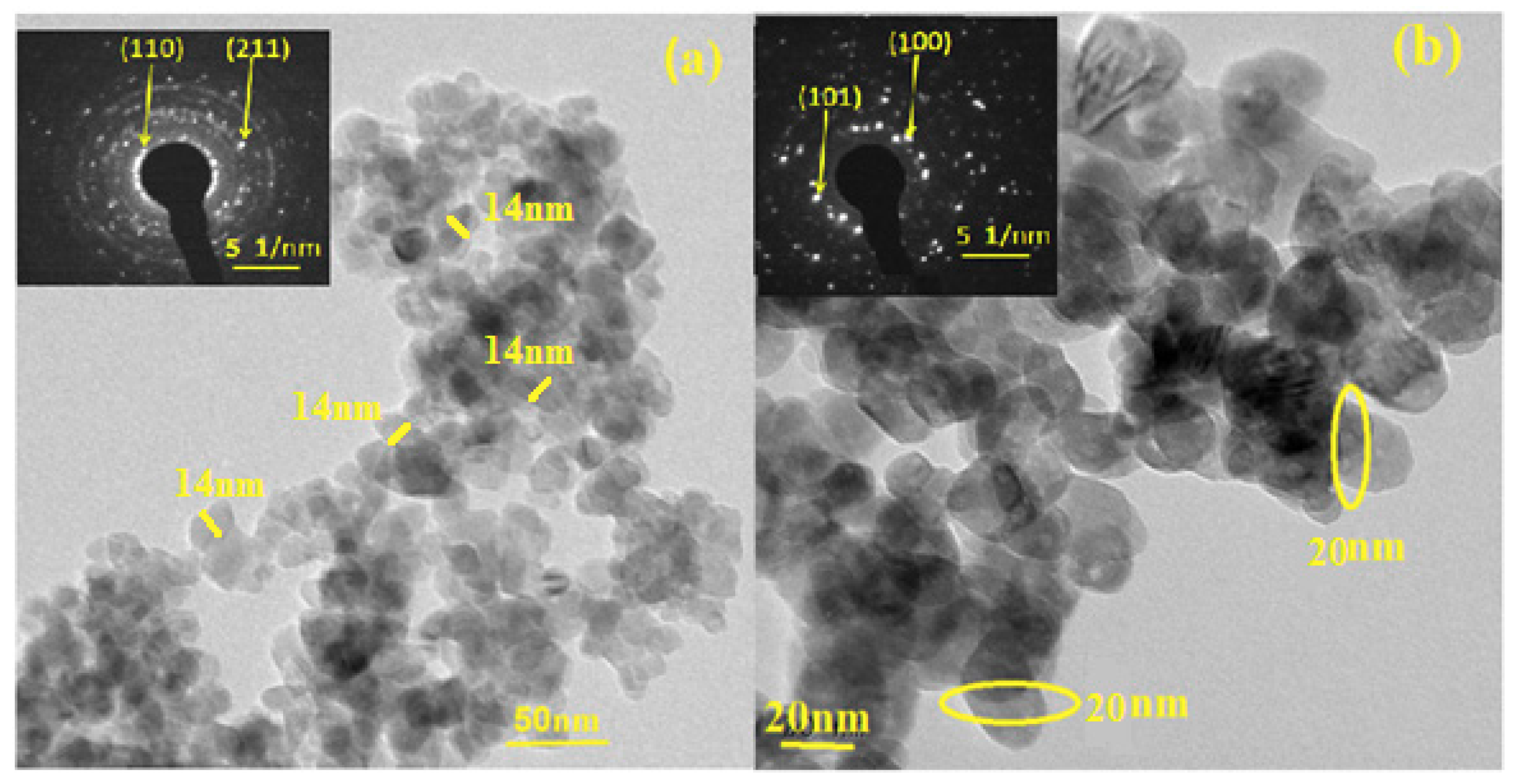
The surface morphology was studied by FE-SEM. The FE-SEM image of FL–ZnO exhibits rod shape morphology given in Figure 2c,d, it is possible due to the inhibition of the growth of nanoparticle in third dimension by fragmented lignin.
The FE-SEM images of pristine ZnO (Figure 2a,b) showed an agglomeration. FL contain many polar functional groups mainly –OH, –OCH3, >C=O and C–O–C which might play an important role in the formation of ZnO rod nanoparticle. Due to the presence of these functional groups lignin fragments may have acted as a complexing, capping or stabilizing agent for Zn+2 ions. The polymeric nature of lignin fragments can create protective and functionalized surrounding for metal ions which might be playing a structure directing role in the formation of rod shaped ZnO nanoparticles [25]. Difference in particle morphology of of pristine TiO2 and FL–TiO2 was observed in Figure 2e,f and Figure 2g,h. FL–TiO2 showed uniformity in grain size and shape. The grains were roughly spherical to spherically elongate in shapes. The average grain size for this FL–TiO2 was 41 nm. Pristine TiO2 showed grains of large and varied size with roughly spherical in shape with an average grain size of 277 nm. Here the anisotropic effect was observed in both samples. The peak of FL–ZnO and FL–TiO2 show broad peak width due to the decrease in crystal size as compare to the pristine ZnO and TiO2.

4.3. TEM

Under TEM, FL–TiO2 showed spherical morphology with average particle size is 14 nm (Figure 3a). FL–ZnO has rod shaped structure composed of spherical particles with self-aligned nature having average size 20 nm diameter. This is one directional growth along (101) plane [26]. XRD showed the highest intensity peak (101) plane of the FL–ZnO, which may be favored by biosynthesis method using fragments lignin as a template given in Figure 3b.

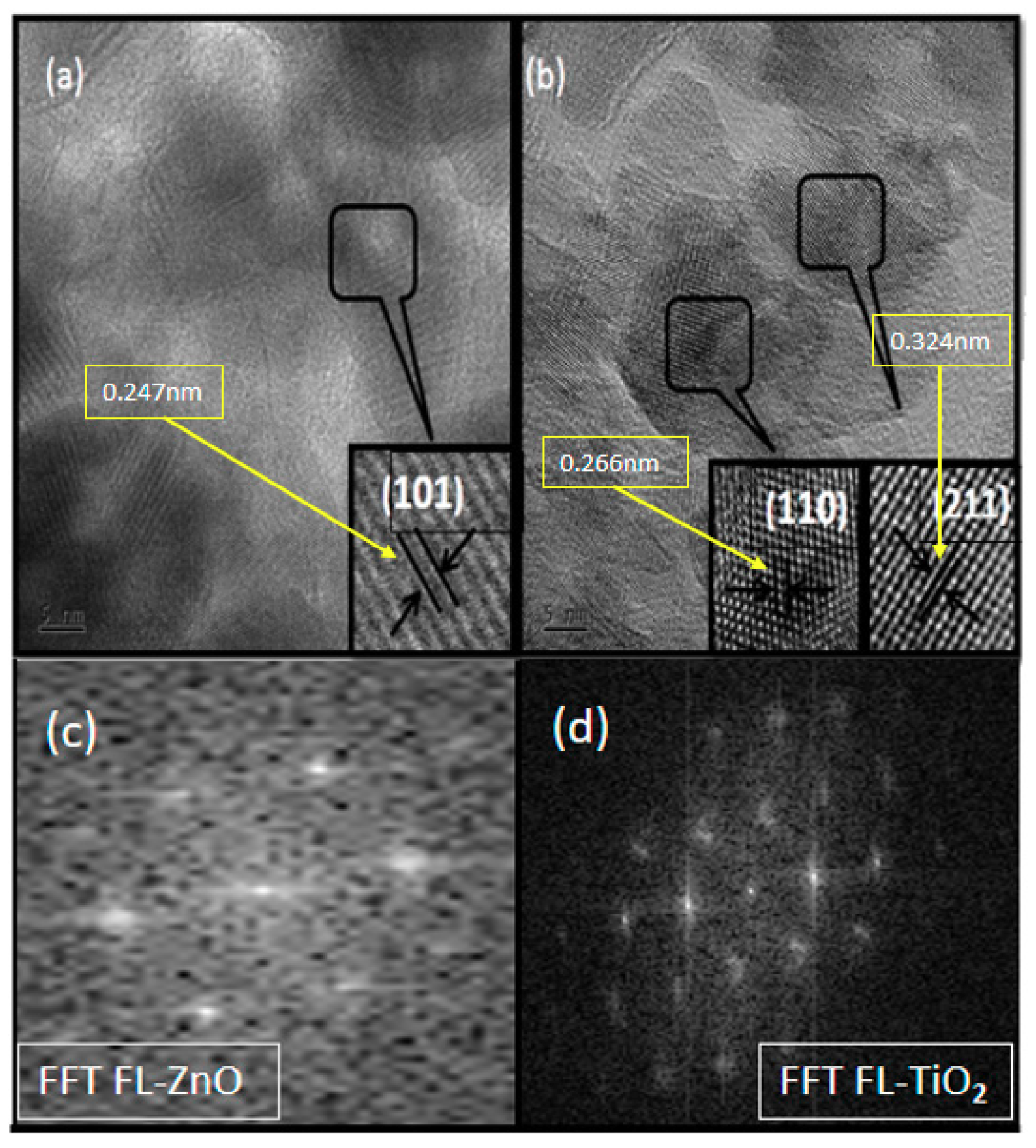
High-Resolution Transmission Electron Microscopy (HRTEM)
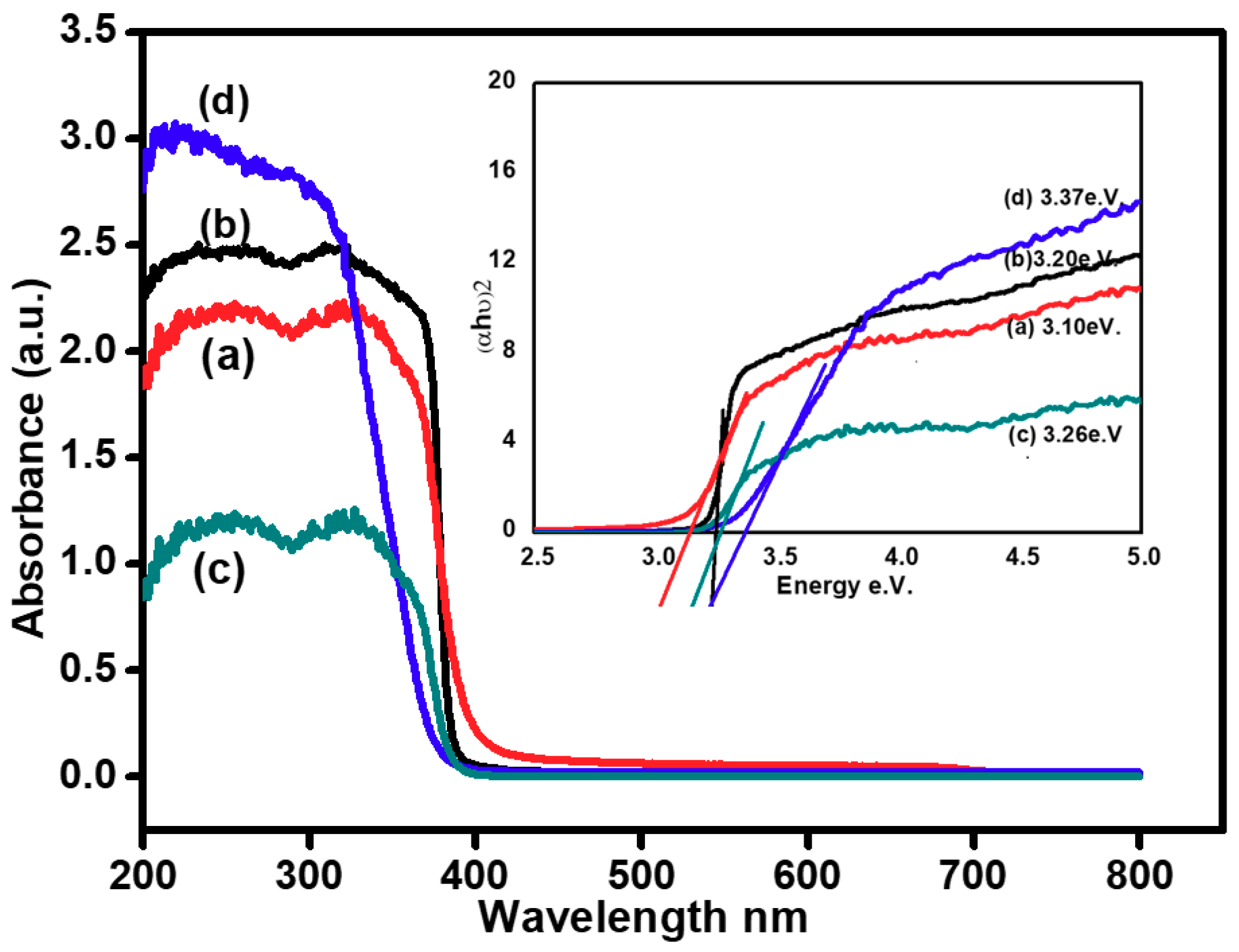
The interplanar distance of FL–ZnO is 0.247 nm for (101) plane. Both the technique XRD and HRTEM shows the mixed phase of FL–TiO2 nanoparticles. For anatase plane (110) with an inter planer distance of 0.266 nm and for the rutile phase (211) the inter planer distance is 0.324 nm. The mixed phase formation of FL–TiO2 and one directional growth of nanoparticle were due to the use of FL which act as a surfactant as shown in Figure 4a,b.
The fast Fourier transformation (FFT) gives the information of the lattice fringes on HRTEM image and it can use to index the observed spots. In given Figure 4c,d corresponding to the fast Fourier transmission (FFT) pattern of the nanoparticle is closed to single crystalline. From these observations, it is reasonable that the as-prepared ZnO and TiO2 nanoparticle is of nanocrystalline structures, which means that they are constituted by well-crystallized ZnO and TiO2 nanoparticles.

4.4. Optical Study

The Ultra-Violet diffuse reflectance spectra (UVDRS) of pristine ZnO, TiO2, FL–ZnO and FL–TiO2 were shown in set of Figure 5. It was calculated from a UV-Visible spectrum and Tauc’s plot. All these materials showed absorbance in a range of 350–400 nm. The calculated band gap of FL–ZnO nanoparticles in presence of FL at 3.10 eV and pristine ZnO is 3.26 eV. More is the oxygen defects lower is the band gap [27]. Deviation in the band gaps of FL–TiO2, and pristine TiO2 was observed in curves b and d in Figure 5. FL–TiO2 showed the band gap of 3.20 eV while pristine TiO2 has band gap of 3.37 eV.

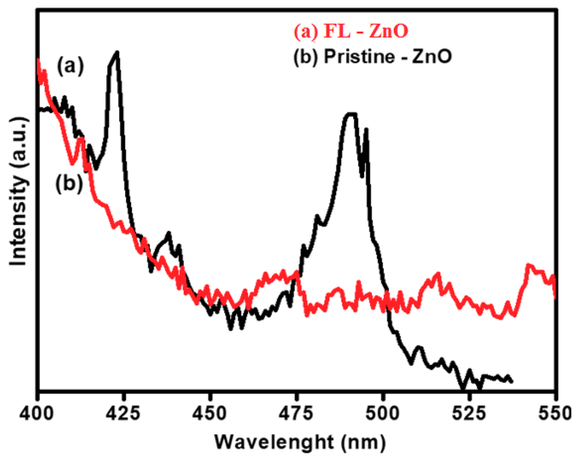
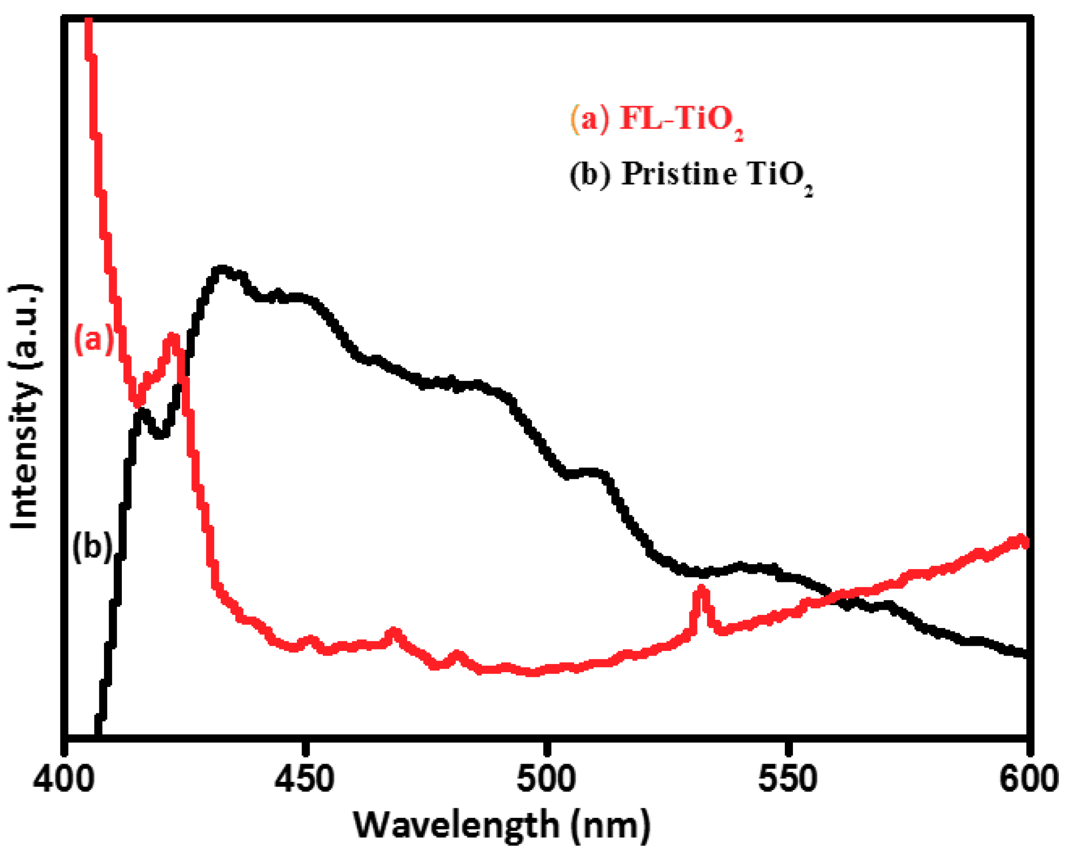
4.5. Photoluminescence Study of Pristine ZnO, FL–ZnO, Pristine TiO2 and FL–TiO2

Photoluminescence study gives us the information of the structural defects in nanoparticles. The Figure 6 and Figure 7 showed photoluminescence property of nanoparticles of pristine ZnO, FL–ZnO, pristine TiO2, and FL–TiO2 synthesized. ZnO samples were excited at 390 nm excitation wavelength and TiO2 samples were excited at 395 nm excitation wavelength. The emission spectrum of pristine ZnO, FL–ZnO, Pristine TiO2 and FL–TiO2 samples were showed in Figure 6 and Figure 7. The different types of peaks were observed in visible region from 400 to 600 nm, such as 400–436 nm (violet region), 436–497 nm (blue region), 497–568 nm (green region) and 568–592 nm (yellow region).
FL–ZnO has highest peak intensity than pristine ZnO where pristine ZnO has no specific peak shoulder in spectrum (Figure 6). For FL–ZnO the peak intensity area was 400–436 nm (violet region), 436–497 nm (blue region) which gives more oxygen defect in visible region. The blue green region are due to the lattice defects which can produce compressive strain at intrinsic crystal lattice [28]. It causes the transition of energy from conduction band or zinc interstitials, which results the recombination of holes and electrons in the valences band and conduction band [29]. Pristine TiO2 showed broad emission band because agglomerated lattice sites and peak intensity shift towards 568–592 nm (yellow–red region) which is significant for increase in particle size (Figure 7) caused by oxygen interstitials zinc vacancies and oxygen interstitials. It may conclude that O-/O2- ion concentration increases intrinsic strain in the lattice of the materials, which gives transition from conduction band to the oxygen interstitial position yellow emission [30,31]. In FL–TiO2 only two peaks were observed, one at 400–436 nm (violet region), and the other at 497–568 nm (green region) due to structural defects related to the deep level emission [32].

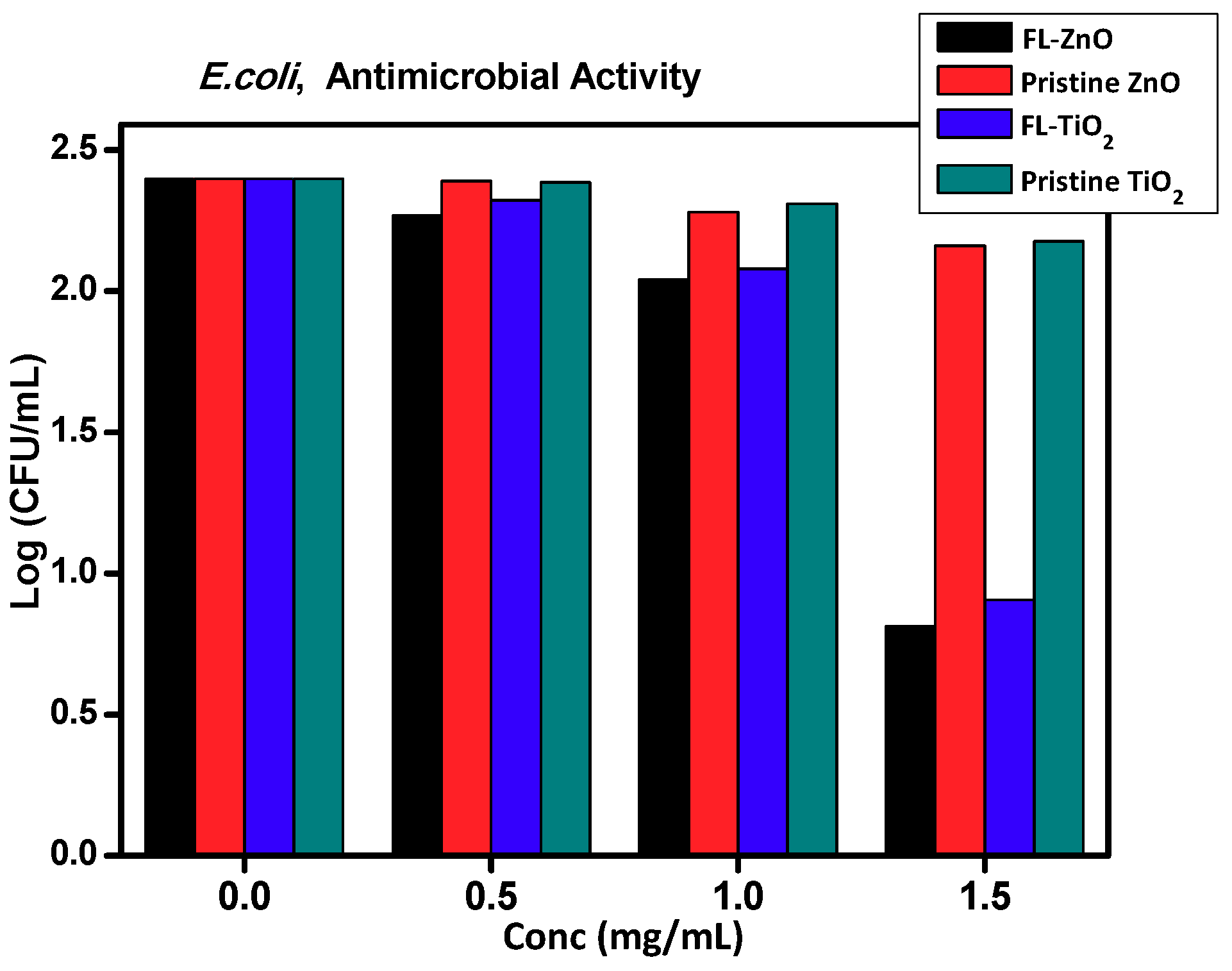
5. Antimicrobial Property

Nanoparticles have emerged as an effective antimicrobial-agent alternative to traditional organic based drugs, primarily due to actions that specifically target and minimize toxicity [33,34,35]. Most studies reported bacteriostatic or bactericidal effect of nanoparticles due to disruption of their cell membrane. The nanoparticle can accommodate a large number of ligands present on microbial cells due to its large surface area to volume ratio. Several types of metal and metal oxide nanoparticles have been already reported to possess anti-microbial property like titanium oxide, gold, silver, copper, iron, zinc oxide, copper oxide and iron oxide nanoparticles [36,37,38,39,40,41,42]. However, of all nanoparticles, TiO2 and ZnO nanoparticles have gained considerable attention because of their unique electronic, optical and medicinal properties [43,44]. These nanoparticles are highly biocompatible and therefore have found various applications in biological field [45]. Recently, a green chemistry approach has been reported to synthesis various nanoparticles including ZnO and TiO2 wherein natural products such as silk fibroin, cellulose, starch, humic acid, carbohydrates, lignin, etc. have been used as reducing and stabilizing agents. Among them, lignin is interesting natural products because: (i) it comes from the wood pulp industry; (ii) it is the second most abundant on earth, just second to cellulose; (iii) it is a nanocrystalline and heterogeneous polymer with a network structure; (iv) it is the only kind of biomass constituent that belong to the aromatic compounds; (v) structurally, it contains many aliphatic and aromatic hydroxyls, several aromatic methoxy, carboxyl, carbonyl and ethereal moieties and (vi) it has many aliphatic hydroxyl groups, therefore, it can be used in reduction of metal salt to metal nanoparticles [46,47,48], for changing surface morphology and nanoroughness of metal oxide, it can affect on for cell adhesion and proliferation [49]. Antibacterial activity of pristine ZnO and TiO2 nanoparticles and FL–ZnO and FL–TiO2 nanocomposites on E. coli and S. aureus were as shown in Figure 8 and Figure 9, respectively.
As can be observed, FL–ZnO and FL–TiO2 nanocomposites showed a better antimicrobial activity than pristine ZnO and TiO2 nanoparticles. The nanocomposites were more effective on cells of S. aureus than on cells of E. coli. The antimicrobial action of FL–ZnO and FL–TiO2 nanocomposites were concentration dependent. When the cells of E. coli and S. aureus were independently subjected to various concentrations of ZnO and TiO2 nanoparticles and FL–ZnO and FL–TiO2 nanocomposites, there was a decrease in the number of viable cells. An initial 2.4 log CFU/mL was reduced to less than 0.5 log CFU/mL in presence of FL–ZnO and 0.7 log CFU/mL in presence of FL–TiO2 nanocomposites. However, the log CFU/mL in presence of pristine ZnO and TiO2 nanoparticles were respectively, 2.2 and 2.1. The reduction was more prominent in presence of FL–ZnO and FL–TiO2 nanocomposites. The colony count of E. coli and S. aureus, in presence of FL–ZnO and FL–TiO2 nanocomposites also, was less (Figure 10). Small particles size of material showed enhancement in the bioactivity because increased in surface area to the volume ratio. The smaller particles can easily bind higher number of bacterial colonies which result in the large number of active oxygen species to burst the cell wall of the bacteria also the structural defects, oxygen defect and oxygen interstitials help to increase the antimicrobial activity of nanomaterial of FL–ZnO and FL–TiO2. Whereas, pristine ZnO and TiO2 nanoparticles have agglomeration, bulky size and less surface to the volume ration can decreases contribution to the antimicrobial activity [50].
The antimicrobial activities of FL–ZnO and FL–TiO2 were in agreement with previous reports. The powder extract of dry ginger rhizome was used as a reducing material as well as surface stabilizing agent to synthesize ZnO nanoparticles and were shown to possess antimicrobial activity against bacteria and fungi [51]. Recently Coptidis rhizoma-mediated ZnO nanoparticles were tested for its antimicrobial activity against four disease-causing pathogens Bacillus megatherium, B. pumilus, B. cereus and E. coli [52]. ZnO nanoparticles adopt a series of mechanisms to act as an anti-bacterial agent. Loss of phospholipid bilayer cell membrane integrity is considered as one of the most important mechanisms of ZnO and TiO2 NPs due to the oxidative stress induced by reactive oxygen species (ROS). This ROS molecule further causes cell death by inhibiting or altering DNA replication, protein synthesis, and membrane potentials [53]. In the present study, FL–TiO2 and FL–ZnO nanocomposites have shown a prominent action on cells of S. aureus because of the difference in the mechanism of attachment of Gram- positive and Gram negative bacteria to nanoparticles, their transport inside the cell, and differences in their different membrane structure. Gram positive bacteria have a thick layer of peptidoglycan and have teichoic acid and lipoteichoic acid, the later serves as binding sites to nanocomposites. They also chelate metal ions from nanoparticles and transport inside the cell. Gram negative bacteria have a triple layer of peptidoglycan in their cell wall which imparts additional barrier for nanoparticles to enter inside cells [54]. Upon illumination, ZnO and TiO2 nanoparticles generate the electron hole pairs, and produce reactive oxygen species (ROS), which oxidizes organic matter and, thus, imparts biocidal property to nanoparticles [55]. However, ZnO and TiO2 nanoparticles are having a very low efficiency for the separation of electron hole pairs due to fast recombination of charge carriers, therefore, it is very essential to suppression of the recombination of photogenerated electron-hole pairs in ZnO and nanoparticles. FL which contains different organic function groups, such as aromatic, phenolic, hydroxyl, etheral, alkene and methoxy groups, are hypothesized to prevent this recombination of electron hole pair. In the present study, the biosynthesized FL–ZnO and FL–TiO2 nanocomposites have porous structure and low band gap with several defect as was observed in photoluminescence spectrum. FL–TiO2 nanocomposites have mixed phase morphology of anatase and rutile geometry. Therefore, such an environment is favorable for ejection and stabilization of electrons to create hole and electron pairs and imparts FL–ZnO and FL–TiO2 nanocomposites as an antimicrobial property.

6. Conclusions

The FL results in five different fragments which contain aldehydic, etheral, alcoholic, aromatic, hydroxyl and phenolic groups, which may act as surfactant and restrict the cluster formations. Hence, these groups play an important role in synthesis of FL–ZnO and FL–TiO2 nanocomposites by a biosynthesis method. These organic materials were coated on the nanomaterial during fabrication which creates defects to generate holes and electrons, which easily react with the cell membrane and DNA to result in cell death. Furthermore, the large surface area and special morphology of ZnO gives a more pronounced effect. In this way, both of these biosynthesized materials show better results in bactericidal activity compared to the pristine material.

Supplementary Materials

The following are available online at https://0-www-mdpi-com.brum.beds.ac.uk/2504-477X/3/3/90/s1. Figure S1. FTIR of Fragmented Lignin, Figure S2. 1H-NMR of Fragmented Lignin, Figure S3. HRMS of Fragmented Lignin.

Author Contributions

Conceptualization, K.M.S.-J., M.G.C.; Methodology, K.M.S.-J., H.B.S., R.P.; Software, K.M.S.-J., Y.A.S., A.A.A.; Validation, K.M.S.-J., R.P.P.; Formal Analysis, K.M.S.-J., R.P.P., S.P.R.; Investigation, B.B.K., M.G.C.; Data Curation, K.M.S.-J., R.P.; Writing, K.M.S.-J., R.P., S.P.R.; Original Draft Preparation, K.M.S.-J., R.P.; Writing–Review & Editing, K.M.S.-J., R.P.; Supervision, R.P., B.B.K., M.G.C.

Funding

This research received no external funding

Acknowledgments

Manohar G. Chaskar would like to thank Prof. Ramakrishna More A.S.C College and B.G. College Sangavi pune for providing research facilities. Also, we thank for C-MET Pune. Rajendra Patil acknowledges Departmental Research and Development Grant 2018-19.

Conflicts of Interest

The authors declare no conflict of interest

References

  1. Kaul, G.; Kapoor, E.; Dasgupta, A.; Chopra, S. Management of multidrug-resistant tuberculosis in the 21st century. Drugs Today 2019, 55, 215–224. [Google Scholar] [CrossRef] [PubMed]
  2. Richardson, L.A. Understanding and overcoming antibiotic resistance. PLoS Biol. 2017, 15, e2003775. [Google Scholar] [CrossRef] [PubMed]
  3. Paule, A.; Frezza, D.; Edeas, M. Microbiota and Phage Therapy: Future Challenges in Medicine. Med. Sci. 2018, 6, 86. [Google Scholar] [CrossRef] [PubMed]
  4. Spaulding, C.N.; Klein, R.D.; Schreiber, H.L.; Janetka, J.W.; Hultgren, S.J. Precision antimicrobial therapeutics the path of least resistance? NPJ Biofilms Microbiomes 2018, 4, 4. [Google Scholar] [CrossRef] [PubMed]
  5. Baker, S.J.; Payne, D.J.; Rappuoli, R.; De Gregorio, E. Technologies to Address Antimicrobial Resistance; National Acadamey of Science of United State of America: New Havan, CT, USA,, 2018; Volume 115, pp. 12887–12895. [Google Scholar]
  6. Gupta, A.; Mumtaz, S.; Li, C.H.; Hussain, I.; Rotello, V.M. Combatting antibiotic-resistant bacteria using nanomaterials. Chem. Soc. Rev. 2019, 48, 415–427. [Google Scholar] [CrossRef]
  7. Raghunath, A.; Perumal, E. Metal oxide nanoparticles as antimicrobial agents: a promise for the future. Int. J. Antimicrob. Agents 2017, 49, 137–152. [Google Scholar] [CrossRef]
  8. Baranwal, A.; Srivastava, A.; Kumar, P.; Bajpai, V.K.; Maurya, P.K.; Chandra, P. Prospects of Nanostructure Materials and Their Composites as Antimicrobial Agents. Front. Microbiol. 2018, 9, 422. [Google Scholar] [CrossRef] [Green Version]
  9. Mantravadi, P.K.; Kalesh, K.A.; Dobson, R.C.J.; Hudson, A.O.; Parthasarathy, A. The Quest for Novel Antimicrobial Compounds: Emerging Trends in Research, Development, and Technologies. Antibiotics 2019, 8, E8. [Google Scholar] [CrossRef]
  10. Mahamuni, P.P.; Patil, M.; Dhanavade, M.J.; Badiger, M.V.; Shadija, P.G.; Lokhande, A.C.; Bohara, R.A. Synthesis and characterization of zinc oxide nanoparticles by using polyol chemistry for their antimicrobial and antibiofilm activity. Biochem. Biophys. Rep. 2018, 17, 71–80. [Google Scholar] [CrossRef]
  11. Siddiqi, K.S.; Rahman, A.; Tajuddin, U.R.; Husen, A. Properties of Zinc Oxide Nanoparticles and Their Activity Against Microbes. Nanoscale Res. Lett. 2018, 13, 141. [Google Scholar] [CrossRef]
  12. Wilhelm, S.; Kaiser, M.; Würth, C.; Heiland, J.; Carrillo-Carrion, C.; Muhr, V.; Wolfbeis, O.S.; Parak, W.J.; Resch-Genger, U.; Hirsch, T. Water dispersible. Upconverting nanoparticles: effects of surface modification on their luminescence and colloidal stability. Nanoscale 2015, 7, 1403–1410. [Google Scholar] [CrossRef] [PubMed]
  13. Xie, Y.; Kocaefe, D.; Chen, C.; Kocaefe, Y. Review of Research on Template Methods in Preparation of Nanomaterials. J. Nanomater. 2016, 2016, 10. [Google Scholar] [CrossRef]
  14. Bhandari, S.; Mondal, D.; Nataraj, S.K.; Balakrishna, R.G. Biomolecule-derived quantum dots for sustainable optoelectronics. Nanoscale Adv. 2019, 1, 913–936. [Google Scholar] [CrossRef]
  15. Wang, J.; Vermerris, W. Antimicrobial nanomaterials derived from natural products—A review. Materials 2016, 9, 255. [Google Scholar] [CrossRef] [PubMed]
  16. Huang, J.; Fu, S.; Gan, L. Chapter 2—Structure and Characteristics of Lignin 2019. In Lignin Chemistry and Applications; Elsevier: Atlanta, GA, USA, 2019; pp. 25–50. [Google Scholar]
  17. Katahira, R.; Elder, T.J.; Beckham, G.T. Chapter 1 A Brief Introduction to Lignin Structure. In Lignin Valorization: Emerging Approaches; The Royal Society of Chemistry: Philadelphia, PA, USA, 2018; Volume 1, p. 20. [Google Scholar]
  18. Acosta, J.E.; Torres-Chávez, L.P.I.; Wong, B.R.; Saiz, C.M.L.; Leyva, B.M. Antioxidant, Antimicrobial, and Antimutagenic Properties of Technical Lignins and Their Applications. J. BioResour. 2016, 2, 5452–5481. [Google Scholar]
  19. Vinardell, M.P.; Mitjans, M. Lignins and Their Derivatives with Beneficial Effects on Human Health. Int. J. Mol. Sci. 2017, 18, 1219. [Google Scholar] [CrossRef] [PubMed]
  20. Gordobil, O.; Herrera, R.; Yahyaoui, M.; Ilk, S.; Kaya, M.; Labidi, J. Potential use of kraft and organosolv lignins as a natural additive for healthcare products. RSC Adv. 2018, 8, 24525–24533. [Google Scholar] [CrossRef] [Green Version]
  21. Ge, Y.; Li, Z. Application of Lignin and Its Derivatives in Adsorption of Heavy Metal Ions in Water: A Review. ACS Sustain. Chem. Eng. 2018, 6, 7181–7192. [Google Scholar] [CrossRef]
  22. Lora, J.H.; Glasser, W.G. Recent Industrial Applications of Lignin: A Sustainable Alternative to Nonrenewable Materials. J. Polym. Environ. 2002, 10, 39–48. [Google Scholar] [CrossRef]
  23. Anand, N. Chapter 2.2 Biological Methods; The Indian Pharmacopeia Commission: Ghaziabad, India, 2010; Volume 1, pp. 32–54. ISBN 81-903436-6-1 (VoU). [Google Scholar]
  24. Joshi, K.M.; Shinde, D.R.; Nikam, L.K.; Panmad, R.; Kale, B.B.; Chaskar, M.G. Fragmented lignin-assisted synthesis of a hierachical ZnO nanostructure for ammonia gas sensing. RSC Adv. 2019, 9, 2487. [Google Scholar] [CrossRef]
  25. .Rubin, J.E.; Ball, K.R.; Trejo, M.C. Antimicrobial susceptibility of Staphylococcus aureus and Staphylococcus pseudintermedius isolated from various animals. Can. Vet. J. 2011, 52, 153. [Google Scholar] [PubMed]
  26. Fu, Y.Q.; Luo, J.K.; Nguyen, N.T.; Walton, A.J.; Flewitt, A.J.; Zu, X.T.; Du, H. Advances in piezoelectric thin films for acoustic biosensors, acoustofluidics and lab-on-chip applications. Prog. Mater. Sci. 2017, 89, 31–91. [Google Scholar] [CrossRef] [Green Version]
  27. Miao, T.T.; Sun, D.X.; Guo, Y.R.; Li, C.; Li, Y.; Ma, G.Z. Low temperature precipitation synthesis of flower like ZnO with lignin amine and its optical properties. Nanoscale Res. Lett. 2013, 8, 431. [Google Scholar] [CrossRef] [PubMed]
  28. Khan, M.F.; Ansari, A.H.; Hameedullah, M.; Ahmad, E.; Husain, F.M.; Zia, Q.; Zaheer, B.M.R.; Alams, M.M.; Khan, A.M.; Alothman, Z.A.; et al. Sole-gel synthesis of thorn like ZnO nanoparticles endorosing mechanical stirring effect and their antimicrobial activity: Potential role as nano-antibiotics. Sci. Rep. 2016, 6, 27689. [Google Scholar] [CrossRef] [PubMed]
  29. Roy, J.S.; Majumder, T.P.; Dabrowski, R. Photoluminescence behavior of TiO2 nanoparticles doped with liquid crustal. J. Mol. Struct. 2015, 1098, 351–354. [Google Scholar] [CrossRef]
  30. Faixl, O.; Grunwald, C.; Beinhoff, O. Determination of phenolic Hydroxylic group content of Milled Wood Lignins (MWUs) Different Botanical Origins Using Selective Aminolysis, FTIR, and UVSpectoscopy. Int. J. Biol. Chem. Phys. Technol. Wood 1992, 46, 428. [Google Scholar]
  31. Haque, F.Z.; Nandanwar, R.; Singh, P. Evaluating photodegradation properties of anatase and rutile TiO2 nanoparticles for organic compounds. Optik 2017, 128, 191–200. [Google Scholar] [CrossRef]
  32. Shi, L.; Shen, H.; Jiang, L.; Li, X. Co-emission of UV, violet and green photoluminescence of ZnO/TiO2 thin film. Mater. Lett. 2007, 61, 4735–4737. [Google Scholar] [CrossRef]
  33. Chithra, M.J.; Sathya, M.; Pushpanathan, K. Effect of pH on Crystal Size and Photoluminescence Property by Chemical Precipitation Method; Springer: New Delhi, India, 2015; Volume 28, p. 3. [Google Scholar]
  34. Kim, B.Y.S.; Rutka, J.T.; Chan, W.C.W. Effect of Coumarate 3-zhydroxylase Down regulation on lignin structure. Nanomed. N. Engl. J. Med. 2010, 363, 2434–2443. [Google Scholar] [CrossRef]
  35. Akhtar, M.S.; Swamy, M.K.; Umar, A.; Abdullah, A.; Sahli, A. Biosynthesis and characterization of silver nanoparticles from methanol leaf extract of Cassia didymobotyra and assessment of their antioxidant and antibacterial activities. Nanosci. Nanotechnol. 2015, 15, 1–6. [Google Scholar] [CrossRef]
  36. Rudramurthy, G.R.; Swamy, M.K.; Sinniah, U.R.; Ghasemzadeh, A. Nanoparticles: Alternatives against drug-resistant. Molecules 2016, 21, 1–30. [Google Scholar] [CrossRef] [PubMed]
  37. Lee, H.; Ryu, D.; Choi, S.; Lee, D. Antibacterial activity of silver- nanoparticles against Staphylococcus aureus and Escherichia coli. Korea J. Microbiol. Biotechnol. 2011, 39, 77–85. [Google Scholar]
  38. Mohamed, M.M.; Fouad, S.A.; Elshoky, H.A.; Mohammed, G.M.; Alaheldin, T.A. Antibacterial effect of gold nanoparticles against Corynebacterium pseudotuberculosis. Int. J. Vet. Sci. Med. 2017, 5, 23–29. [Google Scholar] [CrossRef] [PubMed]
  39. Pacheco, G.J.; sánchez, M.E.; martínez, A.R.; Ruiz, F.; Jasso, M.E.C. Antimicrobial properties of copper nanoparticles and amino acid chelated copper nanoparticles produced by using a soya extract. Bioinorgan. Chem. Appl. 2017, 15, 17. [Google Scholar]
  40. Naseem, T.; Farrukh, M.A. Antibacterial activity of Green synthesis of iron nanoparticles using lawsonia inermis and gardenia jasminoides leaves extract. J. Chem. 2015, 2015, 7. [Google Scholar] [CrossRef]
  41. Jesline, A.; John, P.N.; Narayanan, P.M.; Vani, C.; Murugan, S. Antimicrobial activity of zinc and titanium dioxide nanoparticles against biofilm-producing methicillinresistant Staphylococcus aureus. Appl. Nanosci. 2015, 5, 157–162. [Google Scholar] [CrossRef]
  42. Ren, G.; Hu, D.; Cheng, E.W.C.; Vargas-reus, M.A.; Reip, P.; Allaker, R.P. Characterisation of copper oxide nanoparticles for antimicrobial applications. Int. J. Antimicrob. Agents 2009, 33, 587–590. [Google Scholar] [CrossRef] [PubMed]
  43. Ismail, R.A.; Sulaiman, G.M.; Abdulrahman, S.A.; Marzoog, T.R. Antibacterial activity of magnetic iron oxide nanoparticles synthesised by laser ablation in liquid. Mater. Sci. Eng. C 2015, 53, 286–297. [Google Scholar] [CrossRef] [PubMed]
  44. Agarwal, H.; Menon, S.; Kumar, S.V.; Rajeshkumar, S. Mechanistic study on antibacterial action of zinc oxide nanoparticles synthesized using green route. Chemico-Biol. Interact. 2018, 286, 60–70. [Google Scholar] [CrossRef] [PubMed]
  45. Singh, A.V.; Mehta, K.K.; Worley, K.; Dordick, J.S.; Kane, R.S.; Wan, L.Q. Carbon nanotube-induced loss of multicellular chirality on micropatternedsubstrate is mediated by oxidative stress. ACS Nano 2014, 8, 2196–2205. [Google Scholar] [CrossRef]
  46. Reddy, L.S.; Nisha, M.M.; Joice, M.; Shilpa, P.N. Antimicrobial activity of zinc oxide(ZnO) nanoparticle against Klebsiella pneumoniae. Pharm. Biol. 2014, 52, 1388–1397. [Google Scholar] [CrossRef] [PubMed]
  47. Huang, Y.; Wu, C.; Aronstam, R.S. Toxicity of transition metal oxide nanoparticles: recent insights from in vitro studies. Materials 2010, 3, 4842–4859. [Google Scholar] [CrossRef] [PubMed]
  48. Boeriu, C.G.; Bravo, D.; Gosselink, R.J.; van Dam, J.E. Characterisation of structure-dependent functional properties of lignin with infrared spectroscopy. Ind. Crops Prod. 2004, 20, 205–218. [Google Scholar] [CrossRef]
  49. Singh, A.V.; Ferri, M.; Tamplenizza, M.; Borghi, F.; Divitini, G.; Ducati, C.; Lenardi, C.; Piazzoni, C.; Merlini, M.; Podest, A.; et al. Bottom- up engineering of the suface roughness of nanostructured cubic zirconia to control cell adhesion. Nanotechnology 2012, 23, 475101. [Google Scholar] [CrossRef] [PubMed]
  50. Padmavathy, N.; Vijayaraghavan, R. Enhanced bioactivity of ZnO nanoparticles—An antimicrobial study. Sci. Technol. Adv. Mater. 2008, 9, 035004. [Google Scholar] [CrossRef] [PubMed]
  51. Gosselink, R.; Snijder, M.; Kranenbarg, A.; Keijsers, E.; de Jong, E.; Stigsson, L.L. Characterisation and application of NovaFiber lignin. Ind. Crops Prod. 2004, 20, 191–203. [Google Scholar] [CrossRef]
  52. Janaki, A.C.; Sailatha, E.; Gunasekaran, S. Synthesis, characteristics and antimicrobial activity of ZnO nanoparticles. Spectrochim. Acta Part A Mol. Biomol. Spectrosc. 2015, 144, 17–22. [Google Scholar] [CrossRef] [PubMed]
  53. Nagajyothi, P.C.; Sreekanth, T.V.M.; Tettey, C.O.; Jun, Y.I.; Mook, S.H. Characterization, antibacterial, antioxidant, and cytotoxic activities of ZnO nanoparticles using Coptidis rhizoma. Bioorg. Med. Chem. Lett. 2014, 24, 4298–4303. [Google Scholar] [CrossRef]
  54. Tiwari, V.; Mishra, N.; Gadani, K.; Solanki, P.S.; Shah, N.A.; Tiwari, M. Mechanism of Anti-bacterial Activity of Zinc Oxide Nanoparticle Against Carbapenem-Resistant Acinetobacter baumannii. Front. Microbiol. 2018, 9, 1218. [Google Scholar] [CrossRef] [PubMed] [Green Version]
  55. Pesci, F.M.; Wang, G.; Klug, D.R.; Li, Y.; Cowan, A.J. Efficient Suppression of Electron–Hole Recombination in Oxygen-Deficient Hydrogen-Treated TiO2 Nanowires for Photoelectrochemical Water Splitting. J. Phys. Chem. C 2013, 117, 25837–25844. [Google Scholar] [CrossRef]
Figure 1. X-ray diffraction (XRD) of (a) Pristine ZnO, (b) FL–ZnO, (c) FL–TiO2 and (d) Pristine TiO2.
Jcs 03 00090 g001
Figure 2. Field emission scanning electron microscopy (FE-SEM): (a,b) Pristine ZnO, (c,d) FL–ZnO, (e,f) Pristine TiO2 and (g,h) FL–TiO2.
Jcs 03 00090 g002
Figure 3. TEM of (a) Spherical morphology of FL–TiO2, (b) self-aligned in one direction Spherical particle of FL–ZnO.
Jcs 03 00090 g003
Figure 4. High-resolution transmission electron microscopy (HRTEM): (a) FL–ZnO, (b) FL–TiO2, (c) FFT FL–ZnO and (d) FFT FL–TiO2.
Jcs 03 00090 g004
Figure 5. UV-Visible spectrum and UV-diffuse reflectance spectrum of ZnO and TiO2 as (a) FL–ZnO 3.10 eV, (b) FL–TiO2, 3.20 eV. (c)Pristine ZnO 3.20 eV and (d) Pristine TiO2 3.37 eV.
Jcs 03 00090 g005
Figure 6. Photoluminescence spectra of ZnO materials excited at 390 nm excitation wavelength (a) FL–ZnO and (b) Pristine ZnO.
Jcs 03 00090 g006
Figure 7. Photoluminescence spectra of TiO2 material excited at 395 nm excitation wavelength (a) FL–TiO2 and (b) Pristine TiO2.
Jcs 03 00090 g007
Figure 8. Antimicrobial activity of E. coli against pristine ZnO (48%), FL–ZnO (5.2%), pristine TiO2 (52%) and FL–TiO2 (6%).
Jcs 03 00090 g008
Figure 9. Antimicrobial activity of S. aureus against; growth shown for Pristine ZnO (32%), FL–ZnO (3.2%), Pristine TiO2 (36%) and FL–TiO2 (5%).
Jcs 03 00090 g009
Figure 10. Antimicrobial activity of E. coli and S. aureus against TiO2 and ZnO nanomaterials. (a) Control of S. aureus; (b) FL–ZnO against S. aureus; (c) FL–ZnO against E. coli; (d). Control of E. coli; (e) TiO2 against S. aureus; (f) TiO2 against E. coli.
Jcs 03 00090 g010

