
Did Dionysius of Fourna Follow the Material Recipes Described in His Own Treatise? A First Analytical Investigation of Four of His Panel Paintings


Abstract
:1. Introduction
2. Materials and Methods
2.1. Non-Invasive Analyses
2.1.1. Imaging Techniques
2.1.2. pXRF
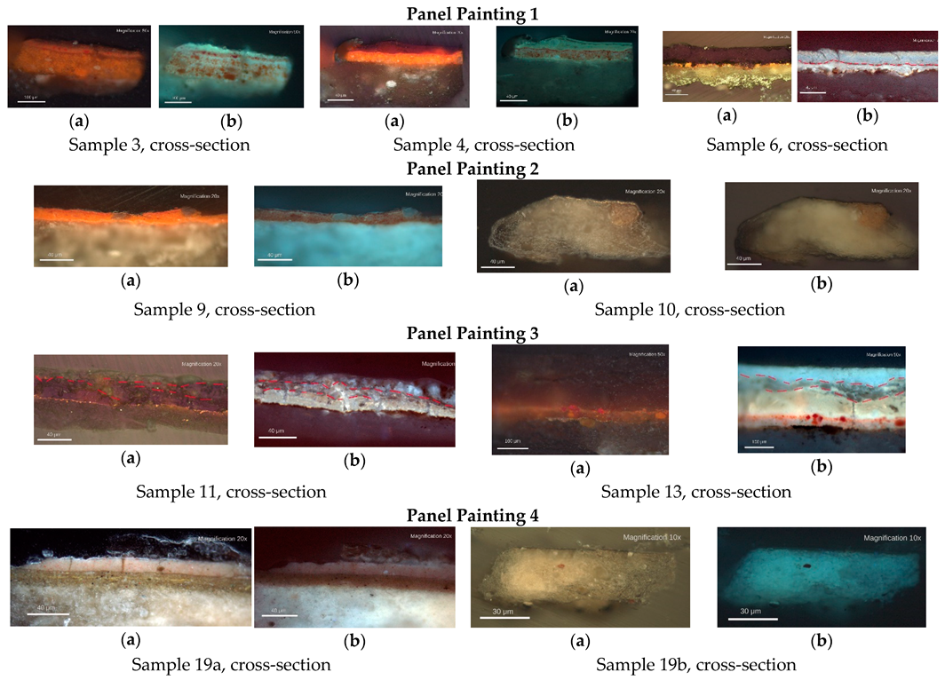
2.2. Microsamples Analyses
2.2.1. Sampling Procedure
2.2.2. Optical Microscopy and Microchemical Testing
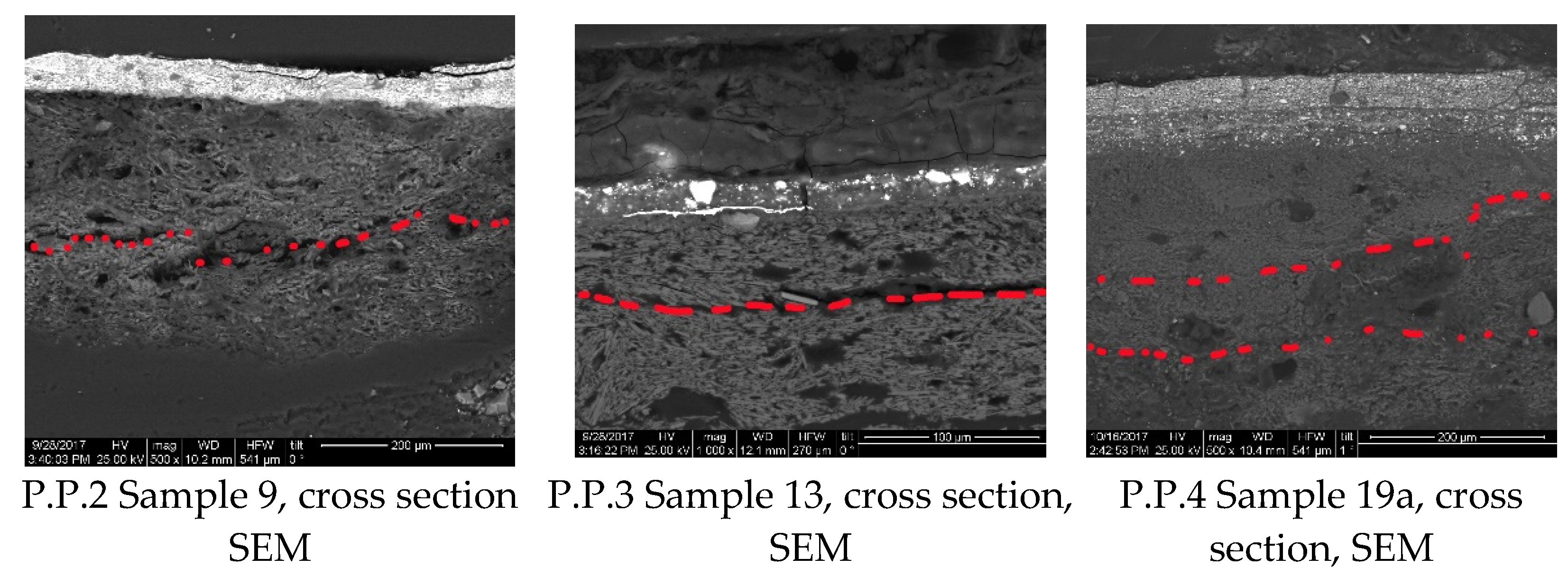
2.2.3. SEM-EDX
2.2.4. FTIR (or Infrared Spectroscopy)
3. Results
3.1. Investigation Scheme
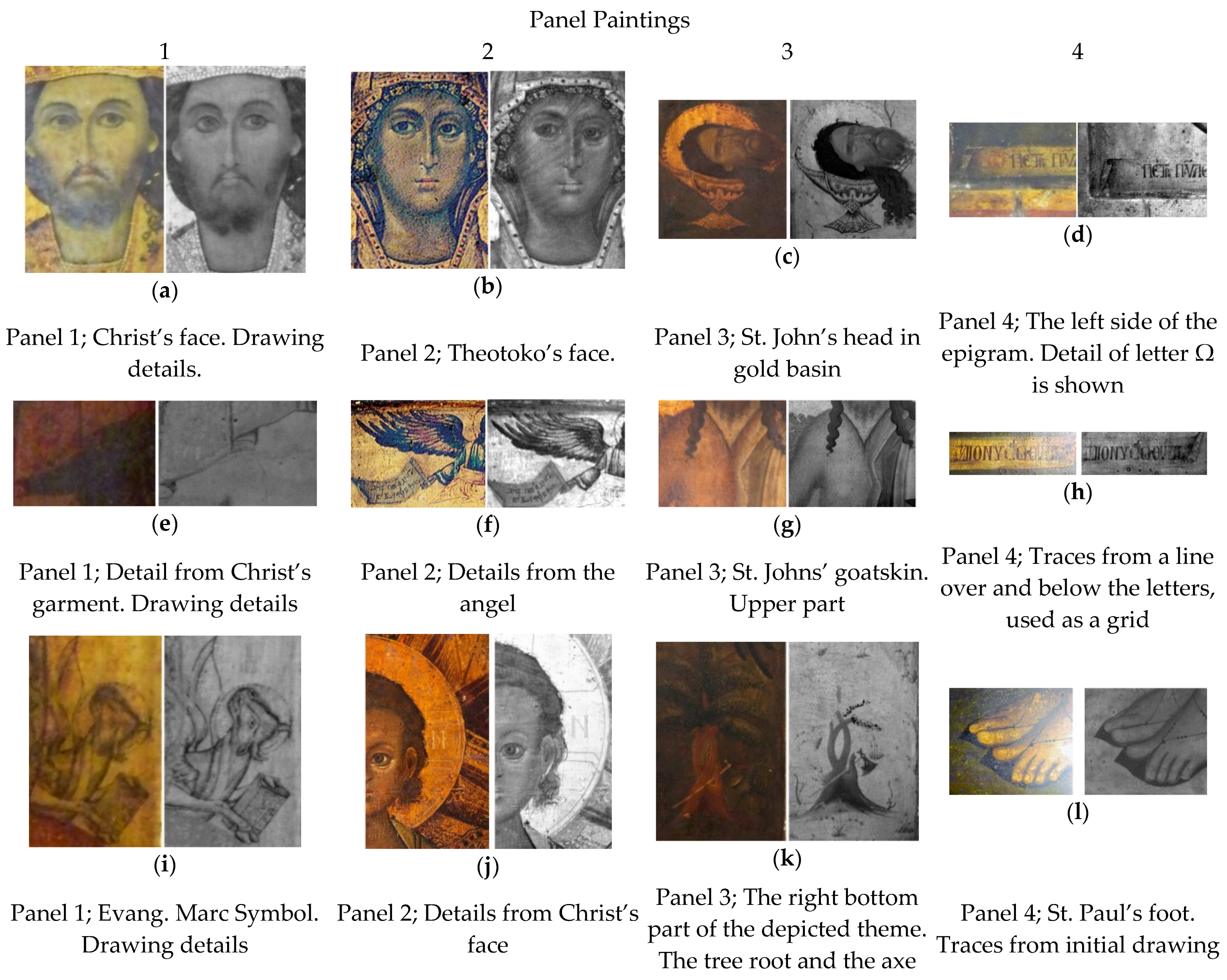
3.2. Underdrawings
3.3. Preparation Layers
3.4. Pigments, Gilding and Binders
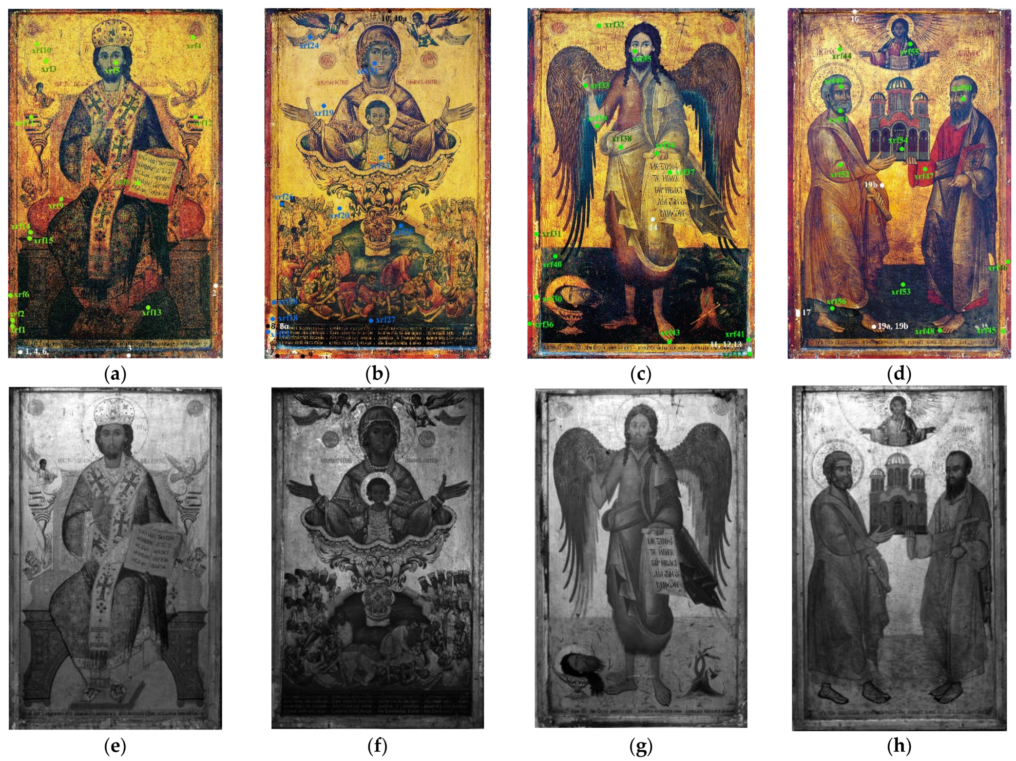
3.4.1. Panel Painting 1: Christ as King of the Kings and Great High Priest
3.4.2. Panel Painting 2. Zoodochos Pigi—The Phaneromeni
3.4.3. Panel Painting 3. Saint John the Baptist—The Forerunner
3.4.4. Panel Painting 4. The Apostles Peter and Paul
3.5. Organic Protective Coating
4. Discussion
5. Conclusions
Author Contributions
Funding
Institutional Review Board Statement
Informed Consent Statement
Data Availability Statement
Acknowledgments
Conflicts of Interest
Glossary
| Bole | a generic term used for a velvety-smooth reddish earth composed of clay and red iron oxide (Fe2O3). It was used in gilding to temper the color and size. |
| Forerunner (or Prodromos) | refers to St. John the Baptist, as the person that precedes the coming of Christ. |
| Hermeneia | Greek word meaning “interpretation”. |
| Phaneromeni | refers to religious icons (often of Virgin Mary) miraculously appeared. |
| Proplasmos | skin shown in painting. |
| Theotokos | Greek word meaning the “mother of God”. |
| Zoodochos Pigi | Greek word meaning the Life-giving fountain, referring to Virgin Mary. |
| Abbreviations | |
| DM | Digital Microscopy. |
| EDX | Energy Dispersive X-ray analysis. |
| FTIR | Fourier Transform Infra-Red spectroscopy. |
| GC/MS | Gas Chromatography-Mass Spectrometry. |
| OM | Optical Microscopy. |
| PP | Panel Painting. |
| pXRF | portable X-ray Fluorescence. |
| SEM | Scanning Electron Microscopy. |
References
- Kakavas, G. Dionysios of Fourna (c. 1670–c. 1745): Artistic Creation and Literary Description, 1st ed.; Alexandros Press: Leiden, The Netherlands, 2008. [Google Scholar]
- Moutafov, E. Post-Byzantine Hermeneia zographikes in the eighteenth century and their dissemination in the Balkans during the nineteenth century. Byz. Mod. Greek Stud. 2006, 30, 69–79. [Google Scholar] [CrossRef]
- Papadopoulos-Kerameas, A. Dionysius of Fourna, Hermeneia of the Painting Art and its Main Unpublished Sources; Russian Imperial Archaeological Society: St Petersbourg, Russia, 1909. [Google Scholar]
- Vassilaki, M. Aκολουθώντας τα βήματα του Διονυσίου. Δελτίο Της Χριστιανικής Και Aρχαιολογικής Εταιρείας 2012, 33, 379–386. [Google Scholar]
- Daniilia, S.; Bikiaris, D.; Burgio, L.; Gavala, P.; Clark, R.J.H.; Chryssoulakis, Y. An Extensive Non-Destructive and Micro-Spectroscopic Study of Two Post-Byzantine Overpainted Icons of the 16th Century. J. Raman Spectrosc. 2002, 33, 807–814. [Google Scholar] [CrossRef]
- Diamantopoulos, A.C. Dionysius of Fourna and the practice of Icon painting: A comparative study of his technical treatise and the painting of the Oberlin Icon, St Gregory the Theologian and St. Artemios. Bull. Allen Meml. Art Mus. 1993, 46, 17–36. [Google Scholar]
- Oltrogge, D. Byzantine recipes and book illumination. Rev. Hist. Arte 2011, 1, 59–61. [Google Scholar]
- Mastrotheodoros, G.; Beltsios, K. Sound Practice and Practical Conservation Recipes as Described in Greek Post-Byzantine Painters’ Manuals. Stud. Conserv. 2018, 64, 42–53. [Google Scholar] [CrossRef]
- Mastrotheodoros, G.; Beltsios, K.; Bassiakos, Y. On the blue and green pigments of post-byzantine greek icons. Archaeometry 2020, 62, 774–795. [Google Scholar] [CrossRef]
- Ferens, M.J. Dionysius of Fourna: Artistic Identity Through Visual Rhetoric. Master’s Thesis, University of California, Riverside, CA, USA, June 2015. [Google Scholar]
- Homar, A.R. Analisis, Contexto y Relaciones entre el Manual de Pintura de Dionisio de Fourna y la Pintura Iconografica y Mural de la Peninsula del Monte Athos. Ph.D. Thesis, Universidad de Murcia, Murcia, Spain, 2016. [Google Scholar]
- Westlake, P.; Siozos, P.; Philippidis, A.; Apostolaki, C.; Derham, B.; Terlixi, A.; Perdikatsis, V.; Jones, R.; Anglos, D. Studying Pigments on Painted Plaster in Minoan, Roman and Early Byzantine Crete. A Multi-Analytical Technique Approach. Anal. Bioanal. Chem. 2012, 402, 1413–1432. [Google Scholar] [CrossRef]
- Martin, E. Some Improvements in Techniques of Analysis of Paint Media. Stud. Conserv. 1977, 22, 63–67. [Google Scholar] [CrossRef]
- Terlixi, A.V.; Doulgeridis, M.; Ioakimoglou, E. Staining and Fluorescent Staining Techniques for the Characterization of Binding Media within Paint Cross-Sections. Examination of Post-Byzantine Icons from the National Gallery of Athens—Alexandros Soutzos Museum’s Collection as a Case Study. In Proceedings of the International Meeting. Icons: Approaches to Research, Conservation and Ethical Issues, Athens, Greece, 3–7 December 2006. [Google Scholar]
- Mastrotheodoros, G.P.; Beltsios, K.G.; Bassiakos, Y.; Papadopoulou, V. On The Grounds of Post-Byzantine Greek Icons. Archaeometry 2016, 58, 830–847. [Google Scholar] [CrossRef]
- Derrick, M.R.; Stulik, D.; Landry, J.M. Scientific Tools. Infrared Spectroscopy in Conservation Science; Getty Conservation Institute: Los Angeles, CA, USA, 1999. [Google Scholar]
- Derrick, M.; Souza, L.; Kieslich, T.; Florsheim, H.; Stulik, D. Embedding paint cross-section samples in polyester resins: Problems and solutions. J. Am. Inst. Conserv. 1994, 33, 227–245. [Google Scholar] [CrossRef]
- Mazzeo, R.; Prati, S.; Sandu, I. Optical Microscopy. In Scientific Examination for the Investigation of Paintings. A handbook for Conservators-Restorers; Pinna, D., Galleotti, M., Mazzeo, R., Eds.; Centro Di: Florence, Italy, 2009; pp. 179–183. [Google Scholar]
- Melo, H.P.; Cruz, A.J.; Candeias, A.; Mirao, J.; Cardoso, A.M.; Oliveira, M.J.; Valadas, S. Problems of analysis by FTIR of calcium sulphate based preparatory layers: The case of group of 16th century Portuguese paintings. Archaeometry 2014, 56, 513–526. [Google Scholar] [CrossRef]
- Ciferri, O. Microbial degradation of paintings. Appl. Environ. Microbiol. 1999, 65, 879–885. [Google Scholar] [CrossRef] [PubMed] [Green Version]
- Eastaugh, N.; Walsh, V.; Chaplin, T.; Siddal, R. Pigment Compendium. A Dictionary and Optical Microscopy of Historical Pigments; Butterworth-Heinemann: Oxford, UK, 2008. [Google Scholar]
- Poli, T.; Chiantore, O.; Diana, E.; Riccirillo, A. Drying Oil and Natural Varnishes in Paintings: A Competition in the Metal Soap Formation. Coatings 2021, 11, 171. [Google Scholar] [CrossRef]
- Hermans, J.J.; Keune, K.; Van Loon, A.; Iedema, P.D. Toward a Complete Molecular Model for the Formation of Metal Soaps in Oil Paints. In Metal Soaps in Art; Springer: Cham, Switzerland, 2019; pp. 47–67. [Google Scholar]
- Hermans, J.J. Metal Soaps in Oil Paint: Structure, Mechanisms and Dynamics; University of Amsterdam: Amsterdam, The Netherlands, 2017. [Google Scholar]
- Otero, V.; Sanches, D.; Montagner, C.; Vilarigues, M.; Carlyle, L.; Lopes, A.J.; Melo, J.M. Characterization of metal carboxylates by Raman and infrared spectroscopy in works of art. J. Raman Spectrosc. 2014, 45, 1197–1206. [Google Scholar] [CrossRef]
- Riaz, T.; Zeeshan, R.; Zarif, F.; Ilyas, K.; Muhammad, N.; Safi, S.Z.; Rahim, A.; Rizvi, S.; Rehman, I.U. FTIR analysis of natural and synthetic collagen. Appl. Spectrosc. Rev. 2018, 53, 703–746. [Google Scholar] [CrossRef]
- De Campos Vidal, B.; Mello, M.L.S. Collagen type I amide I band infrared spectroscopy. Micron 2011, 42, 283–289. [Google Scholar] [CrossRef]
- Payne, K.J.; Veis, A. Fourier transform IR spectroscopy of collagen and gelatin solutions: Deconvolution of the amide I band for conformational studies. Biopolymers 1988, 27, 1749–1760. [Google Scholar] [CrossRef]
- Feller, R.L. Artists’ Pigments. A Handbook of Their History and Characteristics; National Gallery of Art: Washington, DC, USA, 1986. [Google Scholar]
- Χατζηδάκη, Ν.; Phillipon, J.; Ausset, P.; Χρυσουλάκης, Ι.; Aλεξοπούλου, A.θ. Συμβολή Των Φυσικοχημικών Μεθόδων Aνάλυσης Στη Μελέτη 13 Εικόνων Του Βυζαντινού Μουσείου. Δελτίο Της Χριστιανικής Και Aρχαιολογικής Εταιρείας 1988, 13, 215–246. [Google Scholar] [CrossRef]
- Fitzhugh, W.E. Artists’ Pigments; National Gallery of Art: Washington, DC, USA, 1997; Volume 3. [Google Scholar]
- Parry, E.J.; Coste, J.H. The Chemistry of Pigments; Scott, Greenwood & Co.: London, UK, 1902. [Google Scholar]
- Abd el Salam, S. A Binding Media: Methods of Identification: Part 1, Theoretical Work. J. Phys. Sci. Appl. 2011, 1, 204–215. [Google Scholar] [CrossRef]
- Azémard, C.; Vieillescazes, C.; Ménager, M. Effect of photodegradation on the identification of natural varnishes by FT-IR spectroscopy. Microchem. J. 2014, 112, 137–149. [Google Scholar] [CrossRef]
- Bruni, S.; Guglielmi, V. Identification of archaeological triterpenic resins by the non-separative techniques FTIR and 13C NMR: The case of Pistacia resin (mastic) in comparison with frankincense. Spectrochim. Acta A Mol. Biomol. Spectrosc. 2014, 121, 613–622. [Google Scholar] [CrossRef]
- Clark, R.J. Applications of Raman Spectroscopy to the Identification and Conservation of Pigments on Art Objects. Handb. Vib. Spectrosc. 2006, 2977–2992. [Google Scholar] [CrossRef]
- Burgio, L.; Clark, R.J. Library of FT-Raman spectra of pigments, minerals, pigment media and varnishes, and supplement to existing library of Raman spectra of pigments with visible excitation. Spectrochim. Acta A Mol. Biomol. Spectrosc. 2001, 57, 1491–1521. [Google Scholar] [CrossRef]
- Burgio, L.; Clark, R.J.; Theodoraki, K. Raman microscopy of Greek icons: Identification of unusual pigments Spectrohim. Acta A Mol. Biomol. Spectrosc. 2003, 59, 2371–2389. [Google Scholar] [CrossRef]
- Bersani, D.; Lottici, P.P. Raman spectroscopy of minerals and mineral pigments in archaeometry. J. Raman Spectrosc. 2016, 47, 499–530. [Google Scholar] [CrossRef]
- Gettens, R.J.; Stout, G.L. Painting Materials. A Short Encyclopaedia, 3rd ed.; Dover Publications: New York, NY, USA, 1966. [Google Scholar]


| Panel Paintings | |||
|---|---|---|---|
| 1 | 2 | 3 | 4 |
| XRF 1, gold | XRF 18, gold | XRF 30, gold | XRF 44, gold |
| XRF 2, gold | XRF 19, gold | XRF 31, gold | XRF 45, gold |
| XRF 3, gold | XRF 20, gold | XRF 32, gold | XRF 46, red |
| XRF 4, gold | XRF 21, flesh | XRF 33, hand | XRF 47, red |
| XRF 5, flesh | XRF 22, red | XRF 34, hand | XRF 48, black |
| XRF 6, red | XRF 23, white | XRF 35, flesh | XRF 49, flesh |
| XRF 8, white | XRF 24, white | XRF 36, red | XRF 50, flesh |
| XRF 9, red | XRF 25, green | XRF 37, white | XRF 51, grey |
| XRF 10, gold | XRF 26, black | XRF 38, white | XRF 52, white |
| XRF 11, black | XRF 27, brown | XRF 39, green | XRF 53, green |
| XRF 12, black | XRF 28, ground | XRF 40, green | XRF 54, black |
| XRF 13, background | XRF 29, ground | XRF 41, ground | XRF 55, green |
| XRF 14, black | XRF 42, ground | XRF 56, green | |
| XRF 15, brown | XRF 43, black | ||
| XRF 16, ground | |||
| XRF 17, ground | |||
| Microsamples | |||||||
|---|---|---|---|---|---|---|---|
| PP1 | PP2 | PP3 | PP4 | ||||
| # Sample | Sample type | # Sample | Sample type | # Sample | Sample type | # Sample | Sample type |
| 1 | preparation powder | 7 | preparation powder | 11 | cross section | 16 | preparation powder |
| 2 | varnish powder | 8 | pigment powder | 12 | preparation powder | 17 | varnish powder |
| 3 | cross section | 8a | varnish (removed with acetone-loaded cotton swap) | 13 | cross section | 19a | cross section |
| 4 | cross section | 9 | cross section | 14 | varnish powder | 19b | cross section |
| 6 | cross section | 10 | cross section | ||||
| 10a | varnish powder | ||||||
| Layer/Area | Panel Paintings | |||
|---|---|---|---|---|
| 1 | 2 | 3 | 4 | |
| Surface analysis (pXRF) 1 | ||||
| Preparation layer | Spot 16, 17 | Spot 28, 29 | Spot 41, 42 | Spot 44, 45 |
| Ca, S, Pb | Ca, S, Pb | Ca, S | Ca, S | |
| Gold background | Spot 1–4, 10 | Spot 18-20 | Spot 30–32 | Spot 44, 45 |
| Au, Ca, Fe, Pb, Cu, Hg (occasionally) | Au, Ca, Pb, Fe, Cu, Hg (occasionally) | Au, Ca, Fe, Pb | Au, Hg, Ca, Fe, Pb, Cu | |
| Red area | Spot 6, 9 | Spot 21, 22 | Spot 36 | Spot 46 |
| Pb, Hg, Fe | Pb, Hg | Pb, Hg, S | Pb, Hg | |
| White pages from books | Spot 8 | Spot 23, 24 | Spot 37 | |
| Pb | Pb | Pb | ||
| Flesh | Spot 5 | Spot 21 | Spot 34–36, 39 | Spot 49–51 |
| Pb, Fe | Pb, Hg, Fe | Pb, Hg, Fe | Pb, Cu, Fe | |
| Brown area | Spot 15 | Spot 26, 27 | Spot 48, 54 | |
| Pb, Cu | Pb, Cu, Fe, Mn | Pb, Hg, Fe, Cu | ||
| Green area | Spot 13 | Spot 40, 43 | Spot 53, 55–56 | |
| Pb, As, Fe, Cu | Pb, As, Fe, Cu | Cu, As, Pb, Fe | ||
| Blue area | Spot 25 | |||
| Pb, Cu, Fe | ||||
| White garment | Spot 38 | Spot 52 | ||
| Pb | Pb, Fe | |||
| Micro-analysis of cross-section (SEM/EDX) | ||||
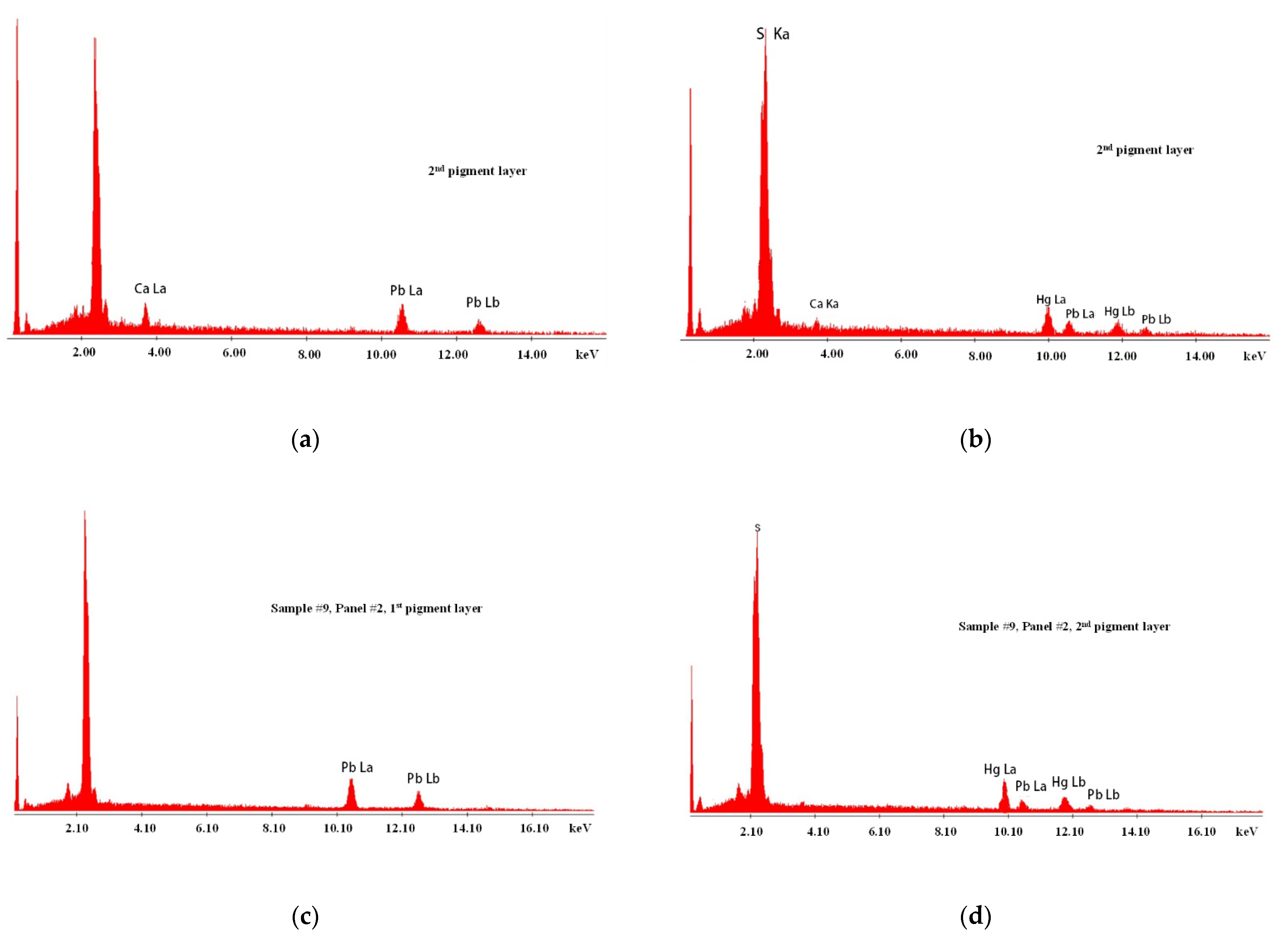
| Preparation | Ca, S | Ca, S | Ca, S | Ca, S |
| Bole layer | Ca, S, Al, Si, Mg, Fe | Ca, S, Al, Si, Mg, Fe | Ca, S, Al, Si, Mg, Fe | |
| Red pigment | Hg, S, Pb | Hg, S, Pb | Hg, S, Pb | |
| Flesh | Ca, Al, Si, Mg, Pb, Fe, Cu | |||
| Material | Vibration 1 | Varnish Samples 1 | Notes | |||
|---|---|---|---|---|---|---|
| PP1 | PP2 | PP3 | PP4 | |||
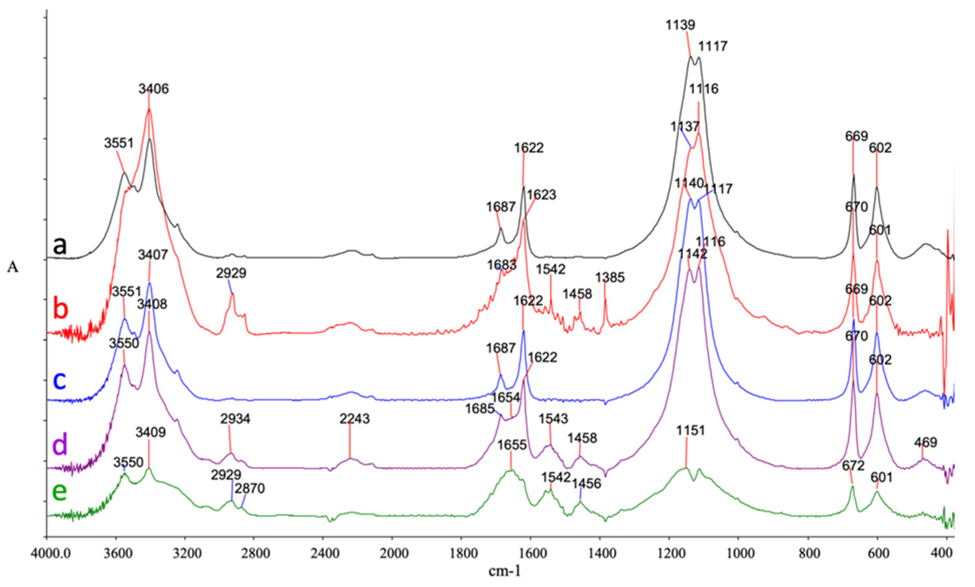
| Gypsum | νO‒H | 3551 3402 | 3410 | 3551 3496 3407 | 3550 3408 | Preparation layers Crystalline water |
| δO‒H | 1687, 1622 | 1685, 1627 | 1687, 1622 | 1688, (1655) 1622 | ||
| νS‒O | 1139 1115 | 1116 | 1140 1116 | 1144 1116 | Preparation layers. Sulfate peaks | |
| δS‒O | 669, 602 | 670, 601 | 669, 602 | 670, 602 | ||
| Nitrates | νN‒O | 1385 | Preparation layers. Biogenically formed | |||
| Proteinaceous material | Amide I (vC = O) | 1655 | 1655 | Preparation layers. Found as add-on in gypsum | ||
| Amide II (vC = O + vC‒N) | 1542 | 1542 | ||||
| δCH2 | 1458 | 1456 | ||||
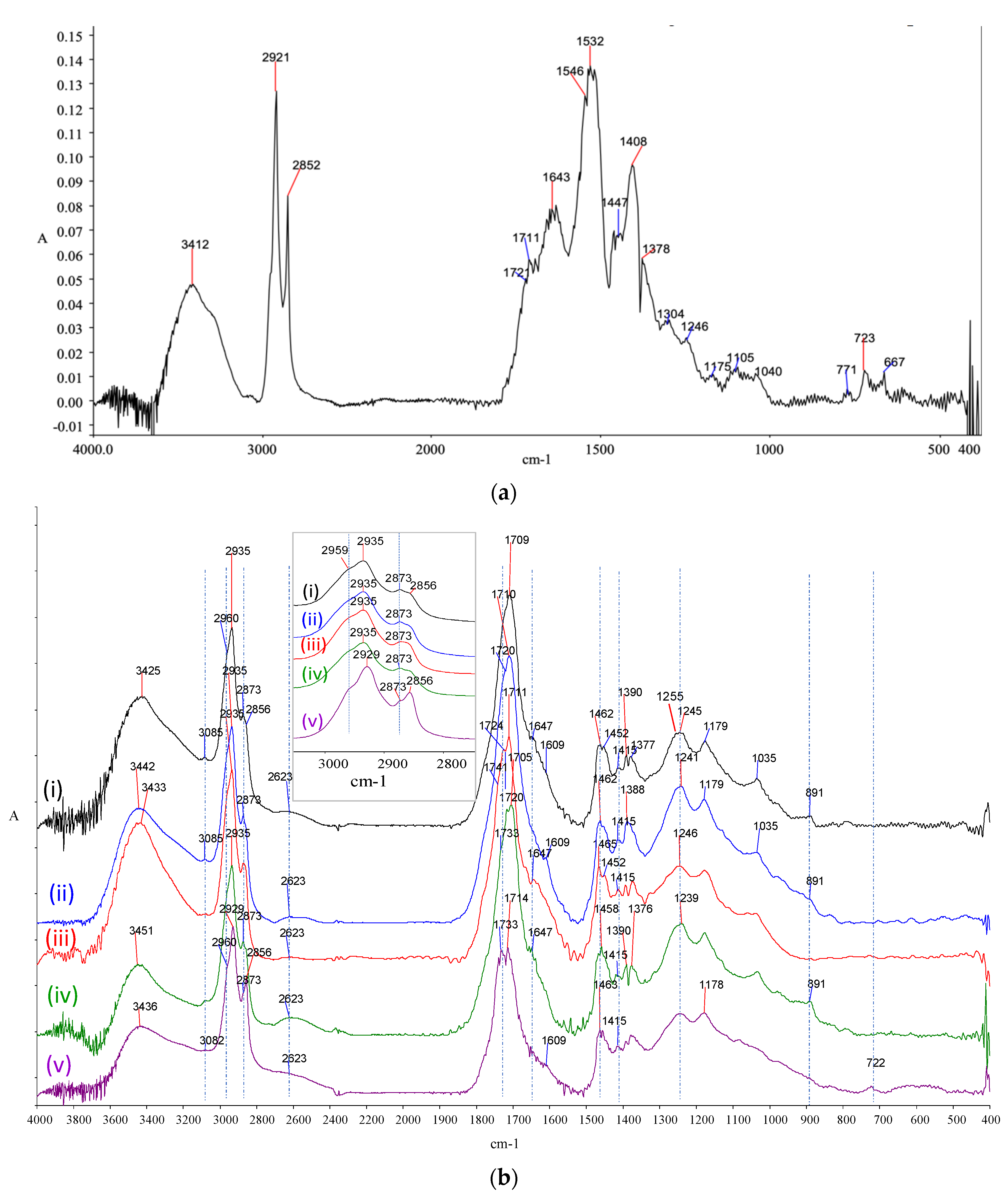
| Diterpenic resin | νO‒H | 3425 | 3442 | 3433 | 3451 | Varnish layers |
| ν C‒H | 30762935 2873 | 30762935 2873 | 30762935 2873 | 307429352873 | ||
| νC=O | 1709 1644 | 1720 (sh) 1710 1641 | 1721(sh) 1711 1644 | 1716(sh) 1706 1644 | ||
| δC‒H | 1462 1451 1391 | 1462 | 1462 1451 1391 | 145813921377 | ||
| δipC‒O‒H | 1413 | 1413 | 1413 | 1415 | ||
| νC‒O | 1255 | 1250–1241 | 1246 1178 | 1239 1178 | ||
| δoopC‒O‒H | 890 | 890 | ||||
| Triterpenic resin | O‒H | 3425 | 3425 | Varnish layers | ||
| νC‒H | 2959 (sh) 2874 | 2959 (sh) 2874 | ||||
| νC=O | 1720 | 1716 | ||||
| δC‒H | 1462 1384 | 1458 1376 | ||||
| νC‒O | 1241 | 1239 | ||||
| Oil binder | νC‒H | 2926 2855 | Paint layers | |||
| νC=O | 1733 | |||||
| δC‒H | 1463 1377 | |||||
| νC‒O | 1250 1181 | |||||
| ρCH2 | 721 | |||||
| Carboxylates | νC‒H | 2925 2859 | 2932 | Paint layers | ||
| δC‒H | 1458 | 1461 | ||||
| νCOO‒ | 1542 | 1547 | ||||
| Materials | PP 1 | PP 2 | PP 3 | PP 4 |
|---|---|---|---|---|
| Gesso layer | ||||
| Calcium sulfate (CaSO4) | Dihydrate | Hydrate | Dihydrate | Dihydrate |
| Bole Layer | ||||
| 2nd or 3rd recipe | √ | √ | √ | √ |
| Gold layer | ||||
| Gold leaf, Au with Cu impurities | √ | √ | √ | √ |
| Pigments | ||||
| White lead [2PbCO3·Pb(OH)2] | √ | √ | √ | √ |
| Orpiment [As2S3] | √ | √ | √ | |
| Cinnabar [HgS] | √ | √ | √ | √ |
| Red lead [Pb3O4] | √ | √ | √ | √ |
| Red Ochre [Fe2O3+SiO2Al2O3+SiO2] | √ | √ | √ | |
| Umber [Fe2O3+Al2O3SiO2+MnO2(8-16%] | √ | |||
| Hematite [Fe2O3] | √ | √ | √ | |
| Verdigris [Cu(CH3COO)2·2Cu(OH)2] | √ | √ | √ | |
| Terra Verde [Fe2+/Fe3+ and Al, Si, Mg, and K] | √ | |||
| Azurite [CuCO3·Cu(OH)2] | √ | √ | √ | √ |
| Binding media | ||||
| Proteinaceous materials | √ | √ | √ | √ |
| Lipids | √ | |||
| Varnish | ||||
| Mastic | √ | √ | √ | √ |
| Sandalwood | √ | √ | √ | √ |
| Oil | √ | |||
| Recipes for Bole | |
|---|---|
| 1st recipe | Bole (=clay) pale red, ochre, red lead, wax, burned paper, mercury |
| 2nd recipe | Bole (=clay) pale red, ochre, soap, egg white |
| 3rd recipe | Bole (=clay) pale red, ochre, red lead, cinnabar, egg white, gal, wax, mercury |
Publisher’s Note: MDPI stays neutral with regard to jurisdictional claims in published maps and institutional affiliations. |
© 2021 by the authors. Licensee MDPI, Basel, Switzerland. This article is an open access article distributed under the terms and conditions of the Creative Commons Attribution (CC BY) license (https://creativecommons.org/licenses/by/4.0/).
Share and Cite
Mafredas, T.; Kouloumpi, E.; Boyatzis, S.C. Did Dionysius of Fourna Follow the Material Recipes Described in His Own Treatise? A First Analytical Investigation of Four of His Panel Paintings. Heritage 2021, 4, 3770-3789. https://0-doi-org.brum.beds.ac.uk/10.3390/heritage4040207
Mafredas T, Kouloumpi E, Boyatzis SC. Did Dionysius of Fourna Follow the Material Recipes Described in His Own Treatise? A First Analytical Investigation of Four of His Panel Paintings. Heritage. 2021; 4(4):3770-3789. https://0-doi-org.brum.beds.ac.uk/10.3390/heritage4040207
Chicago/Turabian StyleMafredas, Thomas, Eleni Kouloumpi, and Stamatis C. Boyatzis. 2021. "Did Dionysius of Fourna Follow the Material Recipes Described in His Own Treatise? A First Analytical Investigation of Four of His Panel Paintings" Heritage 4, no. 4: 3770-3789. https://0-doi-org.brum.beds.ac.uk/10.3390/heritage4040207