Article

Silver Ions Incorporation into Nanofibers for Enhanced hMSC Viability

by
Natalya A. Sitnikova
1,
Anastasiya O. Solovieva
1,*,
Elizaveta S. Permyakova
1,2,
Alexander N. Sheveyko
2,
Dmitry V. Shtansky
2 and
Anton M. Manakhov
1,*
1
Research Institute of Clinical and Experimental Lymphology—Branch of the ICG SB RAS, 2 Timakova St., 630060 Novosibirsk, Russia
2
National University of Science and Technology “MISiS”, Leninsky Prospekt 4, 117198 Moscow, Russia
*
Authors to whom correspondence should be addressed.
Submission received: 8 August 2022 / Revised: 25 August 2022 / Accepted: 27 August 2022 / Published: 29 August 2022
(This article belongs to the Special Issue Recent Advances in Antimicrobial Materials)

Abstract

:
Antimicrobial properties of silver have been known for a long time, but there is also cytotoxicity of high concentrations of silver. Therefore, it is important to select the concentration and shape of silver depending on the goals. The ideal wound dressing should ensure that the wound remains optimally moist, protected from infections, has no toxic compounds, and stimulates regeneration. In the present work, we obtained a series of polycaprolactone-based nanomaterials fabricated by electrospinning and incorporated with silver ions (up to 0.6 at.%). By adjusting the magnetron current (0.3 A) and implanter voltage (5 kV), the deposition of TiO2 and Ag+ implantation into PCL/PEO nanofibers was optimized to achieve implantation of Ag+ without damaging the nanofibrous structure of the biodegradable nanofibers. The obtained results allow us to predict significant protection properties of the developed material not only from mechanical influence but also thanks to the antimicrobial effect due to silver ions, which is important for chronic wounds and injuries with a large area of damage and can activate host cells proliferation.

1. Introduction

Currently, wound healing materials with protective and regenerative properties are in great demand, the development of which is actively being carried out all over the world [1]. To date, active research is underway on regenerative materials with an antibacterial effect. Silver is actively studied as an antibacterial component and is used in various creams, substances and nanomaterials for regenerative medicine [2,3,4,5]. In addition to the antibacterial effect, silver has a regenerative effect, provided by stimulating the proliferation of cells involved in regeneration. The antimicrobial properties of silver have been known for a long time, but there is also cytotoxicity of high concentrations of silver [6].
Silver-ion-releasing polylactide (PLA) nanofibrous scaffolds were developed using a silver-ion-releasing solution, Silvadur ET. This solution comprised AgNO3, a polymer binder, water, and ethanol. Nanofibers coated with solutions having different concentrations of silver ranging from 31.25 to 250 µg/mL have exhibited excellent antimicrobial properties toward both Gram-positive and Gram-negative bacteria while having no effect on silver-resistant bacteria [7]. Nanofibrous scaffolds coated with Silvadur ET solution containing silver less than 62.5 µg/mL maintained the viability and proliferation of both human dermal fibroblasts and human epidermal keratinocytes, although some decrease of the cell viability was evident even for lower concentrations [8].
The cytotoxicity effect of silver ions and silver nanoparticles is mainly due to oxidative stress and is independent of silver ions, while the generation of reactive oxygen species (ROS) on the surface of silver nanoparticles may additionally affect the viability of cells. Indeed, free silver ions play a considerable role in the toxicity of silver nanoparticle suspensions [9]. The Ag+ strongly increases the production of reactive oxygen species, including superoxide anion radicals. These effects correspond to a strong decrease in intracellular reduced glutathione and to an increased susceptibility to H2O2-induced cell death [10]. Therefore, it is important to select the concentration and shape of silver depending on the goals. In the very specific range of silver ions concentration (from 5 to 20 µM) a positive effect on the MSC cell viability was previously shown [11]. Various factors may affect the cytotoxicity of silver ions including type of cell media, the presence of chloride ions, glucose, the form of silver, etc. [12]. The implantation of silver ions instead of incorporation of the Ag nanoparticles may reduce the cytotoxicity of produced materials. Recently, nanofibers coated with TiO2 [13,14] and Cu films [15] were prepared by magnetron sputtering deposition onto biodegradable nanofibrous foils. The most critical issue for this process would be the prevention of structural damage to the nanofibrous scaffold during the deposition process [16].
In this work, we obtained a series of polycaprolactone/polyethyleneoxide PCL/PEO-based nanomaterials fabricated by electrospinning, and incorporated with silver ions (up to 0.6 at.%). By adjusting the magnetron current and implanter voltage, the deposition of TiO2 and Ag+ implantation into PCL/PEO nanofibers was optimized to achieve the implantation of Ag+ without damaging the nanofibrous structure of the PCL/PEO. It was shown that the maximum amount of silver was released from the PCL-Ti0.3-Ag-5 kV sample, and in the first hours there was a rapid release, then a smoother one, and for 7 days it continued to be released more slowly. The aim of this work was to determine the effect of the obtained samples on the viability and proliferative potential of mesenchymal stromal cells.

2. Materials and Methods

2.1. Electrospinning of PCL and PCL/PEO Nanofibers

The overall scheme for preparing functionalized nanofibers is depicted in Figure 1. The electrospun PCL nanofibers were prepared by electrospinning a 9 wt.% solution of polycaprolactone PCL (80,000 g/mol). The processing of the sample can be found elsewhere [17]. Briefly, the granulated PCL was dissolved in a mixture of acetic acid (99%) and formic acid (98%) [15]. All compounds were purchased from Sigma Aldrich (Darmstadt, Germany). The weight ratio of acetic acid (AA) to formic acid (FA) was 2:1. The PCL solutions in AA and FA were stirred at 25 °C for 24 h.
According to our methodology reported elsewhere [18], PCL/PEO nanofibers with a ratio 3:1 PEO prepared by electrospinning of the 9 wt.% solutions containing 75% PCL and 25% PEO (Mw = 100,000, Sigma–Aldrich, Steinheim am Albuch, Germany).
The solutions were electrospun with a 20 cm long wired electrode using a Nanospider™ NSLAB 500 machine (ELMARCO, Liberec, Czech Republic). The applied voltage was 50 kV. The distance between the electrodes was set to 100 mm. The as-prepared and non-treated PCL nanofibers are referred to as PCL-ref throughout the text.

2.2. Deposition of TiO2 Coating and Ag Ion Implantation

The deposition experiments were performed using a vacuum set-up with a magnetron sputtering unit equipped with the MEVVA-type ion implanter [19]. The titania films (~20 nm thick) were deposited by magnetron sputtering of a composite TiC-CaO-Ti3POx target in a gaseous mixture of Ar and 15% N2. The applied magnetron current was 0.5 or 0.3 A, the magnetron voltage was ~450 V, and the bias voltage was kept at −50 V (samples denoted as PCL/PEO-Ti). The target to substrate distance was fixed to 120 mm. Silver ions were implanted using a MEVVA-type implanter operating with the acceleration voltage of 15, 8 and 5 kV and the current of 20 mA. Hereafter, these samples are denoted as PCL/PEO-Ti0.5-Ag15 kV for Ti current of 0.5 A and Ag ion implantation voltage of 15 kV; PCL/PEO-Ti0.3-Ag-8 kV and PCL/PEO-Ti0.3-Ag5 kV for Ti current of 0.3 A and voltages of 8 kV and 5 kV, respectively.

2.3. Characterization of Samples

The sample’s morphology was examined by scanning electron microscopy (SEM), which was carried out with a JSMF 7600 microscope (JEOL Ltd., Tokyo, Japan) equipped with an energy-dispersive X-ray spectrometer. To compensate for surface charge, the samples were coated with a ~5 nm thick Pt layer.
The sample chemical characterization was performed by X-ray photoelectron spectroscopy (XPS), energy-dispersive X-ray spectroscopy (EDXS) and Fourier-transformed infrared (FTIR) spectroscopy. The XPS analysis was carried out using a PHI5500VersaProbeII instrument (PHI) equipped with a monochromatic Al Kα X-ray source (hν = 1486.6 eV) at a pass energy of 23.5 eV and X-ray power of 50 W. The data processing was performed using CASAXPS software as reported elsewhere [20,21,22].
The sample wettability was assessed by measuring the water contact angle (WCA). The measurements were carried out on an Easy Drop Kruss (KRṺSS, Hamburg, Germany) device. For each sample, at least five WCA measurements were performed.

2.4. Cell Tests

Human MSCs 4-6 were taken from the culture bank of RICEL—branch ICG SB RAS, which had been extracted from bone marrow using standard methods [23]. Cells were cultured in DMEM/F12 Medium (Sigma Aldrich, Paisley PA4 9RF, UK) that was supplemented with 10% foetal bovine serum (FBS, Gibco, Carlsbad, CA, USA) under standard culture conditions (humidified atmosphere, 5% CO2 and 95% air, at 37 °C).
Nanofibrous samples were cut into pieces with 0.5 diameter and used in set of experiments. The MTT test was conducted to test the cytotoxicity of used Ag concentrations. 5 × 10³ hMSC were seeded in 96-wells plate, Ag-PCL membrane samples were soaked in culture medium for 24 h, then this medium was transferred to cells for incubation for 3 days. Results were measured by optical density by using Multiscan FC spectrophotometer. To test the proliferation rate, human mesenchymal stromal cells were seeded on Ag-PCL membranes 5 × 10³ per sample and incubated for 3 days in standard conditions (95% humidity, 5% CO2). To further evaluate the total amount, cells were dyed with Hoechst reagent to visualize cell nuclei and with EdU Click iT-reagent to count the number of proliferating cells.

2.5. Statistical Analysis

All experimental data were analysed by Statistica 8.0 software with nonparametric Mann–Whitney U test. The results presented as mean ± standard deviation with statistical significance p < 0.05.

3. Results

3.1. Morphology of Nanofibrous Samples

The SEM micrographs of PCL/PEO-Ti-Ag nanofibers are presented in Figure 1. The Ag+ implantation at 15 kV (Figure 1b) led to a significant destruction of nanofibrous material as compared to PCL/PEO-ref. The damage of the PCL nanofibrous structure at the high current and high implanting voltage conditions was caused by the highly energetic ions as well as “nanodroplets” of the silver nanomaterials approaching the surface. At higher current and higher voltage, stronger thermal heating of the substrate (PCL nanofibers) led to significant damage of the nanostructure. The samples prepared at 5 and 8 kV exhibited morphology similar to the pristine PCL/PEO-ref. High-resolution SEM images of PCL/PEO-ref, PCL/PEO-Ti0.3-Ag-5 kV, and PCL/PEO-Ti0.3-Ag-8 kV are depicted in Figure 2. It is evident that the nanofibers are not damaged, but their surface after Ag+ implantation is smoother as compared to PCL/PEO-ref.

3.2. Chemical Characterization of Biodegradable Nanohybrid Materials

The PCL and PCL/PEO polymers are well known and their compositions were reported elsewhere [18]. The modified nanofibers are of much higher interest, and the compositions of all samples evaluated by XPS are summarized in Table 1. The incorporation of TiO2 and Ag in PCL/PEO-Ti-Ag samples was evident from the surface’s overall atomic composition. The analysis of Ti2p and Ag3d (not reported here) revealed that titanium and silver were both the oxidized states: TiO2 and Ag2O, respectively.
The WCA analysis revealed that the unmodified nanofibers exhibited a hydrophobic nature and the implantation of Ag+ ions led to significant decrease of WCA and, thus, the surface of all modified samples was hydrophilic (see Table 1).

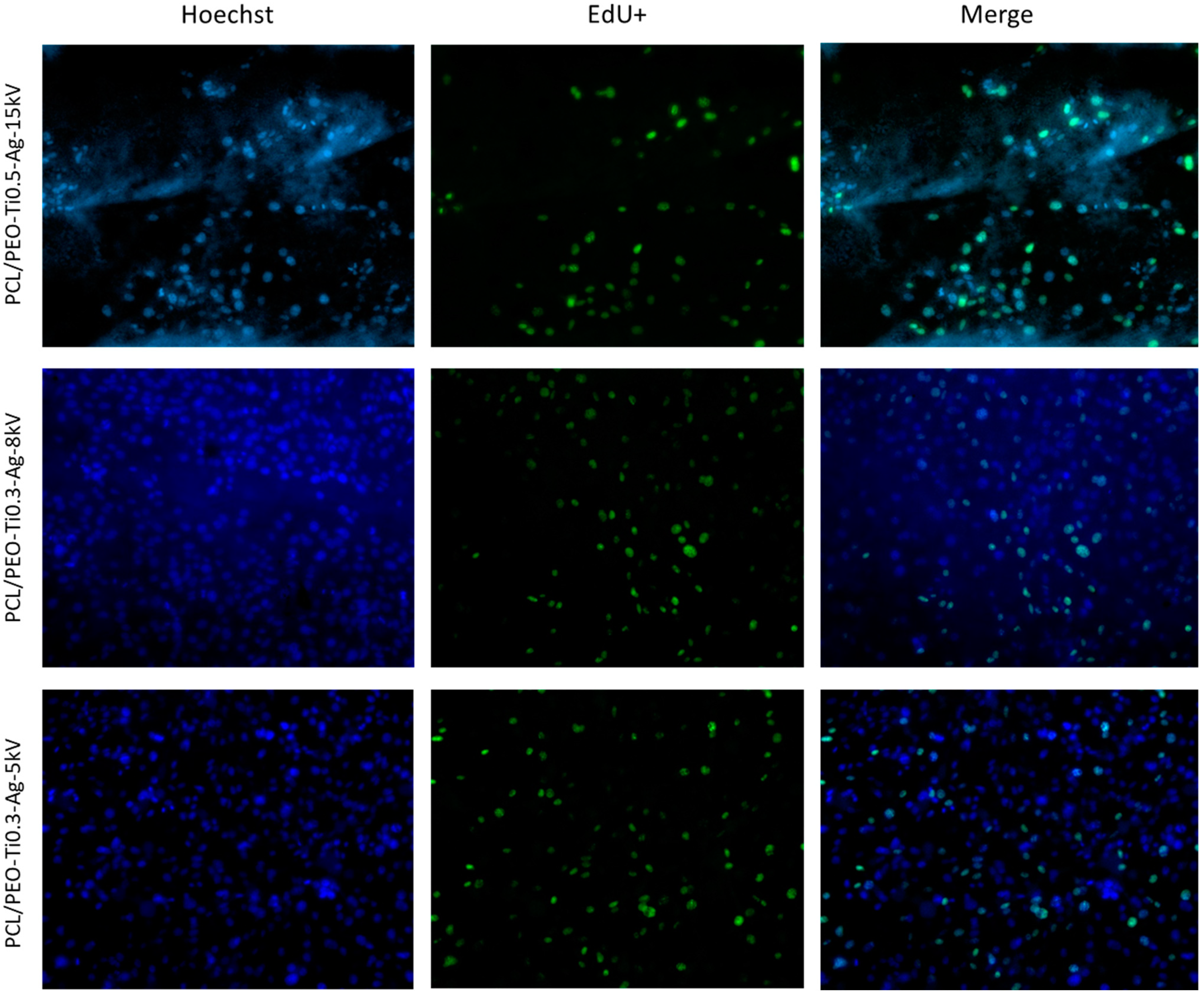
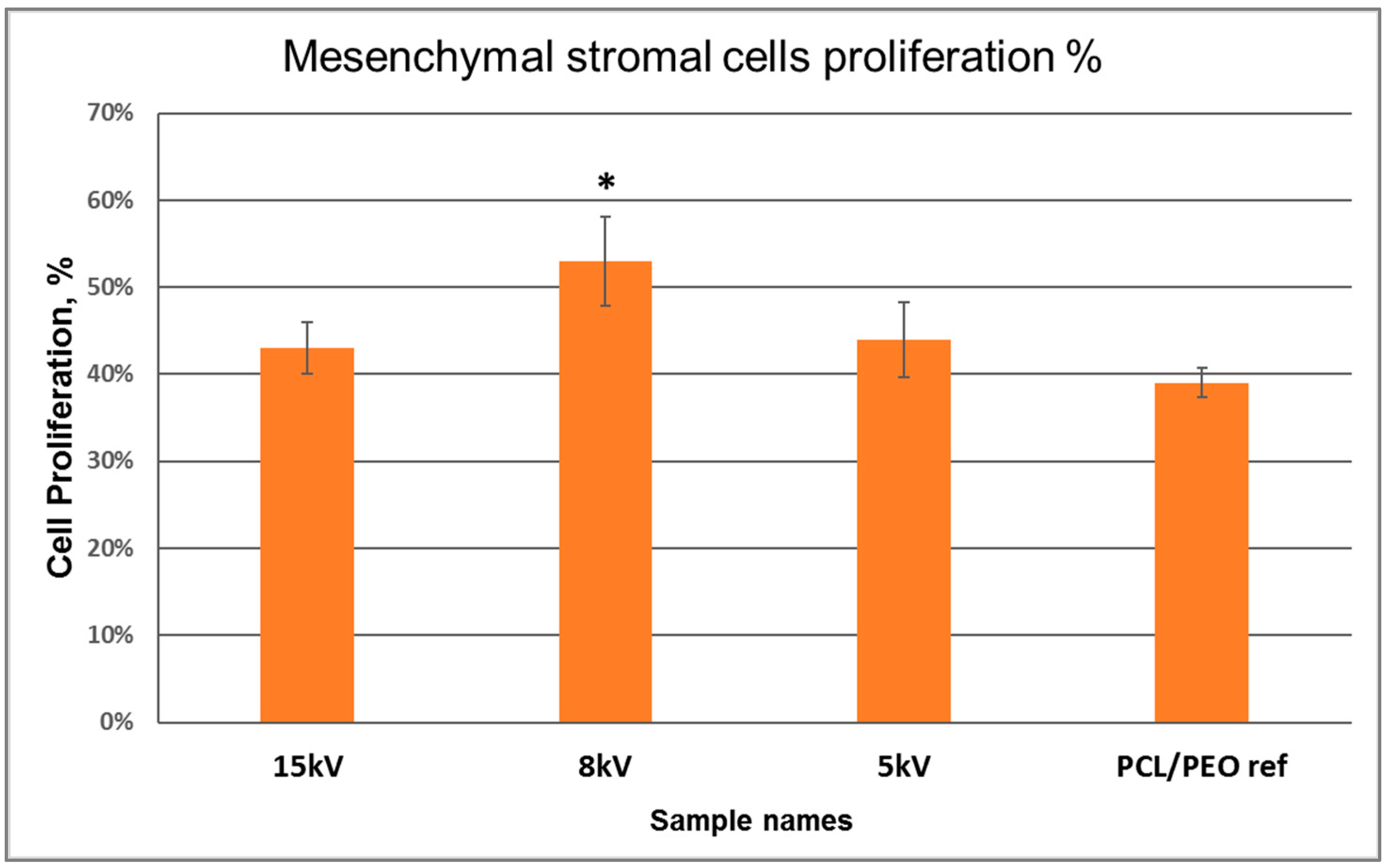
3.3. Analysis of Cell Viability and Proliferation

The cell images are depicted in Figure 3. The first row presents all cell nuclei on the membranes (on PCL/PEO-Ti0.3-Ag-8 kV and PCL/PEO-Ti0.3-Ag-5 kV samples, cells almost create a monolayer). The second row presents proliferating cells that incorporated EdU reagent. Based on the calculations of cells and the EdU+/Dapi ratio, we obtained that all variants of PCL membranes with Ag nanoparticles tend to increase proliferation, especially PCL/PEO-Ti0.3-Ag-8 kV made it significant.
The release of Ag+ ions differed in samples depending on the initial Ag concentrations and was presented in the article [15]. Here, Ag concentrations in all samples had no toxicity on human mesenchymal cells, furthermore, Ag+ ions released in culture medium from PCL/PEO-Ti0.5-Ag-15 kV and PCL/PEO-Ti0.3-Ag-5 kV resulted in an increased percentage of living cells compared to the control (Figure 4). The total cell count was significantly higher in PCL/PEO-Ti0.3-Ag-8 kV samples in comparison to PCL/PEO-ref membranes. However, the proliferation rate was higher in all samples in comparison to PCL/PEO-ref (Figure 5).
The Ag modification of PCL/PEO nanofibers (PCL/PEO-Ti0.3-Ag-8 kV) with a 0.3 A current for TiO2 deposition and Ag+ implantation at 8kV induces proliferation of human mesenchymal cells. Besides that, fast Ag+ ion release from these nanofibers in the first 24 h could have possibly reduced bacterial activity in wound dressings but is low enough for a longer period of time and did not cause cytotoxicity. The obtained results allow us to expect that the material can be applied for tissue engineering as it can provide effective protection not only from mechanical influence but also has an antimicrobial effect because of the Ag [5,15], which is important for chronic wounds and injuries with a large area of damage, and can activate host cell proliferation.

4. Conclusions

In this work, a series of polycaprolactone-based nanomaterials were fabricated by electrospinning and modified by deposition of a TiO2 layer and implantation of silver ions (up to 0.6 at.%). By adjusting the magnetron current (0.3 A) and implanter voltage (5 kV), the deposition of TiO2 and Ag+ implantation into the PCL/PEO nanofibers were optimized to achieve significant implantation of Ag+ without damaging the nanofibrous structure of the biodegradable nanofibers. The nanofibers with the implanted silver ions exhibited enhanced cell viability and proliferation. The obtained results allow us to predict significant protection properties for the developed material not only from mechanical influence but also thanks to an antimicrobial effect due to silver ions, which is important for chronic wounds and injuries with a large area of damage and can activate host cell proliferation.

Author Contributions

Conceptualization, A.M.M. and A.O.S.; methodology, N.A.S. and A.N.S.; investigation, N.A.S. and E.S.P.; writing—original draft preparation, A.M.M. and N.A.S.; writing—review and editing, D.V.S. All authors have read and agreed to the published version of the manuscript.

Funding

The reported study was funded by Russian Foundation for Basic Research RFBR (project number 20-52-26020) Part of this work. (SEM analyses and TiO2 deposition) was carried out during the implementation of the strategic project: “Biomedical materials and bioengineering”, within the framework of the Strategic Academic Leadership Program “Priority 2030” at NUST «MISiS».

Institutional Review Board Statement

The study was approved by the Ethics Committee of the RICEL-branch of ICG SB RAS (No 115 from 24 December 2015).

Informed Consent Statement

Not applicable.

Data Availability Statement

Data are available from the corresponding author upon a reasonable request.

Acknowledgments

The work was performed using the equipment of the Center for Collective Use «Proteomic Analysis» FRC FTM, supported by funding from the Ministry of Science and Higher Education of the Russian Federation (agreement No. 075-15-2021-691).

Conflicts of Interest

The authors declare no conflict of interest.

References

  1. Norouzi, M.; Boroujeni, S.M.; Omidvarkordshouli, N.; Soleimani, M. Advances in Skin Regeneration: Application of Electrospun Scaffolds. Adv. Healthc. Mater. 2015, 4, 1114–1133. [Google Scholar] [CrossRef]
  2. Gallagher, L.; Smith, A.; Kavanagh, K.; Devereux, M.; Colleran, J.; Breslin, C.; Richards, K.; McCann, M.; Rooney, A. Preparation and Antimicrobial Properties of Alginate and Serum Albumin/Glutaraldehyde Hydrogels Impregnated with Silver(I) Ions. Chemistry 2021, 3, 672–686. [Google Scholar] [CrossRef]
  3. Zimet, P.; Valadez, R.; Raffaelli, S.; Estevez, M.B.; Pardo, H.; Alborés, S. Biogenic Silver Nanoparticles Conjugated with Nisin: Improving the Antimicrobial and Antibiofilm Properties of Nanomaterials. Chemistry 2021, 3, 1271–1285. [Google Scholar] [CrossRef]
  4. Thomas, R.; Soumya, K.R.; Mathew, J.; Radhakrishnan, E.K. Electrospun Polycaprolactone Membrane Incorporated with Biosynthesized Silver Nanoparticles as Effective Wound Dressing Material. Appl. Biochem. Biotechnol. 2015, 176, 2213–2224. [Google Scholar] [CrossRef]
  5. Dubey, P.; Bhushan, B.; Sachdev, A.; Matai, I.; Uday Kumar, S.; Gopinath, P. Silver-nanoparticle-Incorporated composite nanofibers for potential wound-dressing applications. J. Appl. Polym. Sci. 2015, 132. [Google Scholar] [CrossRef]
  6. Madhavan, R.V.; Rosemary, M.J.; Nandkumar, M.A.; Krishnan, K.V.; Krishnan, L.K. Silver Nanoparticle Impregnated Poly (ε-Caprolactone) Scaffolds: Optimization of Antimicrobial and Noncytotoxic Concentrations. Tissue Eng. Part A 2011, 17, 439–449. [Google Scholar] [CrossRef]
  7. Mohiti-Asli, M.; Pourdeyhimi, B.; Loboa, E.G. Novel, silver-ion-releasing nanofibrous scaffolds exhibit excellent antibacterial efficacy without the use of silver nanoparticles. Acta Biomater. 2014, 10, 2096–2104. [Google Scholar] [CrossRef]
  8. Zhang, X.F.; Shen, W.; Gurunathan, S. Silver nanoparticle-mediated cellular responses in various cell lines: An in vitro model. Int. J. Mol. Sci. 2016, 17, 1603. [Google Scholar] [CrossRef]
  9. Beer, C.; Foldbjerg, R.; Hayashi, Y.; Sutherland, D.S.; Autrup, H. Toxicity of silver nanoparticles-Nanoparticle or silver ion? Toxicol. Lett. 2012, 208, 286–292. [Google Scholar] [CrossRef]
  10. Cortese-Krott, M.M.; Münchow, M.; Pirev, E.; Heßner, F.; Bozkurt, A.; Uciechowski, P.; Pallua, N.; Kröncke, K.D.; Suschek, C.V. Silver ions induce oxidative stress and intracellular zinc release in human skin fibroblasts. Free Radic. Biol. Med. 2009, 47, 1570–1577. [Google Scholar] [CrossRef]
  11. Souter, P.; Cunningham, J.C.; Horner, A.; Genever, P.G. The variable toxicity of silver ions in cell culture media. Toxicol. Vitr. 2019, 60, 154–159. [Google Scholar] [CrossRef]
  12. Pauksch, L.; Hartmann, S.; Rohnke, M.; Szalay, G.; Alt, V.; Schnettler, R.; Lips, K.S. Biocompatibility of silver nanoparticles and silver ions in primary human mesenchymal stem cells and osteoblasts. Acta Biomater. 2014, 10, 439–449. [Google Scholar] [CrossRef]
  13. Permyakova, E.S.; Kiryukhantsev-Korneev, P.V.; Gudz, K.Y.; Konopatsky, A.S.; Polčak, J.; Zhitnyak, I.Y.; Gloushankova, N.A.; Shtansky, D.V.; Manakhov, A.M. Comparison of Different Approaches to Surface Functionalization of Biodegradable Polycaprolactone Scaffolds. Nanomaterials 2019, 9, 1769. [Google Scholar] [CrossRef]
  14. Manakhov, A.; Permyakova, E.S.; Ershov, S.; Sheveyko, A.; Kovalskii, A.; Polčák, J.; Zhitnyak, I.Y.; Gloushankova, N.A.; Zajíčková, L.; Shtansky, D.V. Bioactive TiCaPCON-coated PCL nanofibers as a promising material for bone tissue engineering. Appl. Surf. Sci. 2019, 479, 796–802. [Google Scholar] [CrossRef]
  15. Manakhov, A.M.; Permyakova, E.S.; Sitnikova, N.A.; Tsygankova, A.R.; Alekseev, A.Y.; Solomatina, M.V.; Baidyshev, V.S.; Popov, Z.I.; Blahová, L.; Eliáš, M.; et al. Biodegradable Nanohybrid Materials as Candidates for Self-Sanitizing Filters Aimed at Protection from SARS-CoV-2 in Public Areas. Molecules 2022, 27, 1333. [Google Scholar] [CrossRef]
  16. Patil, J.V.; Mali, S.S.; Kamble, A.S.; Hong, C.K.; Kim, J.H.; Patil, P.S. Electrospinning: A versatile technique for making of 1D growth of nanostructured nanofibers and its applications: An experimental approach. Appl. Surf. Sci. 2017, 423, 641–674. [Google Scholar] [CrossRef]
  17. Manakhov, A.M.; Sitnikova, N.A.; Tsygankova, A.R.; Alekseev, A.Y.; Adamenko, L.S.; Permyakova, E.; Baidyshev, V.S.; Popov, Z.I.; Blahová, L.; Eliáš, M.; et al. Electrospun Biodegradable Nanofibers Coated Homogenously by Cu Magnetron Sputtering Exhibit Fast Ion Release. Computational and Experimental Study. Membranes 2021, 11, 965. [Google Scholar] [CrossRef]
  18. Kupka, V.; Dvořáková, E.; Manakhov, A.; Michlíček, M.; Petruš, J.; Vojtová, L.; Zajíčková, L. Well-Blended PCL/PEO Electrospun Nanofibers with Functional Properties Enhanced by Plasma Processing. Polymers 2020, 12, 1403. [Google Scholar] [CrossRef]
  19. Permyakova, E.S.; Manakhov, A.M.; Kiryukhantsev-Korneev, P.V.; Sheveyko, A.N.; Gudz, K.Y.; Kovalskii, A.M.; Polčak, J.; Zhitnyak, I.Y.; Gloushankova, N.A.; Dyatlov, I.A.; et al. Different concepts for creating antibacterial yet biocompatible surfaces: Adding bactericidal element, grafting therapeutic agent through COOH plasma polymer and their combination. Appl. Surf. Sci. 2021, 556, 149751. [Google Scholar] [CrossRef]
  20. Manakhov, A.; Moreno-Couranjou, M.; Choquet, P.; Boscher, N.D.; Pireaux, J.-J.J. Diene functionalisation of atmospheric plasma copolymer thin films. Surf. Coatings Technol. 2011, 205, S466–S469. [Google Scholar] [CrossRef]
  21. Manakhov, A.; Permyakova, E.; Ershov, S.; Miroshnichenko, S.; Pykhtina, M.; Beklemishev, A.; Kovalskii, A.; Solovieva, A. XPS Modeling of Immobilized Recombinant Angiogenin and Apoliprotein A1 on Biodegradable Nanofibers. Nanomaterials 2020, 10, 879. [Google Scholar] [CrossRef]
  22. Permyakova, E.S.; Konopatsky, A.S.; Ershov, K.I.; Bakhareva, K.I.; Sitnikova, N.A.; Shtansky, D.V.; Solovieva, A.O.; Manakhov, A.M. Ag-Contained Superabsorbent Curdlan–Chitosan Foams for Healing Wounds in a Type-2 Diabetic Mice Model. Pharmaceutics 2022, 14, 724. [Google Scholar] [CrossRef]
  23. Baghaei, K.; Hashemi, S.M.; Tokhanbigli, S.; Rad, A.A.; Assadzadeh-Aghdaei, H.; Sharifian, A.; Zali, M.R. Isolation, differentiation, and characterization of mesenchymal stem cells from human bone marrow. Gastroenterol. Hepatol. Bed Bench 2017, 10, 208–213. [Google Scholar]
Figure 1. SEM micrographs (magnification 10,000×) of samples PCL/PEO-ref (a) PCL/PEO-Ti0.5-Ag-15 kV (b), PCL/PEO-Ti0.3-Ag-8 kV (c) and PCL/PEO-Ti0.3-Ag-5 kV (d).
Chemistry 04 00064 g001
Figure 2. SEM micrographs (magnification 75,000×) of samples PCL/PEO-ref (a), PCL/PEO-Ti0.3-Ag-8 kV (b) and PCL/PEO-Ti0.3-Ag-5 kV (c).
Chemistry 04 00064 g002
Figure 3. Human mesenchymal cells on the surface of PCL/PEO-Ti0.5-Ag-15 kV, PCL/PEO-Ti0.3-Ag-8 kV and PCL/PEO-Ti0.3-Ag-5 kV nanofibers mats after three days of cultivation. The cell nuclei were stained by Hoechst 33,342 (blue), nuclei of proliferating cells were stained by The Click-iT EdU Alexa Fluor 488 Imaging Kit.
Chemistry 04 00064 g003
Figure 4. Cell viability measured by MTT test for the solution extracted from PCL/PEO-Ti0.5-Ag-15 kV (denoted 15 kV), PCL/PEO-Ti0.3-Ag-8 kV (denoted 8 kV) and PCL/PEO-Ti0.3-Ag-5 kV. Data presented as mean ± std.
Chemistry 04 00064 g004
Figure 5. The hMSC proliferation results in PCL/PEO-Ti0.5-Ag-15 kV (denoted 15 kV), PCL/PEO-Ti0.3-Ag-8 kV (denoted 8 kV) and PCL/PEO-Ti0.3-Ag-5 kV (denoted 5 kV) and PCL/PEO-ref. Data presented as mean ± std. * Statistical significance is p < 0.05.
Chemistry 04 00064 g005
Table 1. Composition of samples (in at. %) derived from XPS analysis and the WCA their surfaces.
SampleC (at.%)O (at.%)Ti (at.%)Ag (at.%)WCA°
PCL/PEO-ref75.025.00.00.091 ± 5
PCL/PEO-Ti0.5-Ag-15 kV31.949.518.00.630 ± 3
PCL/PEO-Ti0.3-Ag-8 kV77.221.11.30.432 ± 1
PCL/PEO-Ti0.3-Ag-5 kV70.327.91.10.730 ± 2
PCL-ref75.025.00.00.0119 ± 3
Publisher’s Note: MDPI stays neutral with regard to jurisdictional claims in published maps and institutional affiliations.

