Article

Minimally Invasive Treatment of Edentulous Maxillae with Overdenture Fully Supported by a Cad/Cam Titanium Bar with a Low-Profile Attachment Screwed on Four or Six Implants: A Case Series

1
Micro-Surgical and Medical Science Department, University of Sassari, 07100 Sassari, Italy
2
Department of Biomedical and Dental Sciences and Morphological and Functional Imaging, Messina University, 98122 Messina ME, Italy
3
Private Practice, 74121 Taranto, Italy
4
Private Practice, 03100 Frosinone, Italy
5
Department of Medical, Surgical and Experimental Sciences, University of Sassari, 07100 Sassari, Italy
*
Author to whom correspondence should be addressed.
Submission received: 7 April 2020 / Revised: 29 April 2020 / Accepted: 6 May 2020 / Published: 7 May 2020

Abstract

:
Rehabilitation of atrophic maxilla still remains a challenge. Fixed implant-supported restorations have become more predictable in the last years; nevertheless, technical and biological complications still occur. Removable overdenture fully supported by a CAD/CAM titanium bar seems to be a viable treatment option for the rehabilitation of completely edentulous patients with a high degree of bone resorption. In these clinical cases, the soft tissues of the lower third of the face need to be respected, and a fixed-removable solution is the only option to have good hygiene control. Nevertheless, there is no consensus about the optimal number and position of the implants. A total of six adult patients were recruited and treated with an overdenture fully supported by a CAD/CAM titanium bar and low-profile attachment, screwed on four or six implants. A detailed step-by-step description of the procedures was presented. Overall, all the patients were successful treated with no relevant complications. With the limitations of this case series, maxillary implant overdenture fully supported by four or six implants seems to be a safer treatment option for the minimally invasive rehabilitation of atrophic maxillae, regardless of the number of implants.

1. Introduction

Rehabilitation of complete edentulous maxilla could be performed with several types of prosthesis. Among these, implant-supported restorations have become the most popular in the last two decades. However, alveolar bone resorption may require bone augmentation procedures before or at implant placement. According to Avrampou et al. [1], an implant-supported prosthesis can be classified according to the retention system (fixed or removable) and according to the prosthetic space that we need to fill. A fixed prosthesis can be delivered with a crown or hybrid design, while a removable denture, or overdenture, can be designed only with a hybrid design. Surprisingly, both prosthesis designs were associated with significant improvements in function, esthetics, and patient’s satisfaction, irrespective of whether the restoration was fixed or removable [2]. The rehabilitation of the completely edentulous maxilla with four implants and a fixed prosthesis is a valuable treatment option for avoiding technique-sensitive augmentation surgeries; nevertheless, biologic and technical complications may occur [3]. Biologic complications are related to plaque accumulation in susceptible patients while technical problems are mostly related to fracture of the provisional prosthesis [3]. Moreover, to increase the predictability of esthetic treatment outcomes, it was demonstrated that the space between the prosthetic crown and the implant platform should be filled with pink prosthetic material in order to fill more prosthetic space. To overcame these drawbacks, in patients with an altered skeletal maxillomandibular relationship and horizontal/vertical bone resorption (Cawood and Howell class IV, V, and VI), a minimally invasive overdenture fully supported by a computer-aided design/computer-aided manufacturing (CAD/CAM) titanium bar seems to be the gold standard [4,5]. This type of prosthesis can be successful delivered on only four implants, placed in the anterior area, which can be placed flapless (using a guided approach) or with a small flap. Nevertheless, there is no evidence if a different number of implants and their positions is better than another to fully support an implant overdenture in the maxilla.
The aim of this case series was to report the two-year implant/prosthetic success and survival rates with four or six splinted implants placed in the maxilla to delivery an implant overdenture fully supported by a CAD/CAM titanium bar.

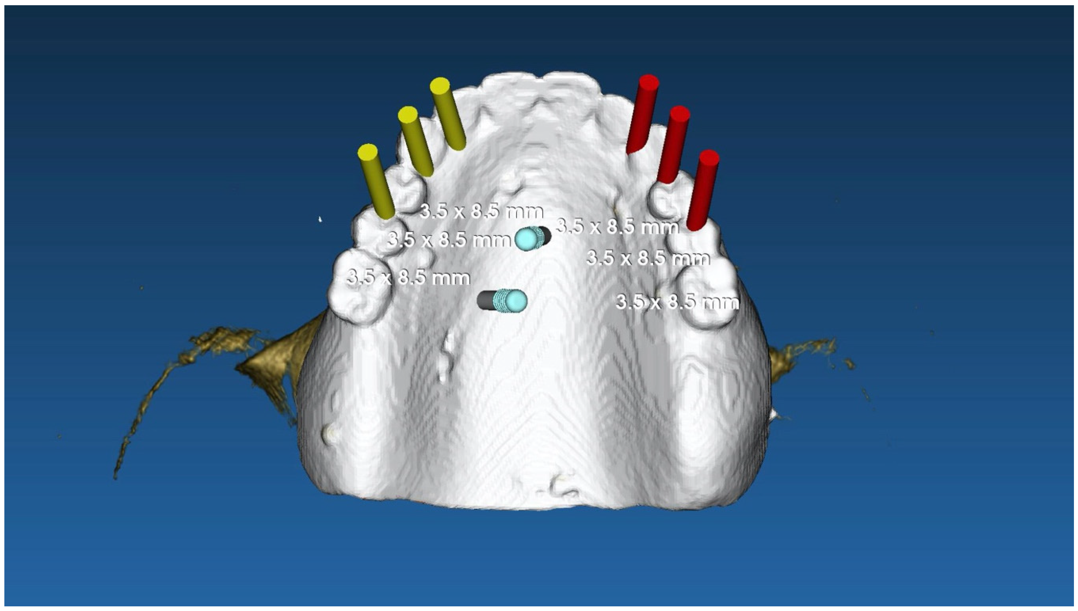
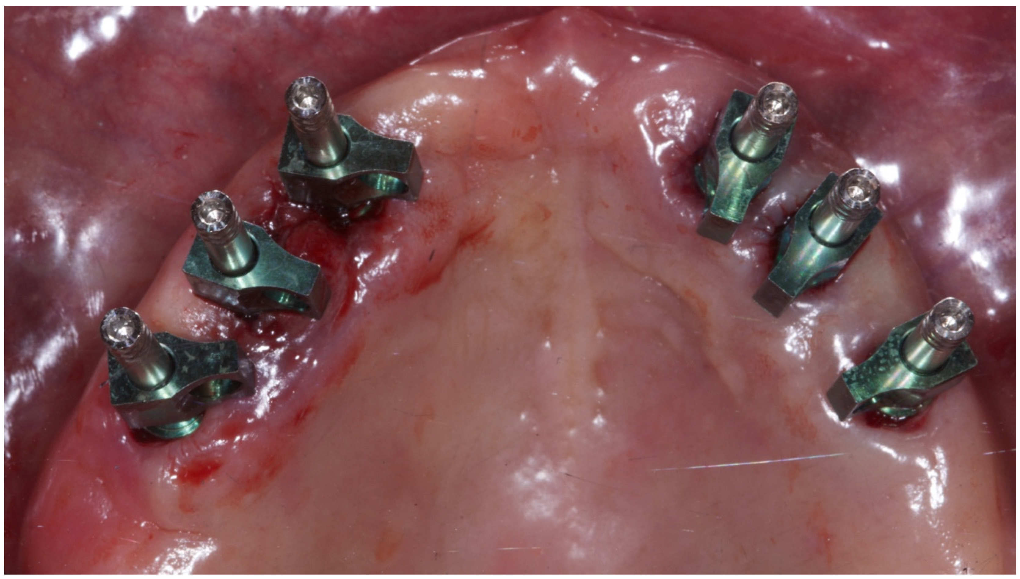
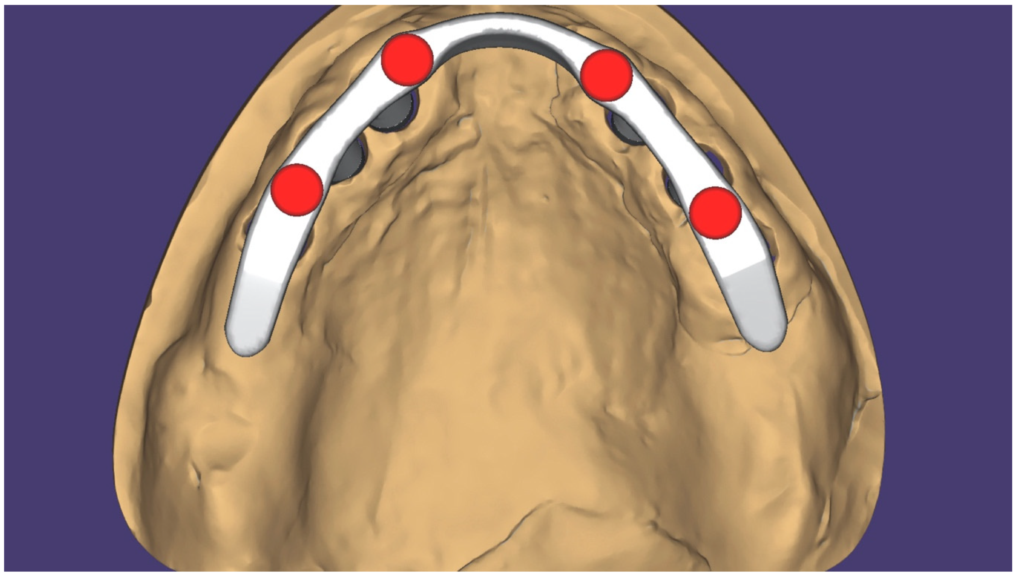
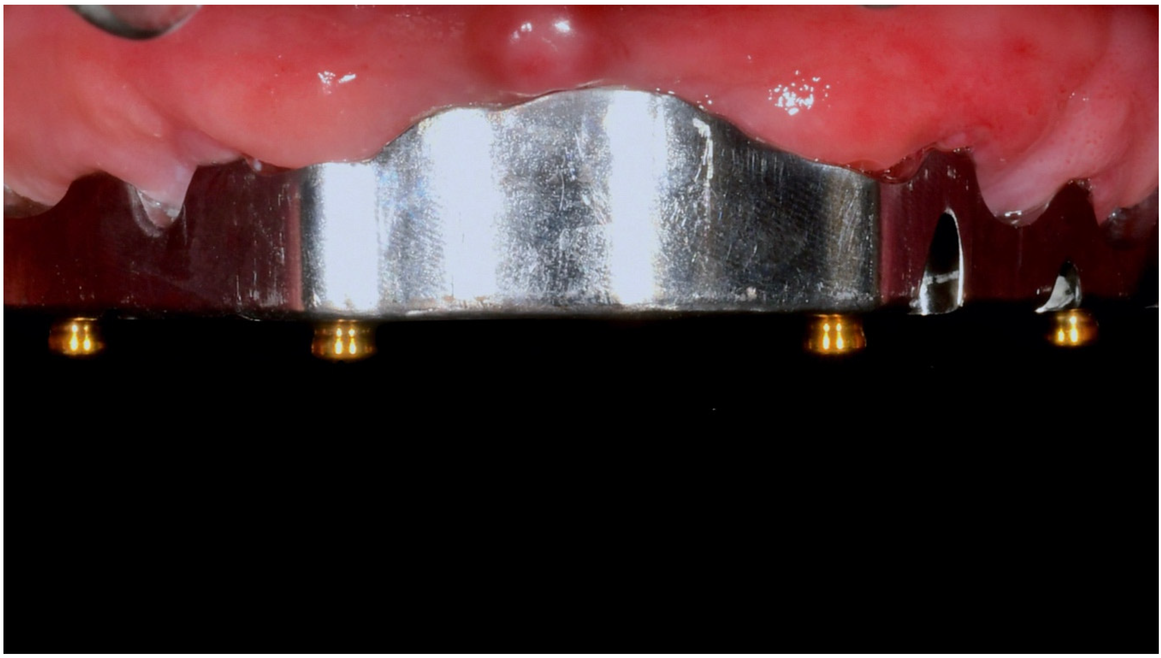
2. Materials and Methods

This paper describes a series of 6 clinical cases, treated with 4 (group 1, n = 3) or 6 (group 2, n = 3) implants of maxillary implant-supported overdentures. Six completely edentulous adult patients were recruited and treated between January and October 2017 by the same surgeon (MT). The investigation was conducted according to the principles embodied in the Helsinki Declaration of 2008, as amended in 2013. After being duly informed about the nature of the treatment, patients were asked to provide written consent. None of the patients presented with uncontrolled diabetes, a history of radiation to the head and neck, pregnancy or nursing, intravenous bisphosphonate therapy, or smoking habits of more than 10 cigarettes a day.
All the patients presented with a pre-existing complete removable denture that was rebased or redone if judged not accurate in its fit and function/esthetics. After, all the patients underwent a cone beam computed tomography (CBCT) scans according to a modified double-scan protocol [6], together with a complete scan of the pre-existing complete removable denture, in order to virtually plan four or six implants in the healed bone, according to the prosthetic volume to be rehabilitated. All the implants were planned as parallel as possible with a dedicated software (RealGuide, 3DEmme, Cantù, Italy). The anterior implants were placed in the lateral position while posterior implants were planned as distal as possible. When six implants were planned, adjunctive implants were placed in the middle, between the anterior and posterior implants. After approval of the virtual plan, a stereolithographic surgical template was ordered (New Ancorvis, Bologna, Italy).
All the patients received professional oral hygiene in the antagonist arch (if needed) and prophylactic antiseptic and antibiotic therapy prior to the implant placement. After local anesthesia, the surgical templates were placed intraorally using the opposing arch as a reference point and stabilized with three to five preplanned anchor pins. Patients received four (Figure 1, Figure 2, Figure 3, Figure 4, Figure 5, Figure 6 and Figure 7) or six (Figure 8, Figure 9, Figure 10, Figure 11, Figure 12, Figure 13 and Figure 14) implants (Osstem TSIII, Osstem Implant, Korea) using a flapless or a miniflap approach, following the drilling protocol recommended by the manufacturer (OneGuide surgical kit, Osstem Implant). Immediately after implant placement, straight Multi Abutments TS (Osstem Implant) were screwed onto the implants. No immediate loading was performed, but the pre-existing prosthesis were adapted using a tissue-conditioning resin to reduce possible symptoms/problems due to an ill-fitting denture (Hydro-cast, SultanHealthcare, USA). After a two-month osseointegration period, a digital impression (Carestream CS 3600) using a specially designed customized prosthetic template was taken, and the digitalized file was super-imposed to the original plan [7]. This is the cross-mounting technique, which helps to transfer the planning information in every prosthetic step. Specifically, it helps us to transfer the position of the implants in the project and to be able to correctly superimpose the data digitally (Figure 15). A one-piece titanium bar with an OT Equator attachment (Rhein’83, Bologna, Italy) was planned in both groups. A secondary framework was designed at the same time using dedicated software (exocad DentalCAD, Exocad GmbH, Germany). Obviously, each step was developed within the prosthetic volume identified during the treatment diagnosis phase. After the finalization, the prostheses were delivered and the patients were followed for hygiene maintenance, with clinical examination every 4 months, and yearly radiographic evaluation, up to two years after loading.
Implant and prosthetic survival rates as well as biologic and technical complications were recorded during the entire follow-up. An implant was considered a failure if it presented with any mobility, progressive marginal bone loss, infection, and/or any mechanical complications, such as implant fracture, which renders the implant unusable although still stable in the bone. A prosthesis was considered a failure if it needed to be replaced. Any biological (pain, swelling, suppuration, etc.) and/or mechanical (screw loosening, fracture of the framework and/or the veneering material, etc.) complications were recorded during the entire follow-up period.
The Fisher exact test was used to compare dichotomous outcomes (survival and complications) between patients rehabilitated with four or six implants. The level of significance was set at 0.05.

3. Results and Discussion

Overall, six patients were treated and followed for two years (three in each group). The patients’ mean age was 68.5 ± 7.2 years. Two women and one man were treated in group 1 (mean age 72.4 ± 5.6 years), while one woman and two men were treated in group 2 (mean age 65.0 ± 7.9 years). All the patients in both groups were not smokers. One patient in group 1 presented controlled type II diabetes. Two patients (one in each group) were under therapy for slightly high blood pressure. During the entire follow-up period, no implants and no prosthetic failed regardless of the number of implants (Fisher exact test: 0/0; 0/0: p = 1.0). At the two-year follow-up examination, two minor complications were experienced in two patients who received four implants. One of the anterior metal matrix bonded on the secondary framework with resin cement de-bonded from the metal part. In both cases, the matrixes were cemented again chairside in about 15 min (Fisher exact test: 0/3; 2/1: p = 0.4).
When comparing these results with technical complications in the mandible, in another paper published by the same authors, no implants and prostheses failed and no major complications were experienced one year after loading. In the present clinical cases, all patients were treated with fixed/removable solution. Several factors should be considered when determining whether a fixed or removable implant-supported complete denture or overdentures is the best option for complete edentulous patients. According to previous studies, bone atrophy represents one of the most important decisional criteria [1,2]. This is due to the need to support facial soft tissue, as well as to provide proper hygiene maintenance. Furthermore, fixed/removable solution represents a resilient system against occlusal force, reducing the overall number of complications.
In the present paper, two minor complications were experienced. This could be explained by the different occlusal force and the kind of residual bone in the atrophic mandible compared to the atrophic maxilla as well as by the longer follow-up in the present paper [5]. Both complications occurred in the patients who received four implants; nevertheless, a statistically significant difference was not reached. This could be explained with the low number of treated patients, as well as the short follow-up.
According to a systematic review by Roccuzzo et al. [8], at the time of writing this manuscript, there were no studies that had addressed the question of how many implants should support an overdenture in the maxilla. In general, posterior implants should be placed as distal as possible, increasing the distance between the anterior and posterior implants (A-P spread) to provide a better distribution of the masticatory force. Improving the implant distribution also allows a reduction of the distal cantilever. The choice to place four or six implants should be made according to the bone atrophy and the residual arch form of the maxillae. In fact, in atrophic patients with no available bone in the posterior area due to bone atrophy and pneumatization of the maxillary sinus, the anterior shape of the maxillary arch influences the implant position, and to increase the A-P spread, sometimes tilted implants are the best choice. A square arch form limits the anterior–posterior distance between implants. In these cases, middle implants could be placed in the pre-maxilla area. Otherwise, in tapering and/or ovoid arches, six implants could be placed, and this represents the gold standard.
Tilting the posterior implants, according to a classic all-on-four protocol, or Marius Bridge by Dr Yvan Fortin, which in 1991 was the first protocol of four tilted implants with a fixed-removable prosthetic, could be another option to reduce the overall number of implants, even in this anatomical scenario, also reducing the distal cantilever, and improving the A-P spread [4]. Pozzi et al. reported no complications one year after loading in a preliminary report on implant-supported overdenture delivered on four implants placed according to the all-on-four protocol [5]. In this study, according to the all-on-four protocol, 17° or 30° Multi-Unit Abutments were used. Different form the presented cases, in the study of Pozzi et al. [5], the metal counterpart was realized in a cobalt-chromium alloy metal framework according to the metal casting technique. In the present paper, the metal counterpart was printed with laser melting technology and then milled. Titanium represents the gold standard due to the lower specific weight that makes titanium lighter. Moreover, titanium is also the gold standard when a composite veneering material is chosen.
In the present case series, low-profile attachments were used to connect and retain the overdenture to the CAD/CAM titanium bar. The low-profile attachments contributed to reducing the technical complications, such as detachment of the teeth or fracture, thus improving the prosthetic materials between the prosthetic crown and bar volume [9,10,11,12,13,14,15,16,17,18,19]. Even if a statistically significant difference was not reached, two minor complications were experienced when four implants were used. A further study should be designed to evaluate the effect of the occlusal forces when few number of implants are used.
Since this study was designed as a comparative case series, the main limitation of this research is the small sample size. Nevertheless, it is possible to assume that a maxillary implant overdenture fully supported by four or six implants is a safer treatment option for the minimally invasive rehabilitation of atrophic maxillae. The take-home message is that when possible, up to six parallel implants (minimum four) should be placed.

Author Contributions

Conceptualization, M.T.; writing—original draft preparation, R.S.; writing—review and editing, G.C. and U.U.; Methodology, A.L.; supervision, S.M.M. All authors have read and agreed to the published version of the manuscript.

Funding

This research received no external funding.

Acknowledgments

In this section you can acknowledge any support given which is not covered by the author contribution or funding sections. This may include administrative and technical support, or donations in kind (e.g., materials used for experiments).

Conflicts of Interest

The authors declare no conflict of interest.

References

  1. Avrampou, M.; Mericske-Stern, R.; Blatz, M.B.; Katsoulis, J. Virtual implant planning in the edentulous maxilla: Criteria for decision making of prosthesis design. Clin. Oral Implant. Res. 2012, 24, 152–159. [Google Scholar] [CrossRef] [PubMed]
  2. Zitzmann, N.U.; Marinello, C.P. Fixed or removable implant-supported restorations in the edentulous maxilla: Literature review. Pract. Periodontics Aesthet. Dent. 2000, 12, 599–608. [Google Scholar] [PubMed]
  3. Tallarico, M.; Canullo, L.; Pisano, M.; Penarrocha-Oltra, D.; Peñarrocha-Diago, M.; Meloni, S.M. An up to 7-Year Retrospective Analysis of Biologic and Technical Complication W ith the All-on-4 Concept. J. Oral Implantol. 2016, 42, 265–271. [Google Scholar] [CrossRef] [PubMed]
  4. Pozzi, A.; Tallarico, M.; Moy, P.K. Four-implant overdenture fully supported by a CAD-CAM titanium bar: A single-cohort prospective 1-year preliminary study. J. Prosthet. Dent. 2016, 116, 516–523. [Google Scholar] [CrossRef] [PubMed] [Green Version]
  5. Tallarico, M.; Xhanari, E.; Kadiu, B.; Scrascia, R. Implant rehabilitation of extremely atrophic mandibles (Cawood and Howell Class VI) with a fixed-removable solution supported by four implants: One-year results from a preliminary prospective case series study. J. Oral Sci. Rehabil. 2017, 3, 32–40. [Google Scholar]
  6. Tallarico, M.; Schiappa, D.; Schipani, F.; Cocchi, F.; Annucci, M.; Xhanari, E. Improved fully digital work-flow to rehabilitate an edentulous patient with an implant overdenture in 4 appointments: A case report. J. Oral Sci. Rehabil. 2017, 25, 38–46. [Google Scholar]
  7. Tallarico, M.; Xhanari, E.; Martinolli, M.; Baldoni, E.; Meloni, S.M. Extraoral chairside digitalization: Clinical reports on a new digital protocol for surgical and prosthetic treatment of completely edentulous patients. J. Oral Sci. Rehabil. 2018, 22, 1–5. [Google Scholar]
  8. Roccuzzo, M.; Bonino, F.; Gaudioso, L.; Zwahlen, M.; Meijer, H.J.A. What is the optimal number of implants for removable reconstructions? A systematic review on implant-supported overdentures. Clin. Oral Implant. Res. 2012, 23, 229–237. [Google Scholar] [CrossRef] [PubMed]
  9. Nastro, E.; Musolino, C.; Allegra, A.; Oteri, G.; Cicciù, M.; Alonci, A.; Quartarone, E.; Alati, C.; De Ponte, F.S. Bisphosphonate-associated osteonecrosis of the jaw in patients with multiple myeloma and breast cancer. Acta Haematol. 2007, 117, 181–187. [Google Scholar] [CrossRef] [PubMed]
  10. Stacchi, C.; Lombardi, T.; Cusimano, P.; Berton, F.; Lauritano, F.; Cervino, G.; Di Lenarda, R.; Cicciù, M. Bone Scrapers Versus Piezoelectric Surgery in the Lateral Antrostomy for Sinus Floor Elevation. J. Craniofacial Surg. 2017, 28, 1191–1196. [Google Scholar] [CrossRef] [PubMed]
  11. Lo Giudice, G.; Lo Giudice, R.; Matarese, G.; Isola, G.; Cicciù, M.; Terranova, A.; Palaia, G.; Romeo, U. Evaluation of magnification systems in restorative dentistry. An in-vitro study. Dent. Cadmos 2015, 83, 296–305. [Google Scholar] [CrossRef]
  12. Matarese, G.; Ramaglia, L.; Fiorillo, L.; Cervino, G.; Lauritano, F.; Isola, G. Implantology and Periodontal Disease: The Panacea to Problem Solving? Open Dent. J. 2017, 11, 460–465. [Google Scholar] [CrossRef] [PubMed]
  13. Cervino, G.; Fiorillo, L.; Spagnuolo, G.; Bramanti, E.; Laino, L.; Lauritano, F.; Cicciu, M. Interface Between MTA and Dental Bonding Agents: Scanning Electron Microscope Evaluation. J. Int. Soc. Prev. Community Dent. 2017, 7, 64–68. [Google Scholar] [CrossRef] [PubMed]
  14. Fiorillo, L.; Cervino, G.; Herford, A.S.; Lauritano, F.; D’Amico, C.; Lo Giudice, R.; Laino, L.; Troiano, G.; Crimi, S.; Cicciu, M. Interferon Crevicular Fluid Profile and Correlation with Periodontal Disease and Wound Healing: A Systemic Review of Recent Data. Int. J. Mol. Sci. 2018, 19, 1908. [Google Scholar] [CrossRef] [PubMed] [Green Version]
  15. Cervino, G.; Fiorillo, L.; Laino, L.; Herford, A.S.; Lauritano, F.; Giudice, G.L.; Fama, F.; Santoro, R.; Troiano, G.; Iannello, G.; et al. Oral Health Impact Profile in Celiac Patients: Analysis of Recent Findings in a Literature Review. Gastroenterol. Res. Pr. 2018, 2018, 7848735. [Google Scholar] [CrossRef] [PubMed]
  16. Fiorillo, L. Oral Health: The First Step to Well-Being. Medicina 2019, 55, 676. [Google Scholar] [CrossRef] [PubMed] [Green Version]
  17. Fiorillo, L.; Musumeci, G. TMJ Dysfunction and Systemic Correlation. J. Funct. Morphol. Kinesiol. 2020, 5, 20. [Google Scholar] [CrossRef] [Green Version]
  18. Cicciù, M.; Risitano, G.; Maiorana, C.; Franceschini, G. Parametric analysis of the strength in the ’’Toronto’’ osseous-prosthesis system. Minerva Stomatol. 2009, 58, 9–23. [Google Scholar]
  19. Cicciù, M.; Risitano, G.; Lo Giudice, G.; Bramanti, E. Periodontal health and caries prevalence evaluation in patients affected by Parkinson’s disease. Parkinsons Dis. 2012, 2012, 541908. [Google Scholar] [CrossRef] [PubMed]
Figure 1. Initial radiographic evaluation.
Prosthesis 02 00006 g001
Figure 2. Four implants with multi abutments after flapless computer-guided surgery.
Prosthesis 02 00006 g002
Figure 3. CAD design of the titanium bar made on four implants according to the prosthetic set-up, and with four low-profile retention systems.
Prosthesis 02 00006 g003
Figure 4. Milled titanium bar with the four OT Equators to retain the implant-supported overdenture.
Prosthesis 02 00006 g004
Figure 5. CAD/CAM Titanium bar screwed in the maxillae.
Prosthesis 02 00006 g005
Figure 6. The definitive prosthesis with pink material to give support to the upper lip and cheeks.
Prosthesis 02 00006 g006
Figure 7. Radiographic evaluation at the last follow-up.
Prosthesis 02 00006 g007
Figure 8. Initial radiographic evaluation.
Prosthesis 02 00006 g008
Figure 9. Virtual planning of six implants according to the prosthetic set-up.
Prosthesis 02 00006 g009
Figure 10. Six implants placed flapless through guided surgery with multi abutments and pick-up transfers.
Prosthesis 02 00006 g010
Figure 11. CAD design of the titanium bar made on six implants according to the prosthetic set-up, and with four low-profile retention systems.
Prosthesis 02 00006 g011
Figure 12. Titanium CAD/CAM bar screwed in the mouth.
Prosthesis 02 00006 g012
Figure 13. The definitive prosthesis with pink material to give support to the upper lip and cheeks.
Prosthesis 02 00006 g013
Figure 14. Radiographic evaluation at the last follow-up.
Prosthesis 02 00006 g014
Figure 15. The digital cross mounting. The digital transfer of information.
Prosthesis 02 00006 g015

