Article

First Detection of Methanogens in Orthopedic Prosthesis Infection: A Four-Case Founding Series

1
Aix Marseille Univ, IRD, APHM, MEPHI, Faculté de Médecine et de Pharmacie, 19-21 boulevard Jean Moulin, CEDEX 05, 13385 Marseille, France
2
IHU Méditerranée Infection, 19-21 boulevard Jean Moulin, CEDEX 05, 13385 Marseille, France
3
Centre de Référence des Infections Ostéoarticulaires (CRIOAC) Interrégional Sud Méditerranée, Service de Chirurgie Orthopédique, Assistance Publique-Hôpitaux de Marseille, 13385 Marseille, France
4
Centre de Référence des Infections Ostéoarticulaires (CRIOAC) Interrégional Sud Méditerranée, Service des Maladies Infectieuses, Hôpital de la Conception, 13385 Marseille, France
*
Author to whom correspondence should be addressed.
Submission received: 15 December 2021 / Revised: 28 January 2022 / Accepted: 29 January 2022 / Published: 7 February 2022

Abstract

:
Orthopedic prosthesis infection must be medically managed after appropriate microbiological documentation. While bacteria and fungi are acknowledged to be causative opportunistic pathogens in this situation, the potential role of methanogens in orthopedic prosthesis infections is still unknown. In a retrospective study, a total of 100 joint and bone samples collected from 25 patients were screened by specific PCR assays for the detection of methanogens. PCR-positive samples were observed by autofluorescence, electron microscopy and tentatively cultured under specific culture conditions. Methanogens were detected by quantitative PCR in 4/100 samples, in the presence of negative controls. Sequencing identified Methanobrevibacter oralis in two cases, Methanobrevibacter smithii in one case and Methanobrevibacter wolinii in one case. Microscopic methods confirmed molecular findings and bacterial culture yielded two strains of Staphylococcus aureus, one strain of Staphylococcus epidermidis and one strain of Proteus mirabilis. These unprecedented data highlight the presence of methanogens in joint and bone samples of patients also diagnosed with bacterial orthopedic prosthesis infection, questioning the role of methanogens as additional opportunistic co-pathogens in this situation.

1. Introduction

Postoperative infection is the principal complication of orthopedic prosthesis implantation, with an estimated prevalence of 0.5 to 1% after hip prosthesis implantation and 1 to 2% after knee prosthesis implantation [1,2,3]. Infections could compromise the functional outcome of the patient, even increasing morbidity and mortality [4,5]. Documentation of orthopedic prosthesis infection relies on microbiological investigations including direct microscopic examination of the samples following Gram staining, PCR-based tests, and culture in an aerobic atmosphere [6,7,8,9]. Microbiological diagnosis of these infections is mandatory, as the antimicrobial susceptibility pattern of the causative opportunistic pathogens guides the medical treatment alongside the surgical orthopedic treatment [10,11,12]. Series of orthopedic prosthesis infections have indicated that bacteria and fungi were responsible for all the currently documented cases, with numerous cases in which mixed infections consisting of several bacteria or mixed bacteria and fungi were identified in the same clinical specimen [13,14].
Methanogenic archaea (here designed as methanogens) are aero-intolerant microorganisms belonging to the Archaea domain and the only known sources of biotic methane [15,16]. They are a common inhabitant of many niches of the human body especially the digestive microbiota [17,18]. Methanogens play important roles in human health such as maintaining gut homeostasis by preventing the accumulation of toxic metabolic end products of bacteria including H2, CO2 and trimethylamine [19,20]. However, their dysbiosis is associated with many diseases including severe acute malnutrition, inflammatory bowel diseases and chronic constipation [21,22,23,24,25].
More recently, methanogens were co-detected with bacteria in various pathological situations, raising questions as to their role as co-pathogen [26,27,28,29,30]. However, no studies have been conducted on the possible implication of methanogens in bone infection.
In this paper, we report on the unprecedented observation that methanogens could also be detected in mixed infections of orthopedic prostheses.

2. Results

2.1. Routine Bacteriological Investigations

A total of 100 samples collected from 25 patients were collected and analyzed. Routine culture found Staphylococcus aureus (n = 55; 11 patients), Staphylococcus epidermidis (n = 12; three patients), Corynebacterium amycolatum (n = 2; one patient), Proteus mirabilis (n = 17; one patient), Pseudomonas aeruginosa (n = 5; two patients), Klebsiella oxytoca (n = 3; two patients), Klebsiella pneumoniae (n = 1; one patient), Escherichia coli (n = 2; one patient), Streptococcus anginosus (n = 1; one patient), Acinetobacter radioresistens (n = 1; one patient) and Enterobacter cloacae (n = 1; one patient).

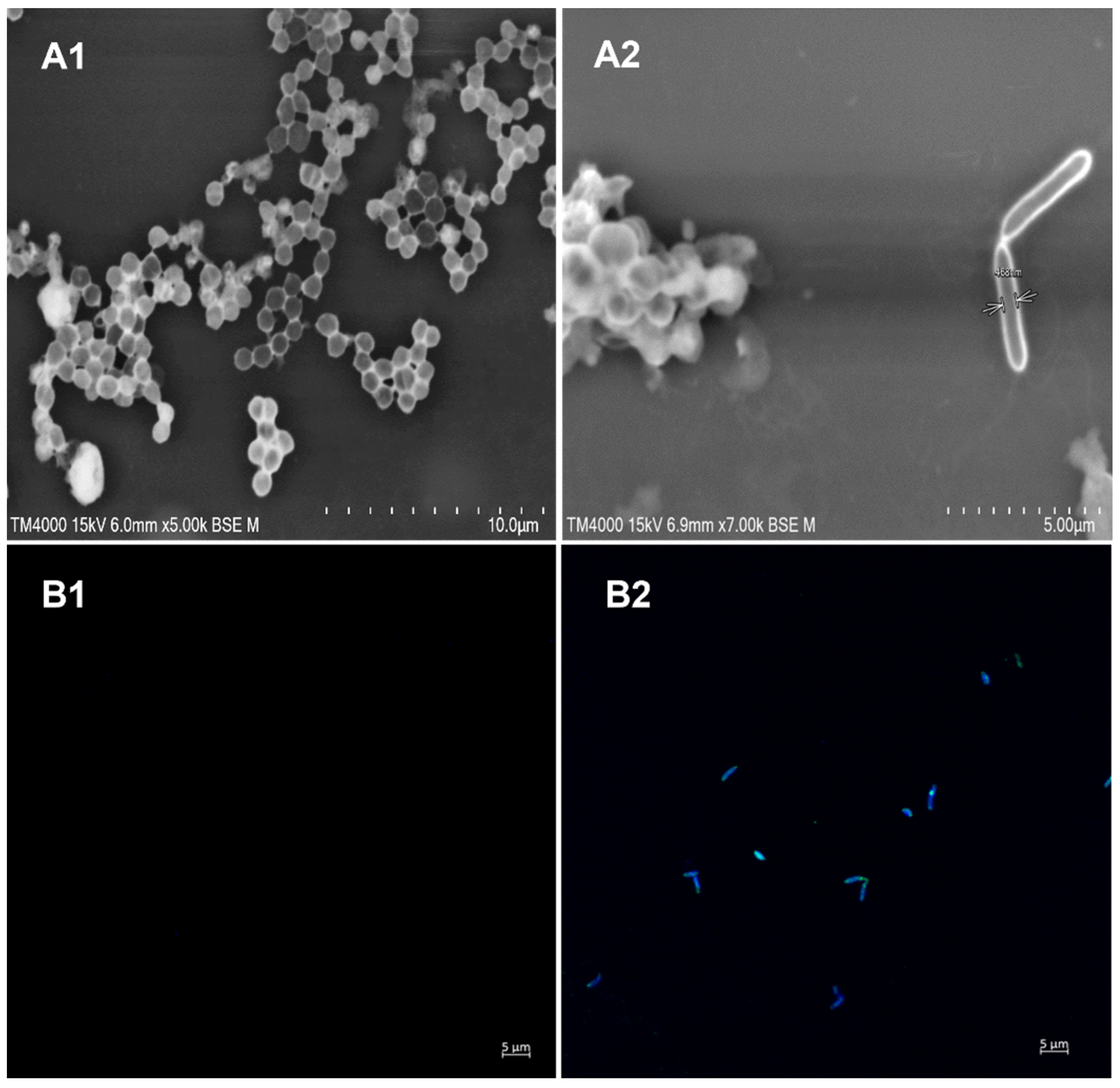
2.2. Methanogen Investigations

The qPCR targeting the actin gene was positive for all samples analyzed. Of the 100 joint fluid and bone samples investigated for the presence of methanogens, four samples (4/100) collected from four different patients were found to be positive in both screening real-time PCR analyses and confirmatory standard PCR. The negative controls remained negative. Quantitative PCR analyses yielded a median Ct of 28.37 (26.77–31.32). The 16S rRNA gene amplicon sequencing identified Methanobrevibacter oralis in two cases, Methanobrevibacter smithii in one case and Methanobrevibacter wolinii in one case. M. oralis was detected in two different samples as was the case in patient no. 2 and M. smithi was detected in two different samples as was the case in patient no. 3, at different times (Table 1). Eight controls (two negative controls for one positive patient) consisting of patients without an orthopedic prosthesis infection were also tested for the presence of methanogens and remained negative.
M.oralis and M. wolinii were co-detected with S. aureus (n = 2), M. smithii were co-detected with S. epidermidis (n = 1) and with P. mirabilis (n = 1). Scanning electron microscopy showed two microscopic microorganisms in PCR-positive joint fluid collected from patient no. 1: the first microorganism presented a bacillary morphology characteristic of M. oralis and the second microorganism corresponded to S. aureus.
M. oralis was also observed by auto-fluorescence in the same PCR-positive joint fluid but not in the PCR-negative samples (Figure 1).

2.3. Medical History

2.3.1. Case 1

An 81-year-old woman had a history of a right knee replacement in 2007. Nine years later, following a fall, she had a prosthetic fracture and surgery with osteosynthesis was performed. One month later, the patient presented an early nosocomial infection of the surgical site with S. aureus and E. coli. Surgical debridement was performed, and the patient was treated with co-trimoxazole for five months, with persistent disabling pain. Two years later, due to increasing pain in the right knee, the hip prosthesis was removed and was replaced by a spacer containing vancomycin [31]. Routine bacterial culture S. aureus was isolated from the knee prosthesis, and methanogen investigation identified M. oralis in the same samples. She was treated with clindamycin and rifampicin for eight months and cured with no relapse at the three-month follow-up examination.

2.3.2. Case 2

A 78-year-old man had a total right hip replacement in 2016. Eight months later the scar became inflamed with discharge. A surgical deep sample taken was positive for S. aureus. The patient was treated with rifampicin and ofloxacin for six weeks and was cured. Three years later, the patient once again suffered right hip pain due to the prosthesis loosening. A CT-guided puncture was performed and S. epidermidis was co-detected with M. oralis from this hip sample. The patient had an indication to remove the prosthesis in two stages.

2.3.3. Case 3

A 46-year-old woman had a total left hip replacement in 2007 which was complicated by an S. aureus infection. One year later, the left hip prosthesis was removed, and a vancomycin spacer was inserted [31]. The spacer was removed, and a new hip prosthesis replacement was done. She had a chronic hip polymicrobial infection, with a chronic hip abscess. A total of four successive surgical interventions were performed between 2007 and 2019. Since 2015, no hip prosthesis has been replaced and she has a chronic inflammatory fistula with discharge. The microorganisms isolated were Staphylococcus lugdunensis, Staphylococcus capitis, Klebsiella pneumoniae and Pseudomonas aeruginosa. Finally, in the latest hip fistula sample, P. mirabilis was co-detected with M. smithii. Over 13 years, she received numerous antibiotics including clindamycin, ofloxacin and imipenem. At the eight-month follow-up examination, the hip fistula persisted.

2.3.4. Case 4

A 74-year-old man received a left knee prosthesis in 2018, and an early S. aureus infection was diagnosed. He was treated with rifampin and ofloxacin for five months. Two years later, the patient relapsed, the prosthesis was removed, and a vancomycin spacer was inserted. S. aureus grew from the intraoperative samples and methanogen investigation identified M. wolinii. S. aureus was treated successively with vancomycin and imipenem, followed by teicoplanin combined with clindamycin and ciprofloxacin. After four months of follow-up, no relapse was noted.

3. Discussion

The documentation of methanogens in the puncture and biopsy samples collected from orthopedic devices and prostheses reported in this study did not merely result from contamination, as methanogens have never been reported in clinical microbiology laboratories. Moreover, the methanogens reported here, have been consistently detected by at least two unrelated laboratory approaches in the same pus sample, including molecular and microscopic methods; and the same methanogen has been consistently detected in time-series samples. The negative controls introduced at every experimental step remained negative, and no methanogen positive controls have ever been introduced in the experimental procedures to avoid contamination. Attempts to culture methanogens have failed, which can be explained by the fact that the bone samples were not collected under anaerobic conditions, having been in contact with the ambient atmosphere. Indeed, several studies have demonstrated the exquisite sensitivity of methanogens to oxygen [31,32,33].
We detected methanogens in 4/100 joint and bone samples from four patients diagnosed with orthopedic prosthesis infection. These observations expand the spectrum of methanogen-associated infections in patients. Indeed, M. smithii and M. oralis are emerging as opportunistic pathogens co-detected with bacteria in cases of skeletal muscular abscesses [28], refractory sinusitis [34] and life-threatening brain abscesses [29]. Methanogens are also part of dysbiosis in the case of vaginosis [35] and urinary tract infections [30]. More recently, we specifically detected M. smithii by adopting a polyphasic approach to the blood of febrile patients [33]. Here, we report for the first time, that the methanogen M. wolinii can be retrieved from clinical samples, as this methanogen was previously only known in the rumen of sheep, having never been detected, not cultured from human microbiota [36]. The obligate detection of methanogens in association with bacteria reported in several studies [34,37,38] suggest syntrophic interactions between methanogens and some bacteria, which could support co-pathogenicity. Here, methanogens have been co-detected with staphylococci which are recognized as the predominant cause of bone infections [39]. In addition, it is known that the formation of biofilm on the surface of the osteoarticular prosthesis is a key step in the physiopathology of bone infections [40,41]. Some studies have reported the ability of methanogens, especially M. smithii and M. oralis, to form a biofilm, suggesting their probable involvement in mixed orthoaedic prosthesis infections [42,43,44].
In this study, the detection of methanogens results from the use of specific laboratory tools. These tools, however, could be implanted in any clinical microbiology laboratory for the routine search of methanogens in orthopedic specimens In particular, the PCR-based detection protocol here reported, could be done in any one laboratory with experience in home-made PCR-based diagnosis of infectious disease pending to the introduction of controls for the accurate interpretation of data, as referenced in this report [45]. However, exploration of the methanogen population in this context and their interactions still require further investigation through large-scale studies to understand their potential role in the infectious process of bone infections, this may lead to a change in the standard therapeutic protocols incorporating antibiotics active against these microorganisms, for optimal management of orthopedic prosthesis infections.

4. Materials and Methods

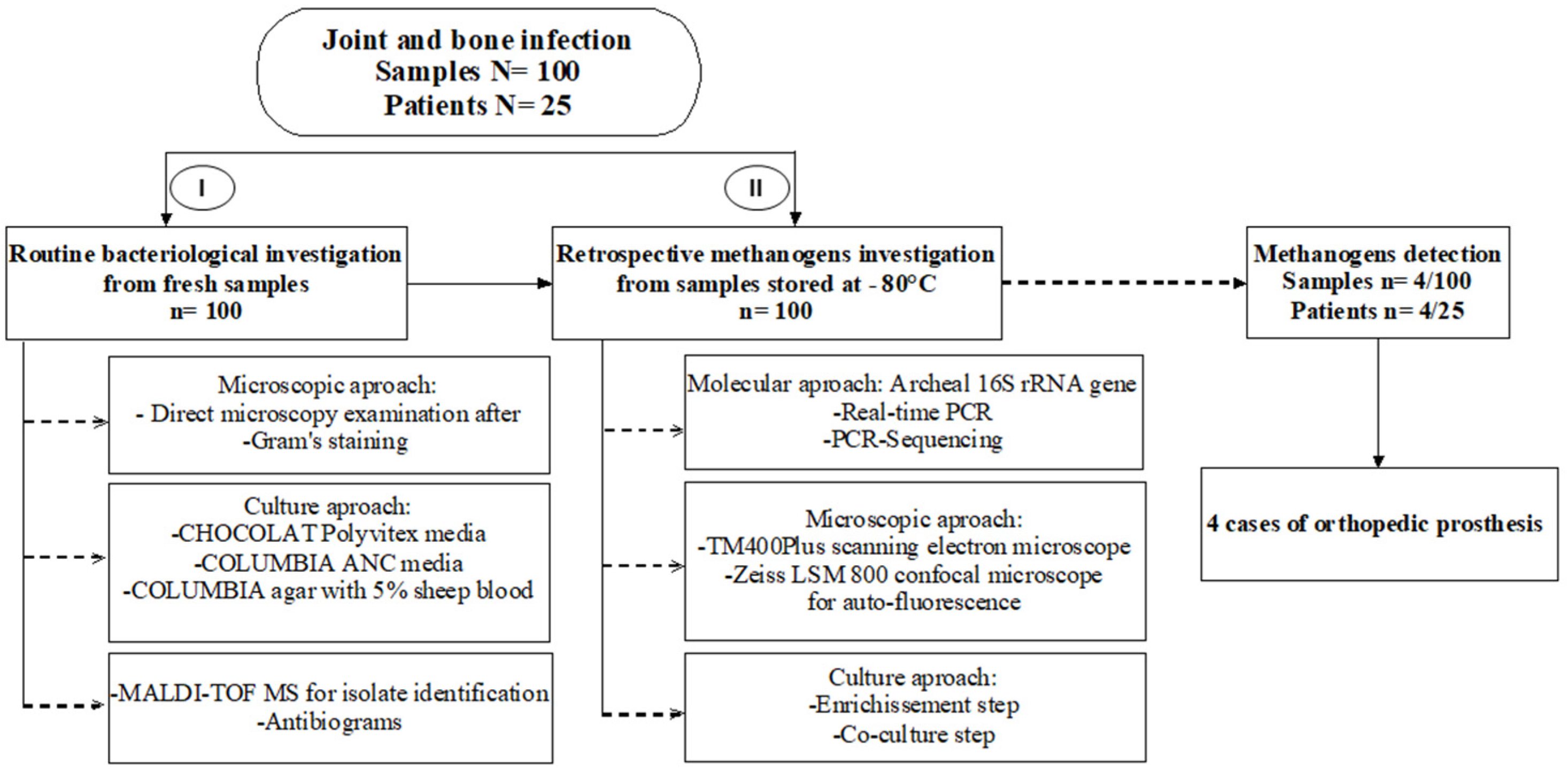
4.1. Patients and Study Design

This retrospective study included a total of 100 orthopedic samples, consisting of 33 joint fluid samples and 67 bone biopsies at the site of orthopedic material implantation samples collected from 25 unrelated patients between January and April 2020 and was approved by the IHU Méditerranée Infection Ethics Committee (No. 2020-032). No samples were taken specifically for this study and, in accordance with European General Data Protection Regulation No. 2016/679, patients were informed of the potential use of their medical data and that they could refuse the use of their data. All the samples were manipulated at the diagnostic laboratory of the IHU Méditerranée Infection, Marseille as part of its routine activities following the reference methodology MR-004 registered on No. PADS20-284 in the AP-HM register. All the patients enrolled in this study had previously presented clinical, biological and radiological pieces of evidence for joint or bone infection. The samples were collected by aspiration or surgical biopsy in sterile tubes or in bottles (BacT/ALERT FN Plus, bioMérieux, Marcy-l’Étoile, France) and stored at −20 °C (Figure 2).

4.2. Routine Bacteriological Investigations

All the samples were examined by microscopy after Gram staining and 200 µL of each sample were cultured using three different culture media: CHOCOLAT Polyvitex (BioMérieux, Marcy-l’Étoile, France) and COLUMBIA ANC (BioMérieux) media, incubated at 37 °C under 5% CO2 for five days to grow aerobic bacteria; and 5% sheep blood Columbia agar medium (BioMérieux) incubated at 37 °C for 10 days to grow anaerobic bacteria. Cultured bacteria were identified by using matrix-assisted laser desorption/ionization time-of-flight mass spectrometry (MALDI-TOF-MS) as previously described [46]. Antibiotic susceptibility testing was performed as previously described [47].

4.3. Methanogen PCR-Based Identification

Manipulations to search for methanogens were performed on the remaining samples preserved at −20 °C after routine microbiological investigations had been completed. For joint fluid samples, a 200-μL volume was transferred into a 2-mL tube and sonicated for 20 min with the Branson 2510 ultrasonic sonicator (Branson, Rungis, France). Total DNA was then extracted using the automat extractor EZ1 advanced XL with EZ1 DNA tissue kit (Qiagen, Courtaboeuf, France). Bone samples were cut into small pieces using a sterile scalpel and transferred into a 2 mL tube and extracted as previously described [47]. Firstly, a quantitative real-time PCR system targeting the 16S rRNA gene of methanogens was used for screening using the following primers and probe: forward 5′-CGAACCGGATTAGATACCCG-3′, Reverse 5′-CCCGCCAATTCCTTTAAGTT-3′, Probe 6FAM-CCTGGGAAGTACGGTCGCAAG. PCR results were validated by the detection of human β-actin gene as an internal control-PCR for each sample. Samples were considered positive when the CT value was equal, or less than 35 CT.
In the second step, clinical samples that were positive by real-time PCR were assessed for the presence of methanogens using PCR-sequencing as described above using the 16S rRNA gene forward primer 5′-CCGGGTATCTAATCCGGTTC-3′ and reverse primer 5′-CTCCCAGGGTAGAGGTGAAA-3′ [47]. The PCR products were purified and sequenced using a Big Dye Terminator v1.1 Cycle Sequencing kit (Applied Biosystems, Foster City, CA, USA), the different sequences were analyzed and assembled with ChromasPro software, then compared to the GenBank database using the online BLAST program of NCBI (www.ncbi.nlm.nih.gov/BLAST/, accessed on 21 May 2020). Two mL tubes filled with 200 µL of sterile phosphate buffered saline (PBS) (Thermo Fisher Scientific, Villebon Sur-Yvette, France) were used as negative controls in each of the above laboratory steps.

4.4. Microscopic Examinations

Samples were observed for the presence of methanogens using the TM4000plus scanning electron microscope (Hitachi, Tokyo, Japan). Briefly, 100 µL of joint fluid and bone samples were fixed with 100 µL of 2.5% glutaraldehyde and 100 µL of that mixture was spotted onto a glass slide by cytocentrifugation (Shandon Cytospin 4, Thermo Scientific, Waltham, MA, USA). Slides were stained using 1% phosphotungstic acid (PTA; Sigma Aldrich, Saint-Louis, MO, USA) for two minutes at room temperature. Image acquisition was performed using the TM4000 plus software (Hitachi). To detect auto-fluorescent methanogens, the PCR-positive samples were excited by an ultraviolet excitation laser, and image acquisition was performed with a Zeiss LSM 800 confocal microscope using a 63×, 1.4 numerical aperture (NA) oil immersion objective, as previously described [33].

Author Contributions

K.D. performed the experiments, interpreted the data drafted the manuscript; F.G. collected the samples, designed the experiments and drafted the manuscript; J.-N.A. collected the samples, analyzed the data and drafted the manuscript; P.S. collected the samples; analyzed the data and drafted the manuscript; A.S. collected the samples, analyzed the data and drafted the manuscript; M.D. designed and implemented the study, analyzed the data, interpreted the results, and wrote the manuscript. All authors have read and agreed to the published version of the manuscript.

Funding

K.D. receives a Ph.D. grant from the Fondation Méditerranée Infection, Marseille. This work was supported by the French Government under the “Investissements d’avenir” (Investments for the Future) programme managed by the Agence Nationale de la Recherche (ANR, fr: National Agency for Research), (reference: Méditerranée Infection 10-IAHU-03).

Institutional Review Board Statement

This work was approved by the IHU Méditerranée Infection Ethics Committee (No. 2020-032). In accordance with European General Data Protection Regulation No. 2016/679, patients were informed of the potential use of their medical data and that they could refuse the use of their data. All the samples were manipulated at the diagnostic laboratory of the IHU Méditerranée Infection, Marseille as part of its routine activities following the reference methodology MR-004 registered on No. PADS20-284 in the AP-HM register.

Informed Consent Statement

In accordance with European General Data Protection Regulation No. 2016/679, patients were informed of the potential use of their medical data and that they could refuse the use of their data, following the reference methodology MR-004 registered on No. PADS20-284 in the AP-HM register.

Data Availability Statement

All data generated or analyzed during this study are included in this published article.

Acknowledgments

This work was supported by the French Government under the «Investissements d’avenir» (Investments for the Future) program managed by the Agence Nationale de la Recherche (ANR, fr: National Agency for Research) (reference: Méditerranée Infection 10-IAHU-03).

Conflicts of Interest

The authors declare no conflict of interest.

References

  1. Pulido, L.; Ghanem, E.; Joshi, A.; Purtill, J.J.; Parvizi, J. Periprosthetic joint infection: The incidence, timing, and predisposing factors. Clin. Orthop. Relat. Res. 2008, 466, 1710–1715. [Google Scholar] [CrossRef] [Green Version]
  2. Pedersen, A.B.; Svendsson, J.E.; Johnsen, S.P.; Riis, A.; Overgaard, S. Risk factors for revision due to infection after primary total hip arthroplasty. A population-based study of 80,756 primary procedures in the Danish Hip Arthroplasty Registry. Acta Orthop. 2010, 81, 542–547. [Google Scholar] [CrossRef]
  3. Tibrewal, S.; Malagelada, F.; Jeyaseelan, L.; Posch, F.; Scott, G. Single-stage revision for the infected total knee replacement: Results from a single centre. Bone Jt. J. 2014, 96, 759–764. [Google Scholar] [CrossRef] [PubMed]
  4. Mahomed, N.N.; Barrett, J.A.; Katz, J.N.; Phillips, C.B.; Losina, E.; Lew, R.A.; Guadagnoli, E.; Harris, W.H.; Poss, R.; Baron, J.A. Rates and outcomes of primary and revision total hip replacement in the United States medicare population. J. Bone Jt. Surg. Am. 2003, 85, 27–32. [Google Scholar] [CrossRef] [PubMed]
  5. Spitzmüller, R.; Gümbel, D.; Güthoff, C.; Zaatreh, S.; Klinder, A.; Napp, M.; Bader, R.; Mittelmeier, W.; Ekkernkamp, A.; Kramer, A.; et al. Duration of antibiotic treatment and risk of recurrence after surgical management of orthopaedic device infections: A multicenter case-control study. BMC Musculoskelet. Disord. 2019, 20, 184. [Google Scholar] [CrossRef] [PubMed] [Green Version]
  6. Parvizi, J.; Zmistowski, B.; Berbari, E.F.; Bauer, T.W.; Springer, B.D.; Della Valle, C.J.; Garvin, K.L.; Mont, M.A.; Wongworawat, M.D.; Zalavras, C.G. New definition for periprosthetic joint infection: From the Workgroup of the Musculoskeletal Infection Society. Clin. Orthop. Relat. Res. 2011, 469, 2992–2994. [Google Scholar] [CrossRef] [Green Version]
  7. Levy, P.Y.; Fournier, P.E.; Fenollar, F.; Raoult, D. Systematic PCR detection in culture-negative osteoarticular infections. Am. J. Med. 2013, 126, 1143.e25–1143.e33. [Google Scholar] [CrossRef]
  8. Osmon, D.R.; Berbari, E.F.; Berendt, A.R.; Lew, D.; Zimmerli, W.; Steckelberg, J.M.; Rao, N.; Hanssen, A.; Wilson, W.R.; Infectious Diseases Society of America. Infectious Diseases Society of America. Diagnosis and management of prosthetic joint infection: Clinical practice guidelines by the Infectious Diseases Society of America. Clin. Infect. Dis. 2013, 56, e1–e25. [Google Scholar] [CrossRef] [Green Version]
  9. Fink, B.; Schuster, P.; Braun, R.; Tagtalianidou, E.; Schlumberger, M. The diagnostic value of routine preliminary biopsy in diagnosing late prosthetic joint infection after hip and knee arthroplasty. Bone Jt. J. 2020, 102, 329–335. [Google Scholar] [CrossRef]
  10. Widmer, A.F. New developments in diagnosis and treatment of infection in orthopedic implants. Clin. Infect. Dis. 2001, 1, 33. [Google Scholar] [CrossRef]
  11. Trampuz, A.; Zimmerli, W. Prosthetic joint infections: Update in diagnosis and treatment. Swiss Med. Wkly. 2005, 135, 243–251. [Google Scholar] [PubMed]
  12. Toms, A.D.; Davidson, D.; Masri, B.A.; Duncan, C.P. The management of peri-prosthetic infection in total joint arthroplasty. J. Bone Jt. Surg. Br. 2006, 88, 149–155. [Google Scholar] [CrossRef] [PubMed]
  13. Tunney, M.M.; Patrick, S.; Curran, M.D.; Ramage, G.; Hanna, D.; Nixon, J.R.; Gorman, S.P.; Davis, R.I.; Anderson, N. Detection of prosthetic hip infection at revision arthroplasty by immunofluorescence microscopy and PCR amplification of the bacterial 16S rRNA gene. J. Clin. Microbiol. 1999, 37, 3281–3290. [Google Scholar] [CrossRef] [PubMed] [Green Version]
  14. Brown, T.S.; Petis, S.M.; Osmon, D.R.; Mabry, T.M.; Berry, D.J.; Hanssen, A.D.; Abdel, M.P. Periprosthetic Joint Infection with Fungal Pathogens. J. Arthroplast. 2018, 33, 2605–2612. [Google Scholar] [CrossRef] [PubMed]
  15. Bapteste, E.; Brochier, C.; Boucher, Y. Higher-level classification of the Archaea: Evolution of methanogenesis and methanogens. Archaea 2005, 1, 353–363. [Google Scholar] [CrossRef] [Green Version]
  16. Liu, Y.; Whitman, W.B. Metabolic, phylogenetic, and ecological diversity of the methanogenic archaea. Ann. N. Y. Acad. Sci. 2008, 1125, 171–189. [Google Scholar] [CrossRef]
  17. Dridi, B.; Henry, M.; El Khéchine, A.; Raoult, D.; Drancourt, M. High prevalence of Methanobrevibacter smithii and Methanosphaera stadtmanae detected in the human gut using an improved DNA detection protocol. PLoS ONE 2009, 4, e7063. [Google Scholar] [CrossRef]
  18. Koskinen, K.; Pausan, M.R.; Perras, A.K.; Beck, M.; Bang, C.; Mora, M.; Schilhabel, A.; Schmitz, R.; Moissl-Eichinger, C. First Insights into the Diverse Human Archaeome: Specific Detection of Archaea in the Gastrointestinal Tract, Lung, and Nose and on Skin. mBio 2017, 8, e00824-17. [Google Scholar] [CrossRef] [Green Version]
  19. Sieber, J.R.; McInerney, M.J.; Gunsalus, R.P. Genomic insights into syntrophy: The paradigm for anaerobic metabolic cooperation. Annu. Rev. Microbiol. 2012, 66, 429–452. [Google Scholar] [CrossRef]
  20. Brugère, J.F.; Borrel, G.; Gaci, N.; Tottey, W.; O’Toole, P.W.; Malpuech-Brugère, C. Archaebiotics: Proposed therapeutic use of archaea to prevent trimethylaminuria and cardiovascular disease. Gut Microbes 2014, 5, 5–10. [Google Scholar] [CrossRef] [Green Version]
  21. Camara, A.; Konate, S.; Tidjani Alou, M.; Kodio, A.; Togo, A.H.; Cortaredona, S.; Henrissat, B.; Thera, M.A.; Doumbo, O.K.; Raoult, D.; et al. Clinical evidence of the role of Methanobrevibacter smithii in severe acute malnutrition. Sci. Rep. 2021, 11, 5426. [Google Scholar] [CrossRef] [PubMed]
  22. Million, M.; Tidjani Alou, M.; Khelaifia, S.; Bachar, D.; Lagier, J.C.; Dione, N.; Brah, S.; Hugon, P.; Lombard, V.; Armougom, F.; et al. Increased Gut Redox and Depletion of Anaerobic and Methanogenic Prokaryotes in Severe Acute Malnutrition. Sci. Rep. 2016, 6, 26051. [Google Scholar] [CrossRef] [PubMed]
  23. Blais Lecours, P.; Marsolais, D.; Cormier, Y.; Berberi, M.; Haché, C.; Bourdages, R.; Duchaine, C. Increased prevalence of Methanosphaera stadtmanae in inflammatory bowel diseases. PLoS ONE 2014, 9, e87734. [Google Scholar] [CrossRef] [PubMed]
  24. Ghavami, S.B.; Rostami, E.; Sephay, A.A.; Shahrokh, S.; Balaii, H.; Aghdaei, H.A.; Zali, M.R. Alterations of the human gut Methanobrevibacter smithii as a biomarker for inflammatory bowel diseases. Microb. Pathog. 2018, 117, 285–289. [Google Scholar] [CrossRef]
  25. Soares, A.C.; Lederman, H.M.; Fagundes-Neto, U.; de Morais, M.B. Breath methane associated with slow colonic transit time in children with chronic constipation. J. Clin. Gastroenterol. 2005, 39, 512–515. [Google Scholar] [CrossRef]
  26. Li, C.L.; Liu, D.L.; Jiang, Y.T.; Zhou, Y.B.; Zhang, M.Z.; Jiang, W.; Liu, B.; Liang, J.P. Prevalence and molecular diversity of Archaea in subgingival pockets of periodontitis patients. Oral Microbiol. Immunol. 2009, 24, 343–346. [Google Scholar] [CrossRef]
  27. Huynh, H.T.; Pignoly, M.; Nkamga, V.D.; Drancourt, M.; Aboudharam, G. The repertoire of archaea cultivated from severe periodontitis. PLoS ONE 2015, 10, e0121565. [Google Scholar] [CrossRef]
  28. Nkamga, V.D.; Lotte, R.; Roger, P.M.; Drancourt, M.; Ruimy, R. Methanobrevibacter smithii and Bacteroides thetaiotaomicron cultivated from a chronic paravertebral muscle abscess. Clin. Microbiol. Infect. 2016, 22, 1008–1009. [Google Scholar] [CrossRef]
  29. Drancourt, M.; Nkamga, V.D.; Lakhe, N.A.; Régis, J.M.; Dufour, H.; Fournier, P.E.; Bechah, Y.; Scheld, W.M.; Raoult, D. Evidence of Archaeal Methanogens in Brain Abscess. Clin. Infect. Dis. 2017, 65, 1–5. [Google Scholar] [CrossRef]
  30. Grine, G.; Lotte, R.; Chirio, D.; Chevalier, A.; Raoult, D.; Drancourt, M.; Ruimy, R. Co-culture of Methanobrevibacter smithii with enterobacteria during urinary infection. EBioMedicine 2019, 43, 333–337. [Google Scholar] [CrossRef] [Green Version]
  31. Argenson, J.N.; Arndt, M.; Babis, G.; Battenberg, A.; Budhiparama, N.; Catani, F.; Chen, F.; de Beaubien, B.; Ebied, A.; Esposito, S.; et al. Hip and Knee Section, Treatment, Debridement and Retention of Implant: Proceedings of International Consensus on Orthopedic Infections. J. Arthroplast. 2019, 34, S399–S419. [Google Scholar] [CrossRef] [PubMed]
  32. Chaudhary, P.P.; Conway, P.L.; Schlundt, J. Methanogens in humans: Potentially beneficial or harmful for health. Appl. Microbiol. Biotechnol. 2018, 102, 3095–3104. [Google Scholar] [CrossRef] [PubMed]
  33. Drancourt, M.; Djemai, K.; Gouriet, F.; Grine, G.; Loukil, A.; Bedotto, M.; Levasseur, A.; Lepidi, H.; Bou-Khalil, J.; Khelaifia, S.; et al. Methanobrevibacter smithii archaemia in febrile patients with bacteremia, including those with endocarditis. Clin. Infect. Dis. 2020, 15, ciaa998. [Google Scholar] [CrossRef] [PubMed]
  34. Sogodogo, E.; Fellag, M.; Loukil, A.; Nkamga, V.D.; Michel, J.; Dessi, P.; Fournier, P.-E.; Drancourt, M. Nine Cases of Methanogenic Archaea in Refractory Sinusitis, an Emerging Clinical Entity. Front. Public Health 2019, 7, 38. [Google Scholar] [CrossRef]
  35. Grine, G.; Drouet, H.; Fenollar, F.; Bretelle, F.; Raoult, D.; Drancourt, M. Detection of Methanobrevibacter smithii in vaginal samples collected from women diagnosed with bacterial vaginosis. Eur. J. Clin. Microbiol. Infect. Dis. 2019, 38, 1643–1649. [Google Scholar] [CrossRef]
  36. Miller, T.L.; Lin, C. Description of Methanobrevibacter gottschalkii sp. nov., Methanobrevibacter thaueri sp. nov., Methanobrevibacter woesei sp. nov. and Methanobrevibacter wolinii sp. nov. Int. J. Syst. Evol. Microbiol. 2002, 52, 819–822. [Google Scholar] [CrossRef] [Green Version]
  37. Matarazzo, F.; Ribeiro, A.C.; Feres, M.; Faveri, M.; Mayer, M.P. Diversity and quantitative analysis of Archaea in aggressive periodontitis and periodontally healthy subjects. J. Clin. Periodontol. 2011, 38, 621–627. [Google Scholar] [CrossRef]
  38. Efenberger, M.; Agier, J.; Pawłowska, E.; Brzezińska-Błaszczyk, E. Archaea prevalence in inflamed pulp tissues. Cent. Eur. J. Immunol. 2015, 40, 194–200. [Google Scholar] [CrossRef] [PubMed] [Green Version]
  39. Wright, J.A.; Nair, S.P. Interaction of staphylococci with bone. Int. J. Med. Microbiol. 2010, 300, 193–204. [Google Scholar] [CrossRef] [Green Version]
  40. Tyner, H.; Patel, R. Propionibacterium acnes biofilm—A sanctuary for Staphylococcus aureus? Anaerobe 2016, 40, 63–67. [Google Scholar] [CrossRef]
  41. McDowell, A.; Patrick, S. Evaluation of nonculture methods for the detection of prosthetic hip biofilms. Clin. Orthop. Relat. Res. 2005, 437, 74–82. [Google Scholar] [CrossRef]
  42. Bang, C.; Schmitz, R.A. Archaea: Forgotten players in the microbiome. Emerg. Top. Life Sci. 2018, 2, 459–468. [Google Scholar] [CrossRef] [PubMed]
  43. Orell, A.; Fröls, S.; Albers, S.V. Archaeal biofilms: The great unexplored. Annu. Rev. Microbiol. 2013, 67, 337–354. [Google Scholar] [CrossRef] [PubMed]
  44. Dame-Teixeira, N.; de Cena, J.A.; Côrtes, D.A.; Belmok, A.; Dos Anjos Borges, L.G.; Marconatto, L.; Giongo, A.; Kyaw, C.M. Presence of Archaea in dental caries biofilms. Arch. Oral Biol. 2020, 110, 104606. [Google Scholar] [CrossRef] [PubMed]
  45. Raskin, L.; Stromley, J.M.; Rittmann, B.E.; Stahl, D.A. Group-specific 16S rRNA hybridization probes to describe natural communities of methanogens. Appl. Environ. Microbiol. 1994, 60, 1232–1240. [Google Scholar] [CrossRef] [PubMed] [Green Version]
  46. Seng, P.; Drancourt, M.; Gouriet, F.; La Scola, B.; Fournier, P.E.; Rolain, J.M.; Raoult, D. Ongoing revolution in bacteriology: Routine identification of bacteria by matrix-assisted laser desorption ionization time-of-flight mass spectrometry. Clin. Infect. Dis. 2009, 49, 543–551. [Google Scholar] [CrossRef]
  47. Haldorsen, B.; Giske, C.G.; Hansen, D.S.; Orri Helgason, K.; Kahlmeter, G.; Löhr, I.H.; Matuschek, E.; Österblad, M.; Rantakokko-Jalava, K.; Wang, M.; et al. Performance of the EUCAST disc diffusion method and two MIC methods in detection of Enterobacteriaceae with reduced susceptibility to meropenem: The NordicAST CPE study. J. Antimicrob. Chemother. 2018, 73, 2905. [Google Scholar] [CrossRef] [PubMed] [Green Version]
Figure 1. Microscopic observations of joint sample of Case 1 and a negative control sample (joint sample PCR-negative to methanogens). (A1) Negative control showing only S. aureus; (A2) two Methanobrevibacter oralis observed in the same field with Staphylococcus aureus using scanning electron microscopy TM4000Plus (Hitachi). All image settings for the magnification, focus and keV mode are shown on micrographs. (B1) Negative control sample; (B2) auto-florescent methanogens in joint sample of Case 1 after exposure to ultraviolet excitation laser with Zeiss LSM 800 confocal microscope at 63×, 1.4 numerical aperture (NA) oil immersion objective. The scale bar represents 5 µm.
Prosthesis 04 00005 g001
Figure 2. Flow chart of this study and the main results.
Prosthesis 04 00005 g002
Table 1. Characteristics of four patients diagnosed with methanogens-positive orthopedic prosthesis.
CasesAgeSexClinical History of Prosthesis InfectionRoutine Bacterial CultureMethanogen DetectionPrevious AntibioticsNumber of Surgeries after Prosthesis ImplantationInfection Evolution (Months)Prosthesis AblationProsthesis
Re-Implantation
Spacer ImplantationCuredRelapse
Case 181FS. aureus and E. coli
right knee prosthesis infection
S. aureusM. oralisRifadin + clindamycin + ofloxacin2111YesYesYESYesNo
Case 278MS. aureus and S. epidermidis right hip prosthesis infectionS. epidermidisM. oralisRifampicin + ofloxacin199YesNoYesNoNo
Case 346FS. aureus and S. lugdunensis and Klebsiella pneumoniae and Proteus mirabilis and S. capitis
left hip prosthesis infection
P. mirabilisM. smithiiImipenem + clindamycinNo358YesYesNoNoYes
Case 474MS. aureus
left Knee prosthesis
S. aureusM. woliniiRifampicin + ofloxacin + teicoplanin274YesYesYesOn goingNo
Publisher’s Note: MDPI stays neutral with regard to jurisdictional claims in published maps and institutional affiliations.

