
Bioprinting of Hydrogel-Based Drug Delivery Systems for Nerve Tissue Regeneration

Rose-Hulman Institute of Technology, Terre Haute, IN 47803, USA
*
Author to whom correspondence should be addressed.
Biophysica 2024, 4(1), 58-73; https://0-doi-org.brum.beds.ac.uk/10.3390/biophysica4010004
Submission received: 7 December 2023
/
Revised: 16 January 2024
/
Accepted: 26 January 2024
/
Published: 31 January 2024
(This article belongs to the Special Issue Molecular Structure and Simulation in Biological System 2.0)
Abstract
:Globally, thousands of people are affected by severe nerve injuries or neurodegenerative disorders. These conditions cannot always be cured because nerve tissue either does not regenerate or does so at a slow rate. Therefore, tissue engineering has emerged as a potential treatment approach. This review discusses 3D bioprinting for scaffold manufacturing, highlights the advantages and disadvantages of common bioprinting techniques, describes important considerations for bioinks, biomaterial inks, and scaffolds, and discusses some drug delivery systems. The primary goal of this review is to bring attention to recent advances in nerve tissue engineering and its possible clinical applications in peripheral nerve, spinal cord, and cerebral nerve regeneration. Only studies that use 3D bioprinting or 3D printing to manufacture hydrogel scaffolds and incorporate the sustained release of a drug or growth factor for nerve regeneration are included. This review indicates that 3D printing is a fast and precise scaffold manufacturing technique but requires printing materials with specific properties to be effective in nervous tissue applications. The results indicate that the sustained release of certain drugs and growth factors from scaffolds can significantly improve post-printing cell viability, cell proliferation, adhesion, and differentiation, as well as functional recovery compared with scaffolds alone. However, more in vivo research needs to be conducted before this approach can be used in clinical applications.
1. Introduction
Severe injuries to the human nervous system can lead to profound consequences, primarily because it shows little to no capacity for self-regeneration. Annually, there are approximately 17,000 new cases of spinal cord injury (SCI), 80,000 cases of severe traumatic brain injury (TBI), and millions of cases of neurodegenerative diseases in the U.S. [1,2,3]. Additionally, 13 to 23 out of every 1 million individuals suffer from peripheral nerve injuries, often with poor prognoses [4]. Unfortunately, prevailing treatments focus primarily on symptom management rather than on tissue regeneration [5]. However, the emerging field of tissue engineering holds promise for developing therapies that can regenerate nerve tissue and restore its functionality.
The objective of tissue engineering is to create biological materials that can replace, restore, improve, or maintain the function of damaged tissues [6]. The most effective tissue engineering strategies use the interdisciplinary triad of tissue engineering: cells, scaffolds, and biochemical/physical signals [6]. Cell sources are autologous, allogeneic, or xenogeneic, ranging from stem cells or differentiated cells [6,7], while scaffolds, constructed from either natural or synthetic biomaterials, are typically fabricated via methods such as freeze-drying, electrospinning, decellularization, or bioprinting [8]. Biochemical signals refer to growth factors or pharmaceuticals [6], while physical signals involve mechanical loading. Efforts towards nerve regeneration present unique problems due to the structural and functional complexity of neural tissues. The limited capacity for self-regeneration creates a powerful impetus for alternative interventions to address nerve damage due to injury and degenerative diseases. Bioprinting, particularly with hydrogel-based bioinks, offers a promising solution to these challenges by enabling the creation of customized, three-dimensional, biomimetic structures. These structures can also be optimized for cell growth and natural tissue integration due to the versatility of polymeric hydrogels. Additionally, by employing hydrogels, drug delivery capabilities can be introduced into bioprinted scaffolds, thereby elevating the therapeutic potential of the system. By enabling the sustained release of neurotrophic factors or specific drugs, the system transitions from passively to actively promoting nerve regeneration and functional recovery. The combination of bioprinting, hydrogels, and drug delivery effectively addresses key issues in nerve tissue regeneration. This approach creates an optimal environment for nerve repair and regeneration and holds the potential to administer precise treatments to improve the regenerative process. Consequently, this strategy not only deals with the structural and functional intricacies of nerve regeneration, but also paves the way for more effective treatment methods for a wider range of neurological disorders. Overall, this review provides a broad overview of bioprinting techniques, considerations for bioinks and scaffolds, sustained-release drug delivery methodologies, and illustrates how these principles culminate in recent research in nerve tissue regeneration. Figure 1 offers a concept map with the proposed considerations for a tissue engineering approach for patient-specific nerve regeneration that are discussed in this paper.
2. Bioprinting
2.1. Overview of 3D Bioprinting
Three-dimensional bioprinting is an additive manufacturing process that uses cell-infused inks, called bioinks, to 3D-print complex tissue and/or organ resembling constructs [9,10]. In tissue engineering, the terms “3D printing” and “3D bioprinting” are often used interchangeably, but it is important to note their differences. Three-dimensional bioprinting uses bioinks that contain living cells and biologics to create tissues, while three-dimensional printing uses completely inert, non-living inks to create porous scaffolds to support cell attachment, proliferation, and differentiation [11]. In recent years, 3D bioprinting has rapidly grown in popularity due to its potential applications in tissue engineering and drug screening [12].
A typical bioprinting technique contains three main steps: pre-processing, processing, and post-processing [9]. Pre-processing consists of gathering imaging data, from computed tomography (CT), magnetic resonance imaging (MRI), ultrasound imaging, and/or optical microscopy [9]. The data are then transferred to computer-aided design (CAD) software to 3D-model the desired, patient-specific tissue, or create free-form models. The processing step of bioprinting includes bioink preparation and the printing process [9]. The most common bioprinting methods are described below and are summarized in Table 1 and the relative benefits and limitations are depicted in Figure 2. The post-processing phase of bioprinting includes maturing the printed tissue, typically by using a bioreactor to foster the growth and development of tissue in ideal environmental and mechanical conditions [13]. In theory, the tissue would then be ready for its desired application. In the context of neural tissue engineering, the bioprinting process is similar, with the exception that the use of bioreactors in the field has not yet been extensively studied [13].
2.2. Bioprinting Methods
2.2.1. Extrusion-Based
Extrusion-based bioprinting (EBB), the most common bioprinting method, is similar to traditional 3D printing, whereby continuous streams of bioink are forced from a screw, either by pneumatic pressure from a piston or mechanical force, to create the layers of a 3D construct imported from a CAD file [9].
EBB has several advantages, including greater cell deposition, fast printing speed, affordability, ability to use a wide variety of bioinks, ability to print structurally stable constructs, and ease of use [11,14]. Combined, these advantages lead to potentially simple scalability. EBB can print high cell densities, similar to those found in natural tissues, making it more feasible to scale current models to human dimensions [9]. However, EBB also has limitations, including a printing resolution of around 100 µm, which is significantly lower than other bioprinting methods [11,14]. As a result, it is not feasible to print fine and intricate tissues using this method. Furthermore, despite its high cell densities, EBB can yield a cell viability as low as 40% after printing due to high nozzle shear stress [15,16], which constitutes a major challenge. Finally, when using higher viscosity bioinks, nozzle clogging is possible. This can be countered by increasing nozzle diameters; however, this further decreases printing resolution [15].
2.2.2. Droplet-Based
In droplet-based bioprinting (DBB), droplets of bioinks are ejected, layer by layer, from a nozzle onto a substrate. The droplets then solidify, forming the final 3D construct [12]. DBB can be separated into inkjet bioprinting, electro-hydrodynamic jetting, acoustic bioprinting, and microvalve-based bioprinting, based on the energy source used to create the droplets [9,17].
Inkjet bioprinting was the first bioprinting technology to emerge and, based on conventional 2D printing processes, it is the most widely used DBB technique [12]. Inkjet bioprinting can be classified into two different groups: continuous inkjet (CIJ) and drop-on-demand (DOD) inkjet bioprinting [9].
CIJ technology employs pressure to expel a continuous flow of bioink droplets through a nozzle; however, it is less favored due to its suboptimal precision [9]. Conversely, DOD technology utilizes thermal, piezoelectric, or electrostatic mechanisms to generate droplets, facilitating more controlled formation on demand and enhancing precision [18]. Furthermore, DOD bioprinters can have multiple fluid containers and nozzles [18], allowing different bioinks to be printed simultaneously.
Inkjet bioprinting has a high printing resolution (around 50 µm), fast printing speeds, cost efficiency, biocompatibility, the ability to create cell concentration gradients, and high post-printing cell viability due to the non-contact nature of the process [9,17,19]. However, inkjet bioprinting requires low-viscosity bioinks to prevent nozzle clogging, and smaller nozzles that can induce shear stresses on cells [9,19]. As a result, the printed constructs often lack structural stability, necessitating an additional cross-linking step to enhance their stability. Furthermore, to reduce the risk of nozzle clogging, bioinks must have lower cell concentrations [9,14].
2.2.3. Laser-Assisted Bioprinting
Laser-assisted bioprinting (LAB) is a vastly different printing modality. Very generally, a typical LAB setup includes a laser source, a laser transparent print ribbon, a laser-absorbing layer, and a cell-laden bioink layer all suspended above a substrate. When the laser is activated, the energy from the laser-absorbing layer creates an area of high pressure that forces a droplet of bioink onto the substrate [9]. LAB can differ significantly depending on whether the laser source is high- or low-powered, the materials used in the print ribbon and laser-absorbing layers, and whether or not the laser-absorbing layer is sacrificial. For instance, metal or biopolymer-based sacrificial layers can be used to protect cells [20].
LAB offers the advantage of a high post-printing cell viability of around 95% [9], has a printing resolution around 10 µm [17], and can print high cell densities [9]. Additionally, LAB does not require a printing nozzle, which eliminates the risk of nozzle-clogging [9]. Unfortunately, the limitations of LAB tend to outweigh its benefits, the most significant being cell exposure to laser radiation, which can cause photonic cell damage [9,14]. The printed constructs may also be exposed to metallic contaminants from the sacrificial layers, which presents a major obstacle to implantation [17]. Additionally, LAB is complex, expensive, requires additional and time-consuming fabrication, and is not a scalable process [9,19]. Therefore, it is not practical to utilize LAB for large tissue constructs or large-scale fabrication.
2.2.4. Stereolithography
Stereolithography uses UV light to polymerize ink to form a 3D construct in a computer-controlled, layer-by-layer fashion [9]. Stereolithography has the highest printing resolution compared with other methods (between 5 and 50 µm) [17], is a nozzle-free process, and allows for the printing of high cell densities [9]. However, these advantages come with several disadvantages. First, only photopolymerizable bioinks can be used; however, the photoinitiators that catalyze polymerization can cause cell damage [9,19]. Additionally, UV radiation can cause cell and DNA damage, causing the post-printing cell viability to be as low as 25% [9]. Finally, only low-viscosity bioinks can be used [9].
Similar to stereolithography, digital light processing (DLP) uses a digital light projector, micro-mirrors, and a digital micromirror device (DMD) to reflect light that polymerizes complete bioink layers one at a time [9,21]. In this method, each of the several thousand micromirrors act as a pixel, and can be turned on or off, which greatly improves the printing resolution [21]. While DLP is typically faster and has higher resolution than stereolithography, cell viability is still an issue [21]. Therefore, both stereolithography and DLP have historically limited applications in neural tissue engineering [17].
3. Bioink and Scaffold Considerations
3.1. Biological Properties
Biocompatibility is the most crucial characteristic for bioinks and biomaterial inks. To avoid adverse effects, these printing materials must be either biologically inert or immuno-compatible to ensure that no significant immune or inflammatory responses are triggered upon scaffold implantation [22]. Natural polymers, often considered biocompatible, are preferred for bioinks; however, numerous synthetic and composite polymers are also recognized for their biocompatibility [10].
Another vital feature of scaffolds is their biodegradability. Scaffolds are designed to gradually degrade, allowing native tissue to regenerate and replace them. Consequently, the degradation byproducts must be non-cytotoxic to avoid harming the cells [22]. Printed scaffolds also need to promote host cell adhesion. Ideally, they should enable host cells to infiltrate the scaffold, proliferate within it as it degrades, and ultimately substitute the damaged or lost tissue. To achieve this, scaffolds should possess a high degree of interconnected porosity [7] and emulate the extracellular matrix (ECM) of the target tissue [10].
3.2. Rheological Properties
Rheology is the study of the flow and deformation of a material under an external force [23]. One important rheological property is viscosity. High-viscosity printing materials can cause nozzle-clogging, but low-viscosity materials will not maintain their shape after printing. Furthermore, shear stress is another important factor related to viscosity. High shear stresses can be caused by printing pressure, nozzle diameter, and the viscosity of the bioink [23], and have been shown to decrease post-printing cell viabilities. Additionally, high-viscosity bioinks themselves can impose high shear stresses on cells [14]. Shear thinning can occur when the viscosity of a material decreases as the shear rate increases, and is another challenge to consider [23]. Overall, rheological properties affect which bioprinting method can be used, as well as the resulting cell viability, printing resolution, and integrity of the printed construct.
3.3. Electrical Properties
One material property unique to neural tissue engineering is electrical conductivity. Electrically conductive scaffolds are necessary because they mimic native neural tissue ECM and can support axon regeneration in vivo [17]. A study by Heo et al. added intrinsically conductive polymers to a GelMA bioink, which enhanced its electrochemical properties and significantly improved the differentiation of dorsal root ganglion cells under electrical stimulation [24]. Other studies have added polypyrrole in their scaffolds to improve their conductive and electrical properties. These studies have determined that enhanced electrical properties in scaffolds improve neurite outgrowth, as well as axonal remyelination and regeneration in vitro [17].
3.4. Mechanical Properties
The mechanical properties of scaffolds should closely mimic those of the native neural tissue. The elastic moduli of scaffolds have been shown to influence cell signaling and affect adhesion, differentiation, and proliferation of neurons [10,17]. Protocols for differentiating stem cells into neurons require hydrogels with elastic moduli between 1 and 10 kPa. Hydrogels with higher stiffness may inadvertently cause stem cells to differentiate into myogenic or osteogenic cells [17]. The mechanical properties of various neural tissues vary: brain tissue typically exhibits a stiffness of about 0.5 kPa, peripheral nerve tissue has an ideal stiffness around 450 kPa, and spinal cord tissue has a stiffness that can range between 200 and 600 kPa [10,17]. Furthermore, creating scaffolds with similar elastic moduli should incorporate other factors to ensure differentiation into neural tissue. These scaffolds need to have similar tensile, compressive, and shear strengths as the native tissue. Thus, it is important to view these properties dynamically, in relation to the duration of implantation, rather than as fixed characteristics [10].
3.5. Bioink Cell Considerations
Bioinks can be defined by the incorporation of cells, and possibly biologically active components and biomaterials, into a mixture suitable for 3D bioprinting. Bioinks should not be confused with biomaterial inks, which are biomaterials suitable for 3D printing and are not cell-laden. However, biomaterial inks can be seeded with cells once printed [25]. Bioinks used in neural tissue engineering commonly include human-induced pluripotent stem cell (hiPSC)-derived neural progenitor cells (NPCs) and other neuronal stem cells (NSCs). The encapsulation of cells in bioinks poses many challenges. High cell concentrations in bioinks can cause nozzle-clogging, but low cell concentrations do not mimic native tissue, and make it less feasible to scale the scaffold to human dimensions [9]. Furthermore, it is important to not introduce cytotoxic agents during the 3D printing process [10] or sterilization processes. Chemical cross-linking agents such as photoinitiators could be cytotoxic, as could the UV radiation commonly used to solidify the gels [10]. Lastly, many sterilization processes would likely cause cell damage, so any future clinical applications of cell-laden scaffolds need to utilize appropriate and non-cytotoxic sterilization methods.
3.6. Hydrogels as Biomaterial Inks and Bioinks
Hydrogels are typically the top candidates for biomaterial inks. Hydrogels are 3D structures made from cross-linked hydrophilic polymers and can swell and absorb up to one thousand times their dry weight with water [22]. Hydrogels are ideal for 3D printing, as they can be printed in a liquid, low-viscosity state, and can be chemically or physically cross-linked to solidify and form a stable 3D structure. Hydrogels are particularly useful as they can encapsulate cells and small molecules, such as drugs and some proteins [5]. They also have many tunable properties, such as biocompatibility, biodegradation, mechanical strength, viscosity, porosity electrical conductivity, responsiveness to environmental stimuli, etc., and can be modified to have ideal properties. Furthermore, hydrogels can mimic the structure of the native ECM [7,23]. Lastly, high-porosity hydrogels provide a suitable environment for nutrient exchange, allowing for cell adhesion, proliferation, and migration [7,23].
4. Drug Delivery
4.1. Overview
The drug delivery aspect of tissue engineering plays a vital role in facilitating cell proliferation and differentiation, which is crucial for promoting functional recovery of the tissue. The core concept of drug delivery in this context involves the sustained release of a drug from an implanted device over a specified period once it is placed in the body. The nature of the delivery device varies depending on the specific method of drug delivery. In tissue engineering, this device is typically the printed scaffold implanted into the body. Commonly, the drug delivered is a growth factor, which plays a critical role in nerve regeneration. Degradation is a primary mechanism of drug delivery. This involves the gradual release of the drug as the material it is embedded in slowly degrades. There are numerous other methods for drug delivery; however, the most relevant to tissue engineering are discussed below and summarized in Table 2.
4.2. Drug Delivery Methods
4.2.1. Microspheres
Another mechanism for drug delivery is the use of microspheres, which can effectively deliver a wide variety of compounds including insoluble drugs, proteins, and small molecules [26]. Microspheres are created using synthetic or natural polymers, including chitosan, polycaprolactone (PCL), and polyvinyl acetate (PVA) [26]. The main disadvantages of this technique are the risk of decreased biological activity, which can arise from immunogenic responses, and unstable or denaturing proteins, potentially leading to the encapsulated item in the microsphere losing its biological activity [27].
4.2.2. Fibrin Glue
A third approach to drug delivery employs a fibrin gel matrix, often referred to as fibrin glue comprising fibrinogen and a thrombin solution with a drug incorporated into one of these solutions. These solutions are then mixed, initiating cross-linking, resulting in the formation of the fibrin gel matrix [28]. Pore diameter and pore size can be adjusted by varying the length of cross-linking time, with increased cross-linking leading to smaller pores. To increase the cross-linking time, the ionic strength or the thrombin concentration may be increased [28]. Other important properties are the shear modulus and tensile strength of the glue, which may be adjusted by changing the amounts of calcium and fibrinogen in the glue [28]. The final method of modifying the properties of the fibrin gel matrix is the addition of antifibrinolytic agents, which slow down fibrinolysis, the process of destroying the gel, therefore slowing drug release [28]. Some limitations of employing a fibrin gel matrix include the risk of inadequate drug integration and the inability to use this method for long-term drug delivery, as fibrin glue is not designed to degrade over extended periods [28].
4.2.3. Nanoparticles
Intuitively, the term “nanoparticles” refers to particles with dimensions in the nanometer range. Nanoparticles that are then infused with drugs to be delivered to different areas of the body are often referred to as nanocarriers [29]. The physical, chemical, and biological properties of nanocarriers can be altered by changing the type or combination of nanoparticles used [29]. Nanocarriers are most commonly made of liposomes, solid lipids, polymers (which are often coated), silica materials, and carbon [29]. The wide variety of materials to choose from allows for a lot of flexibility in what areas of the body nanocarriers can be used to deliver drugs. The extremely small size of these nanocarriers also increases the flexibility and functionality of nanocarriers as they can operate on the cellular level and cross the blood–brain barrier [29]. The main disadvantage of nanoparticles is that they are a new technology with a lot of unknowns. How these particles affect various pathways and processes is unknown; they have the potential to cause disruption and damage [29].
5. Applications in Nerve Tissue Regeneration
The following section summarizes the recent publications of bioprinted drug delivery systems for nerve tissue regeneration. The key features and results of these studies are tabulated in Table 3.
5.1. Peripheral Nerve Regeneration
In a 2020 study, researchers employed extrusion-based bioprinting and UV irradiation to create a gelatin methacryloyl (GelMA) hydrogel embedded with chitosan microspheres loaded with nerve growth factor (NGF) [30]. The bioink was enriched with PC12 cells and RSC96 Schwann cells, which were subsequently cultured and compared against a control group containing GC-MS without NGF. The results showed that post-printing cell viability was marginally higher in the GC-MS + NGF group than in the GC-MS group [30]. Although these differences were not statistically significant, they suggest successful bioink preparation and printing techniques [30]. Staining analysis revealed that the surface environment fostered cell adhesion and growth [30]. Notably, in the GC-MS + NGF group, PC12 cells exhibited significantly longer axon lengths and a higher proportion of cells developed axons [30]. Furthermore, the study recorded a cumulative NGF release of 61 ± 10% over a period of 9 h [30]. Overall, the observed enhancements in neurite growth, cell adhesion, and proliferation demonstrate the hydrogel’s good biocompatibility and potential for improved neural cell function.
In 2023, Wu et al. developed GelMA and silk fibroin glycidyl methacrylate (SF-MA) hydrogel nerve conduits with and without 7,8-DHF nanoassemblies (GFNC and GFC, respectively). In vitro, 7,8-DHF was able to promote neurite elongation in a concentration-dependent manner after co-culture with PC12 cells [31]. In vivo, these conduits were used to bridge 12 mm gaps in rat sciatic nerves, with the GFNC group exhibiting a significantly higher nerve conduction velocity (NCV) compared with the GFC and autograft groups [31]. The GFNC group also demonstrated significant improvements in axon regeneration and remyelination due to the sustained release of 7,8-DHF [31]. This study successfully created GelMA/SF-MA nerve conduits that not only mimicked the mechanical properties of rat sciatic nerves, but also enhanced Schwann cell adhesion, proliferation, and migration. The use of DLP 3D printing was crucial, as it worked well with photocurable and biocompatible hydrogel materials, avoiding cytotoxicity issues.
A different study from Tao et al. used DLP 3D printing to fabricate GelMA nerve conduits photopolymerized using LED light. XMU-MP-1, an inhibitor of the Hippo pathway, was encapsulated within poly(ethylene glycol)-poly(3-caprolactone) (MPEG-PCL) nanoparticles, which were incorporated into the biomaterial ink [32]. The researchers printed nerve guidance conduits (NGCs) with (3DDC) and without (3DC) the drug-loaded nanoparticles and implanted them into rat sciatic nerves. Functional restoration was indicated in the 3DDC group, which demonstrated significantly higher NCV values and compound motor action potential (CMAP) compared with the 3DC and autograft groups [32]. The 3DDC group also showed increased axon diameter and myelin thickness. After 144 h, 83.2% of the XMU-MP-1 was released from the nanoparticles and 47.4% was released from the conduits [32]. The DLP process was effectively employed to print GelMA hydrogels with the drug-loaded nanoparticles, maintaining the sustained release profile of the drug, while directly incorporating XMU-MP-1 into the GelMA hydrogels, leading to aggregation and burst release [32]. This study highlights DLP’s ability to fabricate structures with mechanical properties suitable for cell migration, adhesion, and proliferation, while simultaneously delivering drugs to enhance nerve regeneration.
Xu et al. employed DLP 3D printing to fabricate GelMA NGCs embedded with MPEG-PCL nanoparticles containing RGFP966 [33]. In vitro, RGFP966 effectively promoted neurite elongation in PC12 cells and the remyelination of Schwann cells [33]. When these NGCs were implanted into 10 mm gaps in rat sciatic nerves, the RGFP966-enhanced group exhibited significantly higher NCV compared with the standard NGC group, with results comparable to the autograft group [33]. Histological evaluation revealed a greater thickness and mean diameter of the myelinated fibers in the NGC + RGFP966 versus the NGC group, as well as comparable densities of nerve fibers in the NGC + RGFP966 group, which were similar to the autograft group [33]. RGFP966 release reached a plateau of around 30% after 100 h [33]. These findings suggest that the GelMA NGCs with RGFP966 effectively promoted the functional restoration and regeneration of rat sciatic nerves.
5.2. Spinal Cord Regeneration
In 2018, Chen et al. used mask projection laser stereolithography to 3D-print poly(propylene fumarate) (PPF) scaffolds, some incorporating collagen (PPFC), and others combining collagen with a neurotrophin-3 (NT3) coating (PPFC + NT3) [34]. They found that NT3, when coated on PPFC scaffolds, exhibited stable and controlled release in vitro, indicative of a sustained release system [34]. These scaffolds were implanted into rats with a complete transection spinal cord injury (SCI) model. After 12 weeks, the PPFC + NT3 group showed significantly more new and mature neurons at the lesion site, as well as pronounced axonal regeneration, myelination, remyelination, and synaptic formation [34]. Electrophysiological tests indicated larger motor-evoked potentials (MEPs) and shorter latency periods in the PPFC + NT3 group, although these values were still below pre-SCI levels. Notably, rats in the PPFC + NT3 group exhibited markedly improved hindlimb motor function compared with the other groups, characterized by sweeping motions of the lower limbs without weight support [34].
In 2021, low-temperature extrusion 3D printing was used to create collagen and chitosan scaffolds integrated with brain-derived neurotrophic factor (BDNF) for spinal cord repair. The scaffold with BDNF integrated into the bioink (3D-CC-BDNF) exhibited a stable, long-term release of BDNF in vitro alongside significantly enhanced cell adhesion and growth, while effectively supporting the development of neuronal stem cells into neurons [35]. Post implantation into a transected spinal cord model, assessments at eight weeks showed that the 3D-CC-BDNF group experienced superior motor function recovery, and fostered synaptic connections, nerve fiber regeneration, axonal growth, and myelination at the injury site [35]. The study highlighted the 3D-CC-BDNF scaffold’s excellent biocompatibility and sustained BDNF release, enhancing its regenerative capabilities. The low-temperature 3D printing method preserved BDNF’s bioactivity, suggesting its potential as a simple yet effective manufacturing technique for spinal cord injury treatments [35].
In a 2023 study, Song et al. developed hydrogel using a decellularized rat spinal cord extracellular matrix (ECM) blended with polyethylene glycol diacrylate and gelatin [36]. Polycaprolactone (PCL) microfibers created via electrospinning and oxymatrine (OMT) were integrated into the hydrogels, and were solidified after UV irradiation [36]. The study included three groups—a PCL scaffold, PCL/hydrogel scaffold, and PCL/hydrogel/OMT scaffold—each implanted into semi-transected rat spinal cords and evaluated after eight weeks. The scaffolds remained stable and provided long-term support for nerve regeneration [36]. The in vitro release of OMT was 80.32 ± 7.1% at 30 days, with a rapid initial release followed by a slower subsequent release. The scaffolds created a conducive microenvironment for cell growth, with PCL microfibers directing axonal growth, and OMT enhancing nerve regeneration and neural stem cell differentiation, while also inhibiting astrocyte formation and improving locomotor function in rats. However, the complexity of the fabrication process may limit their clinical applicability [36].
5.3. Cerebral Regeneration
In a 2010 study, DOD inkjet printing was used to bioprint a fibrin gel scaffold surrounded by a collagen type I hydrogel. The bioink was enhanced with neonatal mouse cerebellum cells (C17.2 cell line) and vascular endothelial growth factor (VEGF) [37]. The printed C17.2 cells had a post-printing viability of 93.23 ± 3.77%, comparable to manually plated control cells. Initially, the cells were small and round, but quickly changed shape; by day two, they exhibited an elongated shape with neurite-like extensions, indicating differentiation. By day three, differentiation was more pronounced [37]. In contrast, cells in the control collagen/fibrin scaffold without VEGF showed no signs of differentiation and shrank over time. Cells in the VEGF-infused scaffold migrated towards the VEGF/fibrin gel, forming clusters [37]. This was a pioneering study in using 3D bioprinting with multiple cross-linking methods for neural cell culture and paved the way for using bioactive materials to replace lost or damaged neural tissues [37].
In 2020, Sharma et al. used EBB to create dome-shaped structures using a fibrin-based bioink containing guggulsterone microspheres (GMs) housing human induced pluripotent stem cell (hiPSC)-derived neural progenitor cells (NPCs). Three different bioink groups were compared: NPCs with GM, NPCs with guggulsterone in the media (SG), and NPCs with unloaded spheres (UM). The GM group exhibited the highest post-printing cell viability, although all maintained viability over 80% [38]. After 15 days, all groups expressed TUJ1 (an immature neuronal marker) and FOXA2 (a midbrain dopamine neuron marker). At 30 days, the GM group showed significantly higher expressions of TUJ1, TH (enzyme expressed by dopaminergic neurons), and GFAP (an astrocyte marker), and similar levels of O4 (an oligodendrocyte marker) compared with the UM group [38]. These results suggest that the guggulsterone-releasing microspheres promoted the maturation of NPCs into dopaminergic neurons. The 3D-printed domes had a porous structure conducive to nutrient transfer, more closely resembling the microenvironment of the brain [38]. This study offers promising insights into a tissue engineering strategy potentially applicable for replacing the dopaminergic neurons lost in Parkinson’s disease.
In 2021, De la Vega et al. employed EBB to print cylindrical fibrin-based constructs with encapsulated NPCs that were then cultured for 45 days. The study featured four experimental scaffold groups: NPCs with no drugs (N), NPCs with drugs (retinoic acid (RA) and purmorphamine (puro)) added to the media (P), NPCs with drug-loaded PCL microspheres (LM), and NPCs with unloaded microspheres (UM) [26]. Immunocytochemistry (ICC) analysis on days 15 and 30 revealed that the LM group exhibited higher expressions of neuronal markers compared with other groups [26]. By day 45, bioprinted tissues contained neurons, astrocytes, and oligodendrocytes, with LM showing significantly higher marker expressions [26]. Interestingly, the LM group’s resting membrane potential was higher than -70 mV; however, the membrane potentials rose after exposure to Ach (a stimulant) and decreased after exposure to GABA (an inhibitor), indicating functionality [26]. Within 7 days, 45% of puro was released, sustaining release over 31 days [26]. The study noted that the prolonged drug release improved cellular proliferation and differentiation [26]. However, it faced challenges with lower post-printing cell viabilities in the bioinks containing microspheres, contradictory to previous findings from Sharma et al. Overall, the study demonstrated the successful printing of hiPSCs with controlled-release morphogens, leading to improved cellular growth and functionality.
6. Future Directions
Employing bioprinting to construct hydrogel-based drug delivery systems holds great promise for the future of nervous tissue engineering. Although still in its infancy, it is expected that the following decades will see a major expansion of this field as 3D printing technology develops. To improve our current understanding of the potential impact that this technology holds, it is important that more extended and in vivo studies address the uncertainty around the long-term effects of bioprinted scaffolds and drug delivery systems on nerve regeneration. Such studies would provide valuable data on the longevity, stability, long-term biocompatibility, and effectiveness of these systems. Eventually, these studies should also extend into larger animal models to give insight into how these systems might ultimately behave in human subjects. Additionally, further research is needed to develop bioinks, biomaterial inks, and scaffolds that more closely mimic the electrical and mechanical properties of native neural tissues. This would include the development of new materials or composites that provide the necessary conductivity and mechanical support for nerve regeneration.
To expand the potential impact of bioprinted devices, efforts to integrate multiple growth factors and drugs into a single scaffold should be expended. This approach could provide a more robust and multi-faceted stimulatory environment for nerve regeneration, closely mimicking the complex biological milieu found in vivo. Additionally, the exploration and development of multi-material printing techniques offers the potential to fabricate systems that are more complex and functionally graded. As printing technologies advance, the simultaneous printing of multiple biomaterials will be enabled and allow researchers to better replicate the heterogeneous nature of native neural tissues, potentially leading to more effective regeneration strategies.
Bioprinting also has great potential for applications in treating degenerative diseases. Exploring the use of bioprinting techniques in the context of neurodegenerative diseases such as Alzheimer’s, Parkinson’s, and multiple sclerosis presents a promising and exciting research direction. Tailoring scaffold and drug delivery systems to address the specific challenges of these diseases could open new therapeutic avenues and fit nicely into the emerging efforts towards personalized medicine. In the specific case of Alzheimer’s, 3D printing could be employed to fabricate scaffolds that mimic the brain’s extracellular matrix, providing support for neuron growth. Additionally, these scaffolds could be engineered to slowly release neuroprotective drugs or growth factors to inhibit disease progression, promote neurogenesis, and restore cognitive functions. Similarly, to address Parkinson’s Disease, bioprinted scaffolds could be designed to support the growth and differentiation of stem cells into dopaminergic neurons. Additionally, incorporating sustained-release mechanisms for dopaminergic agents or neurotrophic factors directly into these scaffolds could provide localized and prolonged treatment, potentially halting or reversing the progression of life-altering symptoms such as tremors and rigidity.
For all neurodegenerative conditions, the key advantage of bioprinting lies in its ability to create highly customized and patient-specific tissue constructs. This customization can extend to the precise control of the scaffold’s architecture, mechanical properties, and biochemical environment, ensuring an optimal setting for tissue regeneration and drug delivery. Additionally, the integration of patient-derived cells, such as induced pluripotent stem cells, into bioprinting processes could lead to treatments that are highly personalized and have a reduced risk of immune rejection. Overall, bioprinting in neurodegenerative disease treatment opens the door to high-throughput drug testing and disease modeling. By creating accurate, patient-specific models of neural tissues affected by these diseases, researchers can better understand disease mechanisms and screen potential therapeutic compounds more effectively.
7. Conclusions
Tissue engineering is a burgeoning field with a vast array of potential clinical applications; however, the realm of nervous system repair remains relatively underexplored. As the pace of research accelerates, coupled with technological advances, we expect to see efforts to integrate multiple drugs or growth factors into single scaffolds, improved biomimicry, and extended studies that reveal a better understanding of long-term effects. Additionally, the use of tissue engineering to treat degenerative diseases such as Alzheimer’s and Parkinson’s Disease presents great promise. While substantial research is still required before its clinical application, the field of neural tissue engineering holds considerable potential for significantly improving patient outcomes in the future.
Funding
This research was funded by the Rose-Hulman Summer Undergraduate Research Fellowship Program. The APC was funded by MDPI.
Institutional Review Board Statement
Not applicable.
Informed Consent Statement
Not applicable.
Data Availability Statement
Not applicable.
Conflicts of Interest
The authors declare no conflict of interest.
References
- Anonymous. Neurodegenerative Diseases. n.d. Available online: https://www.niehs.nih.gov/research/supported/health/neurodegenerative/index.cfm (accessed on 2 August 2023).
- Bennett, J.M.; Das, J.; Emmady, P.D. Spinal Cord Injuries. In StatPearls; StatPearls Publishing: Treasure Island, FL, USA, 2023. [Google Scholar]
- Anonymous. Brain Trauma Foundation—Frequently Asked Questions (FAQ). n.d. Available online: https://braintrauma.org/info/faq (accessed on 2 August 2023).
- Zhang, S.; Huang, M.; Zhi, J.; Wu, S.; Wang, Y.; Pei, F. Research Hotspots and Trends of Peripheral Nerve Injuries Based on Web of Science From 2017 to 2021: A Bibliometric Analysis. Front. Neurol. 2022, 13, 872261. [Google Scholar] [CrossRef] [PubMed]
- Hlavac, N.; Kasper, M.; Schmidt, C.E. Progress toward finding the perfect match: Hydrogels for treatment of central nervous system injury. Mater. Today Adv. 2020, 6, 100039. [Google Scholar] [CrossRef]
- Mhanna, R.; Hasan, A. Introduction to Tissue Engineering. In Tissue Engineering for Artificial Organs; Hasan, A., Ed.; Wiley-VCH Verlag GmbH & Co. KGaA: Weinheim, Germany, 2017; pp. 1–34. [Google Scholar] [CrossRef]
- Anonymous. NPTEL: Biotechnology—NOC: Tissue Engineering. n.d. Available online: https://archive.nptel.ac.in/courses/102/106/102106081/ (accessed on 2 August 2023).
- Collins, M.N.; Ren, G.; Young, K.; Pina, S.; Reis, R.L.; Oliveira, J.M. Scaffold Fabrication Technologies and Structure/Function Properties in Bone Tissue Engineering. Adv. Funct. Mater. 2021, 31, 2010609. [Google Scholar] [CrossRef]
- Vijayavenkataraman, S.; Yan, W.-C.; Lu, W.F.; Wang, C.-H.; Fuh, J.Y.H. 3D bioprinting of tissues and organs for regenerative medicine. Adv. Drug Deliv. Rev. 2018, 132, 296–332. [Google Scholar] [CrossRef] [PubMed]
- Knowlton, S.; Anand, S.; Shah, T.; Tasoglu, S. Bioprinting for Neural Tissue Engineering. Trends Neurosci. 2018, 41, 31–46. [Google Scholar] [CrossRef] [PubMed]
- Ozbolat, I.T.; Hospodiuk, M. Current advances and future perspectives in extrusion-based bioprinting. Biomaterials 2016, 76, 321–343. [Google Scholar] [CrossRef]
- Anonymous. Polymers Free Full-Text 3D Bioprinting in Tissue Engineering for Medical Applications: The Classic and the Hybrid. n.d. Available online: https://0-www-mdpi-com.brum.beds.ac.uk/2073-4360/12/8/1717 (accessed on 28 June 2023).
- Wang, W.; Deng, Q.; Li, T.; Liu, Y.; Liu, Y.; Sun, Y.; Deng, C.; Zhou, X.; Ma, Z.; Qiang, L.; et al. Research Update on Bioreactors Used in Tissue Engineering. J. Shanghai Jiaotong Univ. (Sci.) 2021, 26, 272–283. [Google Scholar] [CrossRef]
- Sun, W.; Starly, B.; Daly, A.C.; Burdick, J.A.; Groll, J.; Skeldon, G.; Shu, W.; Sakai, Y.; Shinohara, M.; Nishikawa, M.; et al. The bioprinting roadmap. Biofabrication 2020, 12, 022002. [Google Scholar] [CrossRef]
- Pedde, R.D.; Mirani, B.; Navaei, A.; Styan, T.; Wong, S.; Mehrali, M.; Thakur, A.; Mohtaram, N.K.; Bayati, A.; Dolatshahi-Pirouz, A.; et al. Emerging Biofabrication Strategies for Engineering Complex Tissue Constructs. Adv. Mater. 2017, 29, 1606061. [Google Scholar] [CrossRef]
- Mukherjee, S.; Kumar, J.P. Application of an MCDM Tool for Selection of 3D Bioprinting Processes. In Innovative Product Design and Intelligent Manufacturing Systems; Deepak, B.B.V.L., Parhi, D., Jena, P.C., Eds.; Lecture Notes in Mechanical Engineering; Springer: Singapore, 2020; pp. 787–797. [Google Scholar] [CrossRef]
- Cadena, M.; Ning, L.; King, A.; Hwang, B.; Jin, L.; Serpooshan, V.; Sloan, S.A. Three Dimensional Bioprinting of Neural Tissues. Adv. Healthc. Mater. 2021, 10, e2001600. [Google Scholar] [CrossRef]
- Gudapati, H.; Dey, M.; Ozbolat, I. A comprehensive review on droplet-based bioprinting: Past, present and future. Biomaterials 2016, 102, 20–42. [Google Scholar] [CrossRef] [PubMed]
- Moroni, L.; Boland, T.; Burdick, J.A.; De Maria, C.; Derby, B.; Forgacs, G.; Groll, J.; Li, Q.; Malda, J.; Mironov, V.A.; et al. Biofabrication: A Guide to Technology and Terminology. Trends Biotechnol. 2018, 36, 384–402. [Google Scholar] [CrossRef] [PubMed]
- Schiele, N.R.; Corr, D.T.; Huang, Y.; Raof, N.A.; Xie, Y.; Chrisey, D.B. Laser-based direct-write techniques for cell printing. Biofabrication 2010, 2, 032001. [Google Scholar] [CrossRef] [PubMed]
- Anonymous. Digital Light Processing (DLP) in Tissue Engineering: From Promise to Reality, and Perspectives—IOPscience. n.d. Available online: https://0-iopscience-iop-org.brum.beds.ac.uk/article/10.1088/1748-605X/ac96ba/meta?casa_token=N4IuqNKfRGUAAAAA:OWVbCzg7ne4m5NoCf_ONjAol7xUqtbPPbD8EoFXnjfvLgWymtvaULtS83MaW9NL1-jlzPDnr-qq5lSBxUzM (accessed on 28 June 2023).
- Unagolla, J.M.; Jayasuriya, A.C. Hydrogel-based 3D bioprinting: A comprehensive review on cell-laden hydrogels, bioink formulations, and future perspectives. Appl. Mater. Today 2020, 18, 100479. [Google Scholar] [CrossRef] [PubMed]
- Li, H.; Tan, C.; Li, L. Review of 3D printable hydrogels and constructs. Mater. Des. 2018, 159, 20–38. [Google Scholar] [CrossRef]
- Heo, D.N.; Lee, S.-J.; Timsina, R.; Qiu, X.; Castro, N.J.; Zhang, L.G. Development of 3D printable conductive hydrogel with crystallized PEDOT:PSS for neural tissue engineering. Mater. Sci. Eng. C 2019, 99, 582–590. [Google Scholar] [CrossRef] [PubMed]
- Groll, J.; Burdick, J.A.; Cho, D.-W.; Derby, B.; Gelinsky, M.; Heilshorn, S.C.; Jüngst, T.; Malda, J.; Mironov, V.A.; Nakayama, K.; et al. A definition of bioinks and their distinction from biomaterial inks. Biofabrication 2018, 11, 013001. [Google Scholar] [CrossRef]
- De la Vega, L.; Abelseth, L.; Sharma, R.; Triviño-Paredes, J.; Restan, M.; Willerth, S.M. 3D Bioprinting Human-Induced Pluripotent Stem Cells and Drug-Releasing Microspheres to Produce Responsive Neural Tissues. Adv. NanoBiomed Res. 2021, 1, 2000077. [Google Scholar] [CrossRef]
- Anonymous. Microsphere—An Overview ScienceDirect Topics. n.d. Available online: https://0-www-sciencedirect-com.brum.beds.ac.uk/topics/neuroscience/microsphere (accessed on 9 August 2023).
- Spicer, P.P.; Mikos, A.G. Fibrin Glue as a Drug Delivery System. J. Control Release 2010, 148, 49–55. [Google Scholar] [CrossRef]
- Wilczewska, A.Z.; Niemirowicz, K.; Markiewicz, K.H.; Car, H. Nanoparticles as drug delivery systems. Pharmacol. Rep. 2012, 64, 1020–1037. [Google Scholar] [CrossRef] [PubMed]
- Chen, J.; Huang, D.; Wang, L.; Hou, J.; Zhang, H.; Li, Y.; Zhong, S.; Wang, Y.; Wu, Y.; Huang, W. 3D bioprinted multiscale composite scaffolds based on gelatin methacryloyl (GelMA)/chitosan microspheres as a modular bioink for enhancing 3D neurite outgrowth and elongation. J. Colloid. Interface Sci. 2020, 574, 162–173. [Google Scholar] [CrossRef]
- Wu, W.; Dong, Y.; Liu, H.; Jiang, X.; Yang, L.; Luo, J.; Hu, Y.; Gou, M. 3D printed elastic hydrogel conduits with 7,8-dihydroxyflavone release for peripheral nerve repair. Mater. Today Bio 2023, 20, 100652. [Google Scholar] [CrossRef]
- Tao, J.; Zhang, J.; Du, T.; Xu, X.; Deng, X.; Chen, S.; Liu, J.; Chen, Y.; Liu, X.; Xiong, M.; et al. Rapid 3D printing of functional nanoparticle-enhanced conduits for effective nerve repair. Acta Biomater. 2019, 90, 49–59. [Google Scholar] [CrossRef]
- Xu, X.; Tao, J.; Wang, S.; Yang, L.; Zhang, J.; Zhang, J.; Liu, H.; Cheng, H.; Xu, J.; Gou, M.; et al. 3D printing of nerve conduits with nanoparticle-encapsulated RGFP966. Appl. Mater. Today 2019, 16, 247–256. [Google Scholar] [CrossRef]
- Chen, X.; Zhao, Y.; Li, X.; Xiao, Z.; Yao, Y.; Chu, Y.; Farkas, B.; Romano, I.; Brandi, F.; Dai, J. Functional Multichannel Poly(Propylene Fumarate)-Collagen Scaffold with Collagen-Binding Neurotrophic Factor 3 Promotes Neural Regeneration After Transected Spinal Cord Injury. Adv. Healthc. Mater. 2018, 7, 1800315. [Google Scholar] [CrossRef] [PubMed]
- Liu, X.-Y.; Chen, C.; Xu, H.-H.; Zhang, Y.-S.; Zhong, L.; Hu, N.; Jia, X.-L.; Wang, Y.-W.; Zhong, K.-H.; Liu, C.; et al. Integrated printed BDNF/collagen/chitosan scaffolds with low temperature extrusion 3D printer accelerated neural regeneration after spinal cord injury. Regen. Biomater. 2021, 8, rbab047. [Google Scholar] [CrossRef]
- Song, S.; Zhou, J.; Wan, J.; Hao, X.Z.; Li, K.; Yang, C.; Zheng, C.; Wang, L.; Tang, Y.; Wang, C.; et al. Three-dimensional printing of microfiber- reinforced hydrogel loaded with oxymatrine for treating spinal cord injury. Int. J. Bioprint. 2023, 9, 692. [Google Scholar] [CrossRef]
- Lee, Y.-B.; Polio, S.; Lee, W.; Dai, G.; Menon, L.; Carroll, R.S.; Yoo, S.-S. Bio-printing of collagen and VEGF-releasing fibrin gel scaffolds for neural stem cell culture. Exp. Neurol. 2010, 223, 645–652. [Google Scholar] [CrossRef] [PubMed]
- Sharma, R.; Smits, I.P.M.; De La Vega, L.; Lee, C.; Willerth, S.M. 3D Bioprinting Pluripotent Stem Cell Derived Neural Tissues Using a Novel Fibrin Bioink Containing Drug Releasing Microspheres. Front. Bioeng. Biotechnol. 2020, 8, 57. [Google Scholar] [CrossRef]
Figure 1.
Concept map with proposed considerations for a tissue engineering approach for patient-specific nerve regeneration, all discussed in this paper. This review suggests that optimizing desired characteristics in all three categories—bioinks, bioprinting techniques, and drug delivery mechanisms—leads to promising results. All the characteristics should be considered before embarking on a nerve-tissue engineering project.

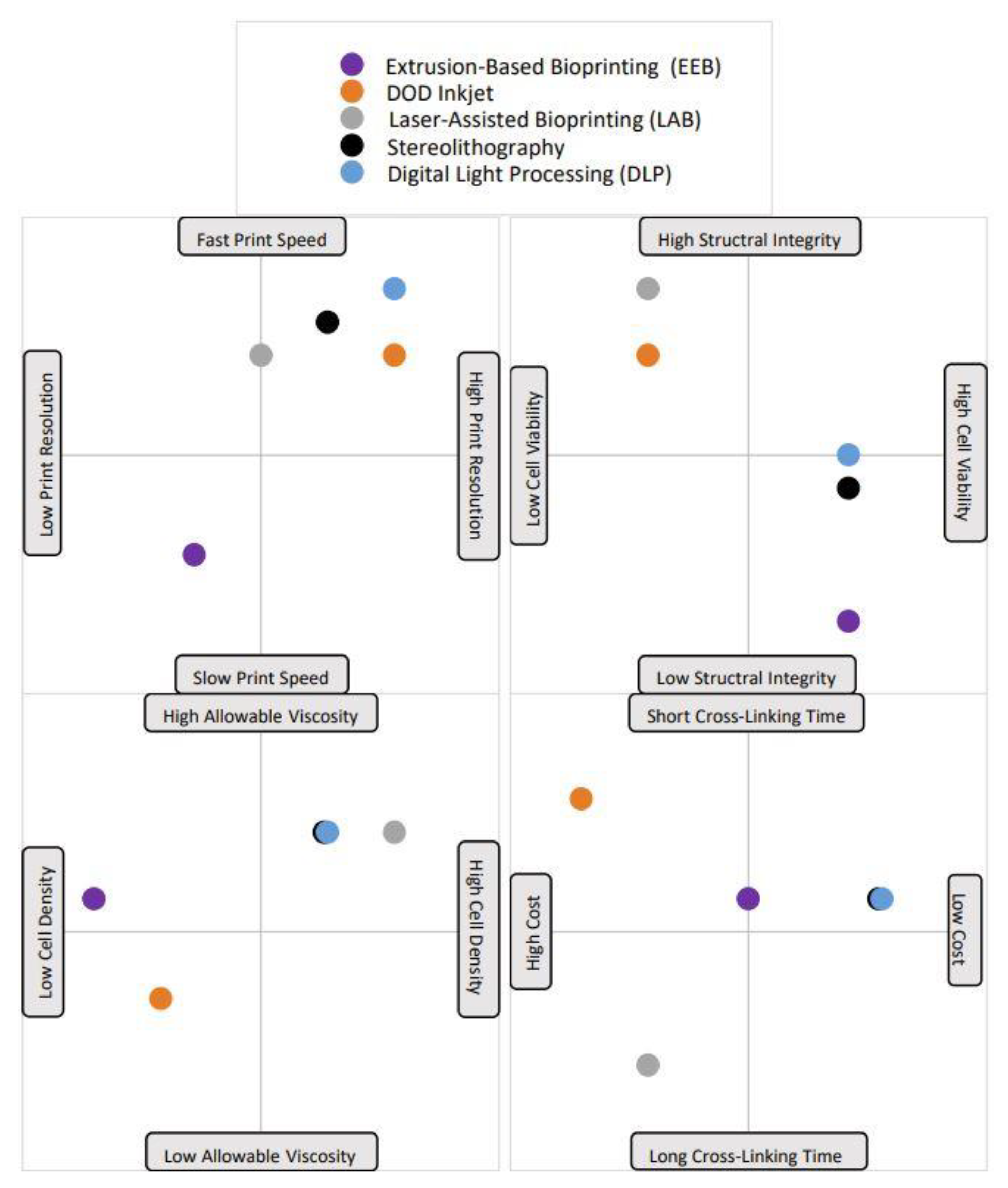
Figure 2.
A visual representation of each bioprinting method’s advantages and disadvantages. Notably, each point is plotted based on the average value for that trait; some printing methods have very variable trait characteristics, but the standard value has been selected and is shown in the plot. For example, the printing speed of the droplet-based DOD inkjet method is highly variable and depends greatly on material viscosity. The majority of the time, low material viscosities are used with DOD, which leads to fast printing speeds, which is what is depicted here.


Table 1.
The five (5) bioprinting process methods, a brief description of each, and a thorough comparison analysis of benefits and limitations. Eight criteria were selected to evaluate each process. The criteria are listed in order as follows: printing speed, cell resolution, structural integrity, post-printing cell viability, cross-linking time, allowable bioink viscosity, cell density, and cost. A ninth is present for methods with additional important criteria to consider.
| Method | Description | Advantages | Neutral | Disadvantages |
|---|---|---|---|---|
| Extrusion-Based Bioprinting (EBB) | Mechanical or pneumatic pressure creates continuous streams of bioink | Slow printing speeds | ||
| Low printing resolution | ||||
| High structural integrity | ||||
| Low cell viability | ||||
| Cross-linking time | ||||
| Must use low-viscosity bioinks (risk of nozzle clogging) | ||||
| High cell density allowable (although nozzle clogging is more likely) | ||||
| Average cost | ||||
| DOD Inkjet | Thermal, piezoelectric, or electrostatic mechanisms create droplets | Fast printing speeds | ||
| High printing resolution | ||||
| Low structural integrity | ||||
| High cell viability | ||||
| High cross-linking time | ||||
| Requires low-viscosity bioinks (risk of nozzle clogging) | ||||
| Low cell density (to reduce nozzle clogging) | ||||
| Low cost | ||||
| Laser-Assisted Bioprinting (LAB) | Laser pulses create areas of high pressure which force droplets onto a substrate | Medium printing speed | ||
| High printing resolution | ||||
| Low structural integrity | ||||
| Very high cell viability | ||||
| High cross-linking time | ||||
| Wide range of viscosities, low to very high (no nozzle, no nozzle clogging) | ||||
| Medium cell density (no nozzle clogging) | ||||
| High cost, not scalable | ||||
| Prone to metallic contaminants | ||||
| Stereolithography | Light polymerizes bioink in a layer-by-layer process | Medium printing speed | ||
| High printing resolution | ||||
| High structural integrity | ||||
| Medium cell viability | ||||
| Low cross-linking time | ||||
| Low to medium viscosity (no nozzle, no nozzle clogging) | ||||
| Medium cell density (no nozzle clogging) | ||||
| Medium cost | ||||
| UV radiation can cause cell and DNA damage | ||||
| Digital-Light Processing (DLP) | Light reflects off thousands of micromirrors and polymerizes whole layers at a time | Fast printing speed | ||
| Very high printing Resolution | ||||
| High structural Integrity | ||||
| Medium cell viability | ||||
| Low cross-linking time | ||||
| Low to medium viscosity (no nozzle, no nozzle clogging) | ||||
| Medium cell density (no nozzle clogging) | ||||
| Medium cost | ||||
| UV radiation can cause cell and DNA damage |
Table 2.
The three main drug delivery system methods discussed A brief description, as well as comparative advantages and disadvantages, are highlighted for each method.
Table 2.
The three main drug delivery system methods discussed A brief description, as well as comparative advantages and disadvantages, are highlighted for each method.
| Method | Description | Advantages | Disadvantages |
|---|---|---|---|
| Microspheres | Small drug-loaded particles made from synthetic or natural polymers | High degree of flexibility in properties that can be chosen based on materials used | Decreased biological activity of item encapsulated by microsphere due to immunogenic responses, instable or denaturing proteins |
| Fibrin Glue | Fibrinogen and thrombin solution are mixed with drug then combined, cross-linking occurs, and the fibrin gel matrix is formed | Adjustable pore diameter, size, shear modulus, and tensile strength | Drug cannot be used if it does not combine with matrix properly; short degradation period |
| Nanoparticles | Particles with dimensions in the nanometer range, often referred to as nanocarriers | Extremely small particle size allows for operation on the cellular level and the ability to cross the blood–brain barrier | Lack of data and results on how nanoparticles effect pathways and processes of human body |
Table 3.
A summary of recently published bioprinted drug delivery systems for nerve tissue regeneration. It includes names of the studies, details on what the study was testing, and the results of each study.
Table 3.
A summary of recently published bioprinted drug delivery systems for nerve tissue regeneration. It includes names of the studies, details on what the study was testing, and the results of each study.
| Study | Nerve Type | Printing Method | Drug Delivery System | Drug or Growth Factor | Cell Type | Results |
|---|---|---|---|---|---|---|
| Chen et al., 2020 | Peripheral | EBB | Chitosan Microspheres | NGF | PC12 and RSC96 Schwann Cells | NGF achieved sustained release; scaffold improved neurite growth and extension [30]. |
| Wu et al., 2023 | Peripheral | DLP | Nanoassemblies | 7,8-DHF | N/A | Sustained release of 7,8-DHF improved axonal elongation; scaffold promoted Schwann cell adhesion, proliferation, and migration [31]. |
| Tao et al., 2019 | Peripheral | DLP | MPEG-PCL Nanoparticles | XMU-MP-1 | N/A | Drug increased axon diameter, myelin thickness, NCV, and MAP [32]. |
| Xu et al., 2019 | Peripheral | DLP | MPEG-PCL Nanoparticles | RGFP966 | N/A | Conduit bridged 10 mm cut in rat sciatic nerves; remyelination of Schwann cells; drug improved functional restoration [33]. |
| Chen et al., 2018 | Spinal Cord | S | Collagen Binding Domain | NT3 | N/A | Conduit + NT3 improved axonal regrowth, remyelination, and hindlimb motor function in rats [34]. |
| Liu et al., 2021 | Spinal Cord | Low-temp EBB | Degradation | BDNF | N/A | BDNF achieved sustained release; conduit + BDNF significantly improved hindlimb motor function in rats after complete spinal cord transection [35]. |
| Song et al., 2023 | Spinal Cord | Not Stated | Degradation | OMT | Cultured with NSCs | OMT achieved sustained release over 4 weeks; NSCs in scaffold + OMT group had higher survival rates; scaffold + OMT improved motor function recovery [36]. |
| Lee et al., 2010 | Cerebral | Inkjet | Fibrin Glue | VEGF | C17.2 cell line | Bioink + VEGF group had post-printing cell viability of 93.23 ± 3.77% and NSCs showed signs of differentiation after 3 days of culture [37]. |
| Sharma et al., 2020 | Cerebral | EBB | Microspheres | Guggul- sterone | hiPSC-derived NSCs | Drug + scaffold + NSC group had highest post-printing cell viability; cells showed markers consistent with dopaminergic neurons at days 30 and 45 [38]. |
| De la Vega et al., 2021 | Cerebral | EBB | PCL Microspheres | RA + puro | hiPSC-derived NPCs | Drug + scaffold + NPC group had the highest cell viability on day 1; NPCs were differentiated into different neuronal types; membrane potentials were responsive to Ach and GABA [26]. |
Disclaimer/Publisher’s Note: The statements, opinions and data contained in all publications are solely those of the individual author(s) and contributor(s) and not of MDPI and/or the editor(s). MDPI and/or the editor(s) disclaim responsibility for any injury to people or property resulting from any ideas, methods, instructions or products referred to in the content. |
© 2024 by the authors. Licensee MDPI, Basel, Switzerland. This article is an open access article distributed under the terms and conditions of the Creative Commons Attribution (CC BY) license (https://creativecommons.org/licenses/by/4.0/).
Share and Cite
MDPI and ACS Style
Steele, E.M.; Carr, Z.L.; Dosmar, E. Bioprinting of Hydrogel-Based Drug Delivery Systems for Nerve Tissue Regeneration. Biophysica 2024, 4, 58-73. https://0-doi-org.brum.beds.ac.uk/10.3390/biophysica4010004
AMA Style
Steele EM, Carr ZL, Dosmar E. Bioprinting of Hydrogel-Based Drug Delivery Systems for Nerve Tissue Regeneration. Biophysica. 2024; 4(1):58-73. https://0-doi-org.brum.beds.ac.uk/10.3390/biophysica4010004
Chicago/Turabian StyleSteele, Eliza Marie, Zacheus L. Carr, and Emily Dosmar. 2024. "Bioprinting of Hydrogel-Based Drug Delivery Systems for Nerve Tissue Regeneration" Biophysica 4, no. 1: 58-73. https://0-doi-org.brum.beds.ac.uk/10.3390/biophysica4010004