Article

Development of Chitosan Films from Edible Crickets and Their Performance as a Bio-Based Food Packaging Material

1
Department of Food Science, Purdue University, 745 Agriculture Mall Dr., West Lafayette, IN 47907, USA
2
School of Engineering Technology, Purdue University, 401 N. Grant St., West Lafayette, IN 47907, USA
3
Department of Entomology, Purdue University, 901 W. State St., West Lafayette, IN 47907, USA
*
Author to whom correspondence should be addressed.
Submission received: 31 August 2021 / Revised: 21 September 2021 / Accepted: 23 September 2021 / Published: 1 October 2021
(This article belongs to the Special Issue Polysaccharides for Application in Packaging)

Abstract

:
Edible insects have gained attention due to their impressive nutritional composition, as well as their efficient use of natural resources. However, a research gap remains on the applications of insect chitosan, especially as it relates to their potential use as food packaging material. Chitosan from two reared cricket species (Acheta domesticus and Gryllodes sigillatus) was evaluated for use as food packaging material. Cricket chitosan films (CCF) were structurally similar to commercial shrimp chitosan films (SCF) at controlled glycerol levels, as seen by shared spectral peaks in FT-IR analyses. Mechanical properties of CCF showed they had equal or greater tensile strength when compared to commercial SCF, although flexibility was lower. Scanning electron microscopy showed increased roughness of microstructure, likely increasing the tortuosity. As a result, CCF had improved water vapor permeability compared to commercial SCF. Melanin complexes present in cricket chitin and chitosan increased hydrophobicity and decreased light transmittance. This study also revealed that intrinsic species differences, which occur during insect and crustacean exoskeleton development, could have effects on the functionality of chitosan packaging materials. Overall, CCF were found to be as effective as commercial SCF, while providing additional advantages. CCF derived from reared crickets have good mechanical and barrier properties, and improved water resistance and light barrier characteristics. Edible cricket chitosan has the potential to be used as bio-based packaging material for food and pharmaceutical applications.

Graphical Abstract

1. Introduction

Currently used in the food industry, petroleum-based food packaging has excellent mechanical and barrier properties to extend food quality and shelf life while providing safe food and convenience to consumers. However, these non-renewable plastics also significantly contribute to the accumulation of waste. This waste has a detrimental effect on the environment by generating a high amount of waste that is destined for landfills or enters ecological systems. Currently, the load of these petroleum-based polymers on the environment is approximately 335 million tons of plastic per year [1]. Therefore, research and production of bio-based packaging materials to replace traditional packaging materials remains a major focus of many studies. Government programs, such as BioPreferred® in the U.S., support and increase the use of bio-based products, including bio-based packaging materials, from renewable sources. Simultaneously, the world is challenged with providing sustainable and resource efficient solutions to reduce food waste and spoilage, which is expected to account for 200 million tons by the year 2050 [1]. The ability to utilize process waste streams for the production of biobased and biodegradable packaging materials is also of great interest since it supports both renewable bio-based packaging while simultaneously reducing waste otherwise destined for landfills [2]. Scientific literature on chitosan biobased polymers shows extensive research performed on the preparation, characterization, and applications of chitosan-based polymer films over the past three decades. This research supports all of these resolutions for decreased food waste and potential substitution of synthetic food packaging.
Chitosan films have been shown to have excellent performance as food packaging materials allowing for shelf-life extension of foods, including antimicrobial, barrier, and sensing films [3]. Commercial chitin and chitosan, sourced from crustacean (e.g., shrimp) food-waste streams, have been used to create food packaging that serves as a physical barrier to food, as well as blended with other active ingredients such as antimicrobials to create active and biodegradable food packaging with shelf-life extension abilities [3,4,5,6,7,8,9,10,11,12,13]. As consumers are beginning to shift to more sustainable protein sources, such as edible insects, new waste streams are becoming available. Like crustaceans, insects are arthropods and have similar chitin rich exoskeletons. As a result, a focus of recent chitosan research sourced from insects was recently reviewed [14,15]. The reviews highlight extraction and purification of insect chitin, and characterize the physicochemical properties of chitin and its modification to chitosan.
Although there is now a foundation of insect chitin and chitosan literature available showing feasibility of alternative sourcing, there is little information on the application and feasibility of insect chitosan for use as food packaging materials. Therefore, this research study focuses on the manufacture of chitosan films sourced from two edible cricket species commonly reared in the United States and their application as food packaging materials. Important properties of food packaging polymers, including mechanical, vapor permeability, optical, light barrier, and hydrophobicity/hydrophilicity properties, were studied on cricket chitosan films (CCF). Shrimp chitosan films (SCF) served as the commercial reference material, to determine if alternative sourcing would improve or worsen the desirable functional properties.

2. Materials and Methods

2.1. Materials

All materials and chemical reagents were purchased from Fisher Scientific (Waltham, MA, USA) and Sigma Aldrich (St. Louis, MO, USA), unless specified. The two cricket species, Acheta domesticus (house cricket) and Gryllodes sigillatus (tropical banded cricket), were obtained from two edible cricket rearing facilities, Ovipost, Inc. (Labelle, FL, USA) and Three Cricketeers, LLC (St. Louis Park, MN, USA), respectively. Commercial shrimp chitosan (~70% deacetylated) was purchased from Sigma Aldrich (St. Louis, MO, USA).

2.2. Methods

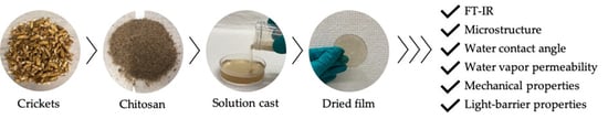
2.2.1. Cricket Chitosan

Cricket chitin was extracted from a chitin-rich by-product produced during the enzymatic proteolysis of cricket proteins for food formulation [16,17]. In our previous study, cricket chitin from each species was effectively demineralized and deproteinized with acidic and alkali treatments [18]. Furthermore, cricket chitosan was deacetylated with concentrated and hot alkali treatment for varying durations. As previously optimized, cricket chitosans were produced with various DDA values as determined by FTIR, and within each cricket species the DDAs produced were found to be significantly different [18]. Deacetylation parameters for each cricket species were then chosen to produce a similar range of DDA values: 72, 76, and 80%. For more specific details on the extraction, deacetylation, optimization process, and molecular weight determination of these samples, the reader is referred to Malm and Liceaga [18].

2.2.2. Solution Casting Films

For each cricket species, Acheta domesticus and Gryllodes sigillatus, chitosan films were prepared with varying degrees of deacetylation: 72, 76, and 80%. Chitosan solutions (1% w/v) were prepared by dissolving cricket chitosan in 1% acetic acid (v/v) solution and stirred for 60 min on a hotplate (100 °C). Evaporation of solutions were minimized by placing a foil lid across the beaker openings, which were then secured with parafilm around the circumference of the beaker. The plasticizing agent, glycerol, was added at 37.5% (w/w chitosan), covered, and mixed without heat for an additional 10 min. Plasticized solutions were sonicated for 5 min (Model CL- 334, QSONICA Sonicators, Newtown, CT, USA) at 30% amplitude followed by centrifugation (Avanti J-26S, Beckman-Coulter, Brea, CA, USA) for 15 min at 17,636× g to degas and remove undissolved particles. For mechanical and water contact analyses, 40 g of supernatant was dispensed in 100 × 15 mm polystyrene dishes and dried for 36 h at 50 °C. For the remaining analyses, 15 g of film solutions were dispensed in 60 × 15 mm polystyrene dishes and dried for 24 h at 50 °C. Afterwards, all films were placed in desiccators until needed. These processes were repeated using commercial shrimp chitosan (~70% deacetylated) and served as the chitosan film reference.

2.2.3. Molecular Characterization

The molecular characterization of all chitosan films was analyzed as previously described [18] using Fourier Transform Infrared spectroscopy (FT-IR) (Thermo Scientific, Waltham, MA, USA) from 3500 to 800 cm−1 with a resolution of 8 cm−1 (n = 4). Spectra were obtained via film transmission using ATR with a diamond prism, and automatically baselined and averaged using OMNIC software (ThermoFisher, Waltham, MA, USA).

2.2.4. Scanning Electron Microscopy (SEM)

Scanning electron microscopy of film surfaces and cross-sections were evaluated for CCF and SCF. For cross-sections, specimens were cryo-fractured prior to analysis. All specimens were sputter coated with platinum for 60 s, and then analyzed (FEI NOVA nanoSEM Field Emission SEM (FEI Company, Hillsboro, OR, USA) with an accelerating voltage of 5 kV and under high vacuum. Film surface images are reported at 500× magnification, and film cross-sections are reported at 2000× and 20,000× magnification.

2.2.5. Water Contact Angle Analysis

The surface hydrophobicity/hydrophilicity of all CCF and SCF was evaluated by water contact angle analysis (Theta Lite Tensiometer, Biolin Scientific, Gothenburg, Sweden) [19]. Duplicate film strips, approximately 5 cm2, were secured onto microscope glass slides with double sided tape. Deionized water (2 µL) was manually placed onto each film surface. Contact angles were recorded by the instrument’s camera, manually baselined during analysis using the OneAttension software (Biolin Scientific) and averaged (n > 7).

2.2.6. Water Vapor Permeability

The water vapor permeability of CCF was measured and compared to the SCF using the procedure established by ASTM standard E96, as previously described [20]. Desiccant (CaCl2), previously dried, was placed in the bottom of the permeability cups. Cups’ openings were sealed with chitosan films and secured by metal clamps. Initial weight of each cup was immediately documented and placed in a 75% RH chamber (saturated NaCl solution) at 25 °C. Weight measurements were recorded twice a day for ~4 days for each permeability cup, resulting in six measurements per cup. Water vapor transmission rate (WVTR) was calculated by dividing the slope of the linear regression of weight measurements (Δm/Δt) by the permeation area of the cup (m2). Water vapor permeability of films was determined as follows:
W V P   ( 10 10 g / s · m · Pa ) = W V T R × d Δ p
where d represents the thickness of chitosan films (m), and Δ p is the partial pressure difference previously determined to be 1753.55 (Pa) [20,21].

2.2.7. Mechanical Properties

Tensile strength and elongation percentage at break measurements were performed on CCF and SCF following ASTM D882 standards with modifications [20,22]. Chitosan films were conditioned at least 48 h in 50% RH desiccators (saturated MgNO3 solution) at 25 °C, and then cut into strips (6 cm × 1 cm). Thickness of each strip was determined in triplicate prior to analysis. A texture analyzer (TA.XT, Stable Microsystems, Surrey, UK) equipped with rubber faced tensile grips was calibrated and experiments were performed using a grip separation of 30 mm with a rate of separation of 0.80 mm/s (n 8). Analysis of data was performed (Exponent Software Stable Microsystems, Surrey, UK), and TS and E% were calculated for each replicate.

2.2.8. Color Measurement

Chitosan films were evaluated for color using a Hunterlab ColorFlex (HunterLab, Reston, VA, USA) (n = 4) following the previously described protocol [20]. Using CIELAB scale, color measurements were reported as lightness, L*, and chromaticity parameters a* and b*. Lightness values were reported between 0 (black) and 100 (white). Chromaticity parameter a* indicates green colors when negative, while positive values represent red colors. Finally, positive chromaticity b* values correspond to blue colors, while negative values correspond to yellow colors. Four films were evaluated for each type of chitosan film, measured against a white tile (L* = 92.22, a* = −0.99, and b* = 0.92).

2.2.9. Light Barrier Properties

To evaluate the light barrier protective properties of chitosan films for food packaging applications, transmittance (%) of each film was scanned from 200 to 800 nm using a spectrophotometer (DU®720, Beckman-Coulter, CA, USA), as previously described [22]. Chitosan films were physically attached to sample holders, where air served as the reference. Scans were repeated on four films for each film formulation.

2.2.10. Opacity

Utilizing the transmittance data obtained from the light barrier property experiments, the opacity of the chitosan films was calculated using the following equation:
O p a c i t y   ( mm 1 ) = A b s o r b a n c e 600   nm t h i c k n e s s   ( mm )
where were Absorbance600nm = 2- log (T %600nm) [23].

2.2.11. Statistical Analysis

All experimental data were replicated at least three times, unless otherwise indicated. Data are reported as average ± standard deviation. Where applicable, statistical analysis of observed differences among means was performed using analysis of variance (ANOVA), followed by Tukey’s pairwise comparison of means at a 5% significance level with the statistical software Minitab 18® (State College, PA, USA).

3. Results

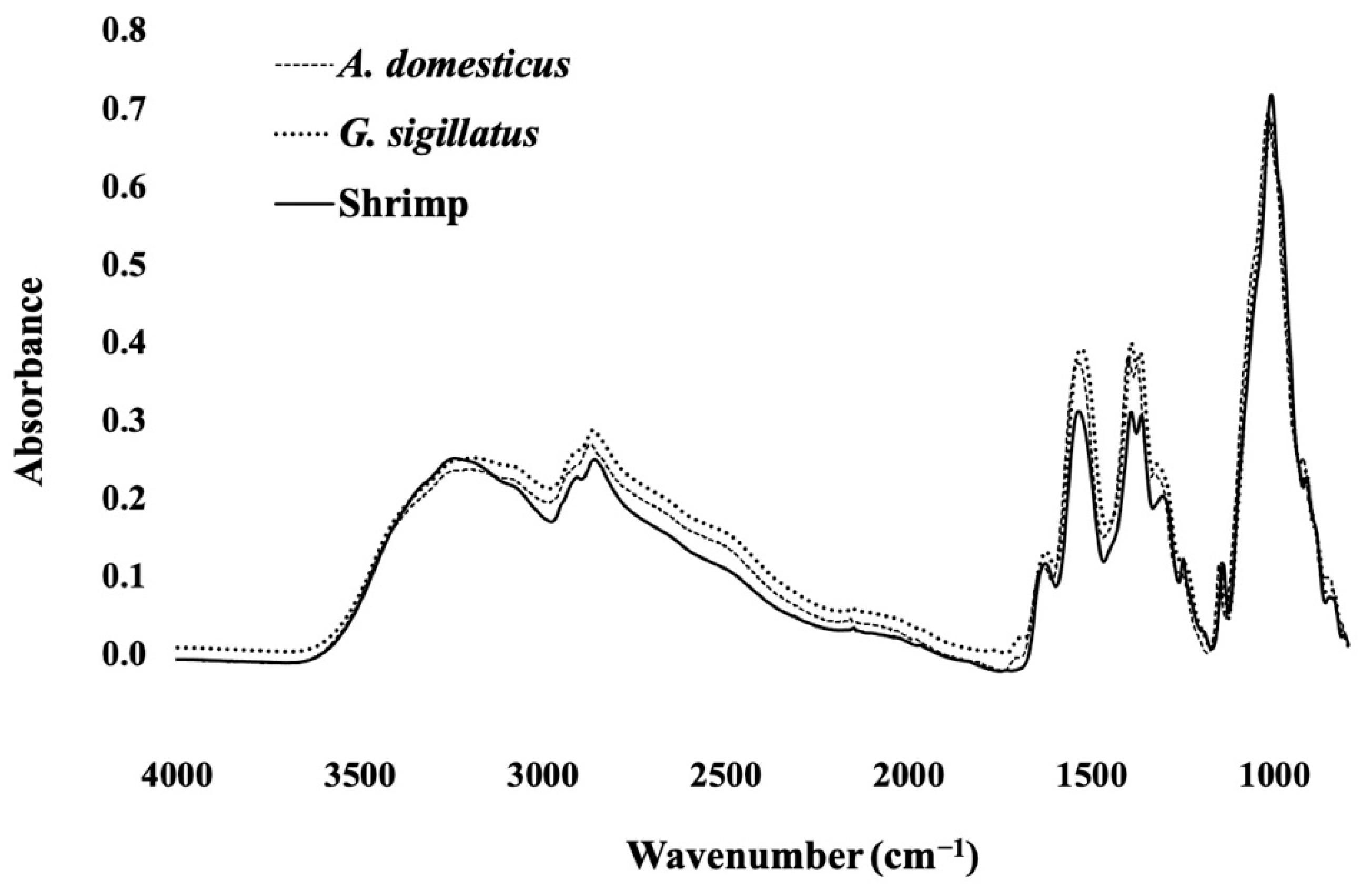
3.1. Molecular Characterization

Molecular characterization of CCF (Figure 1) aligned well with spectra reported in previous chitosan-glycerol plasticized films characterized by FT-IR [18,24,25]. Additionally, CCF were not structurally different that SCF, as shown by the absence of spectral differences between all spectra. Peaks between 3500 and 3000 cm−1 represent N-H and O-H chemistries of both chitosan and glycerol, as well as the hydrogen bonding between the two functional groups due to plasticization [26]. Furthermore, increased intensities of aliphatic stretching moieties (2970 cm−1 to 2750 cm−1), and C–O, C–O–C asymmetrical, and C-O-C symmetrical groups (1120 to 800 cm−1), are also a result of the glycerol’s intrinsic chemistry [24]. Comparison of pristine chitosan powder, previously published [18], and the corresponding plasticized chitosan films, highlighted the increased intensity of two spectral peaks. Specifically, the broad 3500–3000 cm−1 peak mentioned above and the amide II band centered ca. ~1530 cm−1. The increase of these peaks may be associated with the chitosan-glycerol plasticization mechanism. As shown previously by NMR, glycerol’s –OH functional group participates in hydrogen bonding with the acetoamide group of chitosan [27]. Ultimately, these hydrogen bonds allow plasticization of films and may be the cause for the increased 3500–3000 cm−1 peak intensity. Additionally, glycerol interrupts intramolecular hydrogen bonds (N–H functional groups) on chitosan’s polymer structure [27,28]. Therefore, it is possible the disruption of these intramolecular bonds increases their availability and stretching of N–H functional groups. Ultimately, this could lead to an increase of the amide II band of plasticized chitosan films, as the chemical structure of glycerol does not directly contribute to this peak. Additional future experiments with pristine unplasticized chitosan films would be useful to further deconvolute the roles of solution casting and plasticization on these two functional groups of chitosan.
Finally, one difference between CCF and SCF may be a result of the intrinsic differences between the molecular conformation of insect and crustacean exoskeletons. In insects, hardening of the exoskeleton is achieved through a process noted as sclerotization. The exact mechanisms of sclerotization of different insects is yet to be discovered; however, research to date generally accepts that insect exoskeletons are formed through chitin and protein matrices that are crosslinked by catechols to stabilize the cuticle [29]. During sclerotization, catechols undergo a tanning process leading to brown and black pigments, complexed with chitin in the exoskeleton structure [30]. As shown visually in the films’ optical properties, some pigments are still bound to chitin/chitosan structure. The pigmentation is documented in most insect chitin/chitosan studies; however, pigmentation has not been detectable by FT-IR. For example, in one study, chitosan films derived from yellow mealworms (Tenebrio molitor) also showed similar pigmentation as our CCF, but characteristic peaks of such pigments could not be distinguished from the FT-IR spectral peaks of chitosan films plasticized with glycerol [31]. The authors attributed these results to overlapping chemistry with chitosan and glycerol, as well as the fact it is present in low quantities.

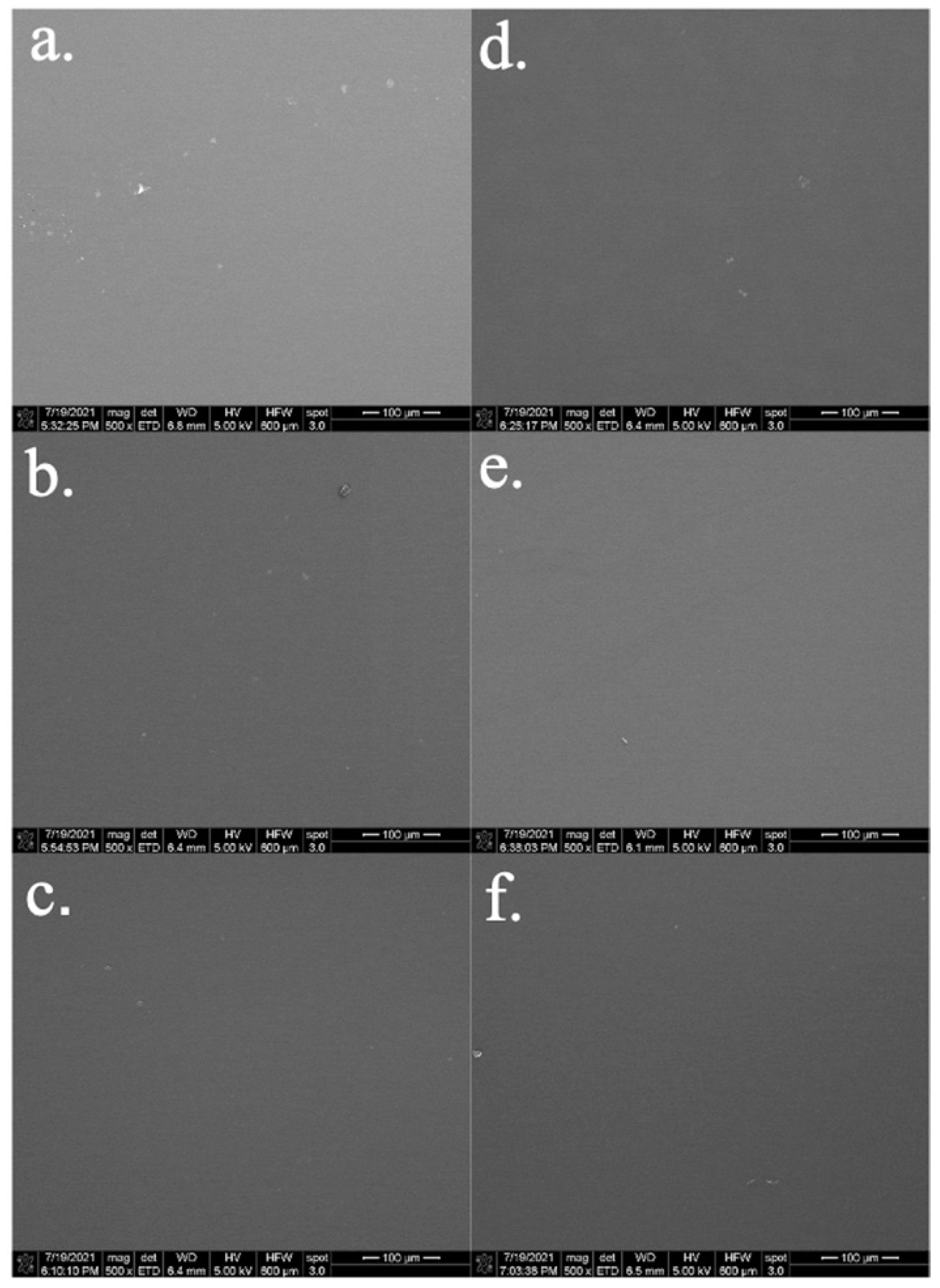
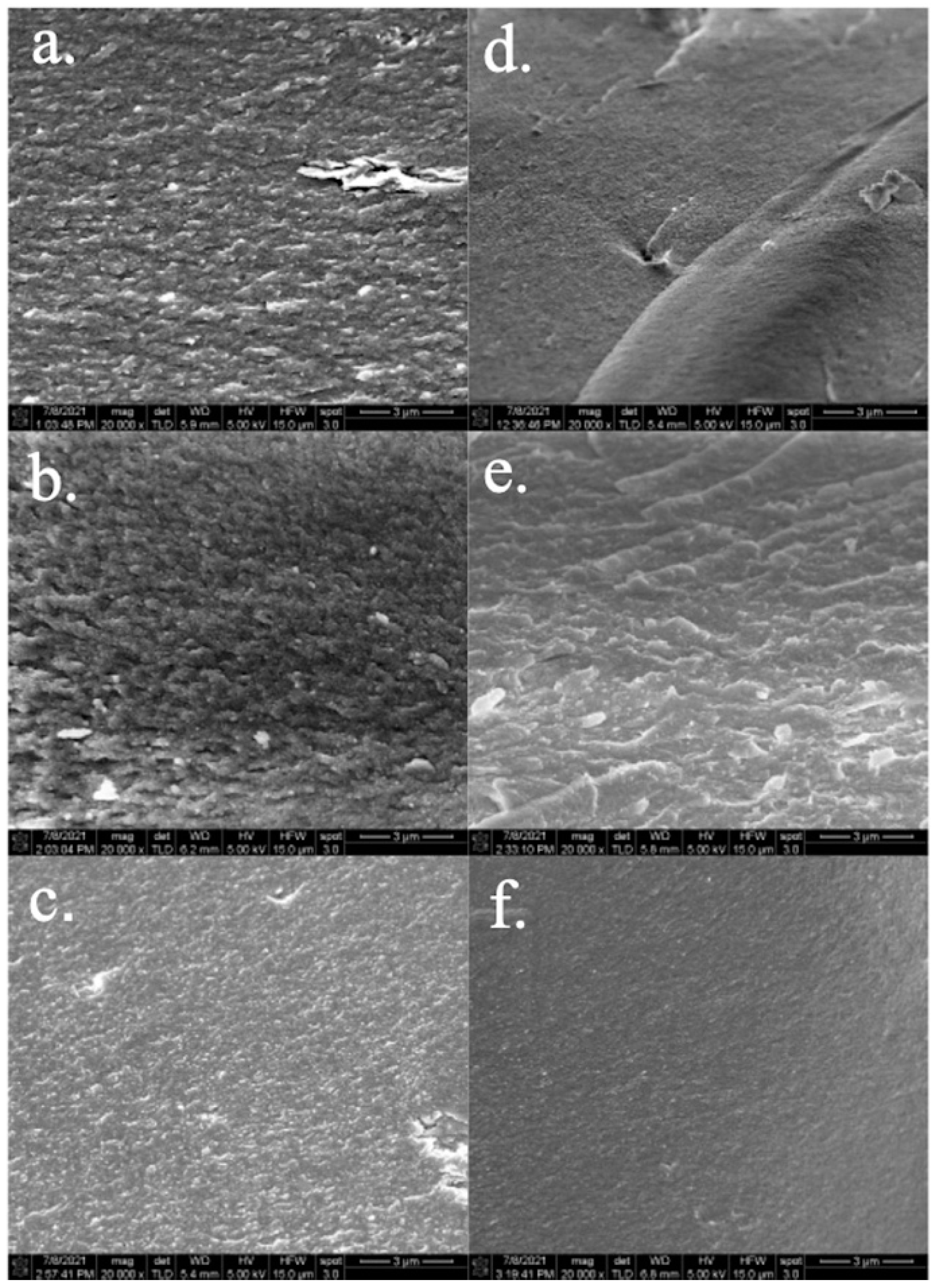
3.2. Film Microstructure by Scanning Electron Microscopy

Surfaces and cross-sections of CCF imaged using scanning electron microscopy are shown in Figure 2 and Figure 3 and compared to SCF (Supplementary material Figure S1). Overall, images of all CCF surfaces (Figure 2) and cross-sections (Figure 3 and Supplementary material Figure S2) of films showed good homogeneity, were smooth, and free of cracks and pores. These results are in alignment with previously studied chitosan films plasticized with glycerol and sorbitol [32,33]. Imaging of cross-sections at 20,000× (Figure 3) elucidates differences in the microstructures of CCF compared to the commercial SCF (Supplementary material Figure S1). Generally, CCF had larger chitosan particles, whereas SCF had smaller and smoother-looking particles. The scanning electron microscopy cross-sections of CCF are visually similar to chitosan-gelatin cross-linked films [34]. The authors described the crosslinked film cross-sections to be more aggregated and irregular compared to the SCF control. The aggregation of the micrographs were attributed to crosslinks formed between chitosan and gelatin in the film’s 3D network [34].
Considering the chitosan–gelatin crosslinking study, the CCF reported here are believed to be complexed with residual melanin, intrinsic to cricket exoskeletons, that could also be more aggregated when compared to the SCF (Supplementary material Figure S1). Furthermore, the differences in structure may also be due to cricket chitosan’s lower molecular weight (344 and 524 kDa) [18]. Liu, Yuan [35] reported that higher molecular weight chitosan (110 kDa) promoted a more compact glycerol-chitosan film structure, seen by their scanning electron microscopy images of smoother cross-sections. Additionally, as the degree of deacetylation of cricket chitosan increased, the cross-section of films became smoother and less aggregated (Figure 3c,f). Previous reports indicate that as the degree of deacetylation of chitosan increases, its polymer chains can pack more closely due to the decreased presence of bulky acetyl groups [36]. Therefore, increasing the degree of deacetylation of CCF may allow greater packing of chitosan polymer chains leading to the compact chitosan film structure at higher deacetylation.

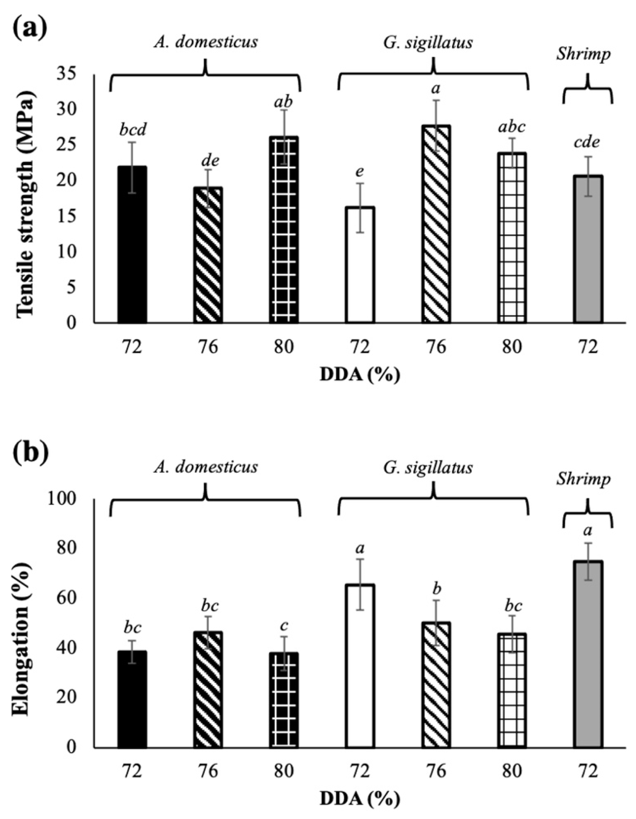
3.3. Mechanical Properties

In this study, glycerol was used as a plasticizing agent at a constant concentration to compare the mechanical effects (tensile strength and elongation) of the different sources of the chitosan polymer, as well as identify any effects caused by the chitosan’s molecular weight and the degree of deacetylation. In all cases, CCF had similar, or greater, tensile strength (TS) when compared to SCF (Figure 4a). These results suggest good intramolecular bonding between cricket chitosan, as previously described by microstructure analysis. Additionally, the degree of deacetylation of cricket chitosan had no clear effect on the corresponding film’s TS. Although all CCF had TS values that were similar, or greater than SCF, there were significant differences in elongation percentages (E%) (Figure 4b). All CCF, with the exception of 72% deacetylated G. sigillatus chitosan films, had significantly lower E% values (p < 0.05). The mechanical results of this study were comparable to other commercial chitosan films prepared similarly [23,37,38]. For example, commercial chitosan plasticized films (30% glycerol w/v), with varying molecular weights, had TS values between 24–32 MPa and E% values of 30–37% [38]. Furthermore, CCF in this study had improved properties compared to chia seed mucilage films we have previously studied, which were plasticized with sorbitol and glycerol and had TS values of 0.38–2.7 MPa and 21–68% E% [20]. The molecular weight of chitosan is also thought to play an important role in the mechanical properties of films. Specifically, an increase in molecular weight typically results in an increased TS and E% due to the increased entanglements of the polymer network [39,40]. As previously reported, A. domesticus and G. sigillatus chitosan have molecular weights of 344 and 524 kDa [18]. However, even though their molecular weights are smaller than commercial shrimp chitosan, the CCF showed excellent mechanical properties as seen by equal or improved TS compared to SCF (Figure 4a).
Currently, only two studies on insect chitosan films that characterize mechanical properties can be found in literature. The first study, chitosan films from mealworms (Tenebrio molitor) (812 kDa) and grasshoppers (Brachystola magna) (696 kDa) were found to have statistically similar TS and E%, when compared to the study’s low (759 kDa) and medium (870 kDa) molecular weight reference chitosan films [31]. In another study, grasshopper (B. magna) chitosan (322 kDa) films were compared to high, medium, and low molecular weight crustacean chitosan films [41]. They showed that grasshopper films had significantly lower TS values than high and medium molecular weight commercial chitosan, but films were statistically similar to low molecular weight crustacean chitosan. Additionally, the authors found that grasshopper chitosan films had lower E% than low and medium molecular weight crustacean chitosan.
One explanation for varied results on mechanical properties of insect chitosan films, compared to crustaceans, may originate from the intrinsic properties of insect exoskeletons. Although not completely understood, it is thought that the matrices of insect cuticles are comprised of proteins and chitin, as well as other components such as melanin. The sclerotization process, responsible for the hardening of insect exoskeleton cuticles, is often associated with the “tanning” of the exoskeleton [42]. Another process, melanogenesis, which occurs during insect cuticle formation, is responsible for the formation of melanin [43]. Although these two processes are separate, they are believed to be closely related and occur simultaneously, with melanin likely to be a product of both processes [42,43]. Research to understand the interactions among of all these components in the exoskeleton of arthropods is currently ongoing; however, studies on chitin extraction from these sources suggest that melanin could covalently bind to chitin [44,45,46]. These assumptions are based on the inability to chemically remove all pigments, presumably melanin, from insect-extracted chitin [45,47]. One research group recently filed a patent, where extractable chitin from the black soldier fly, was chemically converted into chitosan with melanin still covalently bound [30,46]. In our results, similar pigmentation of chitin, chitosan, and corresponding chitosan films can be observed, suggesting that crickets could also have melanin covalently bonded to its chitin, making it unable to be removed during the purification and conversion processing steps [18].
When considering the structural complexity of insect chitin/chitosan, residual melanin may also be a contributing factor to its final properties, including its mechanical characteristics. In an in vitro biomimetic study of insect cuticles, chitosan was found to be covalently crosslinked to melanin, leading to corresponding films (unplasticized) to be stronger and stiffer than commercial crustacean chitosan films [44]. In another study, chitin–melanin complexes were extracted from dung beetles (Catharsius molossus, (Linnaeus)), converted to chitosan–melanin complexes, and formed into unplasticized films [48]. Authors attribute the increased TS of the beetle chitosan films, compared to commercial shrimp chitosan, as a result of the intramolecular interaction between chitosan and melanin. Therefore, the increased TS of CCF may too be a result of the covalent melanin crosslinks, and increased intramolecular bonding that is not present in SCF. Additionally, these chitin–melanin interactions, paired with their lower molecular weights, may explain the decreased elongation of CCF.
Overall, taking into consideration the research available on both insect and crustacean chitosan films plasticized with glycerol, CCF have similar mechanical properties to crustacean films. These findings suggest that CCF may produce packaging materials that are as strong as SCF, but may not be as flexible. However, if high flexibility is required, SCF could be further manipulated to meet performance expectations. For example, additional plasticizing agents could be used and/or their concentrations optimized. As research progresses on insect exoskeleton sclerotization, insect chitin/chitosan and their complexes, greater mechanistic understanding of insect chitosan films is expected.

3.4. Water Vapor Permeability (WVP)

The water vapor permeability is an important parameter when evaluating food packaging materials. Food quality, and safety in some circumstances, require the maintenance of water activity. Therefore, the ability of a food packaging polymer to decrease water vapor migration is an important property to understand and characterize. In this study, the water vapor permeability of chitosan films from crickets was determined and compared to commercial SCF (Table 1). All films derived from cricket chitosan had decreased WVP values compared to SCF. Furthermore, chitosan films from A. domesticus were slightly less water permeable than G. sigillatus films. It is reported that the WVP of chitosan films is directly affected by glycerol, where glycerol allows the permeation of water through the chitosan-glycerol matrix [49].
Little research is available on the water vapor permeability on insect chitosan films, making it difficult to understand the molecular differences responsible for the decreased film WVP. In one study, the WVP of glycerol-chitosan films from grasshoppers was evaluated and compared to low, medium, and high molecular weight commercial chitosan [41]. The authors reported grasshopper chitosan films had the lowest WVP (1 × 10⁻¹⁰ g/m·s·Pa) compared to all shrimp chitosan films (1.6 to 6.43 × 10⁻¹⁰ g/m·s·Pa). The authors attribute the improved WVP of the grasshopper films to be a result of its greater compacted matrix; however, they did not support these conclusions with scanning electron microscopy microstructure analysis. In our study, considering microstructure analysis using scanning electron microscopy, CCF film microstructures appear to be more rough and aggregated, leading to an increased tortuous path length for water vapor to diffuse across the membrane [50,51]. Ultimately, this would lead to decreased water vapor permeability of CCF, compared to the smoother and more compact microstructure of chitosan films which would have a decreased path length for water vapor to travel. Further research on insect chitosan biopolymers, and their microstructures, is needed to make comprehensive conclusions on functional differences between insect and crustacean films.

3.5. Color and Optical Properties

Color measurements showed CCF were light brown, compared to the slight yellow color of the SCF. The color of all films was compared quantitatively using L*, a*, and b* values (Table 1). Cricket films were darker than shrimp films, as shown by their lower L* values, but overall, the cricket films from the different species had very similar lightness values. CCF also had positive a* and larger b* chromaticity parameter values, indicating that films were less green and more yellow in comparison to the commercial shrimp films. Melanin, as already discussed, results in a tan pigment and therefore a likely contributor to the increased darkness and yellowness of the CCF. The appearance and color of CCF films are very similar to crustacean chitosan films with melanin nanoparticles, although the study did not quantitatively study the appearance of the films [52]. The absence of brown/yellowing pigments in shrimp chitin/chitosan is a result of the differing mechanisms crustaceans utilize during the production of their exoskeletons.

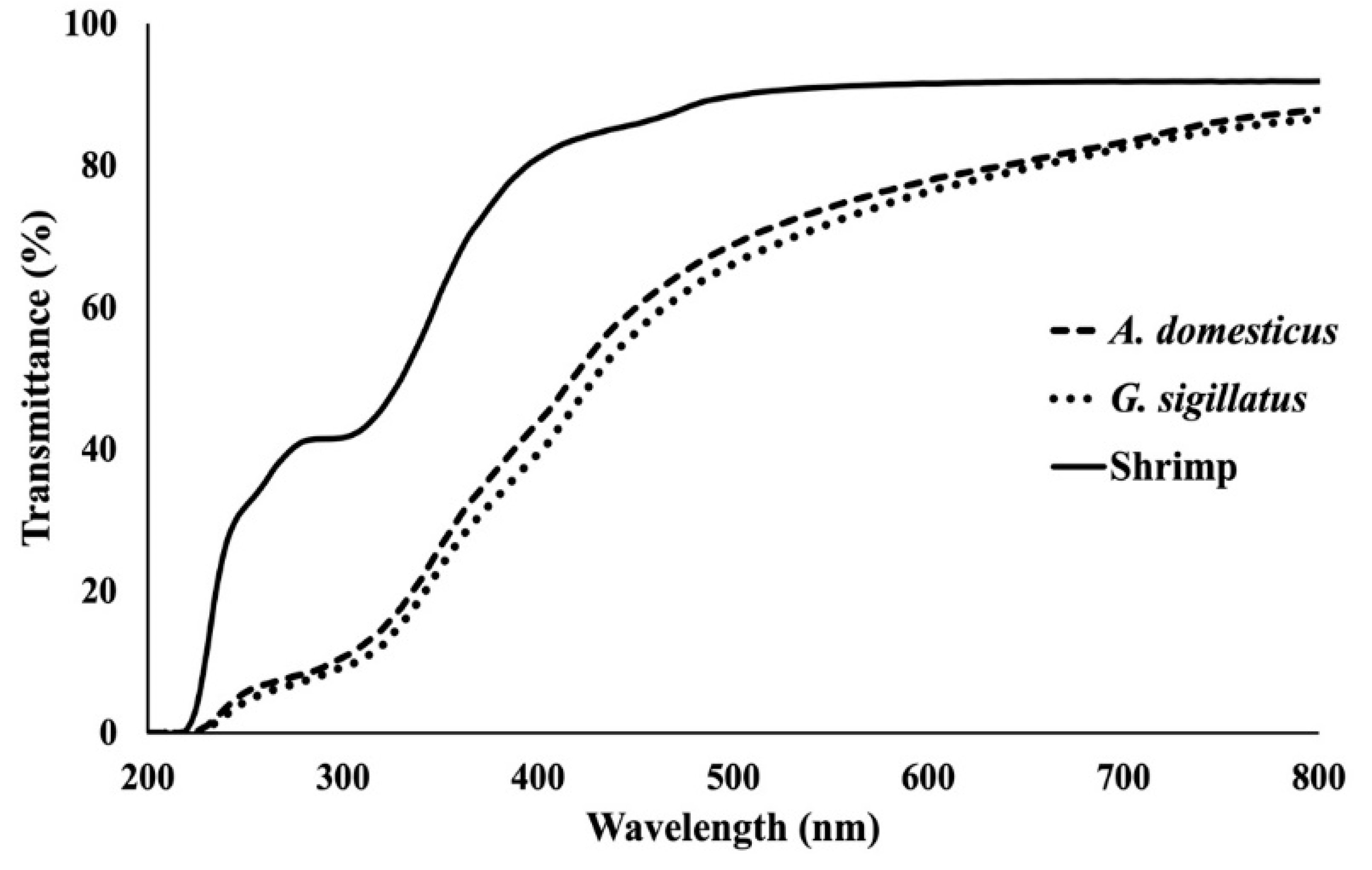
3.6. Light Barrier Properties

As stated previously, a primary function of food packaging is the protection of food materials concealed inside. This can include physical protection, as well as chemical deterioration prevention. Specifically, UV/Vis light that is transmitted through packaging into foods is known to initiate various deleterious chemical reactions, such as increase the rate of lipid oxidation, amongst others. Therefore, one mode of packaging protection is to prevent such reactions by decreasing the light transmitted through packaging.
In this study, transmittance of light (%), from 200 to 800 nm, through chitosan films was evaluated (Figure 5). The percent of light transmitted through CCF was much less than commercial shrimp films, due to the intrinsic properties and functionality of cricket’s chitin rich exoskeletons. Insect exoskeletons serve as a protectant agent from light, such as solar radiation, as well as other oxidative stresses, which is achieved through its incorporation of melanin [43,53]. Recent research studies have incorporated synthetic or naturally extracted melanin in different types of films, as an approach to mimic and achieve UV/Vis protecting materials for food packaging. This has been previously and extensively reviewed by Roy and Rhim [54]. Overall, the outcomes of these studies have shown great improvements in light barrier of packaging due to incorporation of melanin. The decreased transmission of light observed in our study is likely due to the remaining melanin present in CCF as previously hypothesized. The properties of the derivation material, crickets, may lead to chitosan films, which have greater light shielding properties than crustacean chitosan films with a slight increase in opacity, while maintaining a transparency expected of a packaging material.

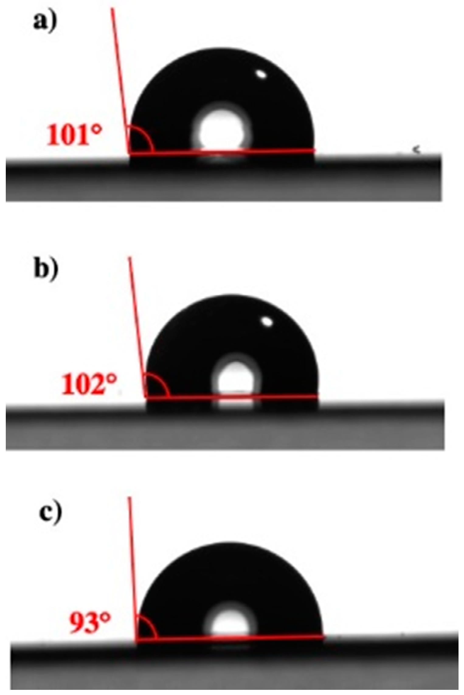
3.7. Water Contact Angles

Overall, CCF derived from both species were more hydrophobic than SCF (Figure 6, Table 1). The degree of deacetylation had no effect on the water contact angle values, although 76% deacetylated CCF were most similar to SCF water contact angles. To the best of our knowledge, this current study is the first to characterize film surface hydrophobicity/hydrophilicity of insect chitosan films.
One study analyzed the functional properties of high and low molecular weight commercial chitosan films, presumably crustacean derived, plasticized with varying concentrations of glycerol [38]. The authors reported that an initial increase from 0 to 15% glycerol did not affect the water contact angle of films (~105°). However, a further increase to 30% glycerol significantly increased the wettability of the surface as seen by a decrease in water contact angle (~98°), which was attributed to the intrinsic hydrophilicity of glycerol. Furthermore, it was found that the molecular weight of chitosan did not play a significant role in the wettability of unplasticized and plasticized films. Considering the study’s results, commercial SCF produced in the present study had a water contact angle of 93°, which aligned well with the previous reported results at a similar glycerol plasticization percentage.
Overall, CCF were more hydrophobic than commercial chitosan films, which we believe could be a result of residual melanin present. In one recent review on insect chitin, chitosan, and their melanin complexes, the authors highlight increased hydrophobicity of melanin–chitosan complexes due to the hydrophobic nature of melanin [45]. Additionally, a biomimetic chitosan film study attributed increased water contact angles due to the presence of melanin [44]. The increased water contact angles of CCF are likely a result of the presumable presence of melanin (hydrophobic), among possible other residual components. Therefore, if melanin crosslinks are present in CCF, intramolecular interactions would increase and lead to decreased ability of the film’s surface to interact with water. In addition, the presence of hydrophobic melanin would lead to the CCF surfaces to have increased water contact angles. Ultimately the increased complexity of cricket chitosan may be an advantage for chitosan biobased food packaging, compared to traditional crustacean chitosan products.

4. Conclusions

In this study, chitosan derived from two cricket species showed excellent film forming properties, comparable to films from commercial shrimp chitosan. Cricket chitosan may be more complex in nature due to the differing intrinsic properties of insects; however, in some cases, this may prove an advantage for biobased food packaging. Increased water resistance, as well as light and vapor barrier properties, were achieved through chitosan films derived from crickets, compared to the shrimp chitosan film.
As edible insect rearing and consumption continues to grow, by-products from this emerging industry may provide advantageous materials for use as biobased food packaging materials. As research is currently being performed at a rapid pace, new insights on insect-based chitin/chitosan and their applications are constantly being revealed. Future research on the insect derived chitosan complexes, such as further investigation and characterization, may provide further insight on the mechanisms responsible for their differing, or similar, functionalities in comparison to that derived from commercial (crustaceans) resources. Based on the promising results of this study, future research can be applied to evaluate insect chitosan films’ effect on shelf-life and quality of food.

Supplementary Materials

The following are available online at https://0-www-mdpi-com.brum.beds.ac.uk/article/10.3390/polysaccharides2040045/s1, Figure S1: Commercial shrimp chitosan film surface (a) at 500×, and cross sections at 2000× (b) and 20,000× (c) magnification, and Figure S2: Cross-sections of chitosan films derived from crickets, A. domesticus (ac) and G. sigillatus (df), shown at 2000× magnification. Chitosan films have varying degrees of deacetylation, including 72% (a,d), 76% (b,e), and 80% (c,f).

Author Contributions

Conceptualization, M.M., F.S.M.-G., O.G.J., I.K., J.M.G.-B. and A.M.L.; Methodology, M.M. and A.M.L.; investigation, M.M.; Resources, A.M.L., F.S.M.-G. and J.M.G.-B., Writing—original draft, M.M.; Writing—review and editing, M.M., F.S.M.-G., O.G.J., I.K., J.M.G.-B. and A.M.L.; Supervision, A.M.L.; Funding acquisition, A.M.L. All authors have read and agreed to the published version of the manuscript.

Funding

This work is supported by the USDA National Institute of Food and Agriculture, Hatch Act formula funds project 1019794.

Institutional Review Board Statement

Not applicable.

Data Availability Statement

All data discussed in this article are presented within the article and supplementary document.

Acknowledgments

The authors acknowledge Claridge’s lab in the Department of Chemistry at Purdue University for providing access to their tensiometer for water contact angle analysis. The authors also thank Robert Seiler at the Purdue University Life Science Microscopy Facility for his assistance with training and cryo-SEM.

Conflicts of Interest

The authors declare no conflict of interest.

References

  1. Guillard, V.; Gaucel, S.; Fornaciari, C.; Angellier-Coussy, H.; Buche, P.; Gontard, N. The next generation of sustainable food packaging to preserve our environment in a circular economy context. Front. Nutr. 2018, 5, 121. [Google Scholar] [CrossRef] [Green Version]
  2. Babu, R.P.; Oconnor, K.; Seeram, R. Current progress on bio-based polymers and their future trends. Prog. Biomater. 2013, 2, 8. [Google Scholar] [CrossRef] [Green Version]
  3. Wang, H.; Qian, J.; Ding, F. Emerging chitosan-based films for food packaging applications. J. Agric. Food Chem. 2018, 66, 395–413. [Google Scholar] [CrossRef]
  4. Siripatrawan, U.; Vitchayakitti, W. Improving functional properties of chitosan films as active food packaging by incorporating with propolis. Food Hydrocoll. 2016, 61, 695–702. [Google Scholar] [CrossRef]
  5. Crizel, T.D.M.; Rios, A.D.O.; Alves, V.D.; Bandarra, N.; Moldao-Martins, M.; Flôres, S.H. Active food packaging prepared with chitosan and olive pomace. Food Hydrocoll. 2018, 74, 139–150. [Google Scholar] [CrossRef]
  6. Wu, C.; Tian, J.; Li, S.; Wu, T.; Hu, Y.; Chen, S.; Sugawara, T.; Ye, X. Structural properties of films and rheology of film-forming solutions of chitosan gallate for food packaging. Carbohydr. Polym. 2016, 146, 10–19. [Google Scholar] [CrossRef] [PubMed]
  7. Râpă, M.; Miteluţ, A.C.; Tănase, E.E.; Grosu, E.; Popescu, P.; Popa, M.E.; Rosnes, J.T.; Sivertsvik, M.; Darie-Niţă, R.N.; Vasile, C. Influence of chitosan on mechanical, thermal, barrier and antimicrobial properties of PLA-biocomposites for food packaging. Compos. Part B Eng. 2016, 102, 112–121. [Google Scholar] [CrossRef]
  8. Rai, S.; Dutta, P.; Mehrotra, G. Lignin incorporated antimicrobial chitosan film for food packaging application. J. Polym. Mater. 2017, 34, 171. [Google Scholar]
  9. Ashrafi, A.; Jokar, M.; Nafchi, A.M. Preparation and characterization of biocomposite film based on chitosan and kombucha tea as active food packaging. Int. J. Biol. Macromol. 2018, 108, 444–454. [Google Scholar] [CrossRef] [PubMed]
  10. Shahbazi, Y. The properties of chitosan and gelatin films incorporated with ethanolic red grape seed extract and Ziziphora clinopodioides essential oil as biodegradable materials for active food packaging. Int. J. Biol. Macromol. 2017, 99, 746–753. [Google Scholar] [CrossRef] [PubMed]
  11. Riaz, A.; Lei, S.; Akhtar, H.M.S.; Wan, P.; Chen, D.; Jabbar, S.; Abid, M.; Hashim, M.M.; Zeng, X. Preparation and characterization of chitosan-based antimicrobial active food packaging film incorporated with apple peel polyphenols. Int. J. Biol. Macromol. 2018, 114, 547–555. [Google Scholar] [CrossRef]
  12. Kaya, M.; Khadem, S.; Cakmak, Y.S.; Mujtaba, M.; Ilk, S.; Akyuz, L.; Salaberria, A.M.; Labidi, J.; Abdulqadir, A.H.; Deligoz, E. Antioxidative and antimicrobial edible chitosan films blended with stem, leaf and seed extracts of Pistacia terebinthus for active food packaging. RSC Adv. 2018, 8, 3941–3950. [Google Scholar] [CrossRef] [Green Version]
  13. Wang, Q.; Tian, F.; Feng, Z.; Fan, X.; Pan, Z.; Zhou, J. Antioxidant activity and physicochemical properties of chitosan films incorporated withLycium barbarumfruit extract for active food packaging. Int. J. Food Sci. Technol. 2014, 50, 458–464. [Google Scholar] [CrossRef]
  14. Mohan, K.; Ganesan, A.R.; Muralisankar, T.; Jayakumar, R.; Sathishkumar, P.; Uthayakumar, V.; Chandirasekar, R.; Revathi, N. Recent insights into the extraction, characterization, and bioactivities of chitin and chitosan from insects. Trends Food Sci. Technol. 2020, 105, 17–42. [Google Scholar] [CrossRef]
  15. Abidin, N.Z.; Kormin, F.; Abidin, N.Z.; Anuar, N.M.; Abu Bakar, M. The Potential of Insects as Alternative Sources of Chitin: An Overview on the Chemical Method of Extraction from Various Sources. Int. J. Mol. Sci. 2020, 21, 4978. [Google Scholar] [CrossRef]
  16. Hall, F.G.; Jones, O.G.; O’Haire, M.E.; Liceaga, A.M. Functional properties of tropical banded cricket (Gryllodes sigillatus) protein hydrolysates. Food Chem. 2017, 224, 414–422. [Google Scholar] [CrossRef] [PubMed]
  17. Luna, G.C.; Martin-Gonzalez, F.S.; Mauer, L.; Liceaga, A. Cricket (Acheta domesticus) protein hydrolysates’ impact on the physicochemical, structural and sensory properties of tortillas and tortilla chips. J. Insects Food Feed 2021, 7, 109–120. [Google Scholar] [CrossRef]
  18. Malm, M.; Liceaga, A. Physicochemical properties of chitosan from two commonly reared edible cricket species, and its application as a hypolipidemic and antimicrobial agent. Polysaccharides 2021, 2, 339–353. [Google Scholar] [CrossRef]
  19. Malm, M.J.; Narsimhan, G.; Kokini, J.L. Effect of contact surface, plasticized and crosslinked zein films are cast on, on the distribution of dispersive and polar surface energy using the Van Oss method of deconvolution. J. Food Eng. 2019, 263, 262–271. [Google Scholar] [CrossRef]
  20. Urbizo-Reyes, U.; Martin-González, M.F.S.; Garcia-Bravo, J.; Liceaga, A.M. Development of chia seed (Salvia hispanica) mucilage films plasticized with polyol mixtures: Mechanical and barrier properties. Int. J. Biol. Macromol. 2020, 163, 854–864. [Google Scholar] [CrossRef] [PubMed]
  21. Garcia, M.; Pinotti, A.; Martino, M.; Zaritzky, N. Characterization of composite hydrocolloid films. Carbohydr. Polym. 2004, 56, 339–345. [Google Scholar] [CrossRef]
  22. Kalaycıoğlu, Z.; Torlak, E.; Akın-Evingür, G.; Özen, I.; Erim, F.B. Antimicrobial and physical properties of chitosan films incorporated with turmeric extract. Int. J. Biol. Macromol. 2017, 101, 882–888. [Google Scholar] [CrossRef]
  23. Souza, V.G.L.; Fernando, A.L.; Pires, J.R.A.; Rodrigues, P.F.; Lopes, A.A.; Fernandes, F.M.B. Physical properties of chitosan films incorporated with natural antioxidants. Ind. Crop. Prod. 2017, 107, 565–572. [Google Scholar] [CrossRef]
  24. Cerqueira, M.A.; Souza, B.W.S.; Teixeira, J.A.; Vicente, A.A. Effect of glycerol and corn oil on physicochemical properties of polysaccharide films—A comparative study. Food Hydrocoll. 2012, 27, 175–184. [Google Scholar] [CrossRef] [Green Version]
  25. Debandi, M.V.; Bernal, D.C.; Francois, N.J. Development of biodegradable films based on chitosan/glycerol blends suitable for biomedical applications. J. Tissue Sci. Eng. 2016, 7, 2157–7552. [Google Scholar] [CrossRef]
  26. Chen, S.; Wei, X.; Sui, Z.; Guo, M.; Geng, J.; Xiao, J.; Huang, D. Preparation of Antioxidant and antibacterial chitosan film from Periplaneta americana. Insects 2021, 12, 53. [Google Scholar] [CrossRef] [PubMed]
  27. Domján, A.; Bajdik, J.; Pintye-Hódi, K. Understanding of the plasticizing effects of glycerol and PEG 400 on chitosan films using solid-state NMR spectroscopy. Macromolecules 2009, 42, 4667–4673. [Google Scholar] [CrossRef]
  28. Rodríguez-Núñez, J.R.; Madera-Santana, T.J.; Sánchez-Machado, D.I.; López-Cervantes, J.; Valdez, H.S. Chitosan/hydrophilic plasticizer-based films: Preparation, physicochemical and antimicrobial properties. J. Polym. Environ. 2013, 22, 41–51. [Google Scholar] [CrossRef]
  29. Schaefer, J.; Kramer, K.; Garbow, J.; Jacob, G.; Stejskal, E.; Hopkins, T.; Speirs, R. Aromatic cross-links in insect cuticle: Detection by solid-state 13C and 15N NMR. Science 1987, 235, 1200–1204. [Google Scholar] [CrossRef]
  30. Khayrova, A.; Lopatin, S.; Varlomov, V. Method of Producing Covalent-Linked Chitosan-Melanin Complex from Black Soldier Fly Hermetia Illucens. Russia Patent RU2728458C1, 29 July 2020. [Google Scholar]
  31. Saenz-Mendoza, A.I.; Zamudio-Flores, P.B.; García-Anaya, M.C.; Velasco, C.R.; Acosta-Muñiz, C.H.; Ornelas-Paz, J.D.J.; Hernández-González, M.; Vargas, A.; Aguilar-González, M.Á.; Salgado-Delgado, R. Characterization of insect chitosan films from Tenebrio molitor and Brachystola magna and its comparison with commercial chitosan of different molecular weights. Int. J. Biol. Macromol. 2020, 160, 953–963. [Google Scholar] [CrossRef]
  32. Flores, Z.; San-Martin, D.; Beldarraín-Iznaga, T.; Leiva-Vega, J.; Villalobos-Carvajal, R. Effect of homogenization method and carvacrol content on microstructural and physical properties of chitosan-based films. Foods 2021, 10, 141. [Google Scholar] [CrossRef]
  33. Liu, M.; Zhou, Y.; Zhang, Y.; Yu, C.; Cao, S. Preparation and structural analysis of chitosan films with and without sorbitol. Food Hydrocoll. 2013, 33, 186–191. [Google Scholar] [CrossRef]
  34. Liu, Z.; Ge, X.; Lu, Y.; Dong, S.; Zhao, Y.; Zeng, M. Effects of chitosan molecular weight and degree of deacetylation on the properties of gelatine-based films. Food Hydrocoll. 2012, 26, 311–317. [Google Scholar] [CrossRef]
  35. Liu, Y.T.; Yuan, Y.; Duan, S.Q.; Li, C.; Hu, B.; Liu, A.P.; Wu, D.T.; Cui, H.Y.; Lin, L.; He, J.L.; et al. Preparation and characterization of chitosan films with three kinds of molecular weight for food packaging. Int. J. Biol. Macromol. 2020, 155, 249–259. [Google Scholar] [CrossRef] [PubMed]
  36. Bumgardner, J.; Murali, V.; Su, H.; Jenkins, O.; Velasquez-Pulgarin, D.; Jennings, J.; Sivashanmugam, A.; Jayakumar, R. Characterization of chitosan matters. In Chitosan Based Biomaterials; Jennings, J.A., Bumgardner, J.D., Eds.; Woodhead Publishing: Cambridge, UK, 2017; Volume 1, pp. 81–114. [Google Scholar]
  37. Suyatma, N.E.; Tighzert, A.L.; Copinet, A.; Coma, V. Effects of Hydrophilic plasticizers on mechanical, thermal, and surface properties of Chitosan films. J. Agric. Food Chem. 2005, 53, 3950–3957. [Google Scholar] [CrossRef] [PubMed]
  38. Leceta, I.; Guerrero, P.; de la Caba, K. Functional properties of chitosan-based films. Carbohydr. Polym. 2013, 93, 339–346. [Google Scholar] [CrossRef] [PubMed]
  39. Ryan, C.C.; Bardosova, M.; Pemble, M.E. Structural and mechanical properties of a range of chitosan-based hybrid networks loaded with colloidal silica and polystyrene particles. J. Mater. Sci. 2017, 52, 8338–8347. [Google Scholar] [CrossRef]
  40. Huei, C.R. Effect of molecular weight of chitosan with the same degree of deacetylation on the thermal, mechanical, and permeability properties of the prepared membrane. Carbohydr. Polym. 1996, 29, 353–358. [Google Scholar] [CrossRef]
  41. Tirado-Gallegos, J.; Zamudio-Flores, P.; Espino-Díaz, M.; Salgado-Delgado, R.; Vela-Gutiérrez, G.; Hernández-Centeno, F.; la Peña, H.L.-D.; Hernández-González, M.; Rendón-Villalobos, J.; Ortega-Ortega, A. Chitosan films obtained from Brachystola magna (Girard) and its evaluation on quality attributes in sausages during storage. Molecules 2021, 26, 1782. [Google Scholar] [CrossRef]
  42. Andersen, S.O. Insect cuticular sclerotization: A review. Insect Biochem. Mol. Biol. 2010, 40, 166–178. [Google Scholar] [CrossRef]
  43. Sugumaran, M.; Barek, H. Critical analysis of the melanogenic pathway in insects and higher animals. Int. J. Mol. Sci. 2016, 17, 1753. [Google Scholar] [CrossRef] [Green Version]
  44. Oh, D.X.; Hwang, D.S. A biomimetic chitosan composite with improved mechanical properties in wet conditions. Biotechnol. Prog. 2013, 29, 505–512. [Google Scholar] [CrossRef]
  45. Khayrova, A.; Lopatin, S.; Varlamov, V. Obtaining chitin, chitosan and their melanin complexes from insects. Int. J. Biol. Macromol. 2021, 167, 1319–1328. [Google Scholar] [CrossRef]
  46. Khayrova, A.; Lopatin, S.; Varlamov, V. Obtaining chitin/chitosan-melanin complexes from black soldier fly Hermetia Illucens. IOP Conf. Ser. Mater. Sci. Eng. 2020, 809, 012020. [Google Scholar] [CrossRef]
  47. Khayrova, A.; Lopatin, S.; Varlamov, V. Black soldier fly hermetia illucens as a novel source of chitin and chitosan. Int. J. Sci. 2019, 8, 81–86. [Google Scholar] [CrossRef] [Green Version]
  48. Ma, J.; Xin, C.; Tan, C. Preparation, physicochemical and pharmaceutical characterization of chitosan from Catharsius molossus residue. Int. J. Biol. Macromol. 2015, 80, 547–556. [Google Scholar] [CrossRef]
  49. Rivero, S.; Damonte, L.; García, M.A.; Pinotti, A. An insight into the role of glycerol in chitosan films. Food Biophys. 2016, 11, 117–127. [Google Scholar] [CrossRef]
  50. Qin, Y.-Y.; Zhang, Z.-H.; Li, L.; Yuan, M.-L.; Fan, J.; Zhao, T.-R. Physio-mechanical properties of an active chitosan film incorporated with montmorillonite and natural antioxidants extracted from pomegranate rind. J. Food Sci. Technol. 2013, 52, 1471–1479. [Google Scholar] [CrossRef] [Green Version]
  51. Rhim, J.-W.; Hong, S.-I.; Park, H.-M.; Ng, P.K.W. Preparation and characterization of chitosan-based nanocomposite films with antimicrobial activity. J. Agric. Food Chem. 2006, 54, 5814–5822. [Google Scholar] [CrossRef] [PubMed]
  52. Roy, S.; Van Hai, L.; Kim, H.C.; Zhai, L.; Kim, J. Preparation and characterization of synthetic melanin-like nanoparticles reinforced chitosan nanocomposite films. Carbohydr. Polym. 2020, 231, 115729. [Google Scholar] [CrossRef] [PubMed]
  53. Cao, W.; Zhou, X.; McCallum, N.C.; Hu, Z.; Ni, Q.Z.; Kapoor, U.; Heil, C.M.; Cay, K.S.; Zand, T.; Mantanona, A.J.; et al. Unraveling the structure and function of melanin through synthesis. J. Am. Chem. Soc. 2021, 143, 2622–2637. [Google Scholar] [CrossRef]
  54. Roy, S.; Rhim, J.-W. New insight into melanin for food packaging and biotechnology applications. Crit. Rev. Food Sci. Nutr. 2021, 2021, 1–27. [Google Scholar] [CrossRef]
Figure 1. FTIR of ~70% deacetylated chitosan films, derived from A. domesticus (dashed lines), G. sigillatus (dashed circles) and commercial shrimp (solid line).
Polysaccharides 02 00045 g001
Figure 2. SEM analysis of chitosan film surfaces derived from crickets, A. domesticus (ac) and G. sigillatus (df), shown at 500× magnification. Chitosan films have varying degrees of deacetylation, including 72% (a,d), 76% (b,e), and 80% (c,f).
Polysaccharides 02 00045 g002
Figure 3. Cross-sections of chitosan films derived from crickets, A. domesticus (ac) and G. sigillatus (df), shown at 20,000× magnification. Chitosan films have varying degrees of deacetylation, including 72% (a,d), 76% (b,e), and 80% (c,f).
Polysaccharides 02 00045 g003
Figure 4. Tensile strength (a), and elongation % at break (b) of cricket chitosan films compared to commercial shrimp chitosan films. Those in a graph which do not share a letter are considered significantly different. DDA (%) indicates degree of deacetylation.
Polysaccharides 02 00045 g004
Figure 5. Transmittance (%) of UV/Vis light through chitosan films derived from A. domesticus crickets (dashed line), G. sigillatus (dotted line), and shrimp (solid line).
Polysaccharides 02 00045 g005
Figure 6. Sessile water droplets on the surface of (a) A. domesticus, (b) G. sigillatus, and (c) commercial shrimp chitosan films (72% deacetylated).
Polysaccharides 02 00045 g006
Table 1. Permeability, surface wettability, color, and opacity properties of cricket chitosan films.
DDA (%) 1Water Vapor Permeability (10−10 g/m·s·Pa)Water Contact Angle (°)Instrumental Color and Opacity Properties 2
A. domesticus chitosan Opacity (mm−1)L*a*b*
722.14 ± 0.07 ab101.18 ± 4 bc1.6 ± 0.10 b71.79 ± 1.04 a0.8 ± 0.42 c26.49 ± 3.02 b
762.29 ± 0.06 ab97.21 ± 2.4 abc1.6 ± 0.04 b74.71 ± 0.43 b−0.02 ± 0.18 b24.64 ± 1.39 b
802.09 ± 0.08 a102.99 ± 5.4 c1.5 ± 0.14 b70.6 ± 0.60 a1.44 ± 0.17 de30.16 ± 0.56 b
G. sigillatus chitosan
722.34 ± 0.06 ab102.58 ± 4.1 c1.6 ± 0.04 b72.23 ± 1.85 ab1.23 ± 0.2 cd25.41 ± 4.75 b
762.40 ± 0.13 b95.48 ± 4.9 ab1.7 ± 0.07 b69.9 ± 1.42 a1.86 ± 0.14 e26.4 ± 3.63 b
802.43 ± 0.17 b101.06 ± 5.2 bc1.7 ± 0.04 b71.96 ± 1 ab1.56 ± 0.19 de23.65 ± 0.91 b
Shrimp chitosan
702.91 ± 0.16 c93.02 ± 2.3 a0.5 ± 0.01 a87.97 ± 0.44 c−2.38 ± 0.23 a8.09 ± 0.4 a
1 DDA (%) = Degree of deacetylation. 2 Values within a column that do not share the same letter (a–e) are statistically different (p < 0.05).
Publisher’s Note: MDPI stays neutral with regard to jurisdictional claims in published maps and institutional affiliations.

