Case Report

Demyelinating Polyradiculoneuropathy in Chronic Lymphocytic Leukemia: A Case Report on BTKis versus Venetoclax-Rituximab

by
Alessandro Cellini
1,
Andrea Visentin
1,
Alessandro Salvalaggio
2,
Mario Cacciavillani
3,
Sergio Ferrari
4 and
Chiara Briani
2,*
1
Hematology and Clinical Immunology Unit, Department of Medicine, University of Padova, 35128 Padova, Italy
2
Neurology Unit, Department of Neurosciences, University of Padova, 35128 Padova, Italy
3
Electromyography Lab, CEMES, Synlab Group, 35128 Padova, Italy
4
Neurology Unit, Department of Neuroscience, Biomedicine and Movement Sciences, University of Verona, 37126 Verona, Italy
*
Author to whom correspondence should be addressed.
Submission received: 2 September 2023 / Revised: 12 November 2023 / Accepted: 30 November 2023 / Published: 27 December 2023
(This article belongs to the Section Leukemias)

Abstract

:
The dysregulation of the immune system in Chronic Lymphocytic Leukemia (CLL) often allows for the development of immune-mediated diseases. Among them, autoimmune cytopenias are the most common, but cases of chronic inflammatory demyelinating polyradiculoneuropathy (CIDP) have been reported. We herein report on a patient who developed a CIDP while undergoing ibrutinib treatment for CLL, prompting drug discontinuation. Steroid treatment and a rituximab course proved to be ineffective at obtaining long-term control of CIDP, but therapy with venetoclax and rituximab, which was started due to CLL progression, led to the progressive amelioration of the symptoms up to complete remission of the neurological disease.

1. Introduction

Chronic Lymphocytic Leukemia (CLL) is the most common leukemia in Western countries [1]. Apart from the neoplastic clone, patients with CLL harbor a dysregulated innate and adaptive immune system which, on one hand, may favor the development of recurrent infections and second malignancies, and, on the other hand, may allow the occurrence of immune-mediated diseases [2]. Autoimmune cytopenias are the most common immune-mediated abnormalities associated with CLL [3], but other rheumatologic, endocrinological or neurological diseases may also occur [4]. Among them, chronic inflammatory demyelinating polyradiculoneuropathy (CIDP) has been reported [5,6]. These immune-mediated diseases might occur concomitantly with symptomatic CLL needing treatment or might emerge during therapy, due to an imbalance among T helper lymphocyte subsets [7].
Ibrutinib, the first in-class Bruton’s tyrosine kinase (BTK) inhibitor, is a targeted therapy approved worldwide for the treatment of several B-cell malignancies, including CLL [8]. This drug has proved to be active in patients suffering from neuropathy with anti-myelin-associated glycoprotein (MAG) antibodies harboring a MYD88-mutated lymphoid clone [9,10] and several immune-mediated diseases have been reported during ibrutinib treatment [11,12,13].
Venetoclax is a BH3-mimetic drug able to restore the BCL2 pro-apoptotic function, triggering the apoptosis of CLL cells [14,15]. The combination of venetoclax plus an anti-CD20 monoclonal antibody, such as rituximab, proved to be highly active in CLL, including in patients who relapse after ibrutinib or are intolerant to it [16]. In addition, venetoclax was active in MYD88-wild type patients with anti-MAG antibody neuropathy [10,17].
We herein report on an elderly CLL patient who developed a CIDP that worsened during ibrutinib treatment, thus prompting drug discontinuation. Transient amelioration of symptoms was achieved with steroids and then rituximab, but the improvement became persistent, up to complete remission after the addition of venetoclax.

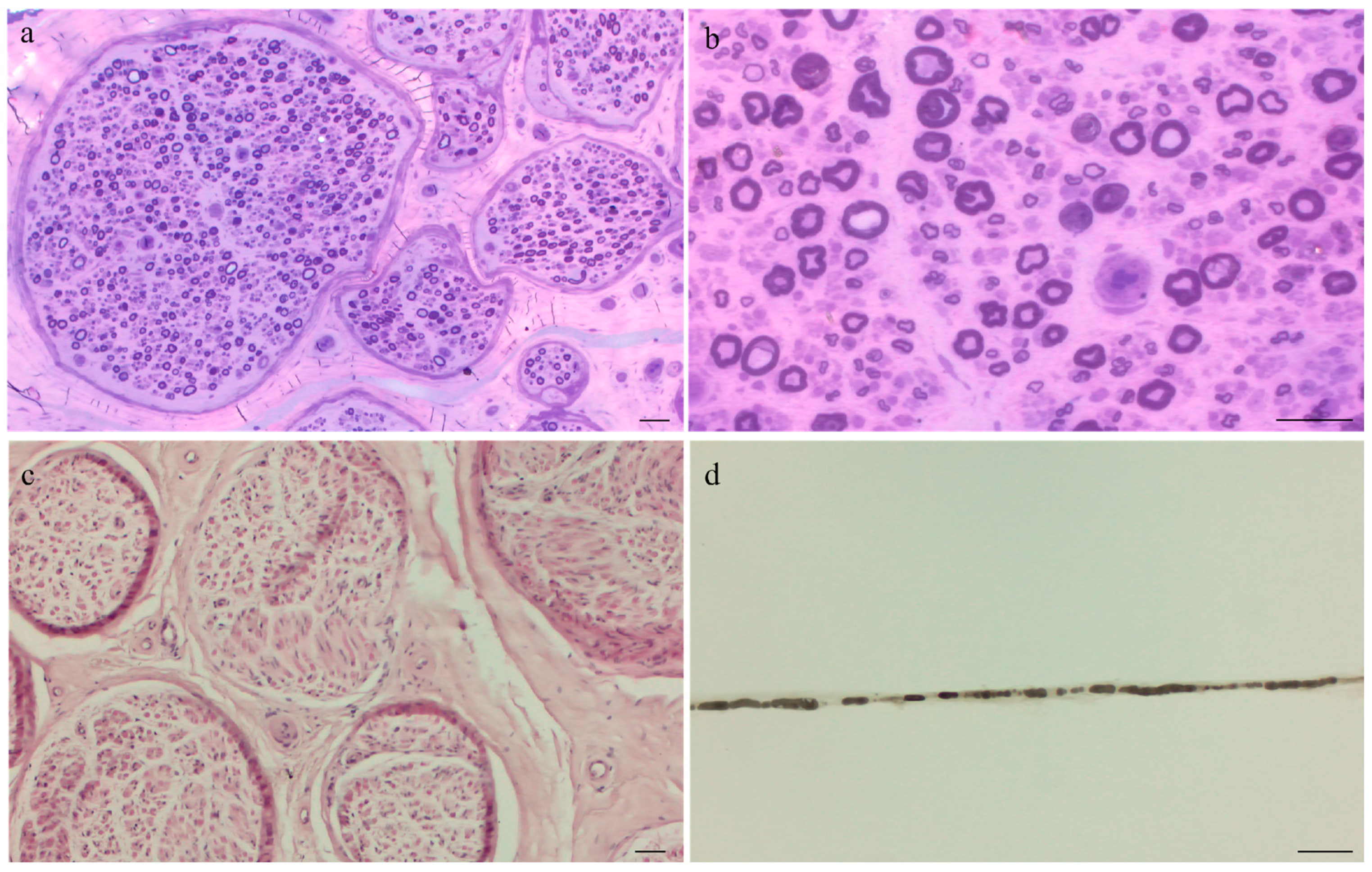
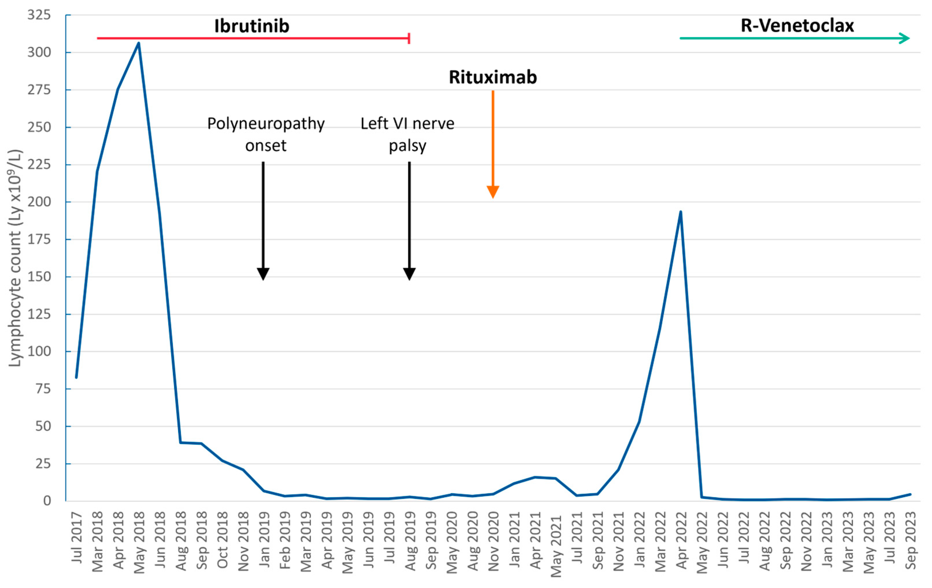
2. Detailed Case Description

In July 2017, a 75-year-old man was diagnosed with high-risk advanced stage CLL (Rai IV, Binet C) after the finding of thrombocytopenia and peripheral blood lymphocytosis with an immunophenotype consistent with CLL on routine blood work (leukocytes 83.30 × 10⁹/L, lymphocytes 75.90 × 10⁹/L, hemoglobin 140 g/L, platelets 98 × 109/L). The man’s medical history was notable for hypertension and paroxysmal atrial fibrillation. His medications included warfarin, simvastatin, enalapril and lercanidipine. He reported no weight loss, night sweats or fever. Enlarged bilateral cervical and supraclavicular lymph-nodes and mild splenomegaly were noted on physical examination. Cytogenetic analysis showed a complex karyotype [18], a 17p deletion was identified by FISH, a TP53 mutation was detected by Sanger sequencing and the IGHV gene was unmutated. No M-spike was detected on serum protein electrophoresis, and IgG and IgM levels were found to be decreased.
The patient was initially put on outpatient follow-up, but due to progressive increase of the lymphocyte count with concurrent worsening of the platelet count and progressive splenomegaly, a CLL-specific therapy was considered. A whole-body CT scan detected multiple centimetric cervical, supraclavicular, axillary, mediastinal, hilar and retroperitoneal lymphadenopathies and documented a spleen diameter of 17 cm. After a cardiology consult, anticoagulation was discontinued due to the lack of documented atrial fibrillation episodes in the past 4 years and the anticoagulating effect of the first generation BTKi, and the patient was put on ibrutinib therapy in March 2018. After 3 months, no lymphadenopathies or splenomegaly were detected on physical examination, and 6 months later, a significant increase in the platelet count occurred (from 67 × 109 to 124 × 109 PLTs/L). Of note, ibrutinib was started at 280 mg daily, and raised to 420 mg daily one month later after an initial increase in the PLT count was documented.
In January 2019, the patient was admitted to the Emergency Department (ED) complaining of gait instability, progressive acral paresthesia and mild distal weakness in the lower limbs. At the time, he was still on ibrutinib and had achieved a stable partial remission (due to the persistent, but still decreasing, lymphocytosis). Due to the subacute progression of the symptoms, he was admitted to the Neurology Unit of our Hospital. Neurological evaluation revealed mild right stepping gait, loss of tactile sensation up to the knees, hand paresthesia, loss of distal vibration and lower limb areflexia. Inflammatory Neuropathy Cause and Treatment (INCAT) Disability Scale [19] score was 2 in lower limbs and 2 in upper limbs (total of 4). Cerebrospinal fluid (CSF) analysis was unremarkable (protein 26 mg/dL, white blood cells 2.4/µL, mirror pattern at isoelectric focusing) and a spinal MRI revealed only vertebral arthrosis.
Neurophysiological evaluation showed signs of demyelinating polyneuropathy with slowing of motor nerve conduction at the four limbs and sural nerves, and the presence of conduction blocks at median and ulnar nerves bilaterally at the elbow–wrist tract, and at the right peroneal nerve in the popliteal–ankle tract. Spontaneous muscle fibrillations were recorded at distal limbs resulting from secondary axonal damage of some distal motor nerve trunks.
An IgM monoclonal protein was detected at serum electrophoresis, no anti-MAG antibodies were present. Sural nerve biopsy showed mild loss of myelin fibers and signs of axonal degeneration; no inflammatory or lymphomatous infiltrates were detected (Figure 1).
Vascular endothelial growth factor (VEGF) levels were within normal values and amyloidosis was ruled out because of the absence of serum free lambda light chain and the lack of amyloid deposit in nerve and bone marrow biopsies. Transformation to an aggressive lymphoma was also excluded by the lack of high metabolic areas on a 18FDG PET-CT scan and the absence of MYD88 L265P mutation in the CSF.
The patient was diagnosed with CIDP and treatment with intravenous steroids was started with amelioration of the symptoms (INCAT upper limbs 1, INCAT lower limbs 1).
Six months later, during steroid tapering, the patient reported worsening of the symptoms. A follow-up neurophysiological evaluation detected a more widespread axonal damage, as well as the persistent signs of demyelination. Nerve ultrasound was also performed, showing a slight diffuse increase in nerve cross-sectional area (CSA). In particular median nerve CSAs at the elbow were 16 mm2 and 14 mm2 (left and right, respectively), sciatic nerve CSAs at the mid-thigh measured 61 mm2 (bilaterally), tibial nerve CSAs were 50 mm2 and 48 mm2 (left and right, respectively) at the popliteal fossa and 22 mm2 and 19 mm2 (left and right, respectively) at the ankle, and brachial plexuses at the supraclavicular space were also enlarged, measuring 110 mm2 and 100 mm2 (left and right, respectively). These ultrasound findings were consistent with the diagnosis of CIDP.
In September 2019, the patient was admitted again to the ED complaining of diplopia and worsening of gait instability (INCAT upper limbs 1, INCAT lower limbs 2). Neurological evaluation documented a left sixth cranial nerve palsy, and the patient was admitted to a Medicine Unit. The blood exams showed no signs of CLL progression, and brain MRI was unremarkable.
Due to the lack of significant findings and suspecting a possible iatrogenic etiology of the neurological abnormalities (symptom onset 8 months after the initiation of ibrutinib), ibrutinib was discontinued and steroid therapy was resumed with progressive improvement of both the cranial nerve palsy and the gait instability.
In November 2020, a new neurophysiological evaluation did not document any new findings, but changes were documented on nerve ultrasound, with disappearance of median nerve and brachial plexuses abnormalities, and a slight reduction in the CSAs of the sciatic nerve and the tibial nerve.
In order to reduce and then discontinue steroids, the patient was treated with six weekly courses of rituximab. The therapy was well tolerated, and the neurological symptoms transiently improved but reappeared soon after rituximab discontinuation, thus needing to restart steroids. A timeline of the patient’s lymphocyte count (used as a surrogate of his CLL activity), treatment and events regarding his neurological symptoms is depicted in Figure 2.
The patient’s CLL maintained the partial remission obtained with the ibrutinib therapy until March 2022, when CLL progression occurred with the reappearance of multiple lymphadenopathies and progressive increase in the spleen diameter and of the lymphocyte count. In April 2022, therapy with venetoclax and rituximab was started with adequate tumor lysis syndrome prophylaxis. After the first week of the venetoclax ramp-up phase, an important reduction in the patient’s lymphocyte count was observed, with transient worsening of his thrombocytopenia. After the second week of ramp-up, no splenomegaly or palpable lymphadenopathies could be documented on physical examination. Neurological symptoms gradually improved, and steroids were discontinued during the second month of therapy. After eight months of venetoclax–rituximab treatment, the patient’s cytopenias resolved, but his IgG levels were still decreased (although with normal IgM and IgA levels), and measurable residual disease was still positive in the peripheral blood.
Concurrently, his neuropathy greatly improved (upper limbs INCAT of 0; lower limbs INCAT of 0). At a follow-up neurological examination, the patient had full strength and sensation in the four limbs, gait was normal and deep tendon reflexes were present apart from the Achilles. More importantly, his functionality was fully recovered, and the patient was able to give up his certificate of disability.

3. Discussion

We here report a rare case of CIDP in CLL that had worsened during ibrutinib therapy but improved after drug withdrawal and completely resolved during therapy with venetoclax–rituximab.
The CIDP worsening after ibrutinib fulfills the criteria for being considered iatrogenic [20], among which the temporal association, the absence of likely alternative explanations and improvement after drug withdrawal.
The peripheral nerve toxicity of ibrutinib has rarely been reported to allow for the search on similarities between the described cases, and data from the phase 3 RESONATE study [21] in relapsed CLL are scarce regarding the characteristics of the neuropathy, which was rarely detected.
Consistently, results from long-term efficacy and safety analysis of first line ibrutinib [22] do not report peripheral neurotoxicity.
In the phase 3 trial of ibrutinib vs. chemoimmunotherapy in older patients with untreated CLL, severe sensory peripheral neuropathy occurred in only one patient in the ibrutinib arm after a median follow-up of 38 months [23]. An additional grade 3 sensory peripheral neuropathy was recorded by Shanafelt et al. in a clinical trial for young patients affected by CLL [24]. Interestingly peripheral neuropathy has not been reported when second generation BTK inhibitors, acalabrutinib or zanubrutinib, are used in relapsed CLL [25,26]
Conversely, no iatrogenic neuropathy or worsening of existing neuropathy when ibrutinib was applied to both relapsed and naive WM patients have been reported. More importantly, ibrutinib has been shown to be effective in ameliorating anti-MAG antibody neuropathies in MYD88L265P-mutated patients with WM [9].
Rituximab, a chimeric anti-CD20 monoclonal antibody that is currently the standard treatment for anti-MAG antibody neuropathy, has occasionally been reported to worsen rather than improve anti-MAG antibody neuropathy [27,28,29,30,31,32]. Conversely, drugs that are well-known to be toxic to the peripheral nerves (e.g., bortezomib [33]) seem to be effective against neuropathies in hematological malignancies, when they target the underlying pathogenic mechanism [34,35].
Since BTK inhibition-induced neuropathy has not been reported with the second generation and highly specific BTK inhibitors as well as in Bruton’s agammaglobulinemia patients (a primary immunodeficiency caused by a defect of BTK), it could be speculated that the pathogenesis of ibrutinib-induced neuropathy might depend on ibrutinib’s off-target effects in some predisposed patients.
Although ibrutinib targets B lymphocytes by inhibiting BTK, it has been reported to modulate the activity of T lymphocytes. In particular, ibrutinib can also inhibit the interleukin-2-inducible kinase (ITK), a proximal member of the T-cell receptor signaling cascade [36], improving T-cell function in CLL, resulting in the expansion of memory T cells, a decrease in the Th1 polarization, a decrease of the T regulatory (Treg)/CD4+ T cell ratio and an improvement in immune synapse formation between T cells and CLL cells [37,38]. Despite this “restoration” of the patient’s immune systems, flares of auto-immune diseases are not surprising during ibrutinib therapy.
Furthermore, the lack of activity of acalabrutinib or zanubrutinib on ITK might justify the lower rates of immune-mediated diseases observed during therapy with these agents.
Conversely, with venetoclax, a recovery from immunosuppressive state after the elimination of leukemic cells has been observed. In particular, a restoration of NK cell function, and a decrease in the T follicular helper cells, Treg, and PD-1+ CD8+ T cells as well as inflammatory cytokines were noted after venetoclax therapy [39].

4. Conclusions

CLL is a disease characterized by a marked dysregulation in the host’s immune system, which in turn can lead to recurring infections or to the development of autoimmune conditions or secondary tumors. Modern targeted therapies are highly effective at treating the disease and can have an influence on the patient’s immune function, both by suppressing the CLL clone and thus inhibiting the negative effect that it exerts on non-pathological immune cells and via the off-target or on-target/off-tumor effects of the novel drugs. The latter might have played a major role in the case we just reported, as our patient could have had a predisposition to the development of CIDP, or even already harbored a latent disease that was then unmasked after the initiation of ibrutinib and the consequent skewing of his immune system polarity by the off-target inhibition of ITK and consequently eliciting a Th1 response.

Author Contributions

A.C. and A.V. are the hematologists in charge of the patient. A.S. and C.B. are the neurologists in charge of the patient. M.C. is the neurophysiologist, who also performed nerve ultrasound. S.F. is the neuropathologist who performed and interpreted the sural nerve biopsy. A.C. drafted the manuscript. All the authors participated in the critical discussion of the case report. All authors have read and agreed to the published version of the manuscript.

Funding

This research was supported by Ricerca per credere nella vita “RCV” odv, grant DIMEDEMATO-2023.

Institutional Review Board Statement

The study was conducted according to the guidelines of the Declaration of Helsinki, and was approved by the local research ethics committee of Padua hospital #4430/AO/18.

Informed Consent Statement

Informed consent has been obtained from the patient to publish this paper.

Data Availability Statement

No new data were created or analyzed in this study. Data sharing is not applicable to this article.

Conflicts of Interest

The authors declare no conflict of interest.

References

  1. The Surveillance E, and End Results (SEER) Program of the National Cancer Institute. Cancer Stat Facts: Leukemia—Chronic Lymphocytic Leukemia (CLL). 2021. Available online: https://seer.cancer.gov/statfacts/html/clyl.html (accessed on 16 August 2023).
  2. Vitale, C.; Boccellato, E.; Comba, L.; Jones, R.; Perutelli, F.; Griggio, V.; Coscia, M. Impact of Immune Parameters and Immune Dysfunctions on the Prognosis of Patients with Chronic Lymphocytic Leukemia. Cancers 2021, 13, 3856. [Google Scholar] [CrossRef] [PubMed]
  3. Hodgson, K.; Ferrer, G.; Montserrat, E.; Moreno, C. Chronic lymphocytic leukemia and autoimmunity: A systematic review. Haematologica 2011, 96, 752–761. [Google Scholar] [CrossRef] [PubMed]
  4. Visentin, A.; Imbergamo, S.; Gurrieri, C.; Frezzato, F.; Trimarco, V.; Martini, V.; Severin, F.; Raggi, F.; Scomazzon, E.; Facco, M.; et al. Major infections, secondary cancers and autoimmune diseases occur in different clinical subsets of chronic lymphocytic leukaemia patients. Eur. J. Cancer 2017, 72, 103–111. [Google Scholar] [CrossRef] [PubMed]
  5. Briani, C.; Visentin, A.; Salvalaggio, A.; Imbergamo, S.; Piazza, F.; Cacciavillani, M.; Campagnolo, M.; Frezzato, F.; Semenzato, G.; Trentin, L. Peripheral neuropathies in chronic lymphocytic leukemia: A single center experience on 816 patients. Haematologica 2017, 102, e140–e143. [Google Scholar] [CrossRef] [PubMed]
  6. Lewis, D.; Osman, C.; Allen, D.; Pinto, A.A.; Duncombe, A.; A Katifi, H. Refractory CIDP with chronic lymphocytic leukaemia responding to chemoimmunotherapy. Pract. Neurol. 2020, 21, 143–146. [Google Scholar] [CrossRef] [PubMed]
  7. Lad, D.P.; Varma, S.; Varma, N.; Sachdeva, M.U.S.; Bose, P.; Malhotra, P. Regulatory T-cell and T-helper 17 balance in chronic lymphocytic leukemia progression and autoimmune cytopenias. Leuk. Lymphoma 2015, 56, 2424–2428. [Google Scholar] [CrossRef] [PubMed]
  8. Ibrutinib [Package Insert]; Janssen Biotech, Inc.: Horsham, PA, USA, 2023.
  9. Castellani, F.; Visentin, A.; Campagnolo, M.; Salvalaggio, A.; Cacciavillani, M.; Candiotto, C.; Bertorelle, R.; Trentin, L.; Briani, C. The Bruton tyrosine kinase inhibitor ibrutinib improves anti-MAG antibody polyneuropathy. Neurol.-Neuroimmunol. Neuroinflamm. 2020, 7, e720. [Google Scholar] [CrossRef]
  10. Castellani, F.; Visentin, A.; Schirinzi, E.; Salvalaggio, A.; Cacciavillani, M.; Candiotto, C.; Baratè, C.; Cellini, A.; Bertorelle, R.; Siciliano, G.; et al. Mutational Profile in 75 Patients with Anti–Myelin-Associated Glycoprotein Neuropathy: Clinical and Hematologic Therapy Response and Hints on New Therapeutic Targets. Neurol.-Neuroimmunol. Neuroinflamm. 2023, 10, e200122. [Google Scholar] [CrossRef]
  11. Shaikh, H.; Khattab, A.; Faisal, M.S.; Chilkulwar, A.; Albrethsen, M.; Sadashiv, S.; Fazal, S. Case series of unique adverse events related to the use of ibrutinib in patients with B-cell malignancies—A single institution experience and a review of literature. J. Oncol. Pharm. Pract. 2018, 25, 1265–1270. [Google Scholar] [CrossRef]
  12. Wanner, D.; Bohn, J.-P.; Rudzki, J.; Stockhammer, G.; Steurer, M. Autoimmune myelitis in a CLL patient undergoing treatment with ibrutinib. Ann. Hematol. 2019, 98, 205–207. [Google Scholar] [CrossRef]
  13. Mato, A.R.; Islam, P.; Daniel, C.; Strelec, L.; Kaye, A.H.; Brooks, S.; Ganetsky, A.; Nasta, S.; Porter, D.L.; Svoboda, J.; et al. Ibrutinib-induced pneumonitis in patients with chronic lymphocytic leukemia. Blood 2016, 127, 1064–1067. [Google Scholar] [CrossRef] [PubMed]
  14. Venetoclax [Package Insert]; Genentech: South San Francisco, CA, USA; AbbVie: North Chicago, IL, USA, 2022.
  15. Anderson, M.A.; Deng, J.; Seymour, J.F.; Tam, C.; Kim, S.Y.; Fein, J.; Yu, L.; Brown, J.R.; Westerman, D.; Si, E.G.; et al. The BCL2 selective inhibitor venetoclax induces rapid onset apoptosis of CLL cells in patients via a TP53-independent mechanism. Blood 2016, 127, 3215–3224. [Google Scholar] [CrossRef]
  16. Jones, J.A.; Mato, A.R.; Wierda, W.G.; Davids, M.S.; Choi, M.; Cheson, B.D.; Furman, R.R.; Lamanna, N.; Barr, P.M.; Zhou, L.; et al. Venetoclax for chronic lymphocytic leukaemia progressing after ibrutinib: An interim analysis of a multicentre, open-label, phase 2 trial. Lancet Oncol. 2018, 19, 65–75. [Google Scholar] [CrossRef] [PubMed]
  17. Briani, C.; Visentin, A.; Castellani, F.; Cacciavillani, M.; Trentin, L. The BCL2 Inhibitor Venetoclax Plus Rituximab Is Active in MYD88 Wild-Type Polyneuropathy with Anti-MAG Antibodies. Neurol.-Neuroimmunol. Neuroinflamm. 2022, 9, e1181. [Google Scholar] [CrossRef] [PubMed]
  18. Visentin, A.; Bonaldi, L.; Rigolin, G.M.; Mauro, F.R.; Martines, A.; Frezzato, F.; Pravato, S.; Gargarella, L.R.; Bardi, M.A.; Cavallari, M.; et al. The complex karyotype landscape in chronic lymphocytic leukemia allows the refinement of the risk of Richter syndrome transformation. Haematologica 2022, 107, 868–876. [Google Scholar] [CrossRef] [PubMed]
  19. Hughes, R.; Bensa, S.; Willison, H.; Bergh, P.V.D.; Comi, G.; Illa, I.; Nobile-Orazio, E.; Van Doorn, P.; Dalakas, M.; Bojar, M.; et al. Randomized controlled trial of intravenous immunoglobulin versus oral prednisolone in chronic inflammatory demyelinating polyradiculoneuropathy. Ann. Neurol. 2001, 50, 195–201. [Google Scholar] [CrossRef] [PubMed]
  20. Miller, F.W.; Hess, E.V.; Clauw, D.J.; Hertzman, P.A.; Pincus, T.; Silver, R.M.; Mayes, M.D.; Varga, J.; Medsger, T.A.; Love, L.A. Approaches for identifying and defining environmentally associated rheumatic disorders. Arthritis Rheumatol. 2001, 43, 241–249. [Google Scholar] [CrossRef]
  21. Munir, T.; Brown, J.R.; O’Brien, S.; Barrientos, J.C.; Barr, P.M.; Reddy, N.M.; Coutre, S.; Tam, C.S.; Mulligan, S.P.; Jaeger, U.; et al. Final analysis from RESONATE: Up to six years of follow-up on ibrutinib in patients with previously treated chronic lymphocytic leukemia or small lymphocytic lymphoma. Am. J. Hematol. 2019, 94, 1353–1363. [Google Scholar] [CrossRef]
  22. Burger, J.A.; Barr, P.M.; Robak, T.; Owen, C.; Ghia, P.; Tedeschi, A.; Bairey, O.; Hillmen, P.; Coutre, S.E.; Devereux, S.; et al. Long-term efficacy and safety of first-line ibrutinib treatment for patients with CLL/SLL: 5 years of follow-up from the phase 3 RESONATE-2 study. Leukemia 2020, 34, 787–798. [Google Scholar] [CrossRef]
  23. Woyach, J.A.; Ruppert, A.S.; Heerema, N.A.; Zhao, W.; Booth, A.M.; Ding, W.; Bartlett, N.L.; Brander, D.M.; Barr, P.M.; Rogers, K.A.; et al. Ibrutinib Regimens versus Chemoimmunotherapy in Older Patients with Untreated CLL. N. Engl. J. Med. 2018, 379, 2517–2528. [Google Scholar] [CrossRef]
  24. Shanafelt, T.D.; Wang, X.V.; Kay, N.E.; Hanson, C.A.; O’brien, S.; Barrientos, J.; Jelinek, D.F.; Braggio, E.; Leis, J.F.; Zhang, C.C.; et al. Ibrutinib–Rituximab or Chemoimmunotherapy for Chronic Lymphocytic Leukemia. N. Engl. J. Med. 2019, 381, 432–443. [Google Scholar] [CrossRef]
  25. Byrd, J.C.; Hillmen, P.; Ghia, P.; Kater, A.P.; Chanan-Khan, A.; Furman, R.R.; O’Brien, S.; Yenerel, M.N.; Illés, A.; Kay, N.; et al. Acalabrutinib Versus Ibrutinib in Previously Treated Chronic Lymphocytic Leukemia: Results of the First Randomized Phase III Trial. J. Clin. Oncol. 2021, 39, 3441–3452. [Google Scholar] [CrossRef] [PubMed]
  26. Brown, J.R.; Eichhorst, B.; Hillmen, P.; Jurczak, W.; Kaźmierczak, M.; Lamanna, N.; O’brien, S.M.; Tam, C.S.; Qiu, L.; Zhou, K.; et al. Zanubrutinib or Ibrutinib in Relapsed or Refractory Chronic Lymphocytic Leukemia. N. Engl. J. Med. 2023, 388, 319–332. [Google Scholar] [CrossRef] [PubMed]
  27. Broglio, L.; Lauria, G. Worsening after rituximab treatment in anti-mag neuropathy. Muscle Nerve 2005, 32, 378–379. [Google Scholar] [CrossRef] [PubMed]
  28. Stork, A.C.J.; Notermans, N.C.; Vrancken, A.F.J.E.; Cornblath, D.R.; van der Pol, W.-L. Rapid worsening of IgM anti-MAG demyelinating polyneuropathy during rituximab treatment. J. Peripher. Nerv. Syst. 2013, 18, 189–191. [Google Scholar] [CrossRef] [PubMed]
  29. Sala, E.; Robert-Varvat, F.; Paul, S.; Camdessanché, J.-P.; Antoine, J.-C. Acute neurological worsening after Rituximab treatment in patients with anti-MAG neuropathy. J. Neurol. Sci. 2014, 345, 224–227. [Google Scholar] [CrossRef] [PubMed]
  30. Benedetti, L.; Franciotta, D.; Vigo, T.; Grandis, M.; Fiorina, E.; Ghiglione, E.; Roccatagliata, L.; Mancardi, G.L.; Uccelli, A.; Schenone, A. Relapses After Treatment with Rituximab in a Patient with Multiple Sclerosis and Anti–Myelin-Associated Glycoprotein Polyneuropathy. Arch. Neurol. 2007, 64, 1531–1533. [Google Scholar] [CrossRef]
  31. Noronha, V.; Fynan, T.M.; Duffy, T. Flare in Neuropathy Following Rituximab Therapy for Waldenstrom’s Macroglobulinemia. J. Clin. Oncol. 2006, 24, e3. [Google Scholar] [CrossRef]
  32. Gironi, M.; Saresella, M.; Ceresa, L.; Calvo, M.; Ferrante, P.; Merli, F.; Nemni, R. Clinical and immunological worsening in a patient affected with Waldenstrom mac-roglobulinemia and anti-mag neuropathy after treatment with rituximab. Haematologica 2006, 91 (Suppl. S6), e51–e52. [Google Scholar]
  33. Velasco, R.; Alberti, P.; Bruna, J.; Psimaras, D.; Argyriou, A.A. Bortezomib and other proteosome inhibitors—Induced peripheral neurotoxicity: From pathogenesis to treatment. J. Peripher. Nerv. Syst. 2019, 24, S52–S62. [Google Scholar] [CrossRef]
  34. Briani, C.; Visentin, A.; Cerri, F.; Quattrini, A. From pathogenesis to personalized treatments of neuropathies in hematological malignancies. J. Peripher. Nerv. Syst. 2020, 25, 212–221. [Google Scholar] [CrossRef] [PubMed]
  35. Riva, M.; Lessi, F.; Berno, T.; Visentin, A.; Campagnolo, M.; Semenzato, G.; Adami, F.; Briani, C. Bortezomib-based regimens in patients with POEMS syndrome: A case series in newly diagnosed and relapsed patients. Leuk. Lymphoma 2019, 60, 2067–2070. [Google Scholar] [CrossRef] [PubMed]
  36. Mhibik, M.; Wiestner, A.; Sun, C. Harnessing the Effects of BTKi on T Cells for Effective Immunotherapy against CLL. Int. J. Mol. Sci. 2019, 21, 68. [Google Scholar] [CrossRef] [PubMed]
  37. Long, M.; Beckwith, K.; Do, P.; Mundy, B.L.; Gordon, A.; Lehman, A.M.; Maddocks, K.J.; Cheney, C.; Jones, J.A.; Flynn, J.M.; et al. Ibrutinib treatment improves T cell number and function in CLL patients. J. Clin. Investig. 2017, 127, 3052–3064. [Google Scholar] [CrossRef]
  38. Puzzolo, M.C.; Del Giudice, I.; Peragine, N.; Mariglia, P.; De Propris, M.S.; Cappelli, L.V.; Trentin, L.; Reda, G.; Cuneo, A.; Molica, S.; et al. TH2/TH1 Shift Under Ibrutinib Treatment in Chronic Lymphocytic Leukemia. Front Oncol. 2021, 11, 637186. [Google Scholar] [CrossRef]
  39. De Weerdt, I.; Hofland, T.; de Boer, R.; Dobber, J.A.; Dubois, J.; van Nieuwenhuize, D.; Mobasher, M.; de Boer, F.; Hoogendoorn, M.; Velders, G.A.; et al. Distinct immune composition in lymph node and peripheral blood of CLL patients is reshaped during venetoclax treatment. Blood Adv. 2019, 3, 2642–2652. [Google Scholar] [CrossRef]
Figure 1. Sural nerve biopsy: (a) semithin cross section showing nerve fascicles with mild loss of myelin fibers. (b) At higher magnification, some images of axonal degeneration are seen. (c) No inflammatory infiltrates are visible in the paraffin sections (H&E stain). (d) Axonal degeneration in teased fibers. Bar: 50 mm.
Hemato 05 00003 g001
Figure 2. Graph depicting the relationship between the patient’s lymphocyte count, the onset and the relapse of his neurological symptoms, and the CLL-directed therapy.
Hemato 05 00003 g002
Disclaimer/Publisher’s Note: The statements, opinions and data contained in all publications are solely those of the individual author(s) and contributor(s) and not of MDPI and/or the editor(s). MDPI and/or the editor(s) disclaim responsibility for any injury to people or property resulting from any ideas, methods, instructions or products referred to in the content.

