
Deposition of Organic-Inorganic Nanocomposite Coatings for Biomedical Applications


Department of Materials Science and Engineering, McMaster University, Hamilton, ON L8S 4L7, Canada
*
Author to whom correspondence should be addressed.
Solids 2022, 3(2), 271-281; https://0-doi-org.brum.beds.ac.uk/10.3390/solids3020019
Submission received: 5 April 2022
/
Revised: 28 April 2022
/
Accepted: 3 May 2022
/
Published: 6 May 2022
Abstract
:Polymethylmethacrylate (PMMA) is a material of choice for many biomedical coating applications. However, such applications are limited due to the toxicity of the traditional solvents used for the solution processing of PMMA coatings and composites. This problem is addressed using an isopropanol-water co-solvent, which allows for the dissolution of high molecular mass PMMA and the fabrication of coatings by a dip-coating method from concentrated PMMA solutions. The use of the co-solvent offers a versatile strategy for PMMA solubilization and coating deposition, despite the insolubility of PMMA in water and isopropanol. Composite coatings are obtained, containing hydroxyapatite, silver oxide, zinc oxide, micron size silica and nanosilica. Such coatings are promising for the manufacturing of implants with enhanced biocompatibility, bioactivity and antimicrobial properties and the fabrication of biosensors. Ibuprofen, tetracycline and amoxicillin are used as model drugs for the fabrication of PMMA-drug composite coatings for drug delivery. The microstructure and composition of the coatings are analyzed. The versatile dip-coating method of this investigation provides a platform for various biomedical applications.
1. Introduction
Polymethylmethacrylate (PMMA) is widely used for various applications in optical devices, solar cells, batteries and supercapacitors [1,2,3,4]. PMMA is a material of choice for various biomedical applications in dental implants, orthopedic devices and biosensors [1,5,6,7]. The interest in PMMA for biomedical applications is attributed to its biocompatibility, chemical stability and good mechanical properties [1]. Many investigations reported the development and successful applications of PMMA composites for the controlled delivery of drugs [1,8], cranioplasty [9], biomedical implants [10] and bone and dental cements [1,11,12]. Composite materials containing TiO2 [13], Al2O3 [14], hydroxyapatite [15,16] and bioglass [17] were developed. Such composites showed enhanced biocompatibility, bioactivity and enhanced mechanical and other functional properties.
PMMA composite films and coatings have generated significant interest, which was fueled by various applications. PMMA composite coatings containing bioactive ceramics were developed [18]. PMMA coatings and films provided a platform for advanced drug delivery applications [19,20]. PMMA exhibits remarkable properties for thin film applications in eye lenses [21,22] and thin film biosensors [23,24]. PMMA films and coatings were deposited by plasma polymerization [25] and laser evaporation [18]. Many investigations focused on the development of solution deposition techniques, such as solution polymerization [26], electrophoretic deposition [27], sol-gel deposition [28], spin coating [29] and dip coating [30,31,32]. Various solvents were used for PMMA, such as toluene, benzene, methyl ethyl ketone and other organic solvents, which are carcinogenic and toxic. The application of such solvents for biomedical applications presents difficulties, because solvent molecules can remain adsorbed on the surface or in the bulk of the PMMA coatings.
Recent studies showed that dip coating of PMMA can be performed using a mixed isopropanol-water co-solvent [33]. Despite the PMMA insolubility in individual solvents, such as water and isopropanol, solutions of high molecular mass PMMA with high concentration were prepared. The use of such solutions was a key factor for successful film deposition by a dip coating method [33]. It should be noted that isopropanol offers benefits for film deposition for biomedical applications due to low evaporation temperature and miscibility with water.
Isopropanol has been utilized in many studies focused on the manufacturing of coatings and thin films for biomedical applications and offered the advantage of low cytotoxicity compared to other organic solvents [34,35,36,37,38]. Good cell proliferation and attachment were observed on the surfaces of the coatings prepared using isopropanol [39,40,41]. The investigations focused on applications of isopropanol for protein purification and extraction [42], the fabrication of fibrous implant materials for tissue engineering [43], the manufacturing of scaffolds for wound healing [44] and the development of thin films for controlled drug delivery [45]. Therefore, the further development of dip coating from a mixed water-isopropanol solvent is a promising strategy for the fabrication of composites containing different functional materials for biomedical applications.
The goal of this investigation was the fabrication of composite coatings containing different functional biomaterials in the PMMA matrix using a water-isopropanol co-solvent. We targeted the fabrication of organic–inorganic composites containing bioactive ceramics, such as hydroxyapatite, silica and materials with antimicrobial properties, such as Ag2O and ZnO. Moreover, the fabrication of PMMA-ZnO composite coatings paves the way for the development of biosensors. Ibuprofen, tetracycline and amoxicillin were used as model drugs for the fabrication of coatings for drug delivery. The results presented below showed that the dip coating method is a versatile strategy for the development of composite coatings.
2. Materials and Methods
Poly(methyl methacrylate) (PMMA, MW = 350,000, Aldrich, Oakville, ON, Canada), ZnO, Ag2O, nanosilica, isopropanol, ibuprofen, tetracycline and amoxicillin were received from the MilliporeSigma company. Micron size silica was obtained from PCR Inc. Hydroxyapatite (HAP) nanoparticles were prepared by a chemical precipitation method, as described in previous investigations [46,47].
PMMA was dissolved in a mixture of water and isopropanol (20% water) at 50 °C, and the obtained solution was cooled to room temperature. The substrates for coating deposition were stainless steel foils (304 type, area 30 × 50 mm, thickness 0.1 mm). It should be noted that PMMA can also be dissolved in ethanol-water mixtures. However, the isopropanol-water solvent allowed for better PMMA solubility. Dip coating was performed at a substrate withdrawal speed of 10 mm min−1 from 10 g L−1 PMMA solutions without and with other functional materials for biomedical applications. The concentrations of such materials in the 10 g L−1 PMMA solutions were 5 g L−1 ZnO, HAP, micron size silica and nanosilica, ibuprofen, tetracycline, amoxicillin and 0.5 g L−1 Ag2O. The thickness of the as-deposited and room-temperature-dried monolayer coatings was 2–3 μm. Coating annealing was performed at 200 °C for 1 h.
The coating microstructure was examined using a JEOL SEM (scanning electron microscope, JSM-7000F). The coating composition was examined using a Bruker Smart 6000 X-ray diffractometer (XRD, CuK radiation). Thermogravimetric analysis (TGA, thermoanalyzer Netzsch STA-409) was carried out in air at a heating rate of 5 °C/min. For the TGA investigations, the deposits were removed from the substrates. A Bruker Vertex 70 spectrometer was used for the Fourier Transform Infrared Spectroscopy (FTIR) experiments.
3. Results
Figure 1 shows SEM images of PMMA coating prepared from 10 g L−1 PMMA solution. The microstructure of the as-deposited coating contained porous surface island networks formed on a relatively dense layer (Figure 1A). Such microstructure can result from the Stranski–Krastanov mode of film growth [48]. It should be also noted that PMMA is soluble in the isopropanol-water mixture in a narrow water concentration range. Therefore, a faster isopropanol evaporation rate during drying can result in a change in the solvent composition and precipitation of PMMA particles in the surface layer to form a porous network. Annealing resulted in the film melting and formation of dense coatings.
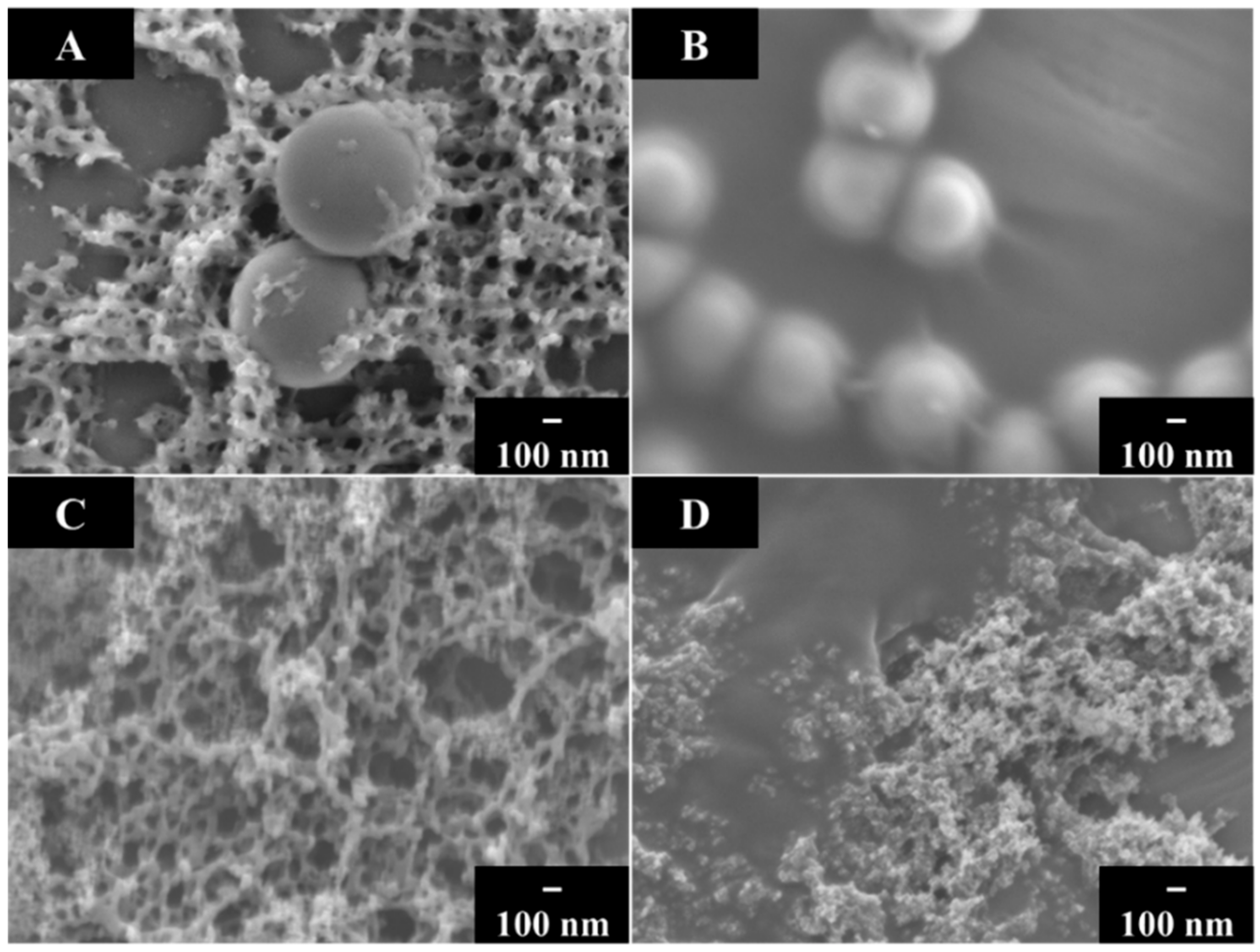
PMMA was successfully co-deposited with inorganic materials. Figure 2 shows SEM images of as-deposited and annealed coating prepared from 10 g L−1 PMMA solutions containing 0.5 g L−1 Ag2O, 5 g L−1 HAP and 5 g L−1 ZnO. The interest in polymer coatings containing Ag2O is attributed to the antimicrobial properties of Ag2O [49,50]. Therefore, small additives of Ag2O can be beneficial for biomedical applications of PMMA coatings. ZnO is widely used for the fabrication of biosensors. This strategy is based on the relatively high isoelectric point of ZnO, which was reported to be about pH = 9 [51]. Electrostatic interactions of ZnO with various biosensing molecules, which have a low isoelectric point and negative charge at pH = 7, facilitated the immobilization of such molecules on the positively charged ZnO particles [51]. The antimicrobial properties of ZnO particles are important for biomedical applications [52,53]. HAP is widely used for implant applications because its chemical composition is similar to that of the mineral part of natural bone [54,55,56]. The concentration of ZnO and HAP in the suspensions was larger than the Ag2O concentration in order to achieve a larger content of ZnO and HAP in the coatings. A larger HAP and ZnO content is necessary for the fabrication of bioactive coatings based on HAP [57] and the immobilization of biosensing molecules in coatings based on ZnO particles [51].
The as-deposited PMMA-Ag2O, PMMA-ZnO and PMMA-HAP coatings were porous (Figure 2). Annealing resulted in reduced coating porosity. The relatively high porosity of as-deposited PMMA-ZnO and PMMA-HAP coatings resulted from the packing of ZnO and HAP particles. The HAP particles had a needle shape. The SEM images of annealed PMMA-ZnO and PMMA-HAP coatings showed ZnO and HAP particles on the coating surface. The annealed films were crack free and relatively dense. The polymer acted as a binding and film-forming agent.
The fabrication of PMMA-silica coatings was motivated by their applications for the corrosion protection of biomedical implants with enhanced biocompatibility [58]. Figure 3 shows SEM images of as-deposited and annealed PMMA-silica coatings. The SEM images of as-deposited coatings showed particles of micron size silica and nanosilica. Annealing resulted in the formation of relatively dense coatings containing silica particles in the PMMA matrix. The nanosilica nanoparticles were incorporated into the PMMA matrix as individual particles or agglomerates.
The XRD and TGA methods were used for the analysis of the composite coatings. Figure 4a shows relatively broad peaks of pure PMMA.
XRD studies showed that the composite PMMA-ZnO, PMMA-HAP and PMMA- Ag2O coatings exhibited peaks of ZnO (Figure 4b), HAP (Figure 4c) and Ag2O (Figure 4d), respectively. Moreover, X-ray diffraction studies (Figure 4e) showed that the deposition from 10 g L−1 PMMA solutions containing 5 g L−1 HAP and 0.5 g L−1 Ag2O resulted in the fabrication of composite coatings containing HAP and Ag2O. Such coatings can potentially be used for the fabrication of biomedical implants with enhanced bioactivity and antimicrobial properties. The XRD studies also confirmed the fabrication of composite PMMA-silica coatings. Figure 5 compares the X-ray diffraction patterns of nanosilica, micron size silica and PMMA with the X-ray diffraction patterns of PMMA composites containing nanosilica and micron size silica. The X-ray diffraction patterns of individual materials showed broad peaks, and the composite materials showed peaks of both PMMA and silica, confirming the fabrication of composites.
The results of TGA studies of the composite coatings are presented in Figure 6. It is known [59] that the decomposition of PMMA in air occurs in the temperature range of 250–400 °C.
The TGA data for PMMA-HAP showed a small variation the sample mass below 300 °C and sharp reduction in the sample mass at higher temperatures due to the burning out of PMMA. The total mass loss at 1000 °C was found to be 61%, which indicated that the HAP content in the composite was 39%. The TGA data for PMMA-Ag2O showed weight losses at lower temperatures, which included two steps. A weight loss can result from dehydration, the decomposition of Ag2O [60] and the burning out of PMMA. The total mass loss at 1000 °C was found to be 94%. The decomposition of PMMA and Ag2O was observed [59,61] at temperatures above 200 °C. Therefore, weight loss in the range of 80–120 °C for PMMA-Ag2O can be attributed to dehydration. It is in this regard that porous and composite materials can accumulate a significant amount of water during synthesis [62,63,64,65]. Therefore, the drying of PMMA-Ag2O coatings at temperatures of 60–100 °C can be beneficial for antimicrobial applications. The TGA studies of the PMMA-ZnO, PMMA-micron size silica and PMMA-nanosilica showed a sharp reduction in mass loss at temperatures above 250–300 °C, and total mass loss was 32, 64 and 81%, respectively. The content of ZnO, micron size silica and nanosilica in the composite coatings was found to be 68, 36 and 19%, respectively. Therefore, the results of the XRD and TGA studies confirmed the formation of composite coating by a dip-coating method. The difference in the thermal behavior of the composites can result from different factors, such as different concentrations of inorganic components, silver reduction, different amounts of adsorbed water, the influence of the inorganic phase on the burning out of polymer and other factors.
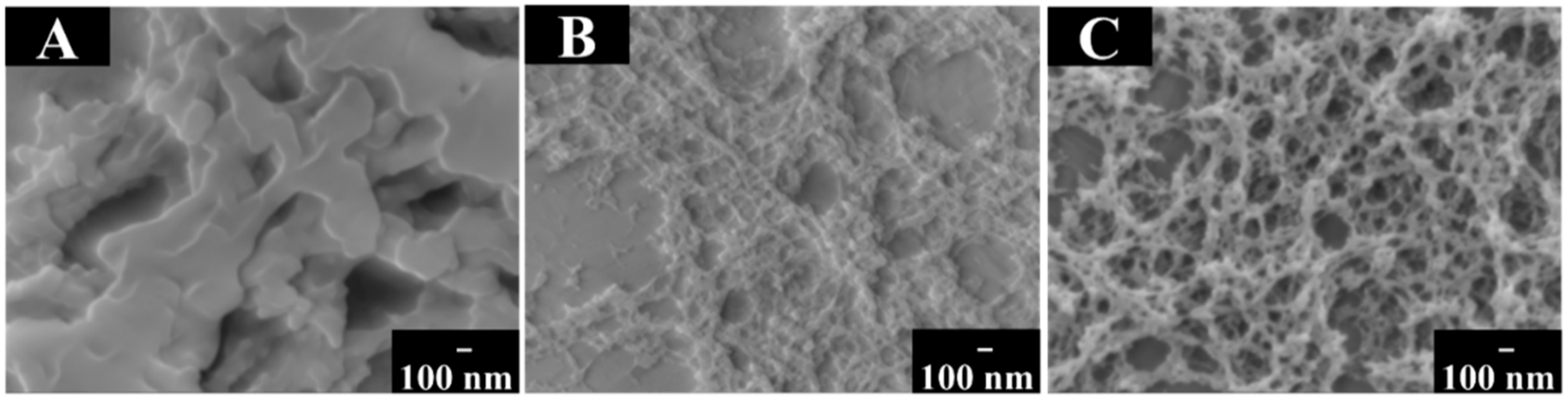
The dip coating methods allowed for the fabrication of composite coatings containing drugs of different types. Ibuprofen, tetracycline and amoxicillin were used as model drugs of different types for the development of the dip coating method. Figure 7 shows SEM images of the composite coatings. The coatings show porous microstructures, which are beneficial for the drug release. Moreover, the biodegradability of PMMA is another beneficial factor for drug delivery [66].
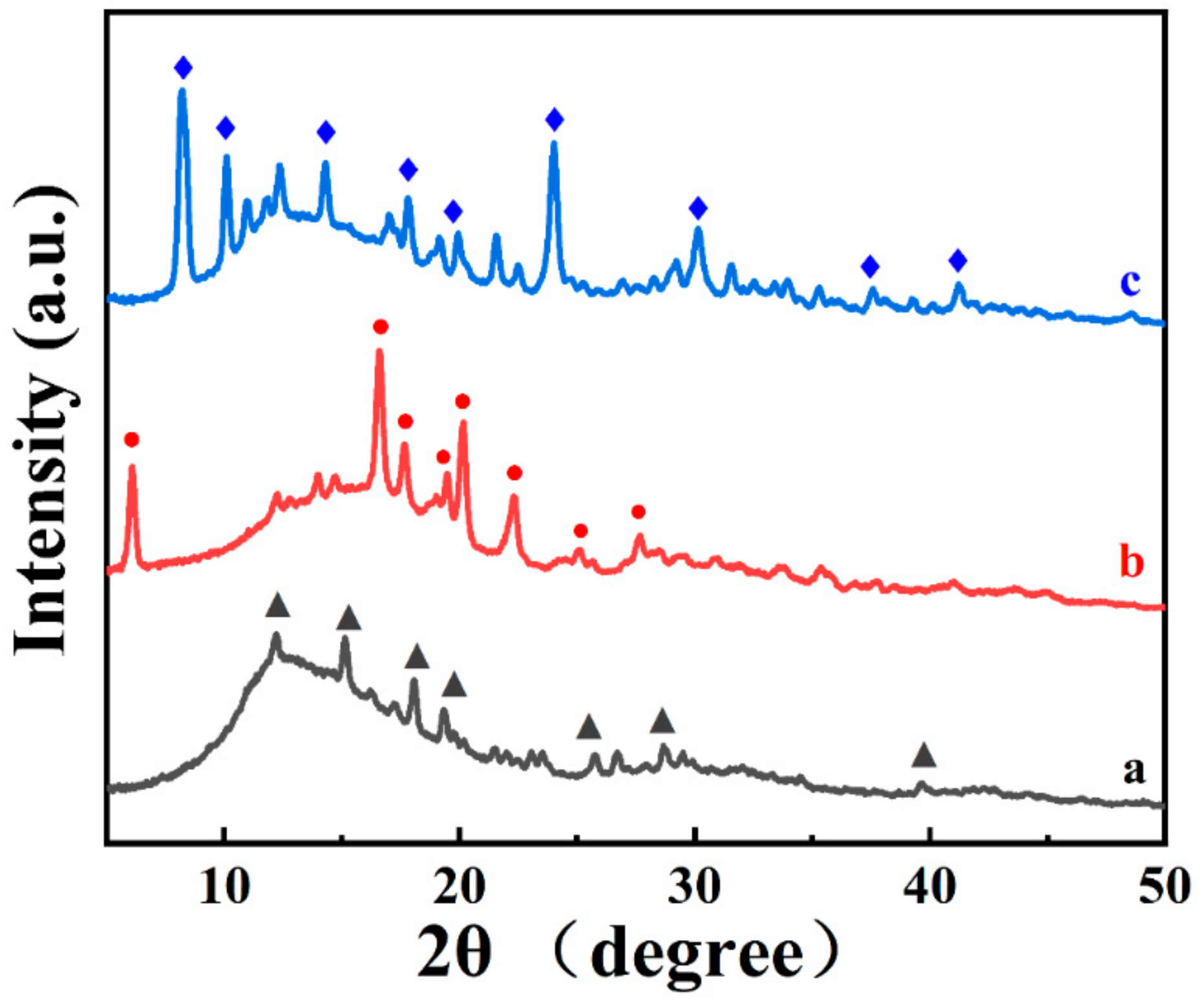
The co-deposition of PMMA with drugs was confirmed by the XRD data presented in Figure 8. The X-ray diffraction patterns showed peaks of the drug materials.
The fabrication of the composite coatings was also confirmed by the results of FTIR spectroscopy. Figure 9 compares the FTIR spectra of as-received PMMA and drugs with the FTIR data for PMMA coatings containing drugs. The most intense bands in the spectrum of PMMA at 1703, 1230 and 1068 cm−1 are attributed to the carbonyl –C=O stretching, C–C–C stretching and skeletal rocking vibration of the polymer backbone, respectively [67]. The absorptions in the range of 1500–1400 cm−1 are related to the bending of the CH2, CH3 and OCH3 groups [67]. Similar absorptions were observed in the spectra of PMMA-ibuprofen, PMMA-tetracycline and PMMA-amoxicillin coatings. The FTIR spectra of ibuprofen [68] showed vibrational peaks at 935 cm−1, which resulted from the O–H bending group of ibuprofen. Carbonyl stretching vibration (C=O) is observed at 1718 cm−1, which corresponds to the carboxyl group (COOH) of ibuprofen. C–O stretching vibration is seen at 1230 cm−1. Such absorptions were observed in the spectrum of PMMA-ibuprofen. The main characteristic peaks of tetracycline [69] are located in the range of 1200–1700 cm−1. The peak at 1575 cm−1 is attributed to the vibration of NH2 amide [69]. The peak at 1446 cm−1 can be assigned to the C-ring-C stretching vibration [69]. Such peaks were observed in the spectrum of PMMA-tetracycline. The FTIR spectrum of amoxicillin [70] showed a C=O stretching band at 1772 cm−1, a C=O stretching band of amide at 1683 cm−1 and absorption due to the asymmetric stretching of carboxylate at 1573 cm−1. Additionally, the C-O bending vibration peak was observed at 1076 cm−1. Similar peaks were observed in the spectrum of PMMA-amoxicillin.
The experimental results described above confirmed the fabrication of composite coatings. The dip coating method developed in this investigation is a versatile strategy for the fabrication of composite coatings containing functional biomaterials of different types. Compared to other coating techniques, such as knife-coating or bar coating, dip coating involved the use of low-cost equipment. The isopropanol-water solvent offers benefits because the use of traditional toxic solvents for PMMA dissolution was avoided. The coatings obtained in this investigation provide a platform for the fabrication of implants with enhanced biocompatibility and antimicrobial properties, coatings for drug delivery and biosensors.
4. Conclusions
The ability to eliminate the use of traditional toxic solvents for PMMA offers benefits for the fabrication of composite coatings for biomedical applications. PMMA and composite coatings were obtained using a water-isopropanol solvent, avoiding the use of traditional toxic solvents. The dip coating method is a versatile strategy for the fabrication of composite coatings containing various functional materials, such as bioceramics, antimicrobial agents and drugs. PMMA coatings containing hydroxyapatite and silica are promising for the fabrication of biomedical implants with enhanced bioactivity and biocompatibility. The incorporation of materials with antimicrobial properties, such as Ag2O and ZnO, into the PMMA matrix can potentially impart antimicrobial properties to the composite coatings. PMMA-ZnO coating can provide a platform for the immobilization of biosensing molecules and the fabrication of biosensors. PMMA-drug composite coatings offer potential for drug delivery. The dip coating method is a simple, low-cost technique, ideally suitable for multilayer processing. Therefore, further development of this method can result in advanced microstructures containing layers of different functional materials. It is expected that future progress in this method will result in the deposition of new coatings containing other functional biomaterials for various applications.
Author Contributions
Conceptualization, Z.W. and I.Z.; methodology, Z.W.; software, Z.W.; validation, Z.W. and I.Z.; formal analysis, Z.W. and I.Z.; investigation, Z.W.; resources, I.Z.; data curation, Z.W.; writing—original draft preparation, Z.W. and I.Z.; writing—review and editing, Z.W. and I.Z.; visualization, Z.W.; supervision, I.Z.; project administration, I.Z.; funding acquisition, I.Z. All authors have read and agreed to the published version of the manuscript.
Funding
This research was funded by the Natural Sciences and Engineering Research Council of Canada, grant number RGPIN-2018-04014, and the CRC program.
Data Availability Statement
All the data is provided in this article.
Acknowledgments
The authors acknowledge the support of the Natural Sciences and Engineering Research Council (NSERC) of Canada, the CRC program and the Canadian Centre for Electron Microscopy.
Conflicts of Interest
The authors declare no conflict of interest.
References
- Ali, U.; Karim, K.J.B.A.; Buang, N.A. A review of the properties and applications of poly (methyl methacrylate)(PMMA). Polym. Rev. 2015, 55, 678–705. [Google Scholar] [CrossRef]
- Wang, H.; Wang, L.; Meng, S.; Lin, H.; Correll, M.; Tong, Z. Nanocomposite of Graphene Oxide Encapsulated in Polymethylmethacrylate (PMMA): Pre-Modification, Synthesis, and Latex Stability. J. Compos. Sci. 2020, 4, 118. [Google Scholar] [CrossRef]
- Saha, D.; Majumdar, M.K.; Das, A.K.; Chowdhury, A.M.S.; Ashaduzzaman, M. Structural Nanocomposite Fabrication from Self-Assembled Choline Chloride Modified Kaolinite into Poly(Methylmethacrylate). J. Compos. Sci. 2019, 3, 83. [Google Scholar] [CrossRef] [Green Version]
- Poon, R.; Zhitomirsky, I. Application of Cyrene as a solvent and dispersing agent for fabrication of Mn3O4-carbon nanotube supercapacitor electrodes. Colloid Interface Sci. Commun. 2020, 34, 100226. [Google Scholar] [CrossRef]
- Dimitrova, M.; Corsalini, M.; Kazakova, R.; Vlahova, A.; Chuchulska, B.; Barile, G.; Capodiferro, S.; Kazakov, S. Comparison between Conventional PMMA and 3D Printed Resins for Denture Bases: A Narrative Review. J. Compos. Sci. 2022, 6, 87. [Google Scholar] [CrossRef]
- Sikkema, R.; Baker, K.; Zhitomirsky, I. Electrophoretic deposition of polymers and proteins for biomedical applications. Adv. Colloid Interface Sci. 2020, 284, 102272. [Google Scholar] [CrossRef]
- Baker, K.; Sikkema, R.; Liang, W.; Zhitomirsky, I. Multifunctional Properties of Commercial Bile Salts for Advanced Materials Engineering. Adv. Eng. Mater. 2021, 23, 2001261. [Google Scholar] [CrossRef]
- Inzana, J.A.; Schwarz, E.M.; Kates, S.L.; Awad, H.A. Biomaterials approaches to treating implant-associated osteomyelitis. Biomaterials 2016, 81, 58–71. [Google Scholar] [CrossRef] [Green Version]
- Leão, R.D.S.; Maior, J.R.S.; Lemos, C.A.D.A.; Vasconcelos, B.C.D.E.; Montes, M.A.J.R.; Pellizzer, E.P.; Moraes, S.L.D. Complications with PMMA compared with other materials used in cranioplasty: A systematic review and meta-analysis. Braz. Oral Res. 2018, 32, e31. [Google Scholar] [CrossRef] [Green Version]
- Jaeblon, T. Polymethylmethacrylate: Properties and contemporary uses in orthopaedics. JAAOS-J. Am. Acad. Orthop. Surg. 2010, 18, 297–305. [Google Scholar] [CrossRef]
- Bistolfi, A.; Ferracini, R.; Albanese, C.; Vernè, E.; Miola, M. PMMA-Based Bone Cements and the Problem of Joint Arthroplasty Infections: Status and New Perspectives. Materials 2019, 12, 4002. [Google Scholar] [CrossRef] [PubMed] [Green Version]
- Lewis, G. Properties of nanofiller-loaded poly (methyl methacrylate) bone cement composites for orthopedic applications: A review. J. Biomed. Mater. Res. Part B Appl. Biomater. 2017, 105, 1260–1284. [Google Scholar] [CrossRef] [PubMed]
- Chatterjee, A. Properties improvement of PMMA using nano TiO2. J. Appl. Polym. Sci. 2010, 118, 2890–2897. [Google Scholar] [CrossRef]
- Hazim, A.; Abduljalil, H.M.; Hashim, A. Analysis of Structural and Electronic Properties of Novel (PMMA/Al2O3, PMMA/Al2O3-Ag, PMMA/ZrO2, PMMA/ZrO2-Ag, PMMA-Ag) Nanocomposites for Low Cost Electronics and Optics Applications. Trans. Electr. Electron. Mater. 2020, 21, 48–67. [Google Scholar] [CrossRef]
- Bai, H.; Walsh, F.; Gludovatz, B.; Delattre, B.; Huang, C.; Chen, Y.; Tomsia, A.P.; Ritchie, R.O. Bioinspired hydroxyapatite/poly (methyl methacrylate) composite with a nacre-mimetic architecture by a bidirectional freezing method. Adv. Mater. 2016, 28, 50–56. [Google Scholar] [CrossRef]
- Carranza, A.; Romero-Perez, D.; Almanza-Reyes, H.; Bogdanchikova, N.; Juarez-Moreno, K.; Pojman, J.A.; Velasquillo, C.; Mota-Morales, J.D. Nonaqueous Synthesis of Macroporous Nanocomposites Using High Internal Phase Emulsion Stabilized by Nanohydroxyapatite. Adv. Mater. Interfaces 2017, 4, 1700094. [Google Scholar] [CrossRef]
- Haach, L.C.D.A.; Purquerio, B.D.M.; Silva Junior, N.F.D.; Gaspar, A.M.M.; Fortulan, C.A. Comparison of two composites developed to be used as bone replacement-PMMA/Bioglass 45S5® microfiber and PMMA/Hydroxyapatite. Bioceram. Dev. Appl. 2014, 4, 1000071. [Google Scholar]
- Floroian, L.; Samoila, C.; Badea, M.; Munteanu, D.; Ristoscu, C.; Sima, F.; Negut, I.; Chifiriuc, M.; Mihailescu, I. Stainless steel surface biofunctionalization with PMMA-bioglass coatings: Compositional, electrochemical corrosion studies and microbiological assay. J. Mater. Sci. Mater. Med. 2015, 26, 195. [Google Scholar] [CrossRef]
- Kettel, M.J.; Heine, E.; Schaefer, K.; Moeller, M. Chlorhexidine Loaded Cyclodextrin Containing PMMA Nanogels as Antimicrobial Coating and Delivery Systems. Macromol. Biosci. 2017, 17, 1600230. [Google Scholar] [CrossRef]
- Neumann, S.E.; Chamberlayne, C.F.; Zare, R.N. Electrically controlled drug release using pH-sensitive polymer films. Nanoscale 2018, 10, 10087–10093. [Google Scholar] [CrossRef]
- Rezaei, F.; Abbasi-Firouzjah, M.; Shokri, B. Investigation of antibacterial and wettability behaviours of plasma-modified PMMA films for application in ophthalmology. J. Phys. D Appl. Phys. 2014, 47, 085401. [Google Scholar] [CrossRef]
- Shanzuo, J.; Ponting, M.; Lepkowicz, R.S.; Rosenberg, A.; Flynn, R.; Beadie, G.; Baer, E. A bio-inspired polymeric gradient refractive index (GRIN) human eye lens. Opt. Express 2012, 20, 26746–26754. [Google Scholar]
- Nugen, S.R.; Asiello, P.J.; Connelly, J.T.; Baeumner, A.J. PMMA biosensor for nucleic acids with integrated mixer and electrochemical detection. Biosens. Bioelectron. 2009, 24, 2428–2433. [Google Scholar] [CrossRef] [PubMed]
- Irawati, N.; Harun, S.W.; Adwan, S.; Alnowami, M.; Ahmad, H. PMMA microfiber coated with Al-doped ZnO nanostructures for detecting uric acid. Microw. Opt. Technol. Lett. 2015, 57, 2455–2457. [Google Scholar] [CrossRef]
- Cools, P.; De Geyter, N.; Vanderleyden, E.; Barberis, F.; Dubruel, P.; Morent, R. Adhesion improvement at the PMMA bone cement-titanium implant interface using methyl methacrylate atmospheric pressure plasma polymerization. Surf. Coat. Technol. 2016, 294, 201–209. [Google Scholar] [CrossRef]
- Coan, T.; Barroso, G.; Machado, R.; De Souza, F.; Spinelli, A.; Motz, G. A novel organic-inorganic PMMA/polysilazane hybrid polymer for corrosion protection. Prog. Org. Coat. 2015, 89, 220–230. [Google Scholar] [CrossRef]
- D’Elia, A.; Deering, J.; Clifford, A.; Lee, B.; Grandfield, K.; Zhitomirsky, I. Electrophoretic deposition of polymethylmethacrylate and composites for biomedical applications. Colloids Surf. B Biointerfaces 2020, 188, 110763. [Google Scholar] [CrossRef]
- Norouzi, M.; Garekani, A.A. Corrosion protection by zirconia-based thin films deposited by a sol–gel spin coating method. Ceram. Int. 2014, 40, 2857–2861. [Google Scholar] [CrossRef]
- Jin, W.; Hao, Q.; Peng, X.; Chu, P.K. Enhanced corrosion resistance and biocompatibilty of PMMA-coated ZK60 magnesium alloy. Mater. Lett. 2016, 173, 178–181. [Google Scholar] [CrossRef]
- Coan, T.; Barroso, G.S.; Motz, G.; Bolzán, A.; Machado, R.A.F. Preparation of PMMA/hBN composite coatings for metal surface protection. Mater. Res. 2013, 16, 1366–1372. [Google Scholar] [CrossRef] [Green Version]
- Negi, Y.; Adhyapak, P.; Damkale, S.; Goyal, R.; Islam, M.; Aiyer, R. Preparation of novel optical-grade metanitroaniline and polymethylmethacrylate-coated single crystals and their optical properties. Mater. Lett. 2004, 58, 3929–3932. [Google Scholar] [CrossRef]
- Sathish, S.; Shekar, B.C. Dip and spin coated nanoscale transparent PMMA thin films for field effect thin film transistors and optoelectronic devices. J. Optoelectron. Adv. Mater 2013, 15, 139–144. [Google Scholar]
- Li, X.; Zhitomirsky, I. Deposition of poly (methyl methacrylate) and composites containing bioceramics and bioglass by dip coating using isopropanol-water co-solvent. Prog. Org. Coat. 2020, 148, 105883. [Google Scholar] [CrossRef]
- Sreekantan, S.; Hassan, M.; Sundera Murthe, S.; Seeni, A. Biocompatibility and Cytotoxicity Study of Polydimethylsiloxane (PDMS) and Palm Oil Fuel Ash (POFA) Sustainable Super-Hydrophobic Coating for Biomedical Applications. Polymers 2020, 12, 3034. [Google Scholar] [CrossRef]
- Cooperstein, M.A.; Canavan, H.E. Assessment of cytotoxicity of (N-isopropyl acrylamide) and poly (N-isopropyl acrylamide)-coated surfaces. Biointerphases 2013, 8, 19. [Google Scholar] [CrossRef] [Green Version]
- Hadidi, M.; Bigham, A.; Saebnoori, E.; Hassanzadeh-Tabrizi, S.; Rahmati, S.; Alizadeh, Z.M.; Nasirian, V.; Rafienia, M. Electrophoretic-deposited hydroxyapatite-copper nanocomposite as an antibacterial coating for biomedical applications. Surf. Coat. Technol. 2017, 321, 171–179. [Google Scholar] [CrossRef]
- Farrokhi-Rad, M. Electrophoretic deposition of titania nanostructured coatings with different porous patterns. Ceram. Int. 2018, 44, 15346–15355. [Google Scholar] [CrossRef]
- Farrokhi-rad, M.; Emamalipour, S.; Mohammadzadeh, F.; Beygi-Khosrowshahi, Y.; Hassannejad, H.; Nouri, A. Electrophoretic deposition of alginate coatings from different alcohol-water mixtures. Surf. Eng. 2021, 37, 1176–1185. [Google Scholar] [CrossRef]
- Narkevica, I.; Stradina, L.; Stipniece, L.; Jakobsons, E.; Ozolins, J. Electrophoretic deposition of nanocrystalline TiO2 particles on porous TiO2-X ceramic scaffolds for biomedical applications. J. Eur. Ceram. Soc. 2017, 37, 3185–3193. [Google Scholar] [CrossRef]
- Bano, S.; Romero, A.R.; Grant, D.; Nommeots-Nomm, A.; Scotchford, C.; Ahmed, I.; Hussain, T. In-vitro cell interaction and apatite forming ability in simulated body fluid of ICIE16 and 13–93 bioactive glass coatings deposited by an emerging suspension high velocity oxy fuel (SHVOF) thermal spray. Surf. Coat. Technol. 2021, 407, 126764. [Google Scholar] [CrossRef]
- Grigaleviciute, G.; Baltriukiene, D.; Bukelskiene, V.; Malinauskas, M. Biocompatibility Evaluation and Enhancement of Elastomeric Coatings Made Using Table-Top Optical 3D Printer. Coatings 2020, 10, 254. [Google Scholar] [CrossRef] [Green Version]
- Zhang, M.; Weng, Y.J.; Zhang, Y.Q. Accelerated desalting and purification of silk fibroin in a CaCl2-EtOH-H2O ternary system by excess isopropanol extraction. J. Chem. Technol. Biotechnol. 2021, 96, 1176–1186. [Google Scholar] [CrossRef]
- Silva, S.S.; Oliveira, N.M.; Oliveira, M.B.; Da Costa, D.P.S.; Naskar, D.; Mano, J.F.; Kundu, S.C.; Reis, R.L. Fabrication and characterization of Eri silk fibers-based sponges for biomedical application. Acta Biomater. 2016, 32, 178–189. [Google Scholar] [CrossRef] [Green Version]
- Pruett, L.J.; Jenkins, C.H.; Singh, N.S.; Catallo, K.J.; Griffin, D.R. Heparin Microislands in Microporous Annealed Particle Scaffolds for Accelerated Diabetic Wound Healing. Adv. Funct. Mater. 2021, 31, 2104337. [Google Scholar] [CrossRef]
- Yu, Q.; Roberts, M.G.; Pearce, S.; Oliver, A.M.; Zhou, H.; Allen, C.; Manners, I.; Winnik, M.A. Rodlike block copolymer micelles of controlled length in water designed for biomedical applications. Macromolecules 2019, 52, 5231–5244. [Google Scholar] [CrossRef]
- Grandfield, K.; Zhitomirsky, I. Electrophoretic deposition of composite hydroxyapatite–silica–chitosan coatings. Mater. Charact. 2008, 59, 61–67. [Google Scholar] [CrossRef]
- Grandfield, K.; Sun, F.; FitzPatrick, M.; Cheong, M.; Zhitomirsky, I. Electrophoretic deposition of polymer-carbon nanotube–hydroxyapatite composites. Surf. Coat. Technol. 2009, 203, 1481–1487. [Google Scholar] [CrossRef]
- Baskaran, A.; Smereka, P. Mechanisms of stranski-krastanov growth. J. Appl. Phys. 2012, 111, 044321. [Google Scholar] [CrossRef]
- Vithiya, K.; Kumar, R.; Sen, S. Antimicrobial activity of biosynthesized silver oxide nanoparticles. J. Pure Appl. Microbiol 2014, 4, 3263–3268. [Google Scholar]
- D’Lima, L.; Phadke, M.; Ashok, V.D. Biogenic silver and silver oxide hybrid nanoparticles: A potential antimicrobial against multi drug-resistant Pseudomonas aeruginosa. New J. Chem. 2020, 44, 4935–4941. [Google Scholar] [CrossRef]
- Arya, S.K.; Saha, S.; Ramirez-Vick, J.E.; Gupta, V.; Bhansali, S.; Singh, S.P. Recent advances in ZnO nanostructures and thin films for biosensor applications. Anal. Chim. Acta 2012, 737, 1–21. [Google Scholar] [CrossRef] [PubMed]
- Janaki, A.C.; Sailatha, E.; Gunasekaran, S. Synthesis, characteristics and antimicrobial activity of ZnO nanoparticles. Spectrochim. Acta Part A Mol. Biomol. Spectrosc. 2015, 144, 17–22. [Google Scholar] [CrossRef] [PubMed]
- Shinde, S.S. Antimicrobial activity of ZnO nanoparticles against pathogenic bacteria and fungi. Sci. Med. Cent. 2015, 3, 1033. [Google Scholar]
- Casas-Luna, M.; Horynová, M.; Tkachenko, S.; Klakurková, L.; Celko, L.; Diaz-de-la-Torre, S.; Montufar, E.B. Chemical Stability of Tricalcium Phosphate–Iron Composite during Spark Plasma Sintering. J. Compos. Sci. 2018, 2, 51. [Google Scholar] [CrossRef] [Green Version]
- Wang, K.; Pasbakhsh, P.; De Silva, R.T.; Goh, K.L. A Comparative Analysis of the Reinforcing Efficiency of Silsesquioxane Nanoparticles versus Apatite Nanoparticles in Chitosan Biocomposite Fibres. J. Compos. Sci. 2017, 1, 9. [Google Scholar] [CrossRef] [Green Version]
- Moura, N.K.D.; Siqueira, I.A.W.B.; Machado, J.P.D.B.; Kido, H.W.; Avanzi, I.R.; Rennó, A.C.M.; Trichês, E.D.S.; Passador, F.R. Production and Characterization of Porous Polymeric Membranes of PLA/PCL Blends with the Addition of Hydroxyapatite. J. Compos. Sci. 2019, 3, 45. [Google Scholar] [CrossRef] [Green Version]
- Pang, X.; Zhitomirsky, I. Electrophoretic deposition of composite hydroxyapatite-chitosan coatings. Mater. Charact. 2007, 58, 339–348. [Google Scholar] [CrossRef]
- Harb, S.V.; Uvida, M.C.; Trentin, A.; Lobo, A.O.; Webster, T.J.; Pulcinelli, S.H.; Santilli, C.V.; Hammer, P. PMMA-silica nanocomposite coating: Effective corrosion protection and biocompatibility for a Ti6Al4V alloy. Mater. Sci. Eng. C 2020, 110, 110713. [Google Scholar] [CrossRef]
- Fateh, T.; Richard, F.; Rogaume, T.; Joseph, P. Experimental and modelling studies on the kinetics and mechanisms of thermal degradation of polymethyl methacrylate in nitrogen and air. J. Anal. Appl. Pyrolysis 2016, 120, 423–433. [Google Scholar] [CrossRef]
- Mu, F.; Zhao, Z.; Zou, G.; Bai, H.; Wu, A.; Liu, L.; Zhang, D.; Norman Zhou, Y. Mechanism of Low Temperature Sintering-Bonding through In-Situ Formation of Silver Nanoparticles Using Silver Oxide Microparticles. Mater. Trans. 2013, 54, 872. [Google Scholar] [CrossRef] [Green Version]
- Ananth, A.; Mok, Y.S. Dielectric barrier discharge (DBD) plasma assisted synthesis of Ag2O nanomaterials and Ag2O/RuO2 nanocomposites. Nanomaterials 2016, 6, 42. [Google Scholar] [CrossRef] [PubMed]
- Zhang, H.; Li, G.; An, L.; Yan, T.; Gao, X.; Zhu, H. Electrochemical lithium storage of titanate and titania nanotubes and nanorods. J. Phys. Chem. C 2007, 111, 6143–6148. [Google Scholar] [CrossRef]
- Porramezan, M.; Eisazadeh, H. Fabrication and characterization of polyaniline nanocomposite modified with Ag2O nanoparticles. Compos. Part B Eng. 2011, 42, 1980–1986. [Google Scholar] [CrossRef]
- Pang, X.; Zhitomirsky, I.; Niewczas, M. Cathodic electrolytic deposition of zirconia films. Surf. Coat. Technol. 2005, 195, 138–146. [Google Scholar] [CrossRef]
- Zhitomirsky, I.; Petric, A. Electrochemical deposition of yttrium oxide. J. Mater. Chem. 2000, 10, 1215–1218. [Google Scholar] [CrossRef]
- Wentao, Z.; Lei, G.; Liu, Y.; Wang, W.; Song, T.; Fan, J. Approach to osteomyelitis treatment with antibiotic loaded PMMA. Microb. Pathog. 2017, 102, 42–44. [Google Scholar] [CrossRef] [PubMed]
- Huszánk, R.; Szilágyi, E.; Szoboszlai, Z.; Szikszai, Z. Investigation of chemical changes in PMMA induced by 1.6 MeV He+ irradiation by ion beam analytical methods (RBS-ERDA) and infrared spectroscopy (ATR-FTIR). Nucl. Instrum. Methods Phys. Res. B 2019, 450, 364–368. [Google Scholar] [CrossRef]
- Carvalho, R.B.; Joshi, S.V. Ibuprofen isobutanolammonium salt. J. Therm. Anal. Calorim. 2020, 139, 1971–1976. [Google Scholar] [CrossRef]
- Zhang, Z.; Liu, H.; Wu, L.; Lan, H.; Qu, J. Preparation of amino-Fe (III) functionalized mesoporous silica for synergistic adsorption of tetracycline and copper. Chemosphere 2015, 138, 625–632. [Google Scholar] [CrossRef]
- Iqbal, D.N.; Ehtisham-ul-Haque, S.; Ahmad, S.; Arif, K.; Hussain, E.A.; Iqbal, M.; Alshawwa, S.Z.; Abbas, M.; Amjed, N.; Nazir, A. Enhanced antibacterial activity of chitosan, guar gum and polyvinyl alcohol blend matrix loaded with amoxicillin and doxycycline hyclate drugs. Arab. J. Chem. 2021, 14, 103156. [Google Scholar] [CrossRef]
Figure 1.
SEM images of PMMA coating: (A) as-deposited and room-temperature-dried, (B) annealed at 200 °C.
Figure 1.
SEM images of PMMA coating: (A) as-deposited and room-temperature-dried, (B) annealed at 200 °C.

Figure 2.
SEM images of (A,B) PMMA-Ag2O, (C,D) PMMA-HAP and PMMA-ZnO coatings, (A,C,E) as-deposited and room-temperature-dried, (B,D,F) annealed at 200 °C.
Figure 2.
SEM images of (A,B) PMMA-Ag2O, (C,D) PMMA-HAP and PMMA-ZnO coatings, (A,C,E) as-deposited and room-temperature-dried, (B,D,F) annealed at 200 °C.

Figure 3.
SEM images of (A,B) PMMA-micron size composites and (C,D) PMMA-nanosilica composites, (A,C) as-deposited and room-temperature-dried, (B,D) annealed at 200 °C.
Figure 3.
SEM images of (A,B) PMMA-micron size composites and (C,D) PMMA-nanosilica composites, (A,C) as-deposited and room-temperature-dried, (B,D) annealed at 200 °C.

Figure 4.
X-ray diffraction patterns of (a) as-received PMMA, and composites (b) PMMA-ZnO, (c) PMMA-HAP, (d) PMMA-Ag2O and (e) PMMA-HAP-Ag2O, ▼—JCPDS file 04-020-9583, ▲—JCPDS file 04-008-4759, ●—JCPDS file 00-041-1104, ♦—PMMA.
Figure 4.
X-ray diffraction patterns of (a) as-received PMMA, and composites (b) PMMA-ZnO, (c) PMMA-HAP, (d) PMMA-Ag2O and (e) PMMA-HAP-Ag2O, ▼—JCPDS file 04-020-9583, ▲—JCPDS file 04-008-4759, ●—JCPDS file 00-041-1104, ♦—PMMA.

Figure 5.
X-ray diffraction patterns of (a) PMMA, (b) micron size silica, (c) PMMA-micron size silica, (d) nanosilica and (e) PMMA-nanosilica (●—silica, ♦—PMMA).
Figure 5.
X-ray diffraction patterns of (a) PMMA, (b) micron size silica, (c) PMMA-micron size silica, (d) nanosilica and (e) PMMA-nanosilica (●—silica, ♦—PMMA).

Figure 6.
TGA data for (A) PMMA-HAP, (B) PMMA-Ag2O, (C) PMMA-ZnO, (D) PMMA-micron size silica and (E) PMMA-nanosilica composites.
Figure 6.
TGA data for (A) PMMA-HAP, (B) PMMA-Ag2O, (C) PMMA-ZnO, (D) PMMA-micron size silica and (E) PMMA-nanosilica composites.

Figure 7.
SEM images of as-deposited and room-temperature-dried coatings: (A) PMMA- ibuprofen, (B) PMMA-tetracycline and (C) PMMA-amoxicillin coatings.
Figure 7.
SEM images of as-deposited and room-temperature-dried coatings: (A) PMMA- ibuprofen, (B) PMMA-tetracycline and (C) PMMA-amoxicillin coatings.

Figure 8.
X-ray diffraction patterns of (a) PMMA-amoxicillin, (b) PMMA-ibuprofen and (c) PMMA-tetracycline; major XRD peaks are labeled: ▲—peaks corresponding to JCPDS file 00-039-1832 of amoxicillin, ●—peaks corresponding to JCPDS file 00-032-1723 of ibuprofen, ♦—peaks corresponding to JCPDS file 00-039-1985 of tetracycline.

Figure 9.
FTIR spectra of (a) as-received PMMA (b) as-received ibuprofen, (c) PMMA-ibuprofen, (d) as-received tetracycline, (e) PMMA-tetracycline, (f) as-received amoxicillin and (g) PMMA-amoxicillin.
Figure 9.
FTIR spectra of (a) as-received PMMA (b) as-received ibuprofen, (c) PMMA-ibuprofen, (d) as-received tetracycline, (e) PMMA-tetracycline, (f) as-received amoxicillin and (g) PMMA-amoxicillin.

Publisher’s Note: MDPI stays neutral with regard to jurisdictional claims in published maps and institutional affiliations. |
© 2022 by the authors. Licensee MDPI, Basel, Switzerland. This article is an open access article distributed under the terms and conditions of the Creative Commons Attribution (CC BY) license (https://creativecommons.org/licenses/by/4.0/).
Share and Cite
MDPI and ACS Style
Wang, Z.; Zhitomirsky, I. Deposition of Organic-Inorganic Nanocomposite Coatings for Biomedical Applications. Solids 2022, 3, 271-281. https://0-doi-org.brum.beds.ac.uk/10.3390/solids3020019
AMA Style
Wang Z, Zhitomirsky I. Deposition of Organic-Inorganic Nanocomposite Coatings for Biomedical Applications. Solids. 2022; 3(2):271-281. https://0-doi-org.brum.beds.ac.uk/10.3390/solids3020019
Chicago/Turabian StyleWang, Zhengzheng, and Igor Zhitomirsky. 2022. "Deposition of Organic-Inorganic Nanocomposite Coatings for Biomedical Applications" Solids 3, no. 2: 271-281. https://0-doi-org.brum.beds.ac.uk/10.3390/solids3020019