


Dent. J. 2024, 12(5), 124; https://0-doi-org.brum.beds.ac.uk/10.3390/dj12050124 - 25 Apr 2024
Abstract
Autotransplantation is a successful technique to replace compromised teeth. This study presents a computer-guided surgical approach for preparing the receiving socket for a mature mandibular third molar donor tooth with a wait-and-see approach instead of prophylactic endodontic treatment. A 42-year-old woman developed root
[...] Read more.
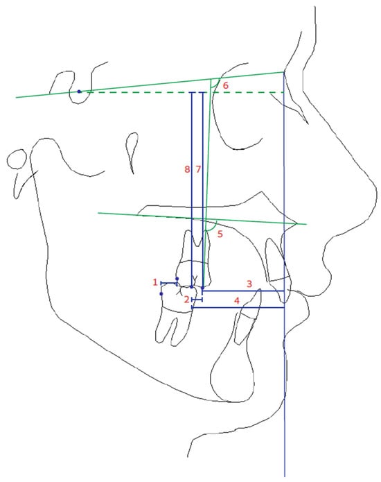
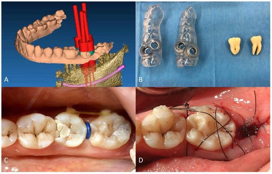
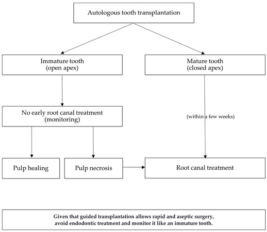
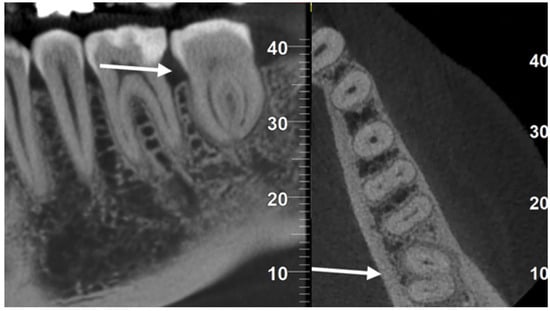
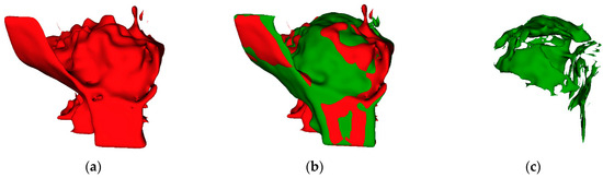
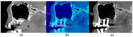
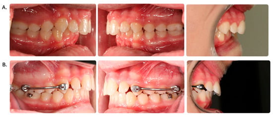
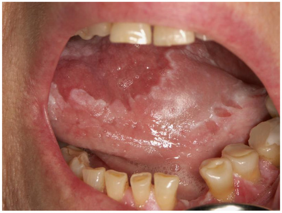
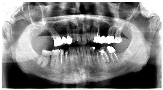
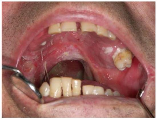
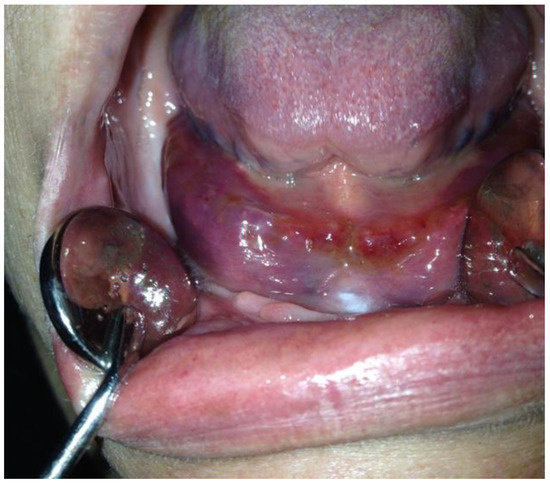
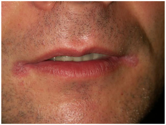
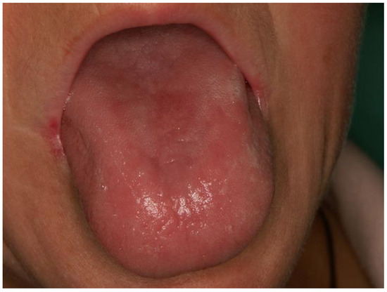
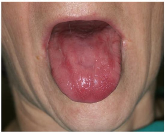
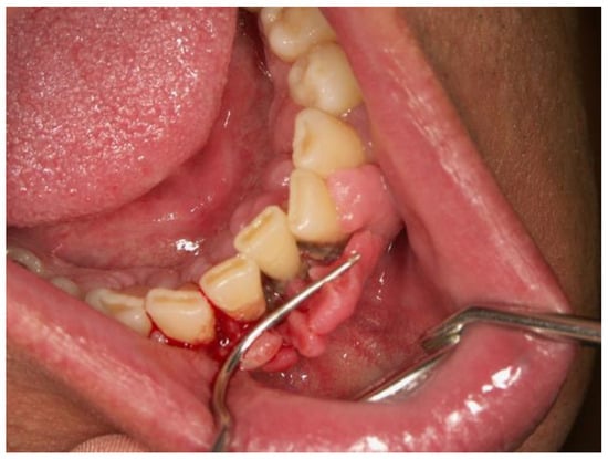
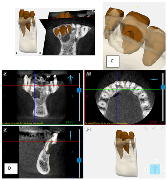
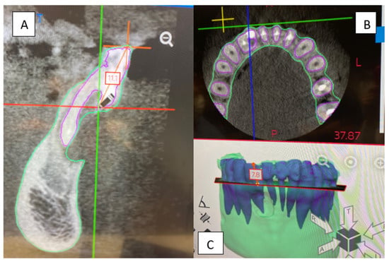
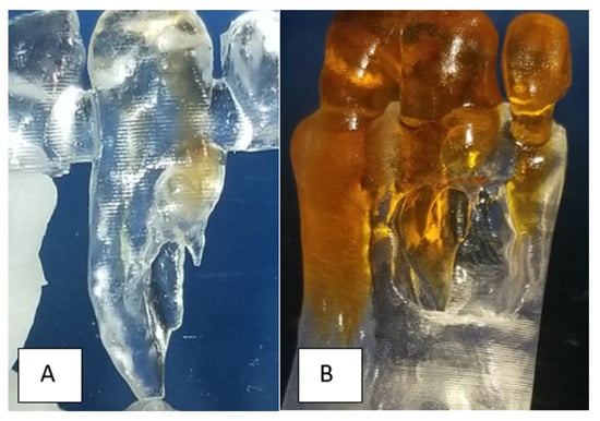
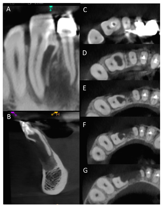
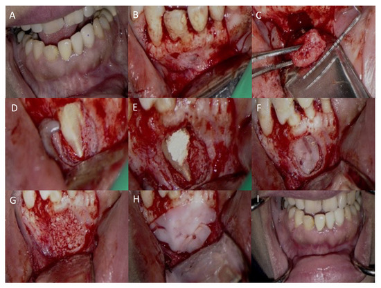
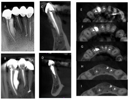
Autotransplantation is a successful technique to replace compromised teeth. This study presents a computer-guided surgical approach for preparing the receiving socket for a mature mandibular third molar donor tooth with a wait-and-see approach instead of prophylactic endodontic treatment. A 42-year-old woman developed root resorption of tooth 3.7. Extraction of 3.7 and autotransplantation of 3.8 was planned, following a 6-week orthodontic phase for periodontal ligament activation and teeth mobilization. Due to the different root morphology between the compromised and donor teeth and the high mandibular bone density, the receiving socket preparation was performed using guided surgery templates. Two surgical splints were designed with a surgical planning software. Tooth 3.7 was extracted, the recipient site was guided-milled, and tooth 3.8 was transplanted into the new socket in approximately one second of extra-alveolar time. The rapidity of the extra-alveolar time facilitated complete healing without resorting to root canal treatment. Five-year radiological control does not show any periapical lesion or root resorption. The surgical procedure for tooth autotransplantation is fundamental: it must be as atraumatic as possible to preserve the periodontal ligament of the tooth and the receiving socket, and the dentist must minimize the extra-alveolar time. Guided surgery is a reliable solution to combine all these aspects.
Full article
(This article belongs to the Special Issue Endodontics and Restorative Sciences)
►
Show Figures
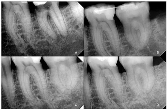
Figure 1


