


Pathogens 2022, 11(8), 824; https://0-doi-org.brum.beds.ac.uk/10.3390/pathogens11080824 - 23 Jul 2022
Cited by 1 | Viewed by 1846
Abstract
►
Show Figures
The spread of pyrethroid resistance in malaria vectors is a major threat affecting the performance of current control measures. However, there is still not enough information on the resistance profile of mosquitoes to carbamates and organophosphates which could be used as alternatives. The
[...] Read more.
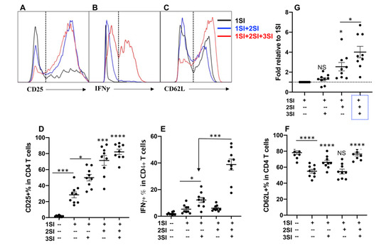
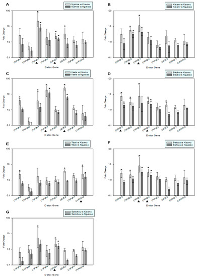
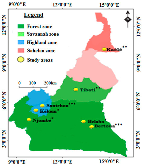
The spread of pyrethroid resistance in malaria vectors is a major threat affecting the performance of current control measures. However, there is still not enough information on the resistance profile of mosquitoes to carbamates and organophosphates which could be used as alternatives. The present study assessed the resistance profile of Anopheles gambiae s.l. to bendiocarb and malathion, at the phenotypic and molecular levels, in different eco-epidemiological settings in Cameroon. Anopheles gambiae s.l. mosquitoes were collected from four eco-epidemiological settings across the country and their susceptibility level to bendiocarb and malathion was determined using WHO tubes bioassays. The ace-1 target site G119S mutation was screened by PCR. Reverse Transcription quantitative PCR 3-plex TaqMan assays were used to quantify the level of expression of eight genes associated with metabolic resistance. Resistance to malathion and/or bendiocarb was recorded in all study sites except in mosquitoes collected in Kaélé and Njombé. The Ace-1 (G119S) mutation was detected in high frequencies (>40%) in Kékem and Santchou. Both An. gambiae and An. coluzzii were detected carrying this mutation. The cytochrome P450s gene Cyp6p3 associated with carbamate resistance and the glutathione S-transferase gene Gste2 associated with organophosphate resistance were found to be overexpressed. Genes associated with pyrethroid (Cyp6m2, Cyp9k1, Cyp6p3) and organochlorine (Gste2, Cyp6z1, Cyp6m2) and cuticle resistance (Cyp4g16) were also overexpressed. The rapid spread of resistance to organophosphates and carbamates could seriously compromise future control strategies based on IRS. It is therefore becoming important to assess the magnitude of bendiocarb and malathion resistance countrywide.
Full article
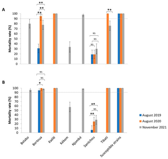
Figure 1